Role of the luxS Quorum-Sensing System in Biofilm Formation and Virulence of Staphylococcus...
10
10.1128/IAI.74.1.488-496.2006. 2006, 74(1):488. DOI: Infect. Immun. Jianping Wang, Yufeng Yao, Michael Otto and Qian Gao Lin Xu, Hualin Li, Cuong Vuong, Viveka Vadyvaloo, Staphylococcus epidermidis in Biofilm Formation and Virulence of Quorum-Sensing System luxS Role of the http://iai.asm.org/content/74/1/488 Updated information and services can be found at: These include: REFERENCES http://iai.asm.org/content/74/1/488#ref-list-1 at: This article cites 58 articles, 38 of which can be accessed free CONTENT ALERTS more» articles cite this article), Receive: RSS Feeds, eTOCs, free email alerts (when new http://journals.asm.org/site/misc/reprints.xhtml Information about commercial reprint orders: http://journals.asm.org/site/subscriptions/ To subscribe to to another ASM Journal go to: on March 24, 2014 by guest http://iai.asm.org/ Downloaded from on March 24, 2014 by guest http://iai.asm.org/ Downloaded from
Transcript of Role of the luxS Quorum-Sensing System in Biofilm Formation and Virulence of Staphylococcus...

10.1128/IAI.74.1.488-496.2006.
2006, 74(1):488. DOI:Infect. Immun. Jianping Wang, Yufeng Yao, Michael Otto and Qian GaoLin Xu, Hualin Li, Cuong Vuong, Viveka Vadyvaloo, Staphylococcus epidermidisin Biofilm Formation and Virulence of
Quorum-Sensing SystemluxSRole of the
http://iai.asm.org/content/74/1/488Updated information and services can be found at:
These include:
REFERENCEShttp://iai.asm.org/content/74/1/488#ref-list-1at:
This article cites 58 articles, 38 of which can be accessed free
CONTENT ALERTS more»articles cite this article),
Receive: RSS Feeds, eTOCs, free email alerts (when new
http://journals.asm.org/site/misc/reprints.xhtmlInformation about commercial reprint orders: http://journals.asm.org/site/subscriptions/To subscribe to to another ASM Journal go to:
on March 24, 2014 by guest
http://iai.asm.org/
Dow
nloaded from
on March 24, 2014 by guest
http://iai.asm.org/
Dow
nloaded from

INFECTION AND IMMUNITY, Jan. 2006, p. 488–496 Vol. 74, No. 10019-9567/06/$08.00�0 doi:10.1128/IAI.74.1.488–496.2006Copyright © 2006, American Society for Microbiology. All Rights Reserved.
Role of the luxS Quorum-Sensing System in Biofilm Formation andVirulence of Staphylococcus epidermidis
Lin Xu,1,2 Hualin Li,1,2 Cuong Vuong,3† Viveka Vadyvaloo,3 Jianping Wang,1,2 Yufeng Yao,3Michael Otto,3 and Qian Gao1,2*
Key Laboratory of Medical Molecular Virology, Shanghai Medical College, Fudan University, Shanghai 200032,People’s Republic of China1; Institute of Medical Microbiology, Fudan University, Shanghai 200032,
People’s Republic of China2; and Rocky Mountain Laboratories, National Institute of Allergy andInfectious Diseases, the National Institutes of Health, Hamilton, Montana 598403
Received 12 June 2005/Returned for modification 18 July 2005/Accepted 5 October 2005
Nosocomial infections caused by Staphylococcus epidermidis are characterized by biofilm formation onimplanted medical devices. Quorum-sensing regulation plays a major role in the biofilm development of manybacterial pathogens. Here, we describe luxS, a quorum-sensing system in staphylococci that has a significantimpact on biofilm development and virulence. We constructed an isogenic �luxS mutant strain of a biofilm-forming clinical isolate of S. epidermidis and demonstrated that luxS signaling is functional in S. epidermidis.The mutant strain showed increased biofilm formation in vitro and enhanced virulence in a rat model ofbiofilm-associated infection. Genetic complementation and addition of autoinducer 2-containing culture fil-trate restored the wild-type phenotype, demonstrating that luxS repressed biofilm formation through a cell-cellsignaling mechanism based on autoinducer 2 secretion. Enhanced production of the biofilm exopolysaccharidepolysaccharide intercellular adhesin in the mutant strain is presumably the major cause of the observedphenotype. The agr quorum-sensing system has previously been shown to impact biofilm development andbiofilm-associated infection in a way similar to that of luxS, although by regulation of different factors. Ourstudy indicates a general scheme of quorum-sensing regulation of biofilm development in staphylococci, whichcontrasts with that observed in many other bacterial pathogens.
For a long time, Staphylococcus epidermidis has been re-garded as an innocuous commensal bacterium on the skin andmucous membranes of the human body. However, it is nowknown to be a major nosocomial pathogen, causing infectionson implanted medical devices such as central venous catheters(CVCs), urinary catheters, prosthetic heart valves, orthopedicdevices, and contact lenses. The investigation of S. epidermidispathogenesis has become of interest in recent years, as S.epidermidis infections have arisen as a major burden to thepublic health system. They are often responsible for prolongedhospitalization and may even be fatal (51).
In contrast to Staphylococcus aureus, S. epidermidis producesonly a limited amount of toxins and degradative exoenzymes(58). The formation of a biofilm, which is a differentiatedstructure of surface-attached cells that are embedded in aself-secreted heterogeneous matrix, is thought to be the lead-ing cause for the persistence of S. epidermidis infection. Abiofilm constitutes a depository that may continuously releasebacteria into the bloodstream. Furthermore, the treatment ofbiofilm-associated infections is especially difficult due to bio-film resistance to antibiotics and attacks from the human im-mune system. To date, several genes have been identified thatare associated with the different stages of biofilm formation,including those carrying autolysin E (atlE) (20), fibrinogen
binding protein (fbe) (41), and the intercellular adhesionoperon (ica) (17, 21). However, the exact roles of these genesduring biofilm formation are not yet entirely clear (11, 26, 32).Possibly, differential expression of these factors in vitro and invivo and under different environmental conditions has so farbeen underestimated.
Quorum sensing (QS) is a bacterial intercommunication sys-tem that controls the expression of multiple genes in responseto population density (16). QS systems use small signal mole-cules called autoinducers (AIs). When the AIs accumulate to athreshold concentration, the system is activated and directly orindirectly controls the transcription of target genes. Gram-negative bacteria normally use acylated homoserine lactones(AHLs) as AIs, while gram-positive bacteria use oligopeptideAIs, which act through two-component phosphorelay cascades(36, 44, 53).
In the last decade, a novel quorum-sensing system was dis-covered in the bioluminescent marine bacterium Vibrio harveyi(3). V. harveyi has three parallel QS systems to induce biolu-minescence. One uses AI-1, a typical AHL, as a signal; asecond uses CAI-1 (V. cholerae autoinducer 1), a signal-medi-ating intercommunication in closely related marine bacteria;and finally there is a novel signaling molecule called AI-2 (22).There is ample data to suggest that AI-2s have been designedfor interspecies cell-cell communication (53). They are foundin both gram-negative and gram-positive bacteria (36, 48, 56).AI-2s are not species specific, as AI-2s produced by otherbacteria species can also trigger the bioluminescence of V.harveyi (47). A gene called luxS is required for the synthesis ofAI-2 (48). AI-2-like activity and luxS homologues are found in
* Corresponding author. Mailing address: Key Laboratory of Med-ical Molecular Virology, Shanghai Medical College, Fudan University,Shanghai 200032, People’s Republic of China. Phone: 86-21-54237195.Fax: 86-21-54237603. E-mail: [email protected].
† Present address: Department of Anesthesiology, University ofMassachusetts, Worster, MA 01655.
488
on March 24, 2014 by guest
http://iai.asm.org/
Dow
nloaded from

a wide range of bacterial species. Database analysis shows thatluxS exists in 35 of the 89 available complete bacterial genomes(National Center for Biotechnology Information) (56). Besideslight production in V. harveyi (4) and Vibrio fischeri (28), luxS/AI-2-dependent QS regulates the growth of Escherichia coli(45), Bacillus anthracis (23), and Actinobacillus actinomycetem-comitans (15). It also plays a role in E. coli flagellum motility(45). Furthermore, it modulates the virulence of Streptococcuspyogenes (29), Clostridium perfringens (39), Vibrio vulnificus(25), and Streptococcus pneumoniae (24, 46). Interestingly,AI-2 from the resident microflora in the lungs of cystic fibrosispatients can up-regulate pathogenicity factors in non-AI-2-producing Pseudomonas aeruginosa (12). In some bacteria, ithas been reported that luxS/AI-2-dependent QS impacts bio-film formation in various ways. For example, a luxS mutant ofStreptococcus gordonii, a major component of dental plaquebiofilm, was unable to form a mixed-species biofilm with aluxS-null strain of the periodontal pathogen Porphyromonasgingivalis (34) despite the wild-type strains readily doing so.Furthermore, a luxS mutant of Helicobacter pylori forms biofilmmore efficiently than the wild type (9). Finally, a luxS mutant ofStreptococcus mutans shows an altered biofilm structure (35).
Staphylococci have one known QS system called agr, foraccessory gene regulator, which secretes modified peptides assignals (38, 40). In S. epidermidis, agr represses biofilm forma-tion and influences the structure of a biofilm (49, 50) by reg-ulation of biofilm factors, such as AtlE and �-toxin (49). luxShomologues are present in the S. aureus and S. epidermidisgenomes (54, 58). However, although Winzer et al. have puri-fied the LuxS protein from S. aureus that can synthesize theAI-2 (54), whether S. epidermidis or other staphylococci use anAI-2-type QS system to control gene expression has not beeninvestigated previously.
In this study, we explored the luxS/AI-2-dependent QS sys-tem of S. epidermidis by construction and analysis of an allelicreplacement mutant of S. epidermidis luxS. We found that theluxS/AI-2-dependent QS system of S. epidermidis is functional.Of note, similarly to the agr system (50), luxS of S. epidermidislimits biofilm formation and virulence in an animal model ofdevice-associated infection. Our data point towards a common
scheme of QS control of biofilm formation and biofilm-asso-ciated infection in staphylococci.
MATERIALS AND METHODS
Bacterial strains and growth media. The bacteria and plasmids used are listedin Table 1. E. coli DH5� was grown in Luria-Bertani medium. Plasmid-contain-ing E. coli strains were grown in the same medium but with ampicillin (100 �g/ml)included. S. epidermidis and its derivative strains were grown in TSB (tryptic soybroth, soybean-casein digest medium USP; Oxoid) medium, and when necessary,erythromycin (10 �g/ml), erythromycin (2.5 �g/ml), and chloramphenicol (20�g/ml) were supplemented. V. harveyi BB170 was kindly provided by B. Bassler(Princeton University) and was grown in autoinducer bioassay (AB) medium at30°C (48). Media were solidified with 1.5% (wt/vol) agar as needed.
DNA manipulation. Genomic DNA of S. epidermidis 1457 was prepared by astandard protocol for gram-positive bacteria (13). Plasmid DNA from E. coli wasextracted using a plasmid purification kit (TianWei Co.) according to the man-ufacturer’s instructions. Plasmid DNA from S. epidermidis and S. aureus wasextracted using the same kit except that the cells were incubated for at least 15min at 37°C in solution P1 with lysostaphin (25 �g/ml; Sigma) before solution P2was added. Taq DNA polymerase and restriction enzymes were obtained fromTakara; and incubation conditions were as recommended by the suppliers. S.epidermidis was transformed by electroporation as described previously (2).
Construction of an S. epidermidis �luxS mutant strain. A 2.5-kb DNA frag-ment from S. epidermidis 1457 genomic DNA with luxS and 1-kb upstream and1-kb downstream flanking sequences was PCR amplified using the primersluxS(a)5 and luxS(a)3, introducing BamHI and EcoRI sites, respectively. Theamplified PCR products were cloned into pcDNA2.1, yielding pQG23. An eryth-romycin resistance (Ermr) gene was PCR-amplified by primers erm5 and erm3from pEC4 (6). The product was ligated to the NdeI and SpeI sites inside theluxS gene on pQG23, yielding pQG24. A BamHI/EcoRI fragment was excised
TABLE 1. Bacterial strains and plasmids used in this study
Strain or plasmid Description Source or reference(s)
StrainsS. epidermidis 1457 luxS wild-type strain, biofilm positive 33S. epidermidis 1457
�luxS mutantluxS mutant (LuxS� Ermr) This study
E. coli DH5� supE44 �lacU169 (�80lacZ�M15) hsdR17 recA1 endA1 gyrA96 thi-1 relA1 �luxS 19, 48V. harveyi BB170 luxN::Tn5 (sensor 1� sensor 2�), AI-2 reporter strain 47
PlasmidspcDNA2.1 Cloning vector, ampicillin resistant InvitrogenpEC4 A vector containing a 1.45-kb ClaI ermB fragment of Tn551 cloned into the ClaI
restriction site of pBluescript KS�6
pBT1 Shuttle vector, temperature sensitive, ampicillin and chloramphenicol resistant 6pQG23 pcDNA2.1 harboring a 2.5-kb DNA fragment containing the luxS gene This studypQG24 pcDNA2.1 harboring an Ermr gene with regions up and down from luxS This studypQG21 pBT1 harboring Ermr gene with up- and downstream sequence of luxS gene This studypQG27 pBT1 harboring 1.0-kb DNA fragment containing the luxS gene This study
TABLE 2. Oligonucleotides used in this study
Oligo-nucleotide DNA sequence (5�33�)a
luxS(a)5 ......CGGGATCCATCAAGTTCTTTCCGTGAAGluxS(a)3 ......CGGAATTCATAGTGGTTATGATGTAATCGluxS(b)5 ......CGGGATCCGCAATATCCGTTTCGATTTCluxS(b)3 ......CGGAATTCAGAGAAACATCGAAGGGAAGerm5............GGAATTGCATATGGATACAAATTCCCCGTAGGCerm3............GGACTAGTGAAATAGATTTAAAAATTTCGCTGicaC5...........TGCTTACACCAACATATTTGAAGATAATACicaC3...........GACGCCTATACAAATTCCTAGAATCATTicaCP ..........TTTCTTGGCGATTTCACT
a Incorporated restriction sites are underlined and boldfaced.
VOL. 74, 2006 EFFECT OF LuxS ON BIOFILM FORMATION IN S. EPIDERMIDIS 489
on March 24, 2014 by guest
http://iai.asm.org/
Dow
nloaded from

from pQG24, purified, and ligated to the vector pBT1 (6) to create pQG21.Allelic replacement of the native luxS gene with the resulting plasmid in genomicDNA of S. epidermidis 1457 with Ermr was performed as described previously(6). Erythromycin-resistant and chloramphenicol-sensitive colonies were se-lected. PCR and sequencing were performed to confirm that the desired geneinactivation had occurred by double-crossover recombination.
Construction of pQG27 for complementation. To complement E. coli DH5�or the �luxS mutant strain of S. epidermidis 1457 with a functional copy of luxS,a 1.0-kb fragment containing an intact luxS open reading frame (ORF), includingits promoter, was amplified from S. epidermidis 1457 genomic DNA by usingprimers luxS(b)5 and luxS(b)3. The amplified product was ligated into pBT1,creating pQG27. pQG27 was then introduced into E. coli DH5� or the S.epidermidis 1457 �luxS mutant strain by electroporation. Clones exhibiting re-sistance to ampicillin (100 �g/ml) or chloramphenicol (20 �g/ml) were chosen forfurther study. The presence of luxS on pQG27 was confirmed by sequencing. Toexclude the possible influence from the vector, the empty vector was electropo-rated into E. coli DH5� or S. epidermidis 1457 as a control strain.
AI-2 assay. The AI-2 reporter assay was performed according to the methodof Surette and Bassler (47) and Surette, Miller, and Bassler (48), which is basedon the reporter strain V. harveyi BB170 responding to the presence of exogenousAI-2 by induction of bioluminescence. Cell-free medium (CM) was prepared asfollows: tester strains were grown overnight at 37°C and then diluted 1:100 intofresh medium, and 1.5-ml samples were taken every 2 h. Cells were pelleted bycentrifugation, and the resulting supernatant was further clarified by filteringthrough a 0.22-mm-pore-size filter (Millipore). Samples (20 �l) of CM wereadded into white 96-well microtiter plates. To determine bioluminescence, the V.harveyi BB170 reporter strain, grown for 16 h with aeration (180 rpm) at 30°C inAB medium, was diluted 1:5,000 in fresh AB medium and then 180 �l was addedto each sample. The bioassay medium was incubated at 30°C. Luminescence wasmonitored hourly by Fluoroskan Ascent FL (Labsystems). In every case, condi-tioned medium from V. harveyi BB170 served as an additional positive control.For all the complementation experiments, strains were grown at 30°C.
Semiquantitative biofilm formation. The method for quantification of biofilmwas performed as described previously (8) and modified as follows. S. epidermidis
strains were grown in TSB for 16 h and diluted 1:100 into fresh TSB. The dilutedcultures were pipetted into sterile 96-well flat-bottomed tissue culture plates andincubated at 37°C for 24 h. Culture supernatants were gently removed with apipette, and the wells were washed four times with PBS (phosphate-bufferedsaline; pH 7.2). The adherent organisms remaining at the bottom of the well werefixed by Bouin fixative. After 1 h, the fixative was removed and wells were washedwith PBS. Following that, the adherent bacteria were stained with crystal violetand the excessive stain was washed off gently by slowly running water. Afterdrying, the stained biofilm was read with a MicroELISA autoreader (Bio-RadCo.) using a wavelength of 570 nm under single-wavelength mode. For allcomplementation experiments, strains were incubated at 30°C.
SEM. For SEM (scanning electron microscopy), biofilm was grown in TSBwith 0.5% glucose for 24 h at 37°C on the surface of sterile glass slides, whichwere deposited in advance in each well of 24-well hydroxylapatite disks. Thecontents of each well then were carefully aspirated with a pipette, rinsed withPBS three times, and then fixed with glutaral fixative solution overnight. Follow-ing dehydration through a graded series of ethanol rinses, the slides were coatedwith gold-palladium. SEM micrographs were taken in the Electron MicroscopyCore Facility at Shanghai Medical College, Fudan University.
Quantitative RT-PCR (TaqMan). Oligonucleotide primers and probes weredesigned using Primer Express version 2.0 (Applied Biosystems) and were syn-thesized by Applied Biosystems. The primers (icaC5 and icaC3) and probes(icaCP) used are listed in Table 2. Fluorescent probes were used to continuouslymonitor formation of PCR products during PCR. Isolated RNA was quantifiedby spectrophotometry. A total of 100 ng of RNA was used per reaction. Reversetranscription of the RNA template and real-time (RT)-PCR were carried out ina single step using TaqMan one-step RT-PCR autoinducer 2 containing mastermix (Applied Biosystems). Reactions were performed in a MicroAmp optical96-well reaction plate with a model 7700 sequence detector (Applied Biosys-tems). Reverse transcription was performed at a temperature of 48°C for 30 min.PCRs were performed as described previously (57). Standard curves were de-termined for each gene by use of purified chromosomal template DNA atconcentrations of 0.0005 to 500 ng/ml. Assays were performed in triplicate using16S rRNA as a control.
FIG. 1. Identification of the luxS gene of S. epidermidis. Shown are the luxS gene and its surrounding region according to genetic information(58).
FIG. 2. Complementation of the AI-2 synthesis deficiency of E. coli DH5� by luxS from S. epidermidis. The capacity to produce AI-2 wasexamined by an AI-2 reporter assay as described previously (47, 48). V. harveyi BB170 (AI-1 sensor�, AI-2 sensor�) served as a positive control.Cell-free medium was prepared from the strains at logarithmic growth phase, when their light production reached maximum. Luminescence wasexpressed as fold induction relative to the background values and normalized to cell OD of 1 at 600 nm. Data were obtained from four independentexperiments.
490 XU ET AL. INFECT. IMMUN.
on March 24, 2014 by guest
http://iai.asm.org/
Dow
nloaded from

Immuno-dot blot assay. To quantify polysaccharide intercellular adhesin(PIA) production, equal amounts of S. epidermidis cells were grown to stationarygrowth phase (24 h) and PIA was extracted by incubation with 0.5 mol/literEDTA (pH 8.0) for 5 min at 100°C. Two aliquots of the samples were spotted ona nitrocellulose membrane, air-dried, and blocked with 5% skim milk in TBSbuffer overnight. PIA was detected using anti-PIA antiserum as described pre-viously (52) by using a scanner and Total Lab version 2003 software (NonlinearUSA, Inc., Durham, NC). Assays were performed in triplicate.
Complementation with conditioned medium. Cell-free conditioned medium(CM) from S. epidermidis 1457 was prepared as follows. Cultures were grownovernight and then incubated and diluted 1:100 followed by resumption ofshaking at 37°C for 4 h for the S. epidermidis wild-type strain and 6 h for theluxS-complemented strain (both achieved exponential phase). Cell-free super-natant was harvested by centrifugation and filtered as described above. CM wasadded to the culture at the start of incubation of biofilm formation for the �luxSmutant in 96-well flat-bottomed tissue culture plates, at 10% (vol/vol) finalconcentration.
A rat model of intravascular catheter-associated infection. A rat model ofCVC-associated infection was developed according to the method of Mack et al.(43). Thirty healthy male Sprague-Dawley rats (weight, 500 g) (Academia Sinica)were surgically dissected, and Silastic lumen-within-lumen catheters (inside di-
ameter, 0.12 mm) were inserted into their right external jugular veins. Theproximal portion of the catheter was tunneled subcutaneously and came out fromthe midscapular space. A metal column was used to block up the end of theproximal portion of the catheter, preventing contamination from outside. Twen-ty-four hours after CVC placement, 4 105 CFU of S. epidermidis 1457 or itsisogenic �luxS mutant were inoculated into the catheters. In our study, 15 ratswere infected with S. epidermidis 1457 and 15 were infected with its �luxSmutant. The catheters were flushed daily with heparin. One week after inocula-tion, the rats were killed and the bacteria recovered from catheters, blood,hearts, kidneys, and livers were counted.
RESULTS
Identification of luxS from S. epidermidis. In order to deter-mine whether S. epidermidis has a luxS/AI-2-dependent QSsystem, we examined the available S. epidermidis genome in-formation (58) and identified a candidate ORF whose pre-dicted protein product was 43% identical and 63% similar tothe V. harveyi LuxS protein. S. epidermidis LuxS is very similar
FIG. 3. Detection of AI-2 activity throughout the growth of S. epidermidis 1457. Cells were grown overnight and diluted 1:100 into fresh TSBmedium. The absorbance (OD) at 600 nm was then examined hourly by spectrophotometry. Every 2 h, conditioned cell-free supernatants wereexamined for the capacity to induce light production in V. harveyi BB170. Data were obtained from four independent experiments.
FIG. 4. AI-2 activity of S. epidermidis 1457, its isogenic �luxS mutant strain and/or the mutant strain with empty vector complemented with luxS.Cell-free medium was prepared from those strains during the log phase. All experiments were performed in triplicate. WT, S. epidermidis 1457wild-type strain; M, S. epidermidis 1457 isogenic �luxS mutant strain; M(pBT1), S. epidermidis �luxS mutant strain containing vector pBT1;M(pBT1-luxS), luxS-complemented S. epidermidis mutant strain.
VOL. 74, 2006 EFFECT OF LuxS ON BIOFILM FORMATION IN S. EPIDERMIDIS 491
on March 24, 2014 by guest
http://iai.asm.org/
Dow
nloaded from
to S. aureus LuxS (identity at the amino acid level of 91%;similarity, 98%) and also shows significant similarity (63%) toLuxS of E. coli. The luxS gene of S. epidermidis is an indepen-dent ORF (Fig. 1), and its promoter is most likely locatedupstream of the ORF.
Functional analysis of the AI-2 QS system in S. epidermidis.To investigate whether the luxS gene of S. epidermidis is func-tional and required for AI-2 production, we complemented anE. coli DH5� strain deficient in AI-2 synthesis (47) with aplasmid (pQG27) containing the luxS gene of S. epidermidisunder control of its putative native promoter. The presence ofthe plasmid containing luxS was sufficient to induce light pro-duction over 3,000-fold, whereas the control E. coli strains didnot produce light (Fig. 2). Furthermore, to determine whetherS. epidermidis produces AI-2 and uses AI-2 QS control, wemeasured AI-2 activity during the growth of the S. epidermidis
wild-type strain 1457 using the same reporter assay. AI-2 ac-cumulates during logarithmic phase and is reduced in station-ary phase in S. epidermidis (Fig. 3). These analyses demon-strated that S. epidermidis contains a pathway to synthesize andsecrete AI-2.
The role of the AI-2 QS system in biofilm formation of S.epidermidis. To investigate the role of luxS in the virulence ofS. epidermidis, a �luxS mutant strain was constructed by anallelic exchange strategy (6). The successful exchange of theluxS gene with the antibiotic resistance cassette was confirmedby PCR and DNA sequencing (data not shown). The mutantand wild-type strains were unaffected in growth rate and extentin a variety of media (data not shown). The �luxS mutant of S.epidermidis 1457 produced no detectable AI-2 activity (Fig. 4),and the difference between the wild type and the �luxS mutantwas most significant after 4 h of growth, at which point both
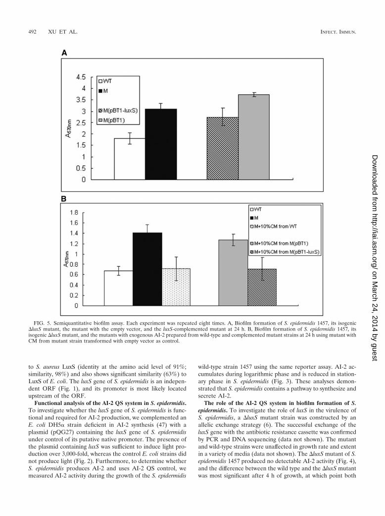
FIG. 5. Semiquantitative biofilm assay. Each experiment was repeated eight times. A, Biofilm formation of S. epidermidis 1457, its isogenic�luxS mutant, the mutant with the empty vector, and the luxS-complemented mutant at 24 h. B, Biofilm formation of S. epidermidis 1457, itsisogenic �luxS mutant, and the mutants with exogenous AI-2 prepared from wild-type and complemented mutant strains at 24 h using mutant withCM from mutant strain transformed with empty vector as control.
492 XU ET AL. INFECT. IMMUN.
on March 24, 2014 by guest
http://iai.asm.org/
Dow
nloaded from

strains entered logarithmic growth phase (optical density,1.2). After complementation of the mutant with plasmidpQG27, AI-2 activity was restored to almost the wild-type level(Fig. 4). These analyses demonstrated that luxS is responsiblefor AI-2 production in S. epidermidis.
Biofilm formation is considered the main virulence factor ofS. epidermidis. To investigate whether luxS influences biofilmformation in S. epidermidis, we first performed semiquantita-tive biofilm assays using microtiter plates. The �luxS mutant ofS. epidermidis showed significantly (P � 0.0001, two-tailed ttest) increased biomass (1.7 times) compared to that of thewild-type strain (Fig. 5A). Biofilm formation of the comple-mented strain was reduced compared to the �luxS mutantstrain (Fig. 5A).
In addition, we used SEM to detect any quantitative orqualitative alteration in the biofilm formation of wild-type and�luxS mutant strains. SEM analysis showed that the �luxSmutant strain generated a more compact and thicker biofilmthan that generated by the wild-type strain (Fig. 6).
To examine whether the increased biofilm formation of theS. epidermidis �luxS mutant strain was due to a genuine AI-2signaling event or a mere result of metabolic changes accom-panied by the inactivation of the luxS gene, we incubated the�luxS mutant strain with exogenous AI-2. To that end, we usedCM prepared from exponential-growth-phase cultures of S.epidermidis 1457, the luxS-complemented mutant strain, andthe mutant strain with the empty vector as control. CM wasmixed with �luxS mutant cells grown overnight, which werethen incubated for 24 h to test for biofilm formation. The �luxSmutant strain showed reduced biofilm formation when 10%CM was added (Fig. 5B) while no growth inhibition was detected(data not shown). These data indicate that the luxS gene of S.epidermidis is involved in repressing biofilm formation througha cell-cell signaling mechanism based on AI-2 secretion.
Secretion of the exopolysaccharide PIA is a major factor thatdetermines biofilm formation in S. epidermidis and other bac-teria (18, 30, 31). To test whether the altered biofilm formationthat we observed in the �luxS mutant strain was due to achange in the transcription of the ica operon responsible for
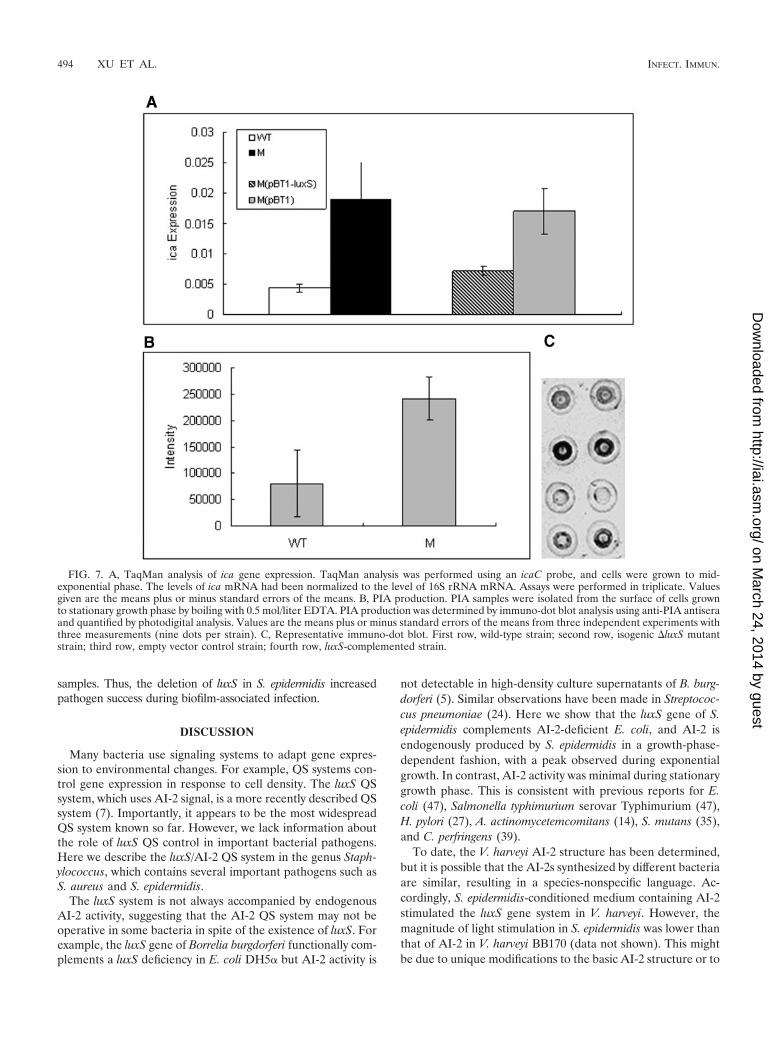
PIA production (21), we analyzed transcription of the icaCgene by quantitative real-time PCR. Expression of icaC duringmid-exponential growth phase was 4.4 times higher (P � 0.01,two-tailed t test) in the �luxS mutant strain than in the wild-type strain (Fig. 7A). The luxS-complemented mutant strainshowed an icaC expression level significantly lower than that ofthe control strain with the empty cloning vector (P � 0.01,two-tailed t test) and comparable to that of the wild-type strain.This indicates that luxS negatively regulates the expression ofthe ica gene at the transcriptional level. To determine whetherluxS regulates the synthesis of PIA, we extracted PIA fromstationary-phase cultures of S. epidermidis 1457, the �luxS mu-tant strain, and the complemented and control strains anddetermined the amount of PIA by use of immuno-dot blotanalysis. PIA production was about three times higher in the�luxS mutant strain than in the wild-type strain (P � 0.03,two-tailed t test). Thus, luxS/AI-2-dependent QS regulates theexpression of PIA. By visual comparison, PIA production wasalso decreased in the luxS-complemented strain compared tothat produced by the mutant strain with the empty vector.Photodigital analysis of this experiment was, however, not pos-sible due to uneven distribution of the reaction on the spotteddots (data not shown).
The role of the AI-2 QS system in the virulence of S. epider-midis. To determine whether luxS has an effect on the patho-genicity of S. epidermidis during biofilm-associated infection,we used a rat intravascular CVC-associated infection model.Twenty-four hours after insertion of Silastic lumen-within-lu-men catheters into the necks of the rats, 4 105 CFU bacteriawere injected into the catheters. Bacteria in blood, the tissuesurrounding the catheters, and other organs were counted aftersacrificing the rats on day 8 after infection. As shown in Table 3,the �luxS mutant had a higher capacity to cause CVC infection (P� 0.006, two-tailed t test) and also showed more significant bac-teremia and a greater burden of metastatic disease in liver (P �0.05, Wilcoxin test). The bacterial counts in other tissues (kidneyand heart) did not differ significantly between the two strains, butthe �luxS mutant strain was found more frequently in tissue
FIG. 6. Scanning electron micrographs of S. epidermidis 1457 and its �luxS mutant. Twenty-four-hour biofilms were grown on hydroxyapatitedisks that were deposited in 24-well cell culture clusters in TSB with 0.5% (wt/vol) glucose. Images shown are at a magnification of 3,000.
VOL. 74, 2006 EFFECT OF LuxS ON BIOFILM FORMATION IN S. EPIDERMIDIS 493
on March 24, 2014 by guest
http://iai.asm.org/
Dow
nloaded from
samples. Thus, the deletion of luxS in S. epidermidis increasedpathogen success during biofilm-associated infection.
DISCUSSION
Many bacteria use signaling systems to adapt gene expres-sion to environmental changes. For example, QS systems con-trol gene expression in response to cell density. The luxS QSsystem, which uses AI-2 signal, is a more recently described QSsystem (7). Importantly, it appears to be the most widespreadQS system known so far. However, we lack information aboutthe role of luxS QS control in important bacterial pathogens.Here we describe the luxS/AI-2 QS system in the genus Staph-ylococcus, which contains several important pathogens such asS. aureus and S. epidermidis.
The luxS system is not always accompanied by endogenousAI-2 activity, suggesting that the AI-2 QS system may not beoperative in some bacteria in spite of the existence of luxS. Forexample, the luxS gene of Borrelia burgdorferi functionally com-plements a luxS deficiency in E. coli DH5� but AI-2 activity is
not detectable in high-density culture supernatants of B. burg-dorferi (5). Similar observations have been made in Streptococ-cus pneumoniae (24). Here we show that the luxS gene of S.epidermidis complements AI-2-deficient E. coli, and AI-2 isendogenously produced by S. epidermidis in a growth-phase-dependent fashion, with a peak observed during exponentialgrowth. In contrast, AI-2 activity was minimal during stationarygrowth phase. This is consistent with previous reports for E.coli (47), Salmonella typhimurium serovar Typhimurium (47),H. pylori (27), A. actinomycetemcomitans (14), S. mutans (35),and C. perfringens (39).
To date, the V. harveyi AI-2 structure has been determined,but it is possible that the AI-2s synthesized by different bacteriaare similar, resulting in a species-nonspecific language. Ac-cordingly, S. epidermidis-conditioned medium containing AI-2stimulated the luxS gene system in V. harveyi. However, themagnitude of light stimulation in S. epidermidis was lower thanthat of AI-2 in V. harveyi BB170 (data not shown). This mightbe due to unique modifications to the basic AI-2 structure or to
FIG. 7. A, TaqMan analysis of ica gene expression. TaqMan analysis was performed using an icaC probe, and cells were grown to mid-exponential phase. The levels of ica mRNA had been normalized to the level of 16S rRNA mRNA. Assays were performed in triplicate. Valuesgiven are the means plus or minus standard errors of the means. B, PIA production. PIA samples were isolated from the surface of cells grownto stationary growth phase by boiling with 0.5 mol/liter EDTA. PIA production was determined by immuno-dot blot analysis using anti-PIA antiseraand quantified by photodigital analysis. Values are the means plus or minus standard errors of the means from three independent experiments withthree measurements (nine dots per strain). C, Representative immuno-dot blot. First row, wild-type strain; second row, isogenic �luxS mutantstrain; third row, empty vector control strain; fourth row, luxS-complemented strain.
494 XU ET AL. INFECT. IMMUN.
on March 24, 2014 by guest
http://iai.asm.org/
Dow
nloaded from

a lower production level of AI-2 in S. epidermidis under thegrowth conditions that were used. In fact, interspecies signalssecreted by Salmonella serovar Typhimurium have recentlybeen demonstrated to be a distinct form of the AI-2 signal,although they are both derived from the same precursor (37).
Inactivation of the luxS gene of S. epidermidis did not resultin noticeable changes in growth patterns compared with thoseof the wild-type strain in different media. This finding indicatesthat luxS has no significant effect on basic cellular metabolicprocesses required for growth of S. epidermidis in vitro. This isin contrast to some other bacteria in which luxS had an effecton growth (23, 29).
Our data showed that biofilm formation, the main virulencemechanism of S. epidermidis, was considerably enhanced in the�luxS mutant strain. This repressive effect on biofilm forma-tion by luxS in vitro was also observed in S. mutans (35) and H.pylori (9), while the �luxS mutant of Salmonella enterica sero-var Typhimurium was unable to develop a complete biofilm(42). Thus, luxS has contrasting effects on biofilm formation indifferent strains. A repressive effect on biofilm formation inStaphylococcus has also been described for the only otherknown QS system of Staphylococcus, agr, suggesting a commonscheme of QS-dependent repression of the biofilm mode ofgrowth in this genus. In contrast to agr, whose impact onbiofilm formation is most likely via regulation of the produc-tion of the autolysin AtlE and the detergent-like phenol-solu-ble modulin peptides and which does not impact PIA produc-tion, luxS of S. epidermidis regulates transcription of the icagenes and production of PIA. This impact on the major biofilmexopolysaccharide PIA is most likely the cause for the differentdegree of biofilm formation of the �luxS mutant.
Decreased virulence has been seen in �luxS mutants ofseveral pathogenic bacteria. A Serratia marcescens 274 �luxSmutant exhibits modestly attenuated virulence in a Caenorhab-ditis elegans model (10), a Vibrio vulnificus �luxS mutant has asignificantly delayed time required to kill mice (25), and aNeisseria meningitidis �luxS mutant is attenuated for bactere-mic infection in an infant rat model (55). In contrast, the S.epidermidis �luxS mutant, similar to the agr mutant of S. epi-dermidis, shows increased virulence in a model of catheter-associated infection. Most likely, the increased virulence maybe partly attributed to the increased synthesis of PIA andmore-intense biofilm formation.
Absent in humans, the LuxS enzyme is an attractive target
for the development of novel therapeutic agents for bacterialinfection. Inhibitors of LuxS have been synthesized (1). How-ever, our present data, showing the increased capacity of bio-film formation of the �luxS mutant of S. epidermidis in vitroand increased virulence of the mutant in a rat model, suggestthat such inhibitors are not suited for the treatment of biofilm-associated S. epidermidis infection.
In conclusion, we show that luxS in S. epidermidis is func-tional and luxS-dependent gene regulation represses biofilmformation in vitro and pathogen success during biofilm-asso-ciated infection. Although by regulating different biofilm fac-tors, the two QS systems of Staphylococcus, agr and luxS, thushave very similar effects on the biofilm mode of growth.
ACKNOWLEDGMENTS
We thank Bonnie Bassler for the gift of V. harveyi strain BB170,Friedrich Gotz for providing S. aureus strain RN4220, and ReinholdBruckner for kindly providing plasmid pBT1. We thank Steven D.Goodman for modification of the manuscript.
This work was supported by the Chinese National Natural ScienceFoundation Grant (30570087) and 211 Project Grant—FunctionalGenomics of Important Pathogenic Microorganisms. This work wasalso supported by the China National 973 Program (2002CB512804).
REFERENCES
1. Alfaro, J. F., T. Zhang, D. P. Wynn, E. L. Karschner, and Z. S. Zhou. 2004.Synthesis of LuxS inhibitors targeting bacterial cell-cell communication. Org.Lett. 6:3043–3046.
2. Augustin, J., and F. Gotz. 1990. Transformation of Staphylococcus epider-midis and other staphylococcal species with plasmid DNA by electropora-tion. FEMS Microbiol. Lett. 54:203–207.
3. Bassler, B. L., E. P. Greenberg, and A. M. Stevens. 1997. Cross-speciesinduction of luminescence in the quorum-sensing bacterium Vibrio harveyi. J.Bacteriol. 179:4043–4045.
4. Bassler, B. L., M. Wright, and M. R. Silverman. 1994. Multiple signallingsystems controlling expression of luminescence in Vibrio harveyi: sequenceand function of genes encoding a second sensory pathway. Mol. Microbiol.13:273–286.
5. Blevins, J. S., A. T. Revel, M. J. Caimano, X. F. Yang, J. A. Richardson, K. E.Hagman, and M. V. Norgard. 2004. The luxS gene is not required for Borreliaburgdorferi tick colonization, transmission to a mammalian host, or inductionof disease. Infect. Immun. 72:4864–4867.
6. Bruckner, R. 1997. Gene replacement in Staphylococcus carnosus andStaphylococcus xylosus. FEMS Microbiol. Lett. 151:1–8.
7. Chen, X., S. Schauder, N. Potier, A. Van Dorsselaer, I. Pelczer, B. L. Bassler,and F. M. Hughson. 2002. Structural identification of a bacterial quorum-sensing signal containing boron. Nature 415:545–549.
8. Christensen, G. D., W. A. Simpson, J. J. Younger, L. M. Baddour, F. F.Barrett, D. M. Melton, and E. H. Beachey. 1985. Adherence of coagulase-negative staphylococci to plastic tissue culture plates: a quantitative modelfor the adherence of staphylococci to medical devices. J. Clin. Microbiol.22:996–1006.
TABLE 3. Metastatic disease caused by S. epidermidis 1457 and its isogenic �luxS mutantin the rat CVC-associated infection modelb
Organ
S. epidermidis 1457 (n � 15) S. epidermidis 1457 �luxS mutant (n � 15)
P valueNo. ratsinfected/total
Median (range)CFU/g tissue
No. ratsinfected/total
Median (range)CFU/g tissue
Livera 4/15 0 (0–66,000) 11/15 333 (0–2,216,667) 0.0164Kidneya 5/15 0 (0–308,000) 9/15 2,000 (0–3,673,333) 0.1002Hearta 3/15 0 (0–66,667) 6/15 0 (0–1,140,000) 0.2195Blooda 3/10 0 (0–130) 8/11 460 (0–10,980) 0.0154Tubea 2,050 (1,750–3,050) 4,525 (2,050–7,600) 0.1084
Infection rateb 27.5 2.8% 61.5 7.8% 0.0064
a The difference in results among liver, kidney, heart, blood, and tube between the two strains was assessed by a Wilcoxon test.b The overall infection rate is defined as recovery of S. epidermidis from the blood, catheter, or organs at the time of sacrifice. The P value for the infection rates
of the mutant versus the wild type was 0.0064. The difference of infection rate between the two strains was assessed by a two-tailed t test.
VOL. 74, 2006 EFFECT OF LuxS ON BIOFILM FORMATION IN S. EPIDERMIDIS 495
on March 24, 2014 by guest
http://iai.asm.org/
Dow
nloaded from
9. Cole, S. P., J. Harwood, R. Lee, R. She, and D. G. Guiney. 2004. Character-ization of monospecies biofilm formation by Helicobacter pylori. J. Bacteriol.186:3124–3132.
10. Coulthurst, S. J., C. L. Kurz, and G. P. Salmond. 2004. luxS mutants ofSerratia defective in autoinducer-2-dependent ‘quorum sensing’ show strain-dependent impacts on virulence and production of carbapenem and prodi-giosin. Microbiology 150:1901–1910.
11. Dobinsky, S., K. Kiel, H. Rohde, K. Bartscht, J. K. Knobloch, M. A. Horst-kotte, and D. Mack. 2003. Glucose-related dissociation between icaADBCtranscription and biofilm expression by Staphylococcus epidermidis: evidencefor an additional factor required for polysaccharide intercellular adhesinsynthesis. J. Bacteriol. 185:2879–2886.
12. Duan, K., C. Dammel, J. Stein, H. Rabin, and M. G. Surette. 2003. Modu-lation of Pseudomonas aeruginosa gene expression by host microflorathrough interspecies communication. Mol. Microbiol. 50:1477–1491.
13. Flamm, R. K., D. J. Hinrichs, and M. F. Thomashow. 1984. Introduction ofpAM�1 into Listeria monocytogenes by conjugation and homology betweennative L. monocytogenes plasmids. Infect. Immun. 44:157–161.
14. Fong, K. P., W. O. Chung, R. J. Lamont, and D. R. Demuth. 2001. Intra- andinterspecies regulation of gene expression by Actinobacillus actinomycetem-comitans LuxS. Infect. Immun. 69:7625–7634.
15. Fong, K. P., L. Gao, and D. R. Demuth. 2003. luxS and arcB control aerobicgrowth of Actinobacillus actinomycetemcomitans under iron limitation. In-fect. Immun. 71:298–308.
16. Fuqua, W. C., S. C. Winans, and E. P. Greenberg. 1994. Quorum sensing inbacteria: the LuxR-LuxI family of cell density-responsive transcriptionalregulators. J. Bacteriol. 176:269–275.
17. Gerke, C., A. Kraft, R. Sussmuth, O. Schweitzer, and F. Gotz. 1998. Char-acterization of the N-acetylglucosaminyltransferase activity involved in thebiosynthesis of the Staphylococcus epidermidis polysaccharide intercellularadhesin. J. Biol. Chem. 273:18586–18593.
18. Gotz, F. 2002. Staphylococcus and biofilms. Mol. Microbiol. 43:1367–1378.19. Hanahan, D. 1983. Studies on transformation of Escherichia coli with plas-
mids. J. Mol. Biol. 166:557–580.20. Heilmann, C., M. Hussain, G. Peters, and F. Gotz. 1997. Evidence for
autolysin-mediated primary attachment of Staphylococcus epidermidis to apolystyrene surface. Mol. Microbiol. 24:1013–1024.
21. Heilmann, C., O. Schweitzer, C. Gerke, N. Vanittanakom, D. Mack, and F.Gotz. 1996. Molecular basis of intercellular adhesion in the biofilm-formingStaphylococcus epidermidis. Mol. Microbiol. 20:1083–1091.
22. Henke, J. M., and B. L. Bassler. 2004. Three parallel quorum-sensing sys-tems regulate gene expression in Vibrio harveyi. J. Bacteriol. 186:6902–6914.
23. Jones, M. B., and M. J. Blaser. 2003. Detection of a luxS-signaling moleculein Bacillus anthracis. Infect. Immun. 71:3914–3919.
24. Joyce, E. A., A. Kawale, S. Censini, C. C. Kim, A. Covacci, and S. Falkow.2004. LuxS is required for persistent pneumococcal carriage and expressionof virulence and biosynthesis genes. Infect. Immun. 72:2964–2975.
25. Kim, S. Y., S. E. Lee, Y. R. Kim, C. M. Kim, P. Y. Ryu, H. E. Choy, S. S.Chung, and J. H. Rhee. 2003. Regulation of Vibrio vulnificus virulence by theLuxS quorum-sensing system. Mol. Microbiol. 48:1647–1664.
26. Kozitskaya, S., S. H. Cho, K. Dietrich, R. Marre, K. Naber, and W. Ziebuhr.2004. The bacterial insertion sequence element IS256 occurs preferentially innosocomial Staphylococcus epidermidis isolates: association with biofilm for-mation and resistance to aminoglycosides. Infect. Immun. 72:1210–1215.
27. Loh, J. T., M. H. Forsyth, and T. L. Cover. 2004. Growth phase regulation offlaA expression in Helicobacter pylori is luxS dependent. Infect. Immun.72:5506–5510.
28. Lupp, C., and E. G. Ruby. 2004. Vibrio fischeri LuxS and AinS: comparativestudy of two signal synthases. J. Bacteriol. 186:3873–3881.
29. Lyon, W. R., J. C. Madden, J. C. Levin, J. L. Stein, and M. G. Caparon. 2001.Mutation of luxS affects growth and virulence factor expression in Strepto-coccus pyogenes. Mol. Microbiol. 42:145–157.
30. Mack, D., W. Fischer, A. Krokotsch, K. Leopold, R. Hartmann, H. Egge, andR. Laufs. 1996. The intercellular adhesin involved in biofilm accumulation ofStaphylococcus epidermidis is a linear �-1,6-linked glucosaminoglycan: puri-fication and structural analysis. J. Bacteriol. 178:175–183.
31. Mack, D., M. Haeder, N. Siemssen, and R. Laufs. 1996. Association ofbiofilm production of coagulase-negative staphylococci with expression of aspecific polysaccharide intercellular adhesin. J. Infect. Dis. 174:881–884.
32. Mack, D., H. Rohde, S. Dobinsky, J. Riedewald, M. Nedelmann, J. K. Knob-loch, H. A. Elsner, and H. H. Feucht. 2000. Identification of three essentialregulatory gene loci governing expression of Staphylococcus epidermidis poly-saccharide intercellular adhesin and biofilm formation. Infect. Immun. 68:3799–3807.
33. Mack, D., N. Siemssen, and R. Laufs. 1992. Parallel induction by glucose ofadherence and a polysaccharide antigen specific for plastic-adherent Staph-ylococcus epidermidis: evidence for functional relation to intercellular adhe-sion. Infect. Immun. 60:2048–2057.
34. McNab, R., S. K. Ford, A. El-Sabaeny, B. Barbieri, G. S. Cook, and R. J.Lamont. 2003. LuxS-based signaling in Streptococcus gordonii: autoinducer 2controls carbohydrate metabolism and biofilm formation with Porphyromo-nas gingivalis. J. Bacteriol. 185:274–284.
35. Merritt, J., F. Qi, S. D. Goodman, M. H. Anderson, and W. Shi. 2003.Mutation of luxS affects biofilm formation in Streptococcus mutans. Infect.Immun. 71:1972–1979.
36. Miller, M. B., and B. L. Bassler. 2001. Quorum sensing in bacteria. Annu.Rev. Microbiol. 55:165–199.
37. Miller, S. T., K. B. Xavier, S. R. Campagna, M. E. Taga, M. F. Semmelhack,B. L. Bassler, and F. M. Hughson. 2004. Salmonella typhimurium recognizesa chemically distinct form of the bacterial quorum-sensing signal AI-2. Mol.Cell 15:677–687.
38. Novick, R. P. 2003. Autoinduction and signal transduction in the regulationof staphylococcal virulence. Mol. Microbiol. 48:1429–1449.
39. Ohtani, K., H. Hayashi, and T. Shimizu. 2002. The luxS gene is involved incell-cell signalling for toxin production in Clostridium perfringens. Mol.Microbiol. 44:171–179.
40. Otto, M., R. Sussmuth, G. Jung, and F. Gotz. 1998. Structure of the pher-omone peptide of the Staphylococcus epidermidis agr system. FEBS Lett.424:89–94.
41. Pei, L., and J. I. Flock. 2001. Functional study of antibodies against afibrogenin-binding protein in Staphylococcus epidermidis adherence to poly-ethylene catheters. J. Infect. Dis. 184:52–55.
42. Prouty, A. M., W. H. Schwesinger, and J. S. Gunn. 2002. Biofilm formationand interaction with the surfaces of gallstones by Salmonella spp. Infect.Immun. 70:2640–2649.
43. Rupp, M. E., J. S. Ulphani, P. D. Fey, and D. Mack. 1999. Characterizationof Staphylococcus epidermidis polysaccharide intercellular adhesin/hemag-glutinin in the pathogenesis of intravascular catheter-associated infection ina rat model. Infect. Immun. 67:2656–2659.
44. Schneider, K. B., T. M. Palmer, and A. D. Grossman. 2002. Characterizationof comQ and comX, two genes required for production of ComX pheromonein Bacillus subtilis. J. Bacteriol. 184:410–419.
45. Sperandio, V., A. G. Torres, J. A. Giron, and J. B. Kaper. 2001. Quorumsensing is a global regulatory mechanism in enterohemorrhagic Escherichiacoli O157:H7. J. Bacteriol. 183:5187–5197.
46. Stroeher, U. H., A. W. Paton, A. D. Ogunniyi, and J. C. Paton. 2003. Muta-tion of luxS of Streptococcus pneumoniae affects virulence in a mouse model.Infect. Immun. 71:3206–3212.
47. Surette, M. G., and B. L. Bassler. 1998. Quorum sensing in Escherichia coliand Salmonella typhimurium. Proc. Natl. Acad. Sci. USA. 95:7046–7050.
48. Surette, M. G., M. B. Miller, and B. L. Bassler. 1999. Quorum sensing inEscherichia coli, Salmonella typhimurium, and Vibrio harveyi: a new familyof genes responsible for autoinducer production. Proc. Natl. Acad. Sci. USA96:1639–1644.
49. Vuong, C., C. Gerke, G. A. Somerville, E. R. Fischer, and M. Otto. 2003.Quorum-sensing control of biofilm factors in Staphylococcus epidermidis.J. Infect. Dis. 188:706–718.
50. Vuong, C., S. Kocianova, Y. Yao, A. B. Carmody, and M. Otto. 2004. In-creased colonization of indwelling medical devices by quorum-sensing mu-tants of Staphylococcus epidermidis in vivo. J. Infect. Dis. 190:1498–1505.
51. Vuong, C., and M. Otto. 2002. Staphylococcus epidermidis infections. Mi-crobes Infect. 4:481–489.
52. Vuong, C., H. L. Saenz, F. Gotz, and M. Otto. 2000. Impact of the agrquorum-sensing system on adherence to polystyrene in Staphylococcus au-reus. J. Infect. Dis. 182:1688–1693.
53. Winans, S. C., and B. L. Bassler. 2002. Mob psychology. J. Bacteriol. 184:873–883.
54. Winzer, K., K. R. Hardie, N. Burgess, N. Doherty, D. Kirke, M. T. Holden,R. Linforth, K. A. Cornell, A. J. Taylor, P. J. Hill, and P. Williams. 2002.LuxS: its role in central metabolism and the in vitro synthesis of 4-hydroxy-5-methyl-3(2H)-furanone. Microbiology 148:909–922.
55. Winzer, K., Y. H. Sun, A. Green, M. Delory, D. Blackley, K. R. Hardie, T. J.Baldwin, and C. M. Tang. 2002. Role of Neisseria meningitidis luxS in cell-to-cell signaling and bacteremic infection. Infect. Immun. 70:2245–2248.
56. Xavier, K. B., and B. L. Bassler. 2003. LuxS quorum sensing: more than justa numbers game. Curr. Opin. Microbiol. 6:191–197.
57. Yao, Y., D. E. Sturdevant, and M. Otto. 2005. Genomewide analysis of geneexpression in Staphylococcus epidermidis biofilms: insights into the patho-physiology of S. epidermidis biofilms and the role of phenol-soluble modu-lins in formation of biofilms. J. Infect. Dis. 191:289–298.
58. Zhang, Y. Q., S. X. Ren, H. L. Li, Y. X. Wang, G. Fu, J. Yang, Z. Q. Qin, Y. G.Miao, W. Y. Wang, R. S. Chen, Y. Shen, Z. Chen, Z. H. Yuan, G. P. Zhao, D.Qu, A. Danchin, and Y. M. Wen. 2003. Genome-based analysis of virulencegenes in a non-biofilm-forming Staphylococcus epidermidis strain (ATCC12228). Mol. Microbiol. 49:1577–1593.
Editor: F. C. Fang
496 XU ET AL. INFECT. IMMUN.
on March 24, 2014 by guest
http://iai.asm.org/
Dow
nloaded from