PlcRa, a new quorum-sensing regulator from Bacillus cereus ...
18
HAL Id: hal-01019491 https://hal.archives-ouvertes.fr/hal-01019491 Submitted on 28 May 2020 HAL is a multi-disciplinary open access archive for the deposit and dissemination of sci- entific research documents, whether they are pub- lished or not. The documents may come from teaching and research institutions in France or abroad, or from public or private research centers. L’archive ouverte pluridisciplinaire HAL, est destinée au dépôt et à la diffusion de documents scientifiques de niveau recherche, publiés ou non, émanant des établissements d’enseignement et de recherche français ou étrangers, des laboratoires publics ou privés. PlcRa, a new quorum-sensing regulator from Bacillus cereus, plays a role in oxidative stress responses and cysteine metabolism in stationary phase Eugenie Huillet, Marcel H. Tempelaars, Gwenaëlle André-Leroux, Wanapaisan Pagakrong, Ludovic Bridoux, Samira Makhzami, Watanalai Panbangred, Isabelle Martin-Verstraete, Tjakko Abee, Didier Lereclus To cite this version: Eugenie Huillet, Marcel H. Tempelaars, Gwenaëlle André-Leroux, Wanapaisan Pagakrong, Ludovic Bridoux, et al.. PlcRa, a new quorum-sensing regulator from Bacillus cereus, plays a role in oxidative stress responses and cysteine metabolism in stationary phase. PLoS ONE, Public Library of Science, 2012, 7, online (12), Non paginé. 10.1371/journal.pone.0051047. hal-01019491
-
Upload
khangminh22 -
Category
Documents
-
view
1 -
download
0
Transcript of PlcRa, a new quorum-sensing regulator from Bacillus cereus ...
HAL Id: hal-01019491https://hal.archives-ouvertes.fr/hal-01019491
Submitted on 28 May 2020
HAL is a multi-disciplinary open accessarchive for the deposit and dissemination of sci-entific research documents, whether they are pub-lished or not. The documents may come fromteaching and research institutions in France orabroad, or from public or private research centers.
L’archive ouverte pluridisciplinaire HAL, estdestinée au dépôt et à la diffusion de documentsscientifiques de niveau recherche, publiés ou non,émanant des établissements d’enseignement et derecherche français ou étrangers, des laboratoirespublics ou privés.
PlcRa, a new quorum-sensing regulator from Bacilluscereus, plays a role in oxidative stress responses and
cysteine metabolism in stationary phaseEugenie Huillet, Marcel H. Tempelaars, Gwenaëlle André-Leroux, Wanapaisan
Pagakrong, Ludovic Bridoux, Samira Makhzami, Watanalai Panbangred,Isabelle Martin-Verstraete, Tjakko Abee, Didier Lereclus
To cite this version:Eugenie Huillet, Marcel H. Tempelaars, Gwenaëlle André-Leroux, Wanapaisan Pagakrong, LudovicBridoux, et al.. PlcRa, a new quorum-sensing regulator from Bacillus cereus, plays a role in oxidativestress responses and cysteine metabolism in stationary phase. PLoS ONE, Public Library of Science,2012, 7, online (12), Non paginé. �10.1371/journal.pone.0051047�. �hal-01019491�

PlcRa, a New Quorum-Sensing Regulator from Bacilluscereus, Plays a Role in Oxidative Stress Responses andCysteine Metabolism in Stationary PhaseEugenie Huillet1*, Marcel H. Tempelaars2, Gwenaelle Andre-Leroux3, Pagakrong Wanapaisan1,4,
Ludovic Bridoux1, Samira Makhzami5, Watanalai Panbangred4, Isabelle Martin-Verstraete6,7,
Tjakko Abee2, Didier Lereclus1*
1 INRA, UMR1319 Micalis, Genetique microbienne et Environnement, Guyancourt, France, 2Wageningen University, Laboratory of Food Microbiology, Wageningen, The
Netherlands, 3 Institut Pasteur, Microbiologie Structurale, CNRS, UMR 3528, Paris, France, 4Mahidol University, Department of Biotechnology, Faculty of Science, Bangkok,
Thailand, 5 INRA, UMR GABI, ICE, Jouy-en-Josas, France, 6 Institut Pasteur, Laboratoire de Pathogenese des Bacteries Anaerobies, Paris, France, 7Univ. Paris Diderot,
Sorbonne Paris Cite, Cellule Pasteur, Paris, France
Abstract
We characterized a new quorum-sensing regulator, PlcRa, which is present in various members of the B. cereus group andidentified a signaling heptapeptide for PlcRa activity: PapRa7. We demonstrated that PlcRa is a 3D structural paralog of PlcRusing sequence analysis and homology modeling. A comparison of the transcriptomes at the onset of stationary phase ofa DplcRa mutant and the wild-type B. cereus ATCC 14579 strain showed that 68 genes were upregulated and 49 genes weredownregulated in the DplcRa mutant strain (.3-fold change). Genes involved in the cysteine metabolism (putative CymRregulon) were downregulated in the DplcRamutant strain. We focused on the gene with the largest difference in expressionlevel between the two conditions, which encoded -AbrB2- a new regulator of the AbrB family. We demonstrated thatpurified PlcRa bound specifically to the abrB2 promoter in the presence of synthetic PapRa7, in an electrophoretic mobilityshift assay. We further showed that the AbrB2 regulator controlled the expression of the yrrT operon involved in methionineto cysteine conversion. We found that the DplcRa mutant strain was more sensitive to hydrogen peroxide- and disulfide-induced stresses than the wild type. When cystine was added to the culture of the DplcRa mutant, challenged withhydrogen peroxide, growth inhibition was abolished. In conclusion, we identified a new RNPP transcriptional regulator in B.cereus that activated the oxidative stress response and cysteine metabolism in transition state cells.
Citation: Huillet E, Tempelaars MH, Andre-Leroux G, Wanapaisan P, Bridoux L, et al. (2012) PlcRa, a New Quorum-Sensing Regulator from Bacillus cereus, Playsa Role in Oxidative Stress Responses and Cysteine Metabolism in Stationary Phase. PLoS ONE 7(12): e51047. doi:10.1371/journal.pone.0051047
Editor: Adam Driks, Loyola University Medical Center, United States of America
Received July 19, 2012; Accepted October 29, 2012; Published December 11, 2012
Copyright: � 2012 Huillet et al. This is an open-access article distributed under the terms of the Creative Commons Attribution License, which permitsunrestricted use, distribution, and reproduction in any medium, provided the original author and source are credited.
Funding: This work was supported by the French National Research Agency (Cell.com project; NuANR-09-Blan-0253) and by INRA (MICA department). Thefunders had no role in study design, data collection and analysis, decision to publish, or preparation of the manuscript.
Competing Interests: The authors have declared that no competing interests exist.
* E-mail: [email protected] (EH); [email protected] (DL)
Introduction
The Bacillus cereus group includes well known spore-forming
pathogens of mammals (B. anthracis and B. cereus) and insects (B.
thuringiensis). B. cereus is frequently associated with food-borne
infections causing gastroenteritis [1]. The capacity of B. cereus to
sporulate allows this bacterium to resist the usual cleaning
procedures used in the food industry, resulting in the presence of
B. cereus in many raw and processed foods, such as rice, spices,
milk, vegetables, meats and various desserts [1].
At the end of the vegetative growth phase, bacterial cells face
a number of challenges, including a decrease in the nutrient
content of their environment. Under these conditions, spore-
forming bacteria may initiate sporulation, producing spores that
can survive in unfavorable environmental conditions [2]. Bacteria
make use of various strategies to cope with environmental changes
during the transition between the vegetative and sporulation
phases [3]. The production of degradative enzymes and antimi-
crobial compounds responsible for the lysis of targeted cells
provides Bacillus subtilis with new nutrients [3]. In parallel, a general
stress response may be activated during the transition phase, due
to the accumulation of oxidative products and changes in the pH
of the medium [4]. Cellular responses may be controlled by a range
of sensors and activators, including two-component systems,
quorum-sensing systems and other transcriptional regulators [3,5].
Quorum sensing regulation appears to be a consequence of
interbacterial communication by which bacteria of one or even
different species sense about their current population density and
react in a defined way to that information. These communication
systems are based on the secretion and recognition of cell-cell
signalling molecules, termed autoinducers [6]. The PlcR/PapR
quorum sensing system is activated during the transition phase in
most members of the B. cereus group [7]. This system controls the
expression of genes encoding exported virulence factors, including
degradative enzymes, enterotoxins and hemolysins [8]. PlcR is
activated by binding to PapR, a signaling peptide produced as
a propeptide under the control of PlcR. PapR undergoes
extracellular processing, to generate an active heptapeptide [9],
which is then re-imported into the bacterial cell via the
oligopeptide permease system, OppABCDF [10]. Within the cell,
PLOS ONE | www.plosone.org 1 December 2012 | Volume 7 | Issue 12 | e51047

PapR interacts with PlcR and the resulting complex binds PlcR
target sites on DNA [11], resulting in the activation of the PlcR
regulon, which contains 48 genes [7].
The structure of PlcR has been resolved. This molecule has
a unique folding pattern, due to the presence of an HTH DNA-
binding domain and a peptide-binding regulatory domain
composed of five tetratricopeptide repeats (TPR) [12]. A TPR is
a structural 34-amino acid repeat motif present in various
eukaryotic and prokaryotic proteins. There may be from 3 to
more than 16 tandem repeats [13]. Determination of the structure
of PlcR resulted in the identification of a new family of central
regulatory quorum sensors (the RNPP family) found exclusively in
Gram-positive bacteria with a low G+C content [12]. These
quorum sensors include NprR from B. cereus [14], PrgX from
Enteroccocus faecalis [15] and RAP phosphatases from B. subtilis
[16,17]. All these RNPP regulators are activated through a secreted
signaling peptide that interacts with the TPR activation domain.
In this work, we characterized PlcRa, a novel quorum-sensing
type regulator of the RNPP family and identified PapRa7, a new
signaling heptapeptide. We constructed a DplcRa mutant strain
from the B. cereus ATCC 14579 type strain [18] and identified
PlcRa-controlled genes through a whole-genome microarray
approach at the onset of stationary phase. We purified PlcRa
protein and assessed the DNA binding activity of PlcRa using
electrophoretic mobility shift assay. By combining in silico
structural analysis, genetic and biochemical methods, the PlcRa
activity in relation with the presence of PapRa7 was described.
Results
PlcRa is a 3D Structural Homolog of PlcRBLAST searches identified three PlcR paralogs encoded by the
B. cereus ATCC 14579 genome: BC0988 (PlcRa), BC1158 (PlcRb,
previously named PlcR2 in B. anthracis [19]) and BC2443 (PlcRc).
These putative regulators display about 29% sequence identity
and about 50% similarity to PlcR. The PlcRa, PlcRb and PlcRc
proteins have high levels of overall sequence identity (85%). A
small ORF, BC0989, encoding a putative peptide with a potential
signal sequence is located upstream from the plcRa gene. We
named this gene papRa. In contrast, the plcRb and plcRc genes are
not associated with such genes. As all characterized RNPP
regulators are activated by exported peptides with regulatory
functions, we decided to focus on the plcRa/papRa locus. We
describe here the analysis of plcRa in the B. cereus ATCC 14579
strain. Genome comparisons revealed genes for PlcRa (identity
.94%) in B. thuringiensis BMB171, serovar chinensis CT-43, HD-
789, HD-771 strains (4 out of 7 complete genomes) and B. cereus
B4264 and G9842 strains (3, including ATCC 14579, out of 13
complete genomes) but no such gene was present in the genome of
B. anthracis (data not shown). The 297-amino acid PlcRa protein
displays 29% identity and 51% similarity to PlcR (Figure 1A).
Given this high identity score over its entire sequence, PlcR is
a relevant template for homology modeling [20]. We therefore
constructed a homology model for PlcRa, based on the structure of
the PlcR dimer solved at a resolution of 2.6 A [12]. Each PlcR
monomer displays a unique folding pattern, with an HTH domain
at the N-terminus, followed by a linker helix connecting the HTH
domain to the 5 TPR domains and serving as an anchoring
platform for dimerization. The packing of the five TPR domains
defines, for each monomer, a pocket that binds the PapR activator
peptide [12]. The PlcRa homodimer model, composed of A and B
chains, was constructed progressively, beginning with chain A,
followed by the addition of chain B and ending with modeling of
the dimer (Figure 1B). As expected, the homology model of the
PlcRa homodimer was found to be highly helical. Each monomer
has, at its N-terminus, an HTH domain followed by a linker helix
of 27 residues (Figure 1A) that connects the HTH to the five TPRs
and anchors the two monomers together (Figure 1B). Thus, the
homology model of PlcRa constructed here is very similar to the
X-ray structure of PlcR. By analogy with PlcR [12], we suggest
that the five TPR motifs may be arranged similarly, to form
a pocket that is responsible for peptide binding. The papRa gene
encodes a 93-amino acid polypeptide, which is longer than PapR
(45 amino acids). As described for PapR, a typical Gram-positive
N-terminal signal peptide was identified for PapRa with SignalP
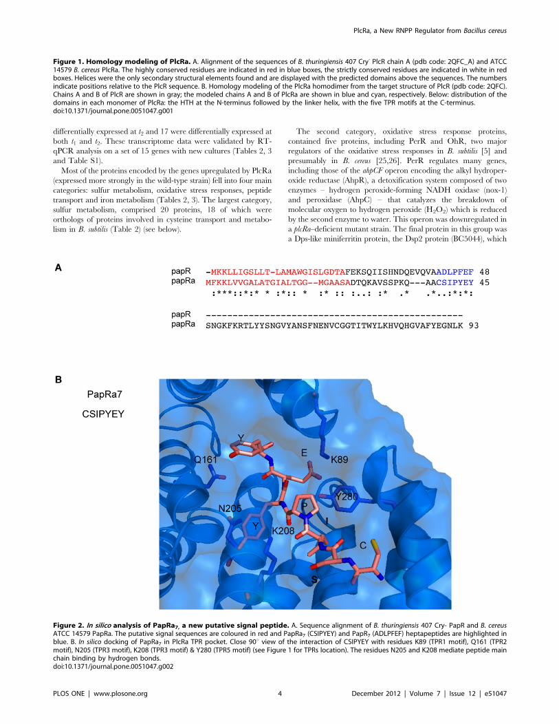
program (Figure 2A) [21]. Interestingly, alignment of PapR and
PapRa sequences showed similarity over a short segment
corresponding to the PapR C-terminus including the heptapeptide
(ADLPFEF), which is the physiological activator of PlcR
(Figure 2A) [9]. Based on this sequence alignment, the CSIPYEY
fragment -PapRa7 - was proposed as a consistent candidate for
a signaling heptapeptide. We docked CSIPYEY into the dedicated
pocket of PlcRa and minimized the energy of the complex with
CHARMm [22]. This docking procedure showed that PapRa7could fit into the PlcRa pocket formed by the five TPRs
(Figure 2B). Overall, these homology modeling and docking
analysis suggested that PlcRa is a 3D structural homolog of PlcR.For confirmation and characterization of PlcRa as a regulator, we
first conducted a genetic analysis of the plcRa gene and searched
for target genes using a comparative transcriptome analysis
approach with the DplcRa strain.
The Expression of plcRa is Activated at the Onset of theStationary Growth PhaseWe investigated the temporal regulation of plcRa gene
expression, by constructing a PplcRa’-lacZ transcriptional fusion in
the low-copy-number plasmid pHT304–18Z [23]. The PplcRa’-lacZ
fusion was introduced into the B. cereus wild-type strain and b-galactosidase activity was measured from t22 to t4 (time zero, t0,
corresponds to the onset of the stationary growth phase, and tn is
the number of hours before (–) or after time zero) during growth in
LB medium. Expression of the PplcRa’-lacZ fusion began at t21 and
increased rapidly from t0.5 to t1.5 (Figure 3A). These findings
suggest that plcRa expression is transiently activated early in the
stationary phase. We then constructed a plcRa mutant (Table 1) as
described in Material and Methods. Expression of the PplcRa’-lacZ
fusion was similar in the wild-type strain and in the DplcRa mutant
strain during growth (data not shown), indicating that the plcRa
gene is not autoregulated. The transcriptional start site identified
by 59RACE was located 46 bp upstream from the predicted start
codon of plcRa (Figure 3B). The plcRa promoter region contains
210 and 235 DNA binding regions resembling those recognized
by SigA [24].
Identification of PlcRa-controlled GenesWe characterized the regulatory role of PlcRa, by comparing
the transcriptomes of the DplcRa and wild-type B. cereus ATCC
14579 strains during early stationary phase in LB medium.
Assessments were carried out one hour after the onset of stationary
phase (t1), when plcRa expression increases, and two hours after the
onset of stationary phase (t2), when plcRa expression has reached
a plateau.
In total, 117 genes were differentially expressed with a more
than three-fold difference between the wild-type strain and the
DplcRa mutant strain (Tables 2, 3). Forty-nine genes were more
strongly expressed in the wild-type strain and 68 genes were less
strongly expressed in the wild-type strain than in the mutant. In
total, 12 genes were differentially expressed at t1, 88 were
PlcRa, a New RNPP Regulator from Bacillus cereus
PLOS ONE | www.plosone.org 2 December 2012 | Volume 7 | Issue 12 | e51047

PlcRa, a New RNPP Regulator from Bacillus cereus
PLOS ONE | www.plosone.org 3 December 2012 | Volume 7 | Issue 12 | e51047
differentially expressed at t2 and 17 were differentially expressed at
both t1 and t2. These transcriptome data were validated by RT-
qPCR analysis on a set of 15 genes with new cultures (Tables 2, 3
and Table S1).
Most of the proteins encoded by the genes upregulated by PlcRa
(expressed more strongly in the wild-type strain) fell into four main
categories: sulfur metabolism, oxidative stress responses, peptide
transport and iron metabolism (Tables 2, 3). The largest category,
sulfur metabolism, comprised 20 proteins, 18 of which were
orthologs of proteins involved in cysteine transport and metabo-
lism in B. subtilis (Table 2) (see below).
The second category, oxidative stress response proteins,
contained five proteins, including PerR and OhR, two major
regulators of the oxidative stress responses in B. subtilis [5] and
presumably in B. cereus [25,26]. PerR regulates many genes,
including those of the ahpCF operon encoding the alkyl hydroper-
oxide reductase (AhpR), a detoxification system composed of two
enzymes – hydrogen peroxide-forming NADH oxidase (nox-1)
and peroxidase (AhpC) – that catalyzes the breakdown of
molecular oxygen to hydrogen peroxide (H2O2) which is reduced
by the second enzyme to water. This operon was downregulated in
a plcRa–deficient mutant strain. The final protein in this group was
a Dps-like miniferritin protein, the Dsp2 protein (BC5044), which
Figure 1. Homology modeling of PlcRa. A. Alignment of the sequences of B. thuringiensis 407 Cry- PlcR chain A (pdb code: 2QFC_A) and ATCC14579 B. cereus PlcRa. The highly conserved residues are indicated in red in blue boxes, the strictly conserved residues are indicated in white in redboxes. Helices were the only secondary structural elements found and are displayed with the predicted domains above the sequences. The numbersindicate positions relative to the PlcR sequence. B. Homology modeling of the PlcRa homodimer from the target structure of PlcR (pdb code: 2QFC).Chains A and B of PlcR are shown in gray; the modeled chains A and B of PlcRa are shown in blue and cyan, respectively. Below: distribution of thedomains in each monomer of PlcRa: the HTH at the N-terminus followed by the linker helix, with the five TPR motifs at the C-terminus.doi:10.1371/journal.pone.0051047.g001
Figure 2. In silico analysis of PapRa7, a new putative signal peptide. A. Sequence alignment of B. thuringiensis 407 Cry- PapR and B. cereusATCC 14579 PapRa. The putative signal sequences are coloured in red and PapRa7 (CSIPYEY) and PapR7 (ADLPFEF) heptapeptides are highlighted inblue. B. In silico docking of PapRa7 in PlcRa TPR pocket. Close 90u view of the interaction of CSIPYEY with residues K89 (TPR1 motif), Q161 (TPR2motif), N205 (TPR3 motif), K208 (TPR3 motif) & Y280 (TPR5 motif) (see Figure 1 for TPRs location). The residues N205 and K208 mediate peptide mainchain binding by hydrogen bonds.doi:10.1371/journal.pone.0051047.g002
PlcRa, a New RNPP Regulator from Bacillus cereus
PLOS ONE | www.plosone.org 4 December 2012 | Volume 7 | Issue 12 | e51047

is homologous to the Dsp2 protein of B. anthracis (BA5290) that has
recently been shown to play a major role in oxidative stress
resistance [27].
The third group comprises four components of an unchar-
acterized oligopeptide permease system, Opp, which is thought to
be required for the import of small molecules into the bacterial
cell. The fourth category consists of five proteins involved in iron
transport and metabolism. In addition to these factors, PlcRa
induces the expression of genes encoding proteins of various
known or unknown functions. These genes include the gene
displaying the strongest upregulation by PlcRa one hour after
entry into stationary phase (Table 3). This gene (BC2444) encodes
a regulatory protein belonging to the AbrB family analyzed in
greater detail below.
Most of the proteins encoded by genes downregulated by PlcRa
(lower expression in the wild-type strain versus plcRa-deficient
mutant strain) belonged to two major categories (Table S1). The
first one consisted of general stress Sigma factor SigB, its associated
regulatory proteins RbsV, RbsW and RbsP and six SigB-
controlled proteins [28]. The second consisted of 31 prophage
proteins encoded by the genes of two prophages, phBC6A52 and
phBC6A51, harbored by the chromosome of strain ATCC 14579
[18]. Our transcriptome analysis indicated that PlcRa down-
regulated the expression of about 50% of phBC6A52 genes (49,
total ORFs number) and 13% of phBC6A51 genes (75, total ORFs
number) [29]. BC1852 and BC1857 encode phBC6A51 prophage
proteins thought to be involved in DNA repair: an SbcC-like
chromosomal ATPase and an SbcD-like protein, both related to
bacterial SMC-like (structural maintenance of chromosome)
proteins [30]. This is the first report of a bacterial regulator
controlling the expression of numerous phage genes in this strain,
or even in B. cereus [29,31,32]. PlcRa downregulated several other
genes unrelated to these two main categories, including a three-
gene operon encoding the components of the Hbl enterotoxin,
a virulence determinant thought to be involved in diarrheal
disease. Expression of the hbl operon is activated by PlcR at the
onset of the stationary phase [1]. PlcRa is a pleiotropic regulator
activated at the onset of stationary phase. Since PlcRa controls
regulators, some PlcRa-controlled genes may be indirect PlcRa
targets.
Figure 3. Analysis of plcRa expression. A. Kinetics of plcRa geneexpression. Specific b-galactosidase activity (U/mg protein) of strain B.cereus ATCC 14579 harboring the transcriptional PplcRa’-lacZ fusion. Timezero corresponds to the onset of the stationary growth phase, and tn isthe number of hours before (–) or after time zero. The cells were grownat 37uC in LB medium. Error bars are shown. B. Determination of thetranscriptional start site of plcRa. The 59 RACE-PCR method was used toidentify the transcriptional start site of plcRa. The start site (+1, in boldtypeface) and the 210 and 235 putative promoter elements from thevegetative sigma factor are shown in bold typeface and underlined. Theputative ribosome-binding site sequence and putative start codon ofplcRa are shown in bold typeface and underlined.doi:10.1371/journal.pone.0051047.g003
Table 1. Strains used.
Strain Relevant genotype Source or reference
BcATCC 14579 Reference wild-type B.cereus strain [18]
DplcRa DplcRa::tet This study
DabrB2 DabrB2::tet This study
DplcRa-plcRa DplcRa::tet, pHT304VplcRa ery This study
plcRa’Z PplcRa’-lacZ ery This study
abrB2’Z PabrB2’-lacZ ery This study
yrrT’Z PyrrT’-lacZ ery This study
plcRa’Z-DplcRa PplcRa’-lacZ ery DplcRa::tet This study
abrB2’Z-DplcRa PabrB2’-lacZ ery DplcRa::tet This study
abrB2’Z-plcRa-DplcRa PabrB2’-lacZ ery pHT1618VplcRa kana DplcRa::tet This study
yrrT’Z-xyl’abrB2 PyrrT’-lacZ ery pHT1618VPxyl’-abrB2 kana This study
yrrT’Z-xyl’abrB2-DplcRa PyrrT’-lacZ ery pHT1618VPxyl’-abrB2 kana DplcRa::tet This study
yrrT’Z-xyl’abrB2-DabrB2 PyrrT’-lacZ ery pHT1618VPxyl’-abrB2 kana DabrB2::tet This study
abrB2’Z-xyl’papRa PabrB2’-lacZ ery pHT1618VPxyl’-papRa kana This study
abrB2’Z-xyl’papRa-DplcRa PabrB2’-lacZ ery pHT1618VPxyl’-papRa kana DplcRa::tet This study
doi:10.1371/journal.pone.0051047.t001
PlcRa, a New RNPP Regulator from Bacillus cereus
PLOS ONE | www.plosone.org 5 December 2012 | Volume 7 | Issue 12 | e51047

PlcRa Upregulates Cysteine Metabolism GenesTwenty of the 49 genes upregulated by PlcRa encode proteins
involved in sulfur metabolism (Table 2). These genes were found
to be differentially expressed only at t2. The sulfur metabolism
genes of B. cereus remain poorly annotated, despite the availability
of several B. cereus genomes. We therefore reconstructed the sulfur
metabolism pathway, by searching for orthologs of B. subtilis genes
in the B. cereus ATCC 14579 genome. The transport of sulfur
sources [33] and the two major cysteine biosynthetic pathways in
B. subtilis – the thiolation pathway, which requires sulfide, and the
reverse transsulfuration pathway, which converts homocysteine to
cysteine, with cystathionine formed as an intermediate [34,35] –
are conserved in B. cereus ATCC 14579. We were able to identify
all the enzymes and transporters required for these pathways other
than those for the reduction of sulfite to sulfide (Table 2, Figure
S1).
Eighteen PlcRa-controlled genes were identified as putative
homologs of genes involved in cysteine metabolism in B. subtilis
(Table 2, Figure S1): a cystine (the oxidized form of cysteine)
transporter (tcyP) [33] and proteins involved in the biosynthesis of
cysteine from sulfate (the cysH operon and cysK gene) or
methionine (the yrrT-mtnN-mccAB operon and luxS gene) [35]. In
B. subtilis, the expression of these genes is repressed by the
transcriptional regulator CymR, in response to cysteine availability
[35,36]. BC4393, which is 76% identical to CymR from B. subtilis,
is probably the global negative regulator of cysteine metabolism in
B. cereus. By screening for the B. subtilis CymR box sequence, we
identified putative CymR-binding motifs upstream from cysK,
cysH, tcyP, yrrT and luxS (Figure S2). Thus, PlcRa upregulates 14
probable members of the CymR regulon in B. cereus. The cymR
gene itself was downregulated in a plcRa mutant strain (ratio 3,
Table 2). A lacZ transcriptional fusion was constructed with the
promoter region of the cymR gene and introduced into the wild-
type strain and the plcRa mutant. We found that the cymR gene
expression was constitutive (Figure S3) as described in B. subtilis (I.
Martin-Verstraete, unpublished results, [37]). A significant but
small difference (ratio 2) in b-galactosidase activity was transiently
observed between the wild-type strain and the plcRa mutant over
a short period, one hour after the onset of stationary phase (Figure
S3). We hypothesized that PlcRa activated CymR-controlled
genes independently of CymR or that PlcRa upregulated CymR-
controlled genes through the modulation of CymR activity.
Transcriptional Control of the BC2444 (abrB2) Gene byPlcRaFor characterization of PlcRa as a transcriptional regulator, we
searched for a direct target gene candidate. Microarray analysis
indicated that the PlcRa-regulated gene BC2444 was differentially
expressed at both t1 and t2 and that this gene presented the highest
differential expression ratio at t1 (6-fold in the microarray analysis
and 50-fold in RT-qPCR analysis) (Table 3). This gene encodes
a putative regulator 50% identical to the AbrB regulator of B.
subtilis [38] and 85% identical to that of B. cereus (BC0042) [39].
Table 2. Sulphur metabolism genes expressed more strongly in the wild-type strain than in the B. cereus plcRa mutant straina.
Wild type/DplcRa expression ratio
microarray analysis qRT-PCR analysis
Locus tagb synonymc Function/similarity t1d t2
d t2
BC1421 cysH Phosphoadenosine phosphosulfate reductase 1 2
BC1422 sat Sulfate adenylyltransferase 1 6 21
BC1423 cysC Adenylylsulfate kinase 1 8
BC1424 Putative ferredoxin-nitrite reductase/sulfite reductase 1 5 32
BC1425 Hypothetical protein 1 5
BC1426 sumT Uroporphyrin-III C-methyltransferase 1 2
BC1427 sirB Sirohydrochlorin ferrochelatase 1 4
BC1428 sirC Precorrin-2 dehydrogenase 1 2
BC4369 yrrT AdoMet-dependent methyltransferase 1 12 8
BC4368 mtnN methylthioadenosine/S-adenosylhomocysteine nucleosidase 1 4
BC4367 mccA Cystathionine ß-synthase 1 9
BC4366 mccB Cystathionine c-lyase 1 16 15
BC4392 yrvO Cysteine desulfhydrase 1 3
BC4393 cymR Cysteine metabolism repressor protein (RRF2 family) 1 3
BC0075 cysK OAS-thiol-lyase 1 3
BC2617 Cysteine dioxygenase 1 2
BC4003 metE Cobalamin-independent methionine synthase 1 3
BC4242 tcyP Sodium-cystine symporter 1 7 3
BC4751 Putative sulfite reductase (flavoprotein alpha-subunit) 1 3
BC4789 luxS S-ribosylhomocysteine lyase/AI2 production 1 5 3
a. Genes with at least a three-fold difference in expression are shown. In the case of operons (probable or demonstrated) ratios below 3 were considered acceptable.b. Locus tag in type strain ATCC 14579.c. The gene names indicated correspond to B. subtilis homologs, with the exception of dps2 (BA5290), which corresponds to a homologous gene in B. anthracis.d. Cultures for RNA extraction were collected one hour (t1) or two hours (t2) after the entry into stationary phase.doi:10.1371/journal.pone.0051047.t002
PlcRa, a New RNPP Regulator from Bacillus cereus
PLOS ONE | www.plosone.org 6 December 2012 | Volume 7 | Issue 12 | e51047

We therefore renamed BC2444, abrB2. Given the high levels of
early activation of this regulator gene observed here, we decided to
investigate its expression kinetics. We analyzed the expression of
a plasmid-borne PabrB2’-lacZ transcriptional fusion in the wild-type
and DplcRa strains (Figure 4). We found that abrB2 expression
increased sharply from t0 to t2. In the plcRa mutant, abrB2
expression was strongly reduced while the introduction of plcRa in
trans restored its expression. Thus, abrB2 expression is activated by
the presence of PlcRa at the onset of stationary phase.
Purified PlcRa Binds Specifically to the abrB2 Promoter inthe Presence of PapRa7To further understand the role of PlcRa on the abrB2 expression
and distinguish between direct or indirect effects, electrophoretic
mobility shift assays (EMSA) were performed with the same
fragment present in plasmid PabrB2’-lacZ (Figure 4) using purified
PlcRa. A biotin end-labeled DNA fragment containing the abrB2
promoter region was incubated in the presence of increasing
PlcRa concentrations (Figure 5A). We did not observe gel-
retardation under these conditions.
Table 3. Genes expressed more strongly in the wild-type strain than in the B. cereus plcRa mutant straina.
Wild type/DplcRa expression ratio
microarray analysis qRT-PCR analysis
Locus tagb synonymc Function/similarity t1d t2
d t2
Oxidative stress response
BC0518 perR Peroxide stress response/metal-dependent repressor protein (Furfamily)
3 1
BC0377 ahpC Alkyl hydroperoxide reductase small subunit 1 3 2
BC0376 ahpF Alkyl hydroperoxide reductase large subunit 1 2
BC5044 dps2 Dps-like miniferritin/antioxidant protein 3 2 3
BC4474 ohrR Organic hydroperoxide resistance regulatory protein 1 3
Peptide/nickel transport
BC0242 Oligopeptide transport system permease protein oppB-like 1 4 3
BC0243 Oligopeptide transport system permease protein oppC-like 1 4
BC0244 Oligopeptide transport ATP-binding protein oppD-like 1 5 3
BC0245 Oligopeptide transport ATP-binding protein oppF-like 1 3
Iron transport/metabolism
BC5380 Ferrichrome-binding protein 3 1
BC5381 Ferrichrome transport ATP-binding protein fhuC 2 1
BC5382 Ferrichrome transport system permease protein fhuG 3 1
BC5383 Ferrichrome transport system permease protein fhuB 2 1
BC1154 Ferrochelatase 4 1
Miscellaneous
BC2444 Putative transition state regulatory protein (AbrB) family) family) 6 3 50 (t1)
BC3727 yrhG Formate/nitrite transporter 3 1
BC3225 Macrolide efflux protein 3 1
BC4925 NADH dehydrogenase 3 1
BC1224 Acetyltransferase 1 3
BC3662 ccdA Ribosomal-protein-alanine acetyltransferase 1 4
BC3338 Hydrolase 1 3
BC4660 acuA Acetoin utilization protein AcuA 1 3
BC1225 2’25’ RNA ligase 1 3
Hypothetical protein
BC2445 Hypothetical protein 3 2
BC2446 Hypothetical membrane-spanning protein 2 2
BC1074 Hypothetical protein 1 4
BC3506 Hypothetical protein 1 4
BC4208 Hypothetical protein 4 1
BC5260 Hypothetical protein 1 3
Total 29
a,b,c,d. See Table 2 for legends.doi:10.1371/journal.pone.0051047.t003
PlcRa, a New RNPP Regulator from Bacillus cereus
PLOS ONE | www.plosone.org 7 December 2012 | Volume 7 | Issue 12 | e51047

All characterized RNPP regulators are activated through
a secreted signaling peptide that interacts with the TPR activation
domain. As described above, the PapRa7 heptapeptide (CSIPYEY)
is a relevant candidate to act as a signaling peptide for PlcRa
activation. Synthetic PapRa7 at different increasing concentrations
was incubated with PlcRa without modifying the binding buffer
(Figure 5B). A protein-DNA complex was formed under these
conditions. In order to confirm that PlcRa binding to the abrB2
promoter region was specific, the EMSA was carried out in the
presence of PapRa7 and a 500-fold excess of the same unlabeled
PCR-amplified DNA (Figure 5C). As shown in Figure 5B, the shift
observed when we incubated the abrB2 promoter DNA fragment
with PlcRa in the presence of PapRa7, disappeared in the presence
of an excess of the same fragment. An additional EMSA control
was carried out in the presence of PapRa7 and a new biotin end-
labeled DNA fragment containing the ilsA promoter region [1].
We did not observe gel-retardation under these conditions (data
not shown). Our results indicated that PlcRa is the direct regulator
of abrB2 and that its binding to the abrB2 promoter region in vitro
requires the presence of PapRa7.
The Addition of PapRa7 Enhanced abrB2 Expressionin vivo in a PlcRa-dependent MannerWe then monitored the expression of the PabrB2’-lacZ fusion in
the wild-type strain, after the addition of synthetic PapRa7 at
various concentrations at the start of the stationary phase
(Figure 6A). b-galactosidase activity increased by a factor of two
to three after the addition of PapRa7 at concentrations of at least
2 mM in wild-type strain cultures whereas the addition of this
peptide to cultures of a DplcRa mutant had no effect (Figure 6A).
We observed no increase in b-galactosidase activity when PapRa7was added to the culture after t2.5. Moreover, when the peptide
was added to the culture during the exponential growth phase, the
increase in b-galactosidase activity coincided strictly with entry
into stationary phase (data not shown). The addition of synthetic
PapRa7 in the culture positively affected abrB2 expression at the
onset of stationary phase in a PlcRa-dependent manner. A plasmid
harboring the papRa gene under the control of a xylose-inducible
promoter was constructed and subsequently introduced into a wild-
type strain containing the PabrB2’-lacZ and into the plcRa-deficient
mutant previously described. When xylose was added at t21, the b-galactosidase activity strictly increased at the onset of stationary
phase compared to the level observed in the culture without xylose
(Figure 6B) whereas in the plcRa mutant we did not observe any
change of the weak b-galactosidase activities (data not shown). We
demonstrated that the level of PapRa production influenced in vivo
the activity of PlcRa.
AbrB2 Controls yrrT ExpressionTo investigate the possible role of AbrB2 in the regulation of
PlcRa-controlled genes, we constructed a deletion mutant strain
(Table 1). We tested the effect of abrB2 deletion on the
transcription of the yrrT operon, which encodes proteins involved
in methionine-to-cysteine conversion [34] (Figure S1). We
constructed a transcriptional fusion between lacZ and the yrrT
promoter region and investigated the kinetics of PyrrT’-lacZ
expression in the wild-type and DplcRa and DabrB2 mutant strains
during growth. To understand the role of AbrB2 on yrrT
expression, we expressed abrB2 under the control of a xylose-
inducible promoter (PxylA) in pHT1618. We introduced this
plasmid into the wild type, DplcRa and DabrB2 strains containing
the PyrrT’-lacZ fusion (Figure 7). In the absence of xylose, an
increase in b-galactosidase activity was detected at the onset of
stationary phase in the wild-type strain, but not in either of the
mutant strains (Figure 7). In the presence of xylose, b-galactosidaseactivity increased at the onset of stationary phase in both the plcRa
and abrB2 mutant strains, reaching levels similar to those for the
wild type. Thus, the down-regulation of yrrT expression due to
abrB2 inactivation was complemented by abrB2 in trans. In
addition, the expression of abrB2 under the control of the xylA
promoter in the presence of xylose also restored the expression of
the yrrT fusion in a DplcRa background. These findings demon-
strated that the PlcRa-dependent control of yrrT is mediated by
AbrB2.
High Sensitivity of the plcRa Mutant Strain to Peroxideand Disulfide StressesPlcRa controls the expression of genes encoding proteins
involved in the oxidative stress response and cysteine biosynthesis.
Previous studies in B. subtilis and S. aureus have established strong
links between cysteine metabolism and oxidative stress
[5,35,37,40]. It has been shown in B. subtilis [5] and B. anthracis
[41,42,43] that cysteine itself and cysteine-containing molecules
such as bacillithiol or CoenzymeA play a key role in protection
against oxidative stress. We therefore evaluated the sensitivity of
the wild-type and plcRa mutant strains to H2O2 and diamide,
a compound that causes thiol oxidation and disulfide stresses. We
first demonstrated that the addition of H2O2 (1 mM) or diamide
(10 mM) to LB medium at the start of stationary phase had no
dramatic effect on plcRa expression (Figure S4). The viability of the
DplcRa and wild-type strains was then assessed after the addition of
H2O2 (1 mM) or diamide (10 mM) to the LB medium (Figure
8AB). Survival rates for the DplcRa strain were lower than those for
the wild-type strain, by a factor of six in the presence of H2O2 and
Figure 4. PlcRa activates abrB2 gene expression early instationary phase. b-galactosidase specific activity (U/mg protein) ofthe wild-type (black circles), DplcRa (white triangles) and complemen-ted DplcRa (white diamonds) strains harboring the transcriptionalPabrB2’-lacZ fusion, in LB. Errors bars are shown.doi:10.1371/journal.pone.0051047.g004
PlcRa, a New RNPP Regulator from Bacillus cereus
PLOS ONE | www.plosone.org 8 December 2012 | Volume 7 | Issue 12 | e51047

10 in the presence of diamide. The introduction of plcRa, in trans,
into the DplcRa strain restored the wild-type phenotype. Thus,
plcRa inactivation led to an increase in sensitivity to H2O22 and
disulfide-induced stresses, suggesting a role for PlcRa in the
regulation of the peroxide and disulfide stress defense system of B.
cereus.
The Addition of Cystine Improves the Stress Resistance ofthe plcRa MutantIn B. subtilis, cysteine depletion induces the expression of
cysteine synthesis genes, such as those of the cysH operon, which is
involved in cysteine production from sulfate, or the yrrT operon,
which is involved in methionine-to-cysteine conversion [34,44].
These operons were downregulated in the plcRa mutant (Table 2,
Figure S1). We hypothesized that this would result in lower
intracellular levels of cysteine in the plcRa mutant. Moreover, we
found that the expression of the PyrrT’-lacZ fusion was induced at
the onset of stationary phase in the wild-type strain, suggesting
cysteine depletion in the growth medium leading to the induction
of the methionine-to-cysteine conversion pathway. No such
induction was observed in the plcRa mutant strain (Figure 7). We
thus investigated the effects of cystine addition during peroxide
stress. Cystine (1 mM) was added to the culture in mid-exponential
growth phase. We first demonstrated that the addition of cystine to
LB medium in mid-exponential growth phase did not modify plcRa
expression (data not shown). No growth difference was observed
for the wild-type and DplcRa mutant strains with or without cystine
(Figure 8C). Two hours later, at the end of exponential phase,
H2O2 (0.4 mM) was added to the medium. Both strains presented
a growth arrest one hour after the H2O2 addition which was
characterized by a OD600 measurements drop. In addition, in the
presence of H2O2, OD600 measures of the DplcRa mutant strain
were lower than the wild type strain. When cystine was added, the
growth arrest for both strains was abolished (Figure 8C). Thus,
cystine significantly reduced the sensitivity of these cells to H2O2
stress in our growth conditions. Moreover, these results strongly
suggest that cystine transport is efficient in the DplcRamutant, as in
the wild type and it might be due at least partly to the TcyABC
system (BC0872–BC0873–BC0874) that is not controlled by
PlcRa (Figure S1). These results demonstrated a role for PlcRa
in the regulation of the oxidative stress defense system of B. cereus in
relation with cysteine biosynthesis.
Discussion
We characterized PlcRa, a new member of the RNPP family of
transcriptional regulators in the B. cereus group. All RNPP
regulators are activated through a secreted signaling peptide that
interacts with the TPR activation domain [12]. Our comparative
modeling of the PlcRa protein indicates a folding similar to PlcR,
with a DNA-binding domain and five TPR motifs putatively
involved in the peptide binding. A small gene, papRa, encoding
a putative exported peptide is present upstream from plcRa. Based
Figure 5. Electrophoretic mobility shift assay to determine conditions of PlcRa binding to abrB2 promoter region. Fragment wasgenerated by PCR amplification and end labeled with biotine. A constant amount of probe (5 fmol) was incubated at room temperature with theindicated concentrations of PlcRa without (A) or with PapRa7 (B, C) at these concentrations: 0.2 mM (well 1, 2), 2 mM (well 3) and 20 mM (well 4) finalconcentration. C. The EMSA was carried out in the presence of 500-fold excess (wells 1–4) of the same unlabeled PCR-amplified DNA. Samples wererun on 6% non-denaturing polyacrilamide gels.doi:10.1371/journal.pone.0051047.g005
PlcRa, a New RNPP Regulator from Bacillus cereus
PLOS ONE | www.plosone.org 9 December 2012 | Volume 7 | Issue 12 | e51047

on a sequence alignment with the PapR peptide, the CSIPYEY
fragment -PapRa72 was proposed as a good candidate for
a signaling heptapeptide. PapRa7 corresponds to an internal
region of the carboxy-terminal part of PapRa. This is dissimilar to
findings for PapR whose mature form corresponds to the C-
terminal end [12]. We first demonstrated that in the plcRa mutant,
abrB2 expression was strongly reduced while the introduction of
plcRa in trans restored its expression. Then, we demonstrated
in vitro that PlcRa binds specifically to the abrB2 promoter, and
that its binding requires the presence of PapRa7. Moreover, the
addition of this heptapeptide in the culture as well as the
overexpression of the papRa gene enhanced abrB2 expression
significantly, in a PlcRa-dependent manner, indicating that PapRa
modulates PlcRa activity. Taken together, our data suggest that
PapRa, in the form of PapRa7, can function as an extracellular
signal. In addition, the expression of plcRa and abrB2 genes was
strongly activated at the onset of stationary phase suggesting
a transcriptional regulation in relation with cell density. Together,
our data indicate that PlcRa/PapRa is probably a new quorum
sensing system in B. cereus. The production and the maturation of
PapRa and the binding of PapRa7 with the TPR activation
domain of PlcRa remain to be established.
The promoter region of PlcR-regulated genes contains a highly
conserved palindromic sequence (TATGNAN4TNCATA), con-
stituting the PlcR binding site [7]. Despite the structural
similarities between PlcR and PlcRa, no palindromic sequence
was found in the promoter regions of PlcRa-regulated genes,
including abrB2, a direct PlcRa target. We were unable to identify
a conserved motif upstream from abrB2 and other PlcRa-
controlled genes, using various bioinformatic tools.
The timing of plcRa expression (t0.5 to t1.5) suggests an additional
regulatory mechanism that prevents constitutive expression by
SigA and determines stationary phase expression. We have shown
that the plcRa gene is not autoregulated. In light of knowledge of
the regulatory network controlling transition state in B. subtilis
model [3] we can speculate for a switch from vegetative sigma to
transition sigma factor or for the activation of an activator or for
the inactivation of a repressor. Overall these results suggest a tightly
controlled plcRa expression at the onset of stationary phase, and
this regulatory mechanism, different from the plcR expression
activation [7,8], remains to be elucidated.
PlcRa principally positively regulates the transcription of genes
involved in regulation, cysteine synthesis and oxidative stress
resistance. It also downregulates the expression of numerous phage
genes and this regulation may be indirect or direct. We
investigated the expression and the role of a major PlcRa directed
gene, abrB2. This gene encodes an AbrB-like regulator, and
displays the highest level of upregulation at t1 whereas most of the
genes (35/49) positively controlled by PlcRa displays upregulation
only at t2. This could suggest that PlcRa may regulate gene
expression indirectly, via AbrB2 at least for the genes induced at t2.
This is the case for the yrrT operon encoding proteins involved in
methionine-to-cysteine conversion: the expression of abrB2 under
the control of a xylose-inducible promoter bypasses the re-
quirement for PlcRa for yrrT expression.
Our results indicate the existence of links between PlcRa and
the response(s) to stress stimuli. Indeed, we showed that the DplcRamutant is more sensitive to H2O2 and diamide stresses than the
isogenic wild-type strain. B. cereus group species respond to
oxidative stress by the activation of different cellular defence
Figure 6. Addition of synthetic PapRa7 or overexpression of papRa enhanced abrB2 gene expression in a PlcRa-dependent manner.A. Expression of the PabrB2’-lacZ fusion in the wild-type and in the DplcRa mutant strains in the presence of synthetic PapRa7. The cells were grown at37uC in LB medium and PapRa7 was added at t0.2 (onset of stationary phase) at different concentrations: 2 mM or 4 mM or 20 mM. Dashed linescorrespond to LB cultures with PapRa7, and thick line corresponds to LB culture without PapRa7. B. Expression of the PabrB2’-lacZ transcriptional fusionin the wild-type strain carrying pHT1618Pxyl’-papRa. The cells were grown at 37uC in HCT medium in the presence or absence of 10 mM xylose. Xylosewas added at t21 as indicated by a white arrow.doi:10.1371/journal.pone.0051047.g006
PlcRa, a New RNPP Regulator from Bacillus cereus
PLOS ONE | www.plosone.org 10 December 2012 | Volume 7 | Issue 12 | e51047

mechanisms. These are composed of scavenging enzymes, as well
as protection and repair systems presumably organized in highly
sophisticated networks [5,25,26,45]. Our transcriptome analysis
showed the downregulation of one iron uptake system in the plcRa-
deficient mutant (Table 3). An induction of iron and manganese
uptake systems in response to H2O2 stress has been demonstrated
in a B. cereus ATCC 14579 transcriptome study [25], and in B.
anthracis combined proteomic and transcriptomic analysis [45].
H2O2 stress induces the synthesis of many proteins and enzymes,
such as catalases, thioredoxin reductase, ferroxidase and perox-
idases, responsible for eliminating H2O2 from the cells [25,45,46].
We therefore suggest that the lower level of resistance to H2O2
stress in the plcRa deficient mutant may be at least partially due to
the lower expression level of AhpCF, a major two-enzyme
detoxification system, and/or Dps2, a Dps-like miniferritin
(Table 3) [27]. One of our results was apparently contradictory:
perR (ratio 3, Table 3) which encodes a repressor, was found to be
weakly expressed in a plcRa mutant strain, together with PerR-
presumably controlled genes, ahpCF operon and dps2 gene. It was
also observed in B.cereus [25] and in B.anthracis [45] that H2O2
treatment modified the expression of both PerR-controlled genes
and the perR gene itself in the same manner, rather than in the
opposite manner as expected. However, this increased RNA level
was not correlated with an increased protein level [45] and it is
well established in B. subtilis that PerR is activated through
conformational change [5].
In the DplcRa mutant, the expression of genes encoding proteins
involved in cysteine synthesis from sulfate or methionine is
downregulated (Table 2, Figure S1), and cystine addition to
a DplcRa culture improved H2O2 stress resistance (Figure 8C). It
has previously been shown that modifications in the intracellular
concentration of cysteine lead to increased sensitivity to oxidative
stresses [5,37,40,47]. For example, in B. subtilis and Staphylococcus
aureus, cymR-deficient strains, which accumulate cysteine due to the
derepression of genes involved in cysteine synthesis, are highly
sensitive to H2O2, disulfide, paraquat, copper- and tellurite-
induced stresses [40,47]. Indeed, the range of acceptable in-
tracellular cysteine concentrations is narrow, as this concentration
must be kept below the toxicity threshold but above the minimum
requirement for protein synthesis and the production of essential
molecules, including compounds required for thiol homeostasis,
which plays an important role in protection against oxidative stress
[5,41]. We suggest that the plcRa mutant had a lower intracellular
cysteine concentration, resulting in a higher susceptibility to both
H2O2 and disulfide stresses generated by the thiol oxidant
diamide. Indeed, cysteine is the direct precursor of low-molecular
weight (LMW) thiol molecules such as bacillithiol [41] and
Coenzyme A [42,43]. These molecules are the key actors in the
maintenance of cytosolic redox balance and in adaptation to the
Figure 7. AbrB2 controls the expression of yrrT operon, involved in methionine to cysteine conversion. b-galactosidase specific activity(U/mg protein) of wild-type (black circles), DplcRa (black triangle) and DabrB2 (black diamonds) strains harboring both pHT304yrrT’-lacZ andpHT1618KVPxyl-abrB2 plasmids, in HCT. See legends figure 5 for growth conditions. White symbols indicate cultures in the presence of xylose.doi:10.1371/journal.pone.0051047.g007
PlcRa, a New RNPP Regulator from Bacillus cereus
PLOS ONE | www.plosone.org 11 December 2012 | Volume 7 | Issue 12 | e51047
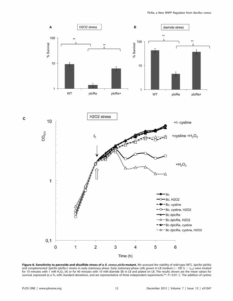
Figure 8. Sensitivity to peroxide and disulfide stress of a B. cereus plcRa mutant. We assessed the viability of wild-type (WT), DplcRa (plcRa)and complemented DplcRa (plcRa+) strains in early stationary phase. Early stationary-phase cells grown in LB medium (, OD 3, , t0.4) were treatedfor 10 minutes with 1 mM H2O2 (A) or for 40 minutes with 10 mM diamide (B) in LB and plated on LB. The results shown are the mean values forsurvival, expressed as a %, with standard deviations, and are representative of three independent experiments.**: P,0.01. C. The addition of cystine
PlcRa, a New RNPP Regulator from Bacillus cereus
PLOS ONE | www.plosone.org 12 December 2012 | Volume 7 | Issue 12 | e51047

presence of reactive oxygen species in the bacteria of the B. cereus
group [41,42,43]. In addition, S-thiolation by cysteine, the most
abundant LMW thiol in cells, constitutes a general mechanism of
thiol protection of proteins in B. subtilis [48] after oxidative stress,
and remains to be characterized in B. cereus group.
The regulation by PlcRa of one of the key operons involved in
cysteine synthesis from methionine is mediated by AbrB2. We
suggest that AbrB2 regulates the expression of genes encoding
proteins involved in cysteine metabolism. These genes probably
belong to the CymR regulon. Further investigations are required
to determine the molecular mechanisms by which AbrB2 regulates
the set of genes encoding proteins involved in cysteine metabolism
which may be direct or may involve CymR control.
Overall, our results demonstrate for the first time the existence
of regulatory connections between cysteine metabolism and the
oxidative stress responses at the onset of stationary phase in the B.
cereus ATCC 14579 strain. These connections are partly controlled
via the PlcRa and AbrB2 regulators, which are found exclusively
in the B. cereus group. We have shown that PlcRa is connected to
regulons which are probably involved either directly (OhR, PerR)
or indirectly (CymR) in the responses to peroxide and disulfide
stresses in B. cereus. Finally, plcRa inactivation had no significant
effect on sporulation capacity in many common laboratory
conditions (data not shown). We therefore suggest that the
involvement of PlcRa in stress responses may ensure bacterial
survival during the transition state preceding the initiation of
sporulation. In conclusion, PlcRa is a new pleiotropic RNPP
regulator, involved in major physiological processes in bacteria of
the species B. cereus: adaptation to poor sulfur source conditions
and oxidative environment early in stationary phase.
Materials and Methods
Bacterial Strains and Growth ConditionsThe type strain B. cereus ATCC 14579 [18] was used throughout
this study. Escherichia coli K-12 strain TG1 was used as a host for
cloning experiments [49]. Plasmid DNA for the electroporation of
B. cereus was prepared from E. coli strain ET12567 (Stratagene, La
Jolla, CA, USA) [50]. E. coli and B. cereus cells were transformed by
electroporation, as previously described. E. coli and B. cereus strains
were grown at 37uC in Luria Bertani (LB) broth and, where
indicated 1 mM cystine was added to the culture medium for B.
cereus. The following antibiotic concentrations were used for
bacterial selection: ampicillin, 200 mg ml21 for E. coli; kanamycin,
200 mg ml21; tetracycline, 15 mg ml21 and erythromycin, 5–10 mgml21 for B. cereus. Bacteria producing b-galactosidase were
identified on LB plates containing X-Gal (40 mg ml21). The xylA
promoter was induced in B. cereus by adding xylose (10 mM, final
concentration) to the HCT 0.2% glucose culture medium. For
microarray analysis, qPCR and b-galactosidase assays, all B. cereusstrains were grown in LB broth at 37uC, in flasks, with an aeration
ratio of 10, on a rotary shaker operating at 175 rpm. Cultures
were inoculated, to an OD600 of 0.05, with cells in the exponential
growth phase in LB broth, and culture pellets were harvested
between 2 h before (t22) and 4 h after (t4) the onset of stationary
phase, for b-galactosidase assays. Cells were harvested at t1 and t2for DNA microarray analysis and for qPCR analysis for DNA
microarray validation and at t0,5, t1,5 and t2,5 for qPCR expression
analysis. The onset of the stationary growth phase (t0) was defined
as the breakpoint in the slope of the exponential phase growth
curve [8].
DNA Manipulation TechniquesChromosomal DNA was extracted from B. cereus cells with the
Puregene DNA Purification kit (Gentra Systems, USA). Plasmid
DNA was extracted from E. coli on QIAprep spin columns
(QIAGEN, France). Restriction enzymes (New England Biolabs,
USA) and T4 DNA ligase (New England Biolabs, USA) were used
in accordance with the manufacturer’s recommendations. Oligo-
nucleotide primers for molecular constructs were synthesized by
Sigma-Proligo (Paris, France) and primers for qPCR were
synthesized by Eurofins-MWG (Paris, France). PCR was per-
formed in an Applied Biosystems 2720 Thermak cycler (Applied
Biosystem, USA). Amplified fragments were purified with the
QIAquick PCR Purification Kit (QIAGEN, France). Digested
DNA fragments were extracted from gels with the QIAquick Gel
Extraction Kit (QIAGEN, France). Nucleotide sequences were
determined by Cogenics (Meylan, France).
Rapid Amplification of 59-cDNA Ends (59RACE)A 59RACE experiment was conducted to map the transcrip-
tional start site of plcRa. B. cereus cultures were grown for four hours
(t1). RNA (4 mg) was used for cDNA synthesis with the Superscript
IITM reverse transcriptase (Invitrogen) and a gene-specific primer
(BC0988GSP1: 59-TCAGAATTAGTTAAAGTCAGTT-39). The
resulting cDNA was purified on S.N.A.P columns and a poly(dC)
tail was added (Invitrogen 59RACE system). The dC-tailed cDNA
was amplified by PCR with an Abridged Anchor primer
(Invitrogen) and a second gene-specific primer (BC0988GSP2:
59-GCAATTGCTTCATGCCACATTAGA-39), complementary
to a region upstream from the binding site of the GSP1 primer.
PCR products were isolated by gel extraction and inserted into the
pGEM-T easy cloning vector (Promega). Three independent PCR
products were sequenced.
Construction of Deletion StrainsThe chromosomal B. cereus plcRa and abrB2 genes were
disrupted by homologous recombination with the pRN5101
heat-sensitive vector [51]. For the B. cereus plcRa and abrB2 mutant
constructs, a tetracycline cassette carrying the tet gene was used for
cloning [52] (Table 1). Each molecular construct, containing the
59- and 39- end flanking regions of the target gene and the positive
selection cassette, was inserted between the HindIII and BamHI
sites of pRN5101.
BamHI-PstI and XbaI-HindIII DNA fragments corresponding to
the regions upstream and downstream from the plcRa gene were
amplified from the B. cereus chromosome by PCR, with the primers
R1–1 (59-GCGGATCCATGTTGAACATGTTTTAAATAC-39)
R1–2 (59-AACTGCAGTTTTTCAATCTTGCTAATTTG-39)
and R1–3 (59-GCTCTAGATGTTTATTAAAAATGAAA-
CAAC-39) R1–4 (59-CCCAAGCTTTGAAAGAAGTTTAGGA-
TATTC-39). External primers R1–V1 (59-GATCAAATCG-
CAAAAAGGCACCTTAG-39), R1–V2 (59-
strongly improved the peroxide stress resistance of the plcRa mutant. We assessed the growth inhibition of wild-type and DplcRa strains in earlystationary phase. Growth curves of the wild-type strain (black circles) and the mutant (black triangles) in LB medium without (solid line) or with 1 mMcystine (dashed line). Hydrogen peroxide (0.4 mM) was added at an OD of 2 (, t20.3). White symbols indicate cultures treated with H2O2. t0 isindicated by a black arrow and hydrogen peroxide addition by a white arrow. This experiment was carried out four times and the results of onerepresentative experiment are shown.doi:10.1371/journal.pone.0051047.g008
PlcRa, a New RNPP Regulator from Bacillus cereus
PLOS ONE | www.plosone.org 13 December 2012 | Volume 7 | Issue 12 | e51047

GGTGAAAGATCATTCGGCAGAGGAGCG-39) were used to
check for correct chromosomal integration of the tet gene.
HindIII-XbaI and EcoRI-BamHI DNA fragments corresponding
to regions upstream and downstream from the abrB2 gene were
generated from the B. cereus chromosome by PCR, with the
primers R3–1 (59-CCCAAGCTTGAGGGAGGAAAGATG-
GAA-39) R3–2 (59-GCTCTAGATGCTATCTGCTTTAC-
GAGT-39) and R3–3 (59-CGGAATTCCAAACGGGATG-
GAACTG-39) R3–4 (59-
GCGGATCCCACAAGTATAAGGATTATG-39). External pri-
mers (R3–V1 59-CCGCTATCTATTGTACAACC-39), R3–V2
(59-ATCGTGTTCGTCTTCGCCAT-39) were used to check for
correct chromosomal integration of the tet gene.
Plasmid ConstructionpHT304plcRa’-lacZ, pHT304abrB2’-lacZ and pHT304yrrT’-
lacZ (Table 1) were obtained by inserting the DNA regions
upstream (corresponding to the intergenic region) from the Bc
ATCC 14579 plcRa, abrB2 and yrrT genes between the XbaI and
PstI cloning sites of pHT304–18Z [23]. The resulting plasmids
were then transferred into B. cereus by electroporation.
pHT1618KVPxyl-papRa and pHT1618KVPxyl-abrB2 contain-
ing the promoterless papRa gene or the promoterless abrB2 gene,
the xylR repressor gene and the inducible promoter of the xylA
gene, were constructed as follows. The papRa gene was amplified
by PCR from chromosomal DNA, using primers 5papRaB (59-
CGGGATCCTAAAGGGGGATTTATTATGTTC-39) har-
bouring a BamHI restriction site and 3papRaE (59–CGGAATTC-GAGGTTCAAAAAATCTACTA-39) harbouring an EcoRI re-
striction site. The abrB2 gene was amplified by PCR from
chromosomal DNA, using primers 5abrB2B (59-CGGGATCC-TAAAGGGTGGCATTTTATGA-39) harbouring a BamHI re-
striction site and 3abrB2E (59–CGGAATTC AGTTTCACTT-
TATTTTAAAAG-39) harbouring an EcoRI restriction site. The
amplified fragments were inserted downstream PxylA between the
BamHI and EcoRI cloning sites of pHT1618KVPxyl (Table 1)
[14].
For complementation studies, the plcRa gene was inserted into
the low-copy number plasmid pHT304 [53] and introduced into
the plcRa mutant. For complementation of the plcRa –deficient
mutant harboring pHT304abrB2’-lacZ, the plcRa gene was inserted
into pHT1618K (Table 1) [14].
PlcRa was overproduced, using pET28a (Novagen). This
plasmid was constructed by inserting an 892 bp NcoI-XhoI
fragment corresponding to the plcRa coding sequence. The DNA
fragment corresponding to the plcRa sequence was generated by
PCR, using oligonucleotides PlcRa1 59-CATGCCATGGAATT-
TAACGATTTGGGT-39 and PlcRa2 59-CCGCTCGAGT-
GAGTGTTTTTTATTTTGTAATTC-39, thus replacing the
TAA stop codon by the XhoI restriction site. This allows the
creation of a translational fusion, adding six C-terminal His
residues and placing expression of the gene under the control of
a T7 promoter. All constructs mentioned above were checked by
sequencing.
Use of Synthetic PeptideCells were cultured at 37uC in LB medium until t0.2. The
culture was then fractionated and synthetic peptide at different
concentrations (1 mM, 2 mM, 4 mM, 8 mM, 20 mM) added to one
fraction. Incubation was pursued and b-galactosidase activity
assayed for each fraction. The peptide CSIPYEY was synthesized,
purified by HPLC and identified by mass spectrophotometry by
Covalab (France).
b-galactosidase Assayb-Galactosidase activity was assayed as described elsewhere [14]
All data are means from assays performed in at least three
independent experiments, and the means+standard deviations are
shown on graphs.
Isolation of Total RNATotal RNA was extracted from samples at the indicated time
points. Total RNA was collected and isolated by a previously
described procedure [28] with minor modifications: for DNA
microarray analysis, 20 ml of each culture was harvested and
centrifuged, and 2 ml of phenol-based RNA extraction buffer
(TRI reagent; Ambion, United Kingdom) was added to the pellet,
after which the cells were snap-frozen in liquid nitrogen. For
qPCR analysis, 2 ml of each culture was harvested and 1 ml of
TRI reagent was added to the cell pellets. RNA quality was
assessed with the Agilent 6000 Nano kit in an Agilent 2100
bioanalyzer (Stratagene, Agilent Technologies, France). RIN
values were in the 8 to 10 range. Total RNA was purified from
two biological replicates for microarray experiments and RT-
qPCR analysis.
Microarray Hybridization and Data AnalysiscDNA synthesis, Cy3/Cy5 labeling and cDNA purification
were carried out as previously described by van Schaik et al. [28].
Microarray experiments comparing the transcriptomes of the wild-
type strain and the plcRa deletion mutant were performed with two
independent biological duplicates, with Cy3/Cy5 dye-swapping
(GEO accession GSE30514). Custom-made Agilent B. cereus
microarrays were hybridized with 200 ng of labeled cDNA for
each sample. The DNA microarrays used in this study were of the
6618K format [28]. Slides were scanned with an Agilent
microarray scanner (G2565BA) and the data were extracted from
the microarrays with Agilent Feature Extraction software (version
8.1.1.1). The data extraction procedure included LOWESS
normalization of the raw data. The data were further processed
as previously described [28], including the use of the web-based
VAMPIRE platform [54] with a P-value threshold of 0.05. For
gene annotation and metabolic routes, we used the PATRIC and
KEGG databases.
RT-quantitative PCR AnalysisWe generated cDNA from 1 mg of total RNA with the
AffinityScript qPCR cDNA Synthesis kit (Stratagene, Agilent
Technologies, France) and random hexamers. We checked cDNA
quality with an Agilent 6000 Pico kit, on an Agilent 2100
bioanalyzer (Stratagene, Agilent Technologies, France). We
carried out qPCR in triplicate, in a reaction volume of 20 mlcontaining 500 pg of cDNA, 15 ml of SYBRH Green PCR Master
Mix (Applied Biosystems, Courtaboeuf, France) and 300 nM of
each gene-specific primer. The primers were designed with Primer
ExpressH (version 2.0), with the following parameters: mean
product length of 70 base pairs (bp), mean primer Tm 59uC and
mean primer size 20 bp. We generated standard curves for each
set of primers, using serial dilutions (four dilutions) of cDNA
obtained from total wild-type strain RNA collected at t2. We
calculated the R2 values for these dilution series and the efficiency
of each primer set. Amplification was achieved with an ABIHPR-ISM 7900 (Applied Biosystems), with the following thermal profile:
2 min at 50uC, 10 min at 95uC followed by 40 cycles of 15 s at
95uC and 60 s at 60uC. The specificity of each amplified PCR
product was checked by melting curve analysis. Two endogenous
controls, 16S and tufA [28] from B. cereus ATCC 14579 were
PlcRa, a New RNPP Regulator from Bacillus cereus
PLOS ONE | www.plosone.org 14 December 2012 | Volume 7 | Issue 12 | e51047
tested, and tufA was found to be the most reliable in our conditions.
We therefore normalized the expression levels of the tested genes
against those for the tufA gene. The relative change in gene
expression was recorded as the ratio of normalized target
concentrations and was calculated by the comparative DDCtmethod [55]. RQ Manager (Applied Biosystems) was used to
generate expression ratios. The mean values for two independent
experiments are presented. Standard deviations were less than 5%
of the mean.
Overproduction and Batch Purification of 6His-tagged-PlcRaWe used pET28a plcRa to transform E.coli strain BL21. The
resulting strain was grown at 30uC in LB with kanamycine 20 mgml-1 until mid-exponential growth phase (OD600 0.7); IPTG was
added (1 mM) and incubation continued for 4 h at 30uC. The cellswere centrifuged at 5000 g for 10 min and resuspended in 1/50 of
the culture volume of Lysis Buffer (50 mM NaH2PO4 pH 8,
300 mM NaCl, 10 mM imidazole). The cells were incubated
during 30 minutes on ice with lysosyme (1 mg/ml) and then
disrupted by sonication, and cell debris was removed by
centrifugation at 12 000 g for 20 min at 4uC. The resulting crude
protein extracts were loaded onto a 0.5 ml Ni-NTA–agarose
column (QIAGEN) during one hour at 4uC. After washing, 6His-
tagged-PlcRa protein was eluted 4 times with 0.5 ml Elution
Buffer (50 mM NaH2PO4 pH 8, 300 mM NaCl, 250 mM imid-
azole). Elution samples 2 and 3 were pooled and a Sephadex G-25
buffer exchange colonn was used (Pharmacia) for recovering 6His-
tagged-PlcRa protein in Storage Buffer (10 mM Tris, pH7.5,
50 mM KCL, 1 mM DTT). Purified PlcRa aliquots were stored at
270uC. All purification steps were analyzed by SDS–PAGE in
a 12% acrylamide gel. The molecular size reference marker was
obtained from Bio-Rad. Protein concentrations were determined
with the Bio-Rad protein assay.
Electrophoretic Mobility Shift Assay (EMSA) AssaysA 175-bp DNA probe of the abrB2 promoter region and a 180-
bp DNA probe of the ilsA promoter region (negative control) were
generated by PCR from BC14579 genomic DNA using 59 end
biotin oligonucleotide primers (Eurofins GENOMICS, LES Ulis,
France). For competition assay, a 175-bp DNA probe of the abrB2
promoter region was generated by PCR from BC14579 genomic
DNA using oligonucleotide primers (Eurofins GENOMICS, LES
Ulis, France). All PCR fragments were extracted from gels with the
QIAquick Gel Extraction Kit (QIAGEN, France) and NanoDrop
2000 spectrophotometer (Thermo scientific) was used for DNA
quantification. EMSA experiments were done according to the
protocol of LightShift Chemiluminescent EMSA kit from Thermo
Fisher Scientific (Brebieres, France) and was performed in a 20 mlreaction volume containing 10 mM Tris, pH7.5, 200 mM KCL,
1 mM DTT, 20 mM or 2 mM or 0.2 mM PapRa7 and a non
specific competitor, 250 ng final salmon sperm DNA. 5 fmol of
DNA biotin probe and 200 nM or 1.2 mM or 2 mM of PlcRa were
used for each reaction. Competition assay was done with abrB2
probe at 2.5 pmol. Electrophoresis was performed with non
denaturing TBE-acrylamide gels (6%).
Stress AssaysViability in the presence of H2O2 and diamide was assessed in
cultures grown in LB medium until the onset of the stationary
phase (,OD 3,,t0,4). The final stock solutions of diamide (1M) or
H2O2 (100 mM) was prepared in sterile demineralized water
immediately before use. Cultures were then split in two, with one
of the two halves exposed to 1 mM hydrogen peroxide (Sigma) for
10 minutes or 10 mM diamide (Sigma) for 40 minutes. Cells were
serially diluted in 0.9% sodium chloride and viability was analyzed
by assessing growth on LB agar. We determined the sensitivity of
growth to hydrogen peroxide, by culturing cells either in LB
medium alone or in LB supplemented with cystine (1 mM) until
the end of exponential growth phase growth phase (,OD 2,
,t20,3). Cultures were split in two, and one half was exposed to
0.4 mM hydrogen peroxide (Sigma). Changes in OD600 were
monitored until t4, to monitor growth arrest and estimate the effect
of stress.
Sequence AnalysisSequences were retrieved with Blast-tp from the NCBI website,
http://blast.ncbi.nlm.nih.gov/Blast.cgi, with PLCRa,
NP_830774.1 used as the query sequence, the Blosum 62 matrix
and all non redundant GenBank CDS translations, PDB,
SwissProt, PIR &PRF databases.
Homology ModelingHomology modeling of the PlcRa homodimer was performed
with Modeler 8v0, using the crystal structure of the complex PlcR/
PapR as the template (group I, PDB entry 2QFC). We sequentially
generated 30 models of PlcRa chain A and PlcRa chain B
satisfying the spatial restraints imposed by the two-dimensional
alignment with the target protein. The best model for each chain
was selected on the basis of the score function in Modeler [20]. To
build the homodimer, the homology model of each chain A and B
was then superimposed to its corresponding A and B chains of the
target structure using DaliLite from the EBI website (http://www.
ebi.ac.uk/DaliLite/). The stereochemistry of the homodimer
PlcRa was finally checked using MolProbity (http://molprobity.
biochem.duke.edu/). Finally, minor repositionings of side chains
were carried out using CHARMm forcefield implemented in
Accelrys�. The binding of PapRa7 was optimized using
CHARMm forcefield.
Microarray Data Accession NumberThe microarray data presented in Tables 2, 3 and in
supplementary data, have been deposited in the Gene Expression
Omnibus repository (http://www.ncbi.nlm.nih.gov/projects/geo),
under accession number GSE30514.
Supporting Information
Figure S1 Reconstruction of the sulfur metabolismpathway in B. cereus: transport and biosynthesis ofsulfur-containing amino acids. The putative proteins in-
volved in the uptake and assimilation of inorganic (sulfate) and
organic sulfur sources (sulfonates, cystine, methionine) are in-
dicated by the corresponding genes. The BC numbers (ATCC
14579 strain) for B. cereus genes are shown, with gene names
according to the orthologs in B. subtilis. ‘?’ indicates genes probably
involved in the pathway or a step for which a gene is lacking or
remains to be identified. All the PlcRa-regulated genes involved in
sulfur metabolism are indicated by a downward black arrow and
the putative functions of all the corresponding proteins are
presented in Table 2. Presumed direct targets of CymR are
indicated in bold typeface. OAS, O-acetyl-serine; AdoMet, S-
adenosyl-methionine.
(PPTX)
Figure S2 Identification of a motif common to thepromoter regions of putative CymR targets in the B.
PlcRa, a New RNPP Regulator from Bacillus cereus
PLOS ONE | www.plosone.org 15 December 2012 | Volume 7 | Issue 12 | e51047

cereus ATCC 14579 strain. An alignment of the promoter
regions of the ssuB, yrrT, tcyP, luxS, cysK, cysH and BC1090 genes is
presented. The consensus sequence for the CymR-binding site was
determined with the WebLogo tool.
(PPTX)
Figure S3 Kinetics of cymR gene expression. b-galactosi-dase specific activity (U/mg protein) of the wild-type (blackcircles), DplcRa (white triangles) strains harboring the transcrip-
tional PcymR’-lacZ fusion, in LB. Errors bars are shown. Time zero
corresponds to the onset of the stationary growth phase, and tn is
the number of hours before (–) or after time zero.
(PPTX)
Figure S4 Kinetics of plcRa gene expression. b-galactosi-dase specific activity (U/mg protein) of the wild-type strain
harboring the transcriptional PplcRa’-lacZ fusion, in LB medium
without or with hydrogen peroxide (0.4 mM) or diamide (10 mM).
Hydrogen peroxide or diamide was added at an OD of 2 (,t20.3).
Errors bars are shown.
(PPTX)
Table S1 a. Genes with differences of 0.33 fold of less are
presented. b. Locus tag in type strain ATCC 14579. c. The gene
names indicated correspond to B. subtilis homologs, with the
exception of hblB, hblL1, hblL2 which correspond to gene names in
B. cereus. *These genes were also analysed with qRT-PCR at t1
and the expression ratio was 0.1.
(DOC)
Acknowledgments
We thank the ICE platform for technical support and V. Brousolle and J.
Brillard for helpful discussions on microarray methods. We thank the
members of the GME team and in particular M. Gohar, for his comments
on the manuscript.
Author Contributions
Conceived and designed the experiments: EH GAL MT TA SM IMV DL.
Performed the experiments: EH GAL MT PW LB SM. Analyzed the data:
EH GAL MT IMV WP DL. Contributed reagents/materials/analysis
tools: EH GAL MT TA IMV SM. Wrote the paper: EH GAL MT TA
IMV DL.
References
1. Stenfors Arnesen LP, Fagerlund A, Granum PE (2008) From soil to gut: Bacilluscereus and its food poisoning toxins. FEMS Microbiol Rev 32: 579–606.
2. Piggot PJ, Hilbert DW (2004) Sporulation of Bacillus subtilis. Curr OpinMicrobiol 7: 579–586.
3. Phillips ZE, Strauch MA (2002) Bacillus subtilis sporulation and stationary phasegene expression. Cell Mol Life Sci 59: 392–402.
4. Price CW, Fawcett P, Ceremonie H, Su N, Murphy CK, et al. (2001) Genome-wide analysis of the general stress response in Bacillus subtilis. Mol Microbiol 41:
757–774.
5. Zuber P (2009) Management of oxidative stress in Bacillus. Annu Rev Microbiol
63: 575–597.
6. Dunny GM, Leonard BA (1997) Cell-cell communication in gram-positive
bacteria. Annu Rev Microbiol 51: 527–564.
7. Gohar M, Faegri K, Perchat S, Ravnum S, Okstad OA, et al. (2008) The PlcRvirulence regulon of Bacillus cereus. PLoS One 3: e2793.
8. Agaisse H, Gominet M, Okstad OA, Kolsto AB, Lereclus D (1999) PlcR isa pleiotropic regulator of extracellular virulence factor gene expression in Bacillus
thuringiensis. Mol Microbiol 32: 1043–1053.
9. Bouillaut L, Perchat S, Arold S, Zorrilla S, Slamti L, et al. (2008) Molecular basis
for group-specific activation of the virulence regulator PlcR by PapRheptapeptides. Nucleic Acids Res 36: 3791–3801.
10. Gominet M, Slamti L, Gilois N, Rose M, Lereclus D (2001) Oligopeptidepermease is required for expression of the Bacillus thuringiensis plcR regulon and
for virulence. Mol Microbiol 40: 963–975.
11. Slamti L, Lereclus D (2002) A cell-cell signaling peptide activates the PlcRvirulence regulon in bacteria of the Bacillus cereus group. EMBO J 21: 4550–
4559.
12. Declerck N, Bouillaut L, Chaix D, Rugani N, Slamti L, et al. (2007) Structure of
PlcR: Insights into virulence regulation and evolution of quorum sensing inGram-positive bacteria. Proc Natl Acad Sci U S A 104: 18490–18495.
13. D’Andrea LD, Regan L (2003) TPR proteins: the versatile helix. TrendsBiochem Sci 28: 655–662.
14. Perchat S, Dubois T, Zouhir S, Gominet M, Poncet S, et al. (2011) A cell-cellcommunication system regulates protease production during sporulation in
bacteria of the Bacillus cereus group. Mol Microbiol 82: 619–633.
15. Shi K, Brown CK, Gu ZY, Kozlowicz BK, Dunny GM, et al. (2005) Structure ofpeptide sex pheromone receptor PrgX and PrgX/pheromone complexes and
regulation of conjugation in Enterococcus faecalis. Proc Natl Acad Sci U S A 102:18596–18601.
16. Solomon JM, Lazazzera BA, Grossman AD (1996) Purification and character-ization of an extracellular peptide factor that affects two different developmental
pathways in Bacillus subtilis. Genes Dev 10: 2014–2024.
17. Parashar V, Mirouze N, Dubnau DA, Neiditch MB (2011) Structural basis of
response regulator dephosphorylation by Rap phosphatases. PLoS Biol 9:
e1000589.
18. Ivanova N, Sorokin A, Anderson I, Galleron N, Candelon B, et al. (2003)
Genome sequence of Bacillus cereus and comparative analysis with Bacillus
anthracis. Nature 423: 87–91.
19. Ross CL, Koehler TM (2006) plcR papR-independent expression of anthrolysinO by Bacillus anthracis. J Bacteriol 188: 7823–7829.
20. Sali A, Blundell TL (1993) Comparative protein modelling by satisfaction ofspatial restraints. J Mol Biol 234: 779–815.
21. Emanuelsson O, Brunak S, von Heijne G, Nielsen H (2007) Locating proteins inthe cell using TargetP, SignalP and related tools. Nat Protoc 2: 953–971.
22. Brooks BR, Brooks CL 3rd, Mackerell AD Jr, Nilsson L, Petrella RJ, et al. (2009)
CHARMM: the biomolecular simulation program. J Comput Chem 30: 1545–
1614.
23. Agaisse H, Lereclus D (1994) Structural and functional analysis of the promoter
region involved in full expression of the cryIIIA toxin gene of Bacillus thuringiensis.
Mol Microbiol 13: 97–107.
24. Moran CP (1993) RNA polymerase and transcription factors. In: Sonenshein
AL, Hoch, J.A and Losick, R., editor. Bacillus subtilis and other gram-positive
bacteria. Washington D.C.: American Society for Microbiology. 653–667.
25. Ceragioli M, Mols M, Moezelaar R, Ghelardi E, Senesi S, et al. (2010)
Comparative transcriptomic and phenotypic analysis of the responses of Bacillus
cereus to various disinfectant treatments. Appl Environ Microbiol 76: 3352–3360.
26. Mols M, Abee T (2011) Primary and secondary oxidative stress in Bacillus.
Environ Microbiol 13: 1387–1394.
27. Tu WY, Pohl S, Gizynski K, Harwood CR (2012) The iron-binding protein
Dps2 confers peroxide stress resistance on Bacillus anthracis. J Bacteriol 194: 925–
931.
28. van Schaik W, van der Voort M, Molenaar D, Moezelaar R, de Vos WM, et al.
(2007) Identification of the sigmaB regulon of Bacillus cereus and conservation of
sigmaB-regulated genes in low-GC-content gram-positive bacteria. J Bacteriol
189: 4384–4390.
29. Smeesters PR, Dreze PA, Bousbata S, Parikka KJ, Timmery S, et al. (2011)
Characterization of a novel temperate phage originating from a cereulide-
producing Bacillus cereus strain. Res Microbiol 162: 446–459.
30. Graumann PL, Knust T (2009) Dynamics of the bacterial SMC complex and
SMC-like proteins involved in DNA repair. Chromosome Res 17: 265–275.
31. Kolsto AB, Tourasse NJ, Okstad OA (2009) What sets Bacillus anthracis apart
from other Bacillus species? Annu Rev Microbiol 63: 451–476.
32. Salvetti S, Faegri K, Ghelardi E, Kolsto AB, Senesi S (2011) Global Gene
Expression Profile for Swarming Bacillus cereus Bacteria. Appl Environ Microbiol
77: 5149–5156.
33. Burguiere P, Auger S, Hullo MF, Danchin A, Martin-Verstraete I (2004) Three
different systems participate in L-cystine uptake in Bacillus subtilis. J Bacteriol
186: 4875–4884.
34. Hullo MF, Auger S, Soutourina O, Barzu O, Yvon M, et al. (2007) Conversion
of methionine to cysteine in Bacillus subtilis and its regulation. J Bacteriol 189:
187–197.
35. Even S, Burguiere P, Auger S, Soutourina O, Danchin A, et al. (2006) Global
control of cysteine metabolism by CymR in Bacillus subtilis. J Bacteriol 188:
2184–2197.
36. Tanous C, Soutourina O, Raynal B, Hullo MF, Mervelet P, et al. (2008) The
CymR regulator in complex with the enzyme CysK controls cysteine
metabolism in Bacillus subtilis. J Biol Chem 283: 35551–35560.
37. Choi SY, Reyes D, Leelakriangsak M, Zuber P (2006) The global regulator Spx
functions in the control of organosulfur metabolism in Bacillus subtilis. J Bacteriol
188: 5741–5751.
38. Strauch MA, Spiegelman GB, Perego M, Johnson WC, Burbulys D, et al. (1989)
The transition state transcription regulator abrB of Bacillus subtilis is a DNA
binding protein. EMBO J 8: 1615–1621.
39. Hadjifrangiskou M, Chen Y, Koehler TM (2007) The alternative sigma factor
sigmaH is required for toxin gene expression by Bacillus anthracis. J Bacteriol 189:
1874–1883.
PlcRa, a New RNPP Regulator from Bacillus cereus
PLOS ONE | www.plosone.org 16 December 2012 | Volume 7 | Issue 12 | e51047

40. Soutourina O, Dubrac S, Poupel O, Msadek T, Martin-Verstraete I (2010) The
pleiotropic CymR regulator of Staphylococcus aureus plays an important role invirulence and stress response. PLoS Pathog 6: e1000894.
41. Newton GL, Rawat M, La Clair JJ, Jothivasan VK, Budiarto T, et al. (2009)
Bacillithiol is an antioxidant thiol produced in Bacilli. Nat Chem Biol 5: 625–627.
42. Nicely NI, Parsonage D, Paige C, Newton GL, Fahey RC, et al. (2007) Structureof the type III pantothenate kinase from Bacillus anthracis at 2.0 A resolution:
implications for coenzyme A-dependent redox biology. Biochemistry 46: 3234–
3245.43. Paige C, Reid SD, Hanna PC, Claiborne A (2008) The type III pantothenate
kinase encoded by coaX is essential for growth of Bacillus anthracis. J Bacteriol190: 6271–6275.
44. Auger S, Danchin A, Martin-Verstraete I (2002) Global expression profile ofBacillus subtilis grown in the presence of sulfate or methionine. J Bacteriol 184:
5179–5186.
45. Pohl S, Tu WY, Aldridge PD, Gillespie C, Hahne H, et al. (2011) Combinedproteomic and transcriptomic analysis of the response of Bacillus anthracis to
oxidative stress. Proteomics 11: 3036–3055.46. Wang L, Chong H, Jiang R (2012) Comparison of alkyl hydroperoxide reductase
and two water-forming NADH oxidases from Bacillus cereus ATCC 14579. Appl
Microbiol Biotechnol.47. Hullo MF, Martin-Verstraete I, Soutourina O (2010) Complex phenotypes of
a mutant inactivated for CymR, the global regulator of cysteine metabolism inBacillus subtilis. FEMS Microbiol Lett 309: 201–207.
48. Hochgrafe F, Mostertz J, Pother DC, Becher D, Helmann JD, et al. (2007) S-
cysteinylation is a general mechanism for thiol protection of Bacillus subtilis
proteins after oxidative stress. J Biol Chem 282: 25981–25985.
49. Dower WJ, Miller JF, Ragsdale CW (1988) High efficiency transformation of E.
coli by high voltage electroporation. Nucleic Acids Res 16: 6127–6145.
50. Lereclus D, Arantes O, Chaufaux J, Lecadet M (1989) Transformation and
expression of a cloned delta-endotoxin gene in Bacillus thuringiensis. FEMS
Microbiol Lett 51: 211–217.
51. Lereclus D, Vallade M, Chaufaux J, Arantes O, Rambaud S (1992) Expansion
of insecticidal host range of Bacillus thuringiensis by in vivo genetic recombination.
Biotechnology (N Y) 10: 418–421.
52. Sanchis V, Agaisse H, Chaufaux J, Lereclus D (1996) Construction of new
insecticidal Bacillus thuringiensis recombinant strains by using the sporulation non-
dependent expression system of cryIIIA and a site specific recombination vector.
J Biotechnol 48: 81–96.
53. Arantes O, Lereclus D (1991) Construction of cloning vectors for Bacillus
thuringiensis. Gene 108: 115–119.
54. Hsiao A, Ideker T, Olefsky JM, Subramaniam S (2005) VAMPIRE microarray
suite: a web-based platform for the interpretation of gene expression data.
Nucleic Acids Res 33: W627–632.
55. Bustin SA, Benes V, Garson JA, Hellemans J, Huggett J, et al. (2009) The MIQE
guidelines: minimum information for publication of quantitative real-time PCR
experiments. Clin Chem 55: 611–622.
PlcRa, a New RNPP Regulator from Bacillus cereus
PLOS ONE | www.plosone.org 17 December 2012 | Volume 7 | Issue 12 | e51047