Abundance of the Quorum-Sensing Factor Ax21 in Four Strains of Stenotrophomonas maltophilia...
10
Abundance of the Quorum-Sensing Factor Ax21 in Four Strains of Stenotrophomonas maltophilia Correlates with Mortality Rate in a New Zebrafish Model of Infection Mario Ferrer-Navarro 1. , Raquel Planell 1. , Daniel Yero 1 , Elı´as Mongiardini 1 , Gerard Torrent 1 , Pol Huedo 1,2 , Paula Martı´nez 1,2 , Nerea Roher 1 , Simon Mackenzie 1,3 , Isidre Gibert 1,2 *, Xavier Daura 1,4 * 1 Institut de Biotecnologia i de Biomedicina (IBB), Universitat Auto ` noma de Barcelona (UAB), Cerdanyola del Valle `s, Barcelona, Spain, 2 Departament de Gene `tica i de Microbiologia, Universitat Auto ` noma de Barcelona (UAB), Cerdanyola del Valle ` s, Barcelona, Spain, 3 Institute of Aquaculture, University of Stirling, Stirling, Scotland, United Kingdom, 4 Catalan Institution for Research and Advanced Studies (ICREA), Barcelona, Spain Abstract Stenotrophomonas maltophilia is a Gram-negative pathogen with emerging nosocomial incidence. Little is known about its pathogenesis and the genomic diversity exhibited by clinical isolates complicates the study of pathogenicity and virulence factors. Here, we present a strategy to identify such factors in new clinical isolates of S. maltophilia, incorporating an adult- zebrafish model of S. maltophilia infection to evaluate relative virulence coupled to 2D difference gel electrophoresis to explore underlying differences in protein expression. In this study we report upon three recent clinical isolates and use the collection strain ATCC13637 as a reference. The adult-zebrafish model shows discrimination capacity, i.e. from very low to very high mortality rates, with clinical symptoms very similar to those observed in natural S. maltophilia infections in fish. Strain virulence correlates with resistance to human serum, in agreement with previous studies in mouse and rat and therefore supporting zebrafish as a replacement model. Despite its clinical origin, the collection strain ATCC13637 showed obvious signs of attenuation in zebrafish, with null mortality. Multilocus-sequence-typing analysis revealed that the most virulent strains, UV74 and M30, exhibit the strongest genetic similitude. Differential proteomic analysis led to the identification of 38 proteins with significantly different abundance in the three clinical strains relative to the reference strain. Orthologs of several of these proteins have been already reported to have a role in pathogenesis, virulence or resistance mechanisms thus supporting our strategy. Proof of concept is further provided by protein Ax21, whose abundance is shown here to be directly proportional to mortality in the zebrafish infection model. Indeed, recent studies have demonstrated that this protein is a quorum-sensing-related virulence factor. Citation: Ferrer-Navarro M, Planell R, Yero D, Mongiardini E, Torrent G, et al. (2013) Abundance of the Quorum-Sensing Factor Ax21 in Four Strains of Stenotrophomonas maltophilia Correlates with Mortality Rate in a New Zebrafish Model of Infection. PLoS ONE 8(6): e67207. doi:10.1371/journal.pone.0067207 Editor: Jamunarani Vadivelu, University of Malaya, Malaysia Received March 3, 2013; Accepted May 15, 2013; Published June 26, 2013 Copyright: ß 2013 Ferrer-Navarro et al. This is an open-access article distributed under the terms of the Creative Commons Attribution License, which permits unrestricted use, distribution, and reproduction in any medium, provided the original author and source are credited. Funding: This work has been supported by funding under the Seventh Research Framework Programme of the European Union (ref. HEALTH-F3-2009-223101) and the Spanish MICINN (ref. BFU2010-17199). I.G. acknowledges support from the Catalan AGAUR (ref. 2009SGR-00108). P.M. and P.H. are recipients of a fellowship from Universitat Auto ` noma de Barcelona. The funders had no role in study design, data collection and analysis, decision to publish, or preparation of the manuscript. Competing Interests: The authors have declared that no competing interests exist. * E-mail: [email protected] (XD); [email protected] (IG) . These authors contributed equally to this work. Introduction Stenotrophomonas maltophilia is a non-fermentative Gram-negative bacterium with increasing incidence in hospital environments [1,2]. This obligate aerobic bacterium can be found in almost any aquatic or humid environment, including drinking-water supplies [3] and is now recognized as an emerging nosocomial pathogen. S. maltophilia has been associated with respiratory infections, septice- mia, biliary sepsis, endocarditis, conjunctivitis, meningitis, urinary tract infections and various wound infections in immunocompro- mised patients as well as in cystic fibrosis (CF) patients [2,4,5]. Currently, S. maltophilia has been isolated from the lungs of approximately 10% of the CF patients in USA and up to 25% of those in Europe [1] and displays significant morbidity and mortality rates among debilitated patients [2,5,6,7,8]. S. maltophilia exhibits high-level intrinsic resistance to a variety of structurally unrelated antibiotics, including b-lactams, quinolones, aminoglycosides, tetracycline, disinfectants and heavy metals [9,10]. Intrinsic resistance may be due to reduced outer- membrane permeability, changes in LPS structure, the production of multidrug efflux pumps and the presence of integrons for site- specific insertion of resistance gene cassettes [11,12]. The production of melanin-like pigments and biofilms have also been linked to antimicrobial resistance [12]. Thus, the adhesion of S. maltophilia to medical implants, catheters and epithelial cells, leading to the formation of biofilms, confers natural protection against different antimicrobial agents and host immune defenses. In this regard, the development of therapies against S. maltophilia infection represents a significant challenge for both clinicians and microbiologists. In addition, knowledge of virulence factors is scarce and limited to homology relationships. Recently, four S. maltophilia genomes have been fully sequenced and assembled (strains K279a, R551–3, JV3 and D457), and PLOS ONE | www.plosone.org 1 June 2013 | Volume 8 | Issue 6 | e67207
-
Upload
independent -
Category
Documents
-
view
0 -
download
0
Transcript of Abundance of the Quorum-Sensing Factor Ax21 in Four Strains of Stenotrophomonas maltophilia...
Abundance of the Quorum-Sensing Factor Ax21 in FourStrains of Stenotrophomonas maltophilia Correlates withMortality Rate in a New Zebrafish Model of InfectionMario Ferrer-Navarro1., Raquel Planell1., Daniel Yero1, Elıas Mongiardini1, Gerard Torrent1,
Pol Huedo1,2, Paula Martınez1,2, Nerea Roher1, Simon Mackenzie1,3, Isidre Gibert1,2*, Xavier Daura1,4*
1 Institut de Biotecnologia i de Biomedicina (IBB), Universitat Autonoma de Barcelona (UAB), Cerdanyola del Valles, Barcelona, Spain, 2 Departament de Genetica i de
Microbiologia, Universitat Autonoma de Barcelona (UAB), Cerdanyola del Valles, Barcelona, Spain, 3 Institute of Aquaculture, University of Stirling, Stirling, Scotland, United
Kingdom, 4 Catalan Institution for Research and Advanced Studies (ICREA), Barcelona, Spain
Abstract
Stenotrophomonas maltophilia is a Gram-negative pathogen with emerging nosocomial incidence. Little is known about itspathogenesis and the genomic diversity exhibited by clinical isolates complicates the study of pathogenicity and virulencefactors. Here, we present a strategy to identify such factors in new clinical isolates of S. maltophilia, incorporating an adult-zebrafish model of S. maltophilia infection to evaluate relative virulence coupled to 2D difference gel electrophoresis toexplore underlying differences in protein expression. In this study we report upon three recent clinical isolates and use thecollection strain ATCC13637 as a reference. The adult-zebrafish model shows discrimination capacity, i.e. from very low tovery high mortality rates, with clinical symptoms very similar to those observed in natural S. maltophilia infections in fish.Strain virulence correlates with resistance to human serum, in agreement with previous studies in mouse and rat andtherefore supporting zebrafish as a replacement model. Despite its clinical origin, the collection strain ATCC13637 showedobvious signs of attenuation in zebrafish, with null mortality. Multilocus-sequence-typing analysis revealed that the mostvirulent strains, UV74 and M30, exhibit the strongest genetic similitude. Differential proteomic analysis led to theidentification of 38 proteins with significantly different abundance in the three clinical strains relative to the reference strain.Orthologs of several of these proteins have been already reported to have a role in pathogenesis, virulence or resistancemechanisms thus supporting our strategy. Proof of concept is further provided by protein Ax21, whose abundance is shownhere to be directly proportional to mortality in the zebrafish infection model. Indeed, recent studies have demonstrated thatthis protein is a quorum-sensing-related virulence factor.
Citation: Ferrer-Navarro M, Planell R, Yero D, Mongiardini E, Torrent G, et al. (2013) Abundance of the Quorum-Sensing Factor Ax21 in Four Strains ofStenotrophomonas maltophilia Correlates with Mortality Rate in a New Zebrafish Model of Infection. PLoS ONE 8(6): e67207. doi:10.1371/journal.pone.0067207
Editor: Jamunarani Vadivelu, University of Malaya, Malaysia
Received March 3, 2013; Accepted May 15, 2013; Published June 26, 2013
Copyright: � 2013 Ferrer-Navarro et al. This is an open-access article distributed under the terms of the Creative Commons Attribution License, which permitsunrestricted use, distribution, and reproduction in any medium, provided the original author and source are credited.
Funding: This work has been supported by funding under the Seventh Research Framework Programme of the European Union (ref. HEALTH-F3-2009-223101)and the Spanish MICINN (ref. BFU2010-17199). I.G. acknowledges support from the Catalan AGAUR (ref. 2009SGR-00108). P.M. and P.H. are recipients of afellowship from Universitat Autonoma de Barcelona. The funders had no role in study design, data collection and analysis, decision to publish, or preparation ofthe manuscript.
Competing Interests: The authors have declared that no competing interests exist.
* E-mail: [email protected] (XD); [email protected] (IG)
. These authors contributed equally to this work.
Introduction
Stenotrophomonas maltophilia is a non-fermentative Gram-negative
bacterium with increasing incidence in hospital environments
[1,2]. This obligate aerobic bacterium can be found in almost any
aquatic or humid environment, including drinking-water supplies
[3] and is now recognized as an emerging nosocomial pathogen. S.
maltophilia has been associated with respiratory infections, septice-
mia, biliary sepsis, endocarditis, conjunctivitis, meningitis, urinary
tract infections and various wound infections in immunocompro-
mised patients as well as in cystic fibrosis (CF) patients [2,4,5].
Currently, S. maltophilia has been isolated from the lungs of
approximately 10% of the CF patients in USA and up to 25% of
those in Europe [1] and displays significant morbidity and
mortality rates among debilitated patients [2,5,6,7,8].
S. maltophilia exhibits high-level intrinsic resistance to a variety of
structurally unrelated antibiotics, including b-lactams, quinolones,
aminoglycosides, tetracycline, disinfectants and heavy metals
[9,10]. Intrinsic resistance may be due to reduced outer-
membrane permeability, changes in LPS structure, the production
of multidrug efflux pumps and the presence of integrons for site-
specific insertion of resistance gene cassettes [11,12]. The
production of melanin-like pigments and biofilms have also been
linked to antimicrobial resistance [12]. Thus, the adhesion of S.
maltophilia to medical implants, catheters and epithelial cells,
leading to the formation of biofilms, confers natural protection
against different antimicrobial agents and host immune defenses.
In this regard, the development of therapies against S. maltophilia
infection represents a significant challenge for both clinicians and
microbiologists. In addition, knowledge of virulence factors is
scarce and limited to homology relationships.
Recently, four S. maltophilia genomes have been fully sequenced
and assembled (strains K279a, R551–3, JV3 and D457), and
PLOS ONE | www.plosone.org 1 June 2013 | Volume 8 | Issue 6 | e67207
putative virulence factors have been identified by homology
relationships [11,13,14]. These factors include type I, II, IV, and
V protein-secretion systems, various pili, fimbriae, putative
adhesins, tissue-degradative exoenzymes, siderophores, quorum-
sensing factors and proteins involved in polysaccharide synthesis
and intracellular signaling. Some fimbrial structures have been
identified and characterized and their role in adhesion to epithelial
cells and abiotic surfaces has been demonstrated [15]. However,
the level of understanding of this bacterium’s pathogenicity and
virulence is still limited and the number of S. maltophilia strains
phenotypically and genotypically analyzed is minor. Furthermore,
there is considerable uncertainty about the route(s) of infection of
S. maltophilia. Additionally, the remarkable diversity of sources
from which S. maltophilia strains have been isolated indicates that
these bacteria exhibit a high level of genomic plasticity and
metabolic heterogeneity, possibly allowing them to expand their
pathogenic potential. Heterogeneity is also illustrated among S.
maltophilia isolates recovered from a single patient, showing
phenotypic variation over time as a consequence of horizontal
gene transfer or high mutation rates [16].
In order to provide answers to some of the above the
development of an appropriate infection models is essential.
Previous studies suggest a limited invasiveness of S. maltophilia in
mice, as indicated by a transient and minimal presence of the
microorganism in animal organs. For example, S. maltophilia CF
strains were shown to cause no mortality in a neonatal mouse
model of respiratory tract infection [17]. Despite this lack of robust
invasiveness, mouse models of S. maltophilia infection have provided
information on the type of host immune response induced by this
opportunistic pathogen [2]. More recently, a model of acute
respiratory infection in DBA/2 mice following a single exposure to
aerosolized bacteria enabled the investigation of bacterial clear-
ance, histological damage, and inflammatory response in the lungs
of infected mice [18]. However, while bacterial colonization and
mortality were achieved in that model, infection disseminated at a
very low rate even using high doses of a virulent strain and most of
the animals were able to resolve S. maltophilia lung colonization in a
relatively short time period. For that reason, animal-weight loss is
often taken as the best criterion for the comparison of pathogenesis
and virulence of tested strains [18,19]. In addition, lung infection
models tend to be time-consuming, labor intensive and have
associated welfare issues. Therefore, alternative, simple models of
S. maltophilia infection are still needed to test the virulence of
phenotypically and genotypically diverse strains.
In recent years the zebrafish (Danio rerio) has emerged as an
important model of vertebrate development, human disease and
microbial infection [20]. Nevertheless, to our knowledge, zebrafish
has not yet been reported as a model of S. maltophilia infection. Our
choice of adult zebrafish as a plausible model was motivated by a
series of observations. The existing literature in relation to other
bacterial pathogen models successfully developed in the zebrafish
[20,21,22]. S. maltophilia as a natural pathogen of fish causing
infectious intussusception syndrome in adult channel catfish [23].
The inclusion of a wild-type phenotypic population diversity as
opposed to inbred mouse lines. The adaptive and innate immune
system of zebrafish has significant similarities to mammalian
systems [24,25]. The zebrafish is recognized as an important
vertebrate model with genomic enablement and last, but not least
relevant, ethical, economic and process-simplicity considerations.
In the study presented here, we have tested a combined
approach that uses an adult-zebrafish model of S. maltophilia
infection for the evaluation of relative virulence and the successive
analysis by fluorescence-based two-dimensional Difference in-Gel
Electrophoresis (DIGE) [26] of the underlying differences in
protein expression, in a quest for virulence factors. This analysis
was applied to three recent clinical isolates and the collection strain
ATCC13637, as a reference. Although a colloidal Coomassie-
stained 2DE proteomic analysis to find heat-induced changes in S.
maltophilia protein abundance has been reported [27], to our
knowledge no quantitative proteomic comparison between S.
maltophilia strains with distinct virulence phenotypes has been
previously performed.
Results
Zebrafish Infection Model Confirms Attenuation ofCollection Strain and Points at Varying Virulence of theClinical Isolates
Adult zebrafish were used as an infection model to determine
the virulence of the individual strains. An intraperitoneal injection
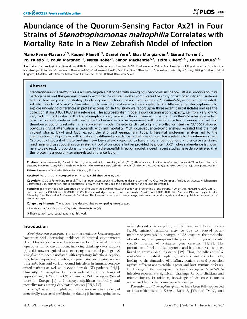
of 108 cfu resulted in strain-dependent mortality rates (Figure 1),
where UV74 was the most aggressive, with 84% mortality 48 h
post-injection (p.i.), followed by strains M30 (55%), E77 (5%) and
ATCC13637 (0%). Although the experimental period was during
7 days, in all cases mortality occurred during the first 48 h.
Clinical symptoms of dead fish included cutaneous hemorrhage
under the lower jaw, on the belly, and around the anus; all dead
fish had a distended abdomen containing bloody or clear fluid and
severe enteritis with intussusception in the lower intestine. All
isolates obtained from post-mortem UV74 infected zebrafish were
identified as S. maltophilia based on morphology, API (analytical
profile index) and 16S rDNA sequence (data not shown). No
zebrafish died when injected with 20 ml of sterile PBS (data not
shown). Interestingly, injection of 108 heat-inactivated UV74
bacteria did not produce mortality in zebrafish. This suggests that
viable bacteria producing thermo-labile proteins are required for
infection and that mortality is not caused by non-specific
activation due to bacterial components such as lipopolysaccha-
rides.
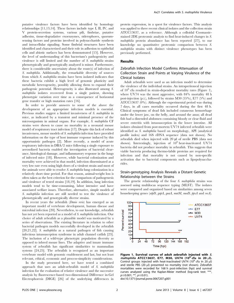
Strain-genotyping Analysis Reveals a Distant GeneticRelationship between the Strains
The genetic relationship of the four S. maltophilia strains was
assessed using multilocus sequence typing (MLST). The isolates
were compared and organized based on similarities among seven
housekeeping genes (atpD, gapA, guaA, mutM, nuoD, ppsA and recA).
Figure 1. Survival curves of adult zebrafish injected with S.maltophilia ATCC13637, E77, M30, UV74 (108 cfu in 20 ml).Control groups injected with heat-inactivated UV74 (108 cfu in 20 ml)and sterile PBS (20 ml) presented no mortality (not shown). Zebrafishmortalities were recorded for 168 h post-infection (hpi) and survivalcurves analyzed using the Kaplan-Meier method (log-rank test: ***,p#0.001; **, p#0.01).doi:10.1371/journal.pone.0067207.g001
Ax21 Correlates with S. maltophilia Virulence
PLOS ONE | www.plosone.org 2 June 2013 | Volume 8 | Issue 6 | e67207
This analysis resulted in the three clinical strains being classified
into new, different sequence types (M30 as ST-76, UV74 as ST-77
and E77 as ST-81), indicating they are not clonally related despite
being isolated from a common hospital setting. Nevertheless,
strains M30 and UV74 share the same allele for genes atpD and
mutM. The collection strain ATCC13637 had been already
characterized by MLST and assigned the ST-14 [28]. In addition,
thirty-five concatenate sequences from different S. maltophilia
strains, isolated from human clinical cases, were obtained from
public databases and used to provide the context for the genotypic
classification of the three clinical isolates and the collection strain.
Strains were chosen such that they cover as far as possible the full
genetic breadth of the species [28]. The phylogenetic analysis
revealed distant genetic relationships between the three clinical
strains and the reference that according to a previous classification
based on AFLP fingerprinting [29] would belong to a different
genomic group (Figure 2). Strains M30 and UV74 show the closest
genetic relationship, clustering within genomic group C as
previously described by Kaiser et al. [28]. The MLST results
imply that the three pair wise (collection:clinical) proteomic
analyses described below are non-redundant.
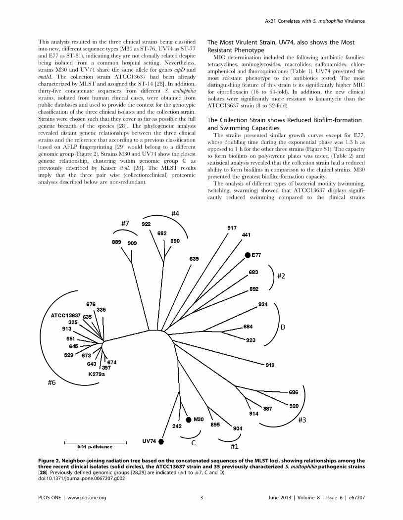
The Most Virulent Strain, UV74, also shows the MostResistant Phenotype
MIC determination included the following antibiotic families:
tetracyclines, aminoglycosides, macrolides, sulfonamides, chlor-
amphenicol and fluoroquinolones (Table 1). UV74 presented the
most resistant phenotype to the antibiotics tested. The most
distinguishing feature of this strain is its significantly higher MIC
for ciprofloxacin (16 to 64-fold). In addition, the new clinical
isolates were significantly more resistant to kanamycin than the
ATCC13637 strain (8 to 32-fold).
The Collection Strain shows Reduced Biofilm-formationand Swimming Capacities
The strains presented similar growth curves except for E77,
whose doubling time during the exponential phase was 1.3 h as
opposed to 1 h for the other three strains (Figure S1). The capacity
to form biofilms on polystyrene plates was tested (Table 2) and
statistical analysis revealed that the collection strain had a reduced
ability to form biofilms in comparison to the clinical strains. M30
presented the greatest biofilm-formation capacity.
The analysis of different types of bacterial motility (swimming,
twitching, swarming) showed that ATCC13637 displays signifi-
cantly reduced swimming compared to the clinical strains
Figure 2. Neighbor-joining radiation tree based on the concatenated sequences of the MLST loci, showing relationships among thethree recent clinical isolates (solid circles), the ATCC13637 strain and 35 previously characterized S. maltophilia pathogenic strains[28]. Previously defined genomic groups [28,29] are indicated (#1 to #7, C and D).doi:10.1371/journal.pone.0067207.g002
Ax21 Correlates with S. maltophilia Virulence
PLOS ONE | www.plosone.org 3 June 2013 | Volume 8 | Issue 6 | e67207
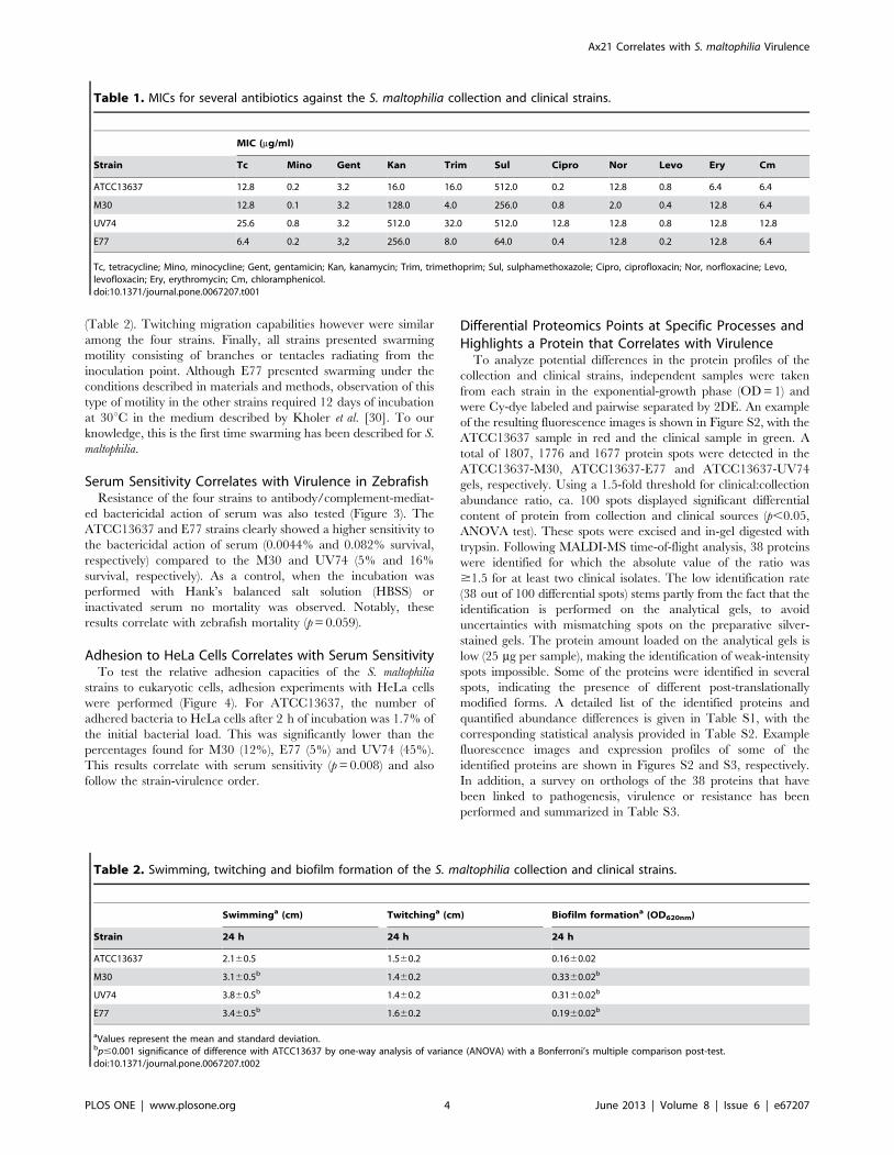
(Table 2). Twitching migration capabilities however were similar
among the four strains. Finally, all strains presented swarming
motility consisting of branches or tentacles radiating from the
inoculation point. Although E77 presented swarming under the
conditions described in materials and methods, observation of this
type of motility in the other strains required 12 days of incubation
at 30uC in the medium described by Kholer et al. [30]. To our
knowledge, this is the first time swarming has been described for S.
maltophilia.
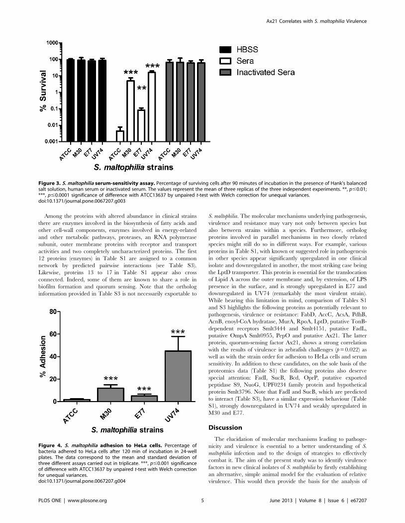
Serum Sensitivity Correlates with Virulence in ZebrafishResistance of the four strains to antibody/complement-mediat-
ed bactericidal action of serum was also tested (Figure 3). The
ATCC13637 and E77 strains clearly showed a higher sensitivity to
the bactericidal action of serum (0.0044% and 0.082% survival,
respectively) compared to the M30 and UV74 (5% and 16%
survival, respectively). As a control, when the incubation was
performed with Hank’s balanced salt solution (HBSS) or
inactivated serum no mortality was observed. Notably, these
results correlate with zebrafish mortality (p = 0.059).
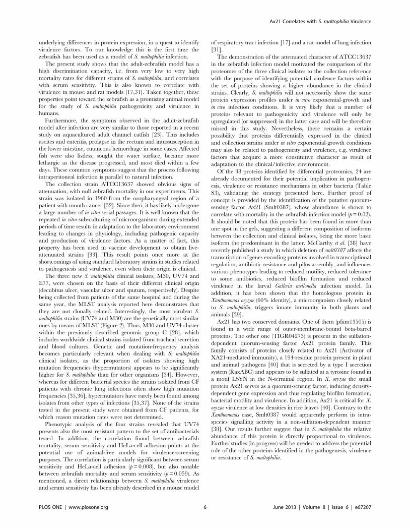
Adhesion to HeLa Cells Correlates with Serum SensitivityTo test the relative adhesion capacities of the S. maltophilia
strains to eukaryotic cells, adhesion experiments with HeLa cells
were performed (Figure 4). For ATCC13637, the number of
adhered bacteria to HeLa cells after 2 h of incubation was 1.7% of
the initial bacterial load. This was significantly lower than the
percentages found for M30 (12%), E77 (5%) and UV74 (45%).
This results correlate with serum sensitivity (p = 0.008) and also
follow the strain-virulence order.
Differential Proteomics Points at Specific Processes andHighlights a Protein that Correlates with Virulence
To analyze potential differences in the protein profiles of the
collection and clinical strains, independent samples were taken
from each strain in the exponential-growth phase (OD = 1) and
were Cy-dye labeled and pairwise separated by 2DE. An example
of the resulting fluorescence images is shown in Figure S2, with the
ATCC13637 sample in red and the clinical sample in green. A
total of 1807, 1776 and 1677 protein spots were detected in the
ATCC13637-M30, ATCC13637-E77 and ATCC13637-UV74
gels, respectively. Using a 1.5-fold threshold for clinical:collection
abundance ratio, ca. 100 spots displayed significant differential
content of protein from collection and clinical sources (p,0.05,
ANOVA test). These spots were excised and in-gel digested with
trypsin. Following MALDI-MS time-of-flight analysis, 38 proteins
were identified for which the absolute value of the ratio was
$1.5 for at least two clinical isolates. The low identification rate
(38 out of 100 differential spots) stems partly from the fact that the
identification is performed on the analytical gels, to avoid
uncertainties with mismatching spots on the preparative silver-
stained gels. The protein amount loaded on the analytical gels is
low (25 mg per sample), making the identification of weak-intensity
spots impossible. Some of the proteins were identified in several
spots, indicating the presence of different post-translationally
modified forms. A detailed list of the identified proteins and
quantified abundance differences is given in Table S1, with the
corresponding statistical analysis provided in Table S2. Example
fluorescence images and expression profiles of some of the
identified proteins are shown in Figures S2 and S3, respectively.
In addition, a survey on orthologs of the 38 proteins that have
been linked to pathogenesis, virulence or resistance has been
performed and summarized in Table S3.
Table 1. MICs for several antibiotics against the S. maltophilia collection and clinical strains.
MIC (mg/ml)
Strain Tc Mino Gent Kan Trim Sul Cipro Nor Levo Ery Cm
ATCC13637 12.8 0.2 3.2 16.0 16.0 512.0 0.2 12.8 0.8 6.4 6.4
M30 12.8 0.1 3.2 128.0 4.0 256.0 0.8 2.0 0.4 12.8 6.4
UV74 25.6 0.8 3.2 512.0 32.0 512.0 12.8 12.8 0.8 12.8 12.8
E77 6.4 0.2 3,2 256.0 8.0 64.0 0.4 12.8 0.2 12.8 6.4
Tc, tetracycline; Mino, minocycline; Gent, gentamicin; Kan, kanamycin; Trim, trimethoprim; Sul, sulphamethoxazole; Cipro, ciprofloxacin; Nor, norfloxacine; Levo,levofloxacin; Ery, erythromycin; Cm, chloramphenicol.doi:10.1371/journal.pone.0067207.t001
Table 2. Swimming, twitching and biofilm formation of the S. maltophilia collection and clinical strains.
Swimminga (cm) Twitchinga (cm) Biofilm formationa (OD620nm)
Strain 24 h 24 h 24 h
ATCC13637 2.160.5 1.560.2 0.1660.02
M30 3.160.5b 1.460.2 0.3360.02b
UV74 3.860.5b 1.460.2 0.3160.02b
E77 3.460.5b 1.660.2 0.1960.02b
aValues represent the mean and standard deviation.bp#0.001 significance of difference with ATCC13637 by one-way analysis of variance (ANOVA) with a Bonferroni’s multiple comparison post-test.doi:10.1371/journal.pone.0067207.t002
Ax21 Correlates with S. maltophilia Virulence
PLOS ONE | www.plosone.org 4 June 2013 | Volume 8 | Issue 6 | e67207
Among the proteins with altered abundance in clinical strains
there are enzymes involved in the biosynthesis of fatty acids and
other cell-wall components, enzymes involved in energy-related
and other metabolic pathways, proteases, an RNA polymerase
subunit, outer membrane proteins with receptor and transport
activities and two completely uncharacterized proteins. The first
12 proteins (enzymes) in Table S1 are assigned to a common
network by predicted pairwise interactions (see Table S3).
Likewise, proteins 13 to 17 in Table S1 appear also cross
connected. Indeed, some of them are known to share a role in
biofilm formation and quorum sensing. Note that the ortholog
information provided in Table S3 is not necessarily exportable to
S. maltophilia. The molecular mechanisms underlying pathogenesis,
virulence and resistance may vary not only between species but
also between strains within a species. Furthermore, ortholog
proteins involved in parallel mechanisms in two closely related
species might still do so in different ways. For example, various
proteins in Table S1, with known or suggested role in pathogenesis
in other species appear significantly upregulated in one clinical
isolate and downregulated in another, the most striking case being
the LptD transporter. This protein is essential for the translocation
of Lipid A across the outer membrane and, by extension, of LPS
presence in the surface, and is strongly upregulated in E77 and
downregulated in UV74 (remarkably the most virulent strain).
While bearing this limitation in mind, comparison of Tables S1
and S3 highlights the following proteins as potentially relevant to
pathogenesis, virulence or resistance: FabD, AccC, AcsA, PdhB,
AcnB, enoyl-CoA hydratase, MurA, RpoA, LptD, putative TonB-
dependent receptors Smlt3444 and Smlt4151, putative FadL,
putative OmpA Smlt0955, PepO and putative Ax21. The latter
protein, quorum-sensing factor Ax21, shows a strong correlation
with the results of virulence in zebrafish challenges (p = 0.022) as
well as with the strain order for adhesion to HeLa cells and serum
sensitivity. In addition to these candidates, on the sole basis of the
proteomics data (Table S1) the following proteins also deserve
special attention: FadI, SucB, Bcd, OprP, putative exported
peptidase S9, NuoG, UPF0234 family protein and hypothetical
protein Smlt3796. Note that FadI and SucB, which are predicted
to interact (Table S3), have a similar expression behaviour (Table
S1), strongly downregulated in UV74 and weakly upregulated in
M30 and E77.
Discussion
The elucidation of molecular mechanisms leading to pathoge-
nicity and virulence is essential to a better understanding of S.
maltophilia infection and to the design of strategies to effectively
combat it. The aim of the present study was to identify virulence
factors in new clinical isolates of S. maltophilia by firstly establishing
an alternative, simple animal model for the evaluation of relative
virulence. This would then provide the basis for the analysis of
Figure 3. S. maltophilia serum-sensitivity assay. Percentage of surviving cells after 90 minutes of incubation in the presence of Hank’s balancedsalt solution, human serum or inactivated serum. The values represent the mean of three replicas of the three independent experiments. **, p#0.01;***, p#0.0001 significance of difference with ATCC13637 by unpaired t-test with Welch correction for unequal variances.doi:10.1371/journal.pone.0067207.g003
Figure 4. S. maltophilia adhesion to HeLa cells. Percentage ofbacteria adhered to HeLa cells after 120 min of incubation in 24-wellplates. The data correspond to the mean and standard deviation ofthree different assays carried out in triplicate. ***, p#0.001 significanceof difference with ATCC13637 by unpaired t-test with Welch correctionfor unequal variances.doi:10.1371/journal.pone.0067207.g004
Ax21 Correlates with S. maltophilia Virulence
PLOS ONE | www.plosone.org 5 June 2013 | Volume 8 | Issue 6 | e67207
underlying differences in protein expression, in a quest to identify
virulence factors. To our knowledge this is the first time the
zebrafish has been used as a model of S. maltophilia infection.
The present study shows that the adult-zebrafish model has a
high discrimination capacity, i.e. from very low to very high
mortality rates for different strains of S. maltophilia, and correlates
with serum sensitivity. This is also known to correlate with
virulence in mouse and rat models [17,31]. Taken together, these
properties point toward the zebrafish as a promising animal model
for the study of S. maltophilia pathogenicity and virulence in
humans.
Furthermore, the symptoms observed in the adult-zebrafish
model after infection are very similar to those reported in a recent
study on aquacultured adult channel catfish [23]. This includes
ascites and enteritis, prolapse in the rectum and intussusception in
the lower intestine, cutaneous hemorrhage in some cases. Affected
fish were also listless, sought the water surface, became more
lethargic as the disease progressed, and most died within a few
days. These common symptoms suggest that the process following
intraperitoneal infection is parallel to natural infection.
The collection strain ATCC13637 showed obvious signs of
attenuation, with null zebrafish mortality in our experiments. This
strain was isolated in 1960 from the oropharyngeal region of a
patient with mouth cancer [32]. Since then, it has likely undergone
a large number of in vitro serial passages. It is well known that the
repeated in vitro sub-culturing of microorganisms during extended
periods of time results in adaptation to the laboratory environment
leading to changes in physiology, including pathogenic capacity
and production of virulence factors. As a matter of fact, this
property has been used in vaccine development to obtain live-
attenuated strains [33]. This result points once more at the
shortcomings of using standard laboratory strains in studies related
to pathogenesis and virulence, even when their origin is clinical.
The three new S. maltophilia clinical isolates, M30, UV74 and
E77, were chosen on the basis of their different clinical origin
(decubitus ulcer, vascular ulcer and sputum, respectively). Despite
being collected from patients of the same hospital and during the
same year, the MLST analysis reported here demonstrates that
they are not clonally related. Interestingly, the most virulent S.
maltophilia strains (UV74 and M30) are the genetically most similar
ones by means of MLST (Figure 2). Thus, M30 and UV74 cluster
within the previously described genomic group C [28], which
includes worldwide clinical strains isolated from tracheal secretion
and blood cultures. Genetic and mutation-frequency analysis
becomes particularly relevant when dealing with S. maltophilia
clinical isolates, as the proportion of isolates showing high
mutation frequencies (hypermutators) appears to be significantly
higher for S. maltophilia than for other organisms [34]. However,
whereas for different bacterial species the strains isolated from CF
patients with chronic lung infections often show high mutation
frequencies [35,36], hypermutators have rarely been found among
isolates from other types of infections [35,37]. None of the strains
tested in the present study were obtained from CF patients, for
which reason mutation rates were not determined.
Phenotypic analysis of the four strains revealed that UV74
presents also the most resistant pattern to the set of antibacterials
tested. In addition, the correlation found between zebrafish
mortality, serum sensitivity and HeLa-cell adhesion points at the
potential use of animal-free models for virulence-screening
purposes. The correlation is particularly significant between serum
sensitivity and HeLa-cell adhesion (p = 0.008), but also notable
between zebrafish mortality and serum sensitivity (p = 0.059). As
mentioned, a direct relationship between S. maltophilia virulence
and serum sensitivity has been already described in a mouse model
of respiratory tract infection [17] and a rat model of lung infection
[31].
The demonstration of the attenuated character of ATCC13637
in the zebrafish infection model motivated the comparison of the
proteomes of the three clinical isolates to the collection reference
with the purpose of identifying potential virulence factors within
the set of proteins showing a higher abundance in the clinical
strains. Clearly, S. maltophilia will not necessarily show the same
protein expression profiles under in vitro exponential-growth and
in vivo infection conditions. It is very likely that a number of
proteins relevant to pathogenicity and virulence will only be
upregulated (or suppressed) in the latter case and will be therefore
missed in this study. Nevertheless, there remains a certain
possibility that proteins differentially expressed in the clinical
and collection strains under in vitro exponential-growth conditions
may also be related to pathogenicity and virulence, e.g. virulence
factors that acquire a more constitutive character as result of
adaptation to the clinical/infective environment.
Of the 38 proteins identified by differential proteomics, 24 are
already documented for their potential implication in pathogen-
esis, virulence or resistance mechanisms in other bacteria (Table
S3), validating the strategy presented here. Further proof of
concept is provided by the identification of the putative quorum-
sensing factor Ax21 (Smlt0387), whose abundance is shown to
correlate with mortality in the zebrafish infection model (p = 0.02).
It should be noted that this protein has been found in more than
one spot in the gels, suggesting a different composition of isoforms
between the collection and clinical isolates, being the more basic
isoform the predominant in the latter. McCarthy et al. [38] have
recently published a study in which deletion of smlt0387 affects the
transcription of genes encoding proteins involved in transcriptional
regulation, antibiotic resistance and pilus assembly, and influences
various phenotypes leading to reduced motility, reduced tolerance
to some antibiotics, reduced biofilm formation and reduced
virulence in the larval Galleria mellonella infection model. In
addition, it has been shown that the homologous protein in
Xanthomonas oryzae (60% identity), a microorganism closely related
to S. maltophilia, triggers innate immunity in both plants and
animals [39].
Ax21 has two conserved domains. One of them (pfam13505) is
found in a wide range of outer-membrane-bound beta-barrel
proteins. The other one (TIGR04273) is present in the sulfation-
dependent quorum-sensing factor Ax21 protein family. This
family consists of proteins closely related to Ax21 (Activator of
XA21-mediated immunity), a 194-residue protein present in plant
and animal pathogens [40] that is secreted by a type I secretion
system (RaxABC) and appears to be sulfated at a tyrosine found in
a motif LSYN in the N-terminal region. In X. oryzae the small
protein Ax21 serves as a quorum-sensing factor, inducing density-
dependent gene expression and thus regulating biofilm formation,
bacterial motility and virulence. In addition, Ax21 is critical for X.
oryzae virulence at low densities in rice leaves [40]. Contrary to the
Xanthomonas case, Smlt0387 would apparently perform its intra-
species signalling activity in a non-sulfation-dependent manner
[38]. Our results further suggest that in S. maltophilia the relative
abundance of this protein is directly proportional to virulence.
Further studies (in progress) will be needed to address the potential
role of the other proteins identified in the pathogenesis, virulence
or resistance of S. maltophilia.
Ax21 Correlates with S. maltophilia Virulence
PLOS ONE | www.plosone.org 6 June 2013 | Volume 8 | Issue 6 | e67207
Materials and Methods
Bacterial Strains, Media, and Growth ConditionsThe three clinical S. maltophilia strains characterized in this work
were isolated from different patients at the Hospital Municipal de
Badalona (Barcelona, Spain) during the year 2009. The clinical
origin of M30, E77 and UV74 strains were decubitus ulcer,
sputum and vascular ulcer, respectively. Species identification was
confirmed biochemically using the API NE system (bioMerieux).
The collection strain ATCC13637, isolated in 1960 from the
oropharyngeal region of a patient with mouth cancer [32], was
also included in the experiments. S. maltophilia strains were
routinely cultured o/n in Luria-Bertani (LB) media at 37uC and
250 rpm unless otherwise stated, and growth curves were
monitored following the optical density at 550 nm (Figure S1).
Zebrafish Infection AssayAdult (9–12 months) wild-type zebrafish (D. rerio) were kept in a
12 h light:12 h dark cycle at 28uC and fed twice daily with dry
feed. All fish used in infection experiments were transferred to an
isolated system and acclimated for three days before infection.
Adult zebrafish (n = 12/each condition) were infected by intra-
peritoneal injection (i. p.) [41] with 20 ml of a 56109 cfu/ml
suspension of S. maltophilia strain ATCC13637 and the clinical
isolates M30, E77 and UV74. These strains were previously grown
at 28uC in blood agar plates (BioMerieux) for 20 h and collected
directly from the plates with phosphate buffered saline (PBS). Two
control groups were injected with PBS and with a heat-inactivated
UV74 strain (incubation at 100uC for 30 min, at which time no
viable bacteria were detectable), respectively. Fish were observed
daily for signs of disease and mortality. All living injected fish were
sacrificed after 7 days by MS-222 overdose. All experiments were
repeated independently twice.
Multilocus Sequence Typing and Phylogenetic AnalysisPCR amplification and sequencing of the seven housekeeping
genes included in the MLST scheme was performed as previously
described [28,42]. The detailed MLST procedure and set of
primers used were obtained from the S. maltophilia MLST database
(http://pubmlst.org/smaltophilia/). Briefly, genomic DNA was
extracted with GenElute Bacterial Genomics DNA Kit (Sigma-
Aldrich). Fragments of the seven genes atpD, gapA, guaA, mutM,
nuoD, ppsA, and recA were amplified using FastStart Taq DNA
Polymerase (Roche, Diagnostics) as follows: initial denaturation at
95uC for 9 min followed by 30 cycles of denaturation at 94uC for
20 s; annealing at the appropriate temperature for 1 min;
extension at 72uC for 50 s; final elongation at 72uC for 5 min.
PCR products were treated with ExoSAP-IT (USB Products) and
sequenced at Macrogen Inc. (Seoul, Korea) in both directions
using standard conditions. The allele numbers for each locus and
sequence type (ST) were determined by comparison with the
available sequences at the S. maltophilia MLST database, where the
data has been deposited. Phylogenetic relationships were estab-
lished on the basis of the concatenated seven gene sequences
without further corrections. Cluster analysis was performed with
the Neighbour-Joining method, using uncorrected p-distance. The
phylogenetic tree of S. maltophilia strains was constructed by use of
MEGA software version 4 [43], together with 35 already analyzed
pathogenic S. maltophilia strains [28] with sequence information
available at the S. maltophilia MLST database.
Determination of MICsThe susceptibility of S. maltophilia to the following antimicrobial
agents was tested: tetracycline, minocycline, gentamicin, kanamy-
cin, trimethoprim (from Apollo Scientific Ltd), sulfamethoxazole,
norfloxacin, ciprofloxacin, erythromycin, levofloxacin (from Sig-
ma-Aldrich), and chloramphenicol (from Roche Diagnostics). The
MICs for these antibiotics were determined by microdilution test
using 96-well plates by serial two-fold dilutions of each drug in
100 ml of LB. 100 ml of bacterial suspension (final
OD550nm = 0,005) were added and the antibiotic dilutions and
the organism suspension were mixed and incubated at 37uC for
16 h before developing with resazurin (30 ml 0.01%) [44]. The
MIC was defined as the lowest drug concentration that prevented
bacterial growth. The microdilution assay followed the Clinical
and Laboratory Standards Institute (CLSI, www.clsi.org) guide-
lines for antimicrobial susceptibility testing.
HeLa Cell Adherence AssayHeLa cells were grown for 24 h on 24-well tissue culture plates
(TPP Techno Plastic Products AG) containing 2 ml of Minimum
Essential Medium a (Invitrogen) with 10% (v/v) inactivated fetal
bovine serum (Invitrogen) and GlutamaxTM (Invitrogen) to 90–
95% of confluence. Bacterial cultures of ATCC13637, M30, E77
and UV74 strains were grown o/n in LB medium at 37uC without
agitation and resuspended in HeLa medium. Bacteria were added
at a multiplicity of infection (MOI) of 50:1 in triplicate to the
confluent 24-well plates and were incubated at 37uC in a
humidified atmosphere of 5% carbon dioxide for 120 minutes.
Wells were gently washed five times with 2 ml of Dulbecco’s
Phosphate-Buffered Saline (Invitrogen) to remove non-adherent
bacteria. The number of cell-attached bacteria was quantified by
lysis with 0.1% Triton X-100 and serial dilution were plated onto
LB plates. Adhesion was measured as a percentage between
adhered and the initial cells.
Serum Sensitivity AssayHuman serum sensitivity assay was performed following the
protocol described by Waters et al. [17]. Briefly, bacteria were
grown on LB medium to an OD550nm of 0.5, washed in Hanks’
balanced salt solution (HBSS) (Invitrogen), and incubated in
HBSS, 60% serum or 60% heat-inactivated serum with agitation
at 37uC with agitation for 90 min. The serum inactivation was
performed at 56uC during 30 min. After incubation, bacteria were
plated on LB plates after serial dilution. The survival was
measured as a percentage between the surviving and initial cells.
Biofilm Formation on Polystyrene PlatesQuantification of S. maltophilia biofilm formation was assessed by
crystal violet (CV) staining in 96-well polystyrene plates. Bacterial
cultures of the four different S. maltophilia strains were grown o/n
at 37uC. Absorbance at 550 nm was adjusted to 0.1 and 200 ml
were grown in a 96-well plate during 24 h at 30uC. Cells were
washed three times with water, fixed at 60uC for 1 h and stained
during 15 min with 200 ml of 0.1% CV. The dye was discarded,
and the plate was rinsed in standing water and allowed to dry for
30 min at 37uC. Stained biofilms were exposed to 250 ml of 95%
ethanol for 15 min, and the OD of the extracted dye was
measured at 620 nm.
Motility AssaysOvernight cultures of the different S. maltophilia strains were
grown (plate or liquid medium) in LB under standard conditions.
The swimming motility was determined in TrA plates (1%
tryptone, 0.5% NaCl, 0.25% agar) [45]. Thus, 5 ml of adjusted o/
n cultures of the different S. maltophilia strains were spotted on TrA
plates. The twitching and swarming motility plates were LB plates
Ax21 Correlates with S. maltophilia Virulence
PLOS ONE | www.plosone.org 7 June 2013 | Volume 8 | Issue 6 | e67207
at 1% agar and BM2 at 0.5% agar [46], respectively. Twitching
was assessed via subagar stab inoculations (stab assay) from o/n
fresh plates as previously described [47]. The twitching zones were
then visualized by staining with 1% (wt/vol) crystal violet and their
diameters measured. Noble agar (DifcoTM) was used in the
preparation of the three motility assay plates. The growth halos
were measured in cm after 24 h of incubation at 30uC for the
swimming and twitching motilities and after 7 days at 30uC for
swarming.
StatisticsStatistical analyses were performed using the GraphPad Prism
program version 5.00. Comparison of strain phenotypic data was
performed by one-way analysis of variance (ANOVA) with a
Bonferroni’s multiple comparison post-test or unpaired t-test with
Welch correction for unequal variances, as indicated in figure and
table captions. Survival curves of zebrafish infection experiments
were analyzed using the Kaplan-Meier method. Differences were
evaluated using the log-rank test. The relationship between
relative protein abundance in the different strains and pathoge-
nicity variables, such as zebrafish mortality rates and adhesion to
human cells, was evaluated with Pearson’s chi-squared test.
p#0.05 was considered significant.
Sample Preparation for Two-dimensional GelElectrophoresis
20 ml of exponential culture (OD = 1) were washed with PBS
1X three times and resuspended in lysis solution (8 M urea, 2 M
thiourea, 2.5% 3-[(3-cholamidopropyl) dimethylammonio]-1 pro-
panesulfonate (CHAPS), 2% ASB-14, 40 mM Tris-HCl, pH 8.8).
ASB-14 was used to increase the presence of membrane proteins
in the 2DE [48]. Then, samples were disrupted by sonication and
centrifuged in order to discard any insoluble cellular debris. In
order to remove salts and other contaminants, samples were
cleaned with 2D Clean-Up Kit (GE Healthcare). Resulting pellets
were resuspended in the above-mentioned lysis solution. Protein
concentration was determined with 2D-Quant Kit (GE Health-
care) and adjusted to 2 mg/ml by the addition of a DIGE labeling
buffer (7 M urea, 2 M thiourea, 2.5% w/v CHAPS, 40 mM Tris,
pH 8.8). A pool consisting of equal amounts of each of the two
samples analyzed in the experiment was prepared as an internal
standard for quantitative comparisons [49]. The clinical isolates
were labeled with Cy3 and the collection strain ATCC13637 was
labeled with Cy5. A third fluorescent dye, Cy2, was used to label
the internal standard sample. Labeling was carried out by the
addition of 400 pmol of the required Cydye in 1 ml of anhydrous
N,N-dimethylformamide per 50 mg of protein. After 30 min of
incubation on ice in the dark, the reaction was quenched with
10 mM lysine and the samples incubated for a further 10 min.
Samples were combined according to the experimental design,
using 50 mg of protein per Cy dye per gel, and diluted two-fold
with isoelectric focusing (IEF) sample buffer (7 M urea, 2 M
thiourea, 4% w/v CHAPS, 2% dithiothreitol [DTT], 2%
pharmalytes, pH 3–10). One clinical strain and the ATCC13637
strain sample, together with an aliquot of the internal standard
pool, were then separated by two-dimensional electrophoresis (2-
DE) in each of the gels. This experimental design allows the
accurate quantification and statistical assessment of the differences
in protein abundances observed between the two sample groups.
2D Difference Gel ElectrophoresisThe 2-DE was performed using GE Healthcare reagents and
equipment. First-dimension isoelectric focusing was performed on
immobilized pH gradient strips (24 cm, pH 3–10) using an Ettan
IPGphor System (GE Healthcare). Samples were applied near the
basic end of the strips by cup-loading, after being incubated o/n in
450 ml of rehydration buffer (7 M urea, 2 M thiourea, 2.5% w/v
CHAPS, 2% ASB-14 w/v, 0.5% pharmalytes, pH 3–10, 100 mM
DeStreak reagent). After focusing at 70 kVh, strips were equil-
ibrated, first for 15 min in 10 ml of reducing solution (6 M urea,
100 mM Tris-HCl, pH 8, 30% v/v glycerol, 2% w/v SDS, 5 mg/
ml dithiothreitol [DTT]) and then in 10 ml of alkylating solution
(6 M urea, 100 mM Tris-HCl, pH 8, 30% v/v glycerol, 2% w/v
SDS, 22.5 mg/ml iodoacetamide) for 15 min on a rocking
platform. Second dimension SDS-PAGE was performed by laying
the strips on 12.5% isocratic Laemmli gels (24620 cm), cast in low
fluorescence glass plates, on an Ettan DALT Six system. Gels were
run at 20uC at a constant power of 2.5 W per gel for 60 min
followed by 17 W per gel until the bromophenol blue tracking
front had run off the end of the gel. Triplicate gels were run for
each sample using independent biological replicates. Fluorescence
images of the gels were obtained on a Typhoon 9400 scanner (GE
Healthcare). Cy2, Cy3 and Cy5 images were scanned at
excitation/emission wavelengths of 488/520 nm, 532/580 nm
and 633/670 nm, respectively, at a resolution of 100 mm. Both
image analysis and statistical quantification of relative protein
levels were performed using Progenesis SameSpots V.4 (Nonlinear
Dynamics) (See Table S1 for detailed statistics of each spot). The
data were analyzed as pairwise comparisons.
Protein Identification by Mass SpectrometryIn order to excise the spots of interest, gels were silver stained as
described elsewhere [50]. Protein spots of interest were excised
from the gel using a cut tip. The selected spots are those that are
differentially expressed in at least two clinical isolates. In-gel
trypsin digestion was performed as described previously [51].
MALDI-MS analysis of tryptic peptides was performed on an
Ultraflex time-of-flight instrument (Bruker Daltonics). Samples
were prepared using a-cyano-4-hydroxy-cinnamic acid. Calibra-
tion was performed in the external mode using a peptide
calibration standard kit (Bruker Daltonics). The spectra were
processed using Flex Analysis 2.2 software (Bruker Daltonics). Peak
lists were generated using the signals in the 800–4000 mass:charge
ratio (m/z) range, with a signal:noise threshold .3. The SNAP
algorithm included in the software was used to select the
monoisotopic peaks from the isotopic distributions observed. After
removing m/z values corresponding to commonly observed matrix
cluster ions, an internal statistical calibration was applied. Peaks
corresponding to frequently seen keratin and trypsin autolysis
peptides were then removed. The resulting final peak list was used
for the identification of the proteins by peptide mass fingerprint.
The Mascot 2.0 program (Matrix Science) was used to search the
NCBI non-redundant database (http://ncbi.nlm.nih.gov, March
2010), with no limitation on taxonomy. Search parameters were as
follows: trypsin cleavages excluding N-terminal to P, one missed
cleavage permission, carbamidomethylation and methionine
oxidation as variable modification, mass tolerance ,50 ppm,
monoisotopic mass values. Criteria for positive identification were
a significant Mascot probability score (p,0.05), and at least five
matching peptide masses. A minimum score of 83, and a .50-
point difference between this score and the score of the second
ranked non-homologous match was obtained for all the identified
differentially expressed proteins.
Ax21 Correlates with S. maltophilia Virulence
PLOS ONE | www.plosone.org 8 June 2013 | Volume 8 | Issue 6 | e67207
Ethics StatementStenotrophomonas maltophilia strains were obtained from the
internal collection of Hospital Municipal de Badalona (Barcelona,
Spain) and have no link with patient data.
Zebrafish were handled in compliance with Directive 2010/63/
EU of the European Parliament and of the Council on the
protection of animals used for scientific purposes and with Decree
214/1997 of the Government of Catalonia, which regulates the
use of animals for experimental and other scientific purposes.
Experimental protocols have been reviewed and approved by the
Animal and Human Experimentation Ethics Committee
(CEEAH) of the Universitat Autonoma de Barcelona (UAB),
Spain (ref # CEEAH-1968).
Supporting Information
Figure S1 Growth curves of the four S. maltophilia strains in
Luria-Bertani (LB) media at 37uC and 250 rpm.
(TIF)
Figure S2 Representative image of the gels obtained for M30 vs.
ATCC13637. Superimposed images in pseudo-color from Cy3
(green, clinical isolate) and Cy5 (red, ATCC13637 strain) labeled
samples run on a two-dimensional DIGE gel. The horizontal
dimension corresponds to isoelectric point (pI) and ranges from 3
(left) to 10 (right). The vertical dimension corresponds to mass and
ranges from <15 kDa (bottom) to <200 kDa (top).
(TIF)
Figure S3 DIGE image analysis from the M30/ATCC13637
comparison. (a) Superimposed images in pseudo-color from Cy3
(green, clinical isolate) and Cy5 (red, ATCC13637 strain) labeled
samples run on the two-dimensional gel. The horizontal
dimension corresponds to isoelectric point (pI) and ranges from 3
(left) to 10 (right). The vertical dimension corresponds to mass and
ranges from <15 kDa (bottom) to <200 kDa (top). The six spots
with largest difference in protein abundance between the two
samples are marked. Three-dimensional images representing the
intensity of these spots, corresponding to the Cy3 image (M30
strain, left-hand panel for each protein) and the Cy5 image
(collection strain, right-hand panel for each protein), are shown. (b)
Standardized abundance plots for the six proteins. Each graph
displays the abundance observed for the spot in each of the three
gel images corresponding to the clinical isolate sample (pink) and
the ATCC13637 strain sample (blue), after standardizing the
values using the internal standard pool images (Cy2) of each of the
three gels. The line links the average abundance values for each
group of samples. ANOVA’s test for the difference in abundance
between each two groups results in p values ,0.05 in all cases
shown.
(TIF)
Table S1 S. maltophilia proteins presenting significant abundance
difference in the clinical and ATCC13637 strains.
(DOCX)
Table S2 Statistical report for each identified DIGE spot.
(DOCX)
Table S3 Orthologs of the differentially abundant proteins
reported to be involved in pathogenesis, virulence or resistance
mechanisms.
(DOCX)
Acknowledgments
The authors thank A. Calderon and T. Falgueras, from Badalona Serveis
Assistencials (Hospital Municipal de Badalona), for providing clinical
isolates and F. Canals, from Hospital Vall d’Hebron, for assistance in
image acquisition.
Author Contributions
Conceived and designed the experiments: MFN RP DY NR SM IG XD.
Performed the experiments: MFN RP DY EM GT PH PM NR. Analyzed
the data: MFN RP DY NR SM IG XD. Wrote the paper: MFN RP DY
NR SM IG XD.
References
1. Looney WJ, Narita M, Muhlemann K (2009) Stenotrophomonas maltophilia: an
emerging opportunist human pathogen. Lancet Infect Dis 9: 312–323.
2. Brooke JS (2012) Stenotrophomonas maltophilia: an emerging global opportunistic
pathogen. Clin Microbiol Rev 25: 2–41.
3. Cervia J, Ortolano G, Canonica F (2008) Hospital tap water as a source of
Stenotrophomonas maltophilia infection. Clin Infect Dis 46: 1485–1487.
4. Marshall WF, Keating MR, Anhalt JP, Steckelberg JM (1989) Xanthomonas
maltophilia: an emerging nosocomial pathogen. Mayo Clin Proc 64: 1097–1104.
5. Denton M, Kerr KG (1998) Microbiological and clinical aspects of infection
associated with Stenotrophomonas maltophilia. Clin Microbiol Rev 11: 57–80.
6. Jang TN, Wang FD, Wang LS, Liu CY, Liu IM (1992) Xanthomonas maltophilia
bacteremia: an analysis of 32 cases. J Formos Med Assoc 91: 1170–1176.
7. Khardori N, Elting L, Wong E, Schable B, Bodey GP (1990) Nosocomial
infections due to Xanthomonas maltophilia (Pseudomonas maltophilia) in patients with
cancer. Rev Infect Dis 12: 997–1003.
8. Vartivarian SE, Papadakis KA, Palacios JA, Manning JT, Anaissie EJ (1994)
Mucocutaneous and soft tissue infections caused by Xanthomonas maltophilia: a new
spectrum. Ann Intern Med 121: 969–973.
9. Toleman MA, Bennett PM, Bennett DM, Jones RN, Walsh TR (2007) Global
emergence of trimethoprim/sulfamethoxazole resistance in Stenotrophomonas
maltophilia mediated by acquisition of sul genes. Emerg Infect Dis 13: 559–565.
10. Zhang L, Li XZ, Poole K (2000) Multiple antibiotic resistance in Stenotrophomonas
maltophilia: involvement of a multidrug efflux system. Antimicrob Agents
Chemother 44: 287–293.
11. Crossman LC, Gould VC, Dow JM, Vernikos GS, Okazaki A, et al. (2008) The
complete genome, comparative and functional analysis of Stenotrophomonas
maltophilia reveals an organism heavily shielded by drug resistance determinants.
Genome Biol 9: R74.
12. Liaw SJ, Lee YL, Hsueh PR (2010) Multidrug resistance in clinical isolates of
Stenotrophomonas maltophilia: roles of integrons, efflux pumps, phosphoglucomutase
(SpgM), and melanin and biofilm formation. Int J Antimicrob Agents 35: 126–
130.
13. Lira F, Hernandez A, Belda E, Sanchez MB, Moya A, et al. (2012) Whole-
genome sequence of Stenotrophomonas maltophilia D457, a clinical isolate and a
model strain. J Bacteriol 194: 3563–3564.
14. Rocco F, De Gregorio E, Colonna B, Di Nocera PP (2009) Stenotrophomonas
maltophilia genomes: a start-up comparison. Int J Med Microbiol 299: 535–546.
15. de Oliveira-Garcia D, Dall’Agnol M, Rosales M, Azzuz AC, Alcantara N, et al.
(2003) Fimbriae and adherence of Stenotrophomonas maltophilia to epithelial cells
and to abiotic surfaces. Cell Microbiol 5: 625–636.
16. Valdezate S, Vindel A, Martın-Davila P, Del Saz BS, Baquero F, et al. (2004)
High genetic diversity among Stenotrophomonas maltophilia strains despite their
originating at a single hospital. J Clin Microbiol 42: 693–699.
17. Waters VJ, Gomez MI, Soong G, Amin S, Ernst RK, et al. (2007)
Immunostimulatory properties of the emerging pathogen Stenotrophomonas
maltophilia. Infect Immun 75: 1698–1703.
18. Di Bonaventura G, Pompilio A, Zappacosta R, Petrucci F, Fiscarelli E, et al.
(2010) Role of excessive inflammatory response to Stenotrophomonas maltophilia lung
infection in DBA/2 mice and implications for cystic fibrosis. Infect Immun 78:
2466–2476.
19. Pompilio A, Pomponio S, Crocetta V, Gherardi G, Verginelli F, et al. (2011)
Phenotypic and genotypic characterization of Stenotrophomonas maltophilia isolates
from patients with cystic fibrosis: genome diversity, biofilm formation, and
virulence. BMC Microbiol 11: 159.
20. Meijer AH, Spaink HP (2011) Host-pathogen interactions made transparent
with the zebrafish model. Curr Drug Targets 12: 1000–1017.
21. Patterson H, Saralahti A, Parikka M, Dramsi S, Trieu-Cuot P, et al. (2012) Adult
zebrafish model of bacterial meningitis in Streptococcus agalactiae infection. Dev
Comp Immunol 38: 447–455.
22. Vojtech LN, Sanders GE, Conway C, Ostland V, Hansen JD (2009) Host
immune response and acute disease in a zebrafish model of Francisella
pathogenesis. Infect Immun 77: 914–925.
Ax21 Correlates with S. maltophilia Virulence
PLOS ONE | www.plosone.org 9 June 2013 | Volume 8 | Issue 6 | e67207
23. Geng Y, Wang K, Chen D, Huang X, He M, et al. (2010) Stenotrophomonas
maltophilia, an emerging opportunist pathogen for cultured channel catfish,
Ictalurus punctatus, in China. Aquaculture 308: 132–135.
24. Postlethwait JH, Yan YL, Gates MA, Horne S, Amores A, et al. (1998)
Vertebrate genome evolution and the zebrafish gene map. Nat Genet 18: 345–
349.
25. Trede NS, Zapata A, Zon LI (2001) Fishing for lymphoid genes. Trends
Immunol 22: 302–307.
26. Gorg A, Walter W, Michael JD (2004) Current two-dimensional electrophoresis
technology for proteomics. Proteomics 4: 3665–3685.
27. De Carolis E, Posteraro B, Florio AR, Colonna B, Prosseda G, et al. (2011)
Analysis of heat-induced changes in protein expression of Stenotrophomonas
maltophilia K279a reveals a role for GroEL in the host-temperature adaptation.
Int J Med Microbiol 301: 273–281.
28. Kaiser S, Biehler K, Jonas D (2009) A Stenotrophomonas maltophilia multilocus
sequence typing scheme for inferring population structure. J Bacteriol 191:
2934–2943.
29. Hauben L, Vauterin L, Moore ERB, Hoste B, Swings J (1999) Genomic
diversity of the genus Stenotrophomonas. Int J Syst Evol Microbiol 49: 1749–1760.
30. Kohler T, Curty LK, Barja F, van Delden C, Pechere JC (2000) Swarming of
Pseudomonas aeruginosa is dependent on cell-to-cell signaling and requires flagella
and pili. J Bacteriol 182: 5990–5996.
31. McKay GA, Woods DE, MacDonald KL, Poole K (2003) Role of
phosphoglucomutase of Stenotrophomonas maltophilia in lipopolysaccharide biosyn-
thesis, virulence, and antibiotic resistance. Infect Immun 71: 3068–3075.
32. Hugh R, Ryschenkow E (1961) Pseudomonas maltophilia, an alcaligenes-like species.
J Gen Microbiol 26: 123–132.
33. Ellis RW, Brodeur, Bernard R. (2003) New bacterial vaccines: Eurekah.com and
Luwer academic/Plenum Publishers.
34. Turrientes MC, Baquero MR, Sanchez MB, Valdezate S, Escudero E, et al.
(2010) Polymorphic mutation frequencies of clinical and environmental
Stenotrophomonas maltophilia populations. Appl Environ Microbiol 76: 1746–58.
35. Oliver A, Canton R, Campo P, Baquero F, Blazquez J (2000) High Frequency of
Hypermutable Pseudomonas aeruginosa in Cystic Fibrosis Lung Infection. Science
288: 1251–1253.
36. Prunier AL, Malbruny B, Laurans M, Brouard J, Duhamel JF, et al. (2003) High
rate of macrolide resistance in Staphylococcus aureus strains from patients with
cystic fibrosis reveals high proportions of hypermutable strains. J Infect Dis 187:
1709–1716.
37. Gutierrez O, Juan C, Perez JL, Oliver A (2004) Lack of association between
hypermutation and antibiotic resistance development in Pseudomonas aeruginosa
isolates from intensive care unit patients. Antimicrob Agents Chemother 48:
3573–3575.38. McCarthy Y, Dow JM, Ryan RP (2011) The Ax21 protein is a cell-cell signal
regulating virulence in the nosocomial pathogen Stenotrophomonas maltophilia.
J Bacteriol 193: 6375–6378.39. Lee S-W, Han S-W, Sririyanum M, Park C-J, Seo Y-S, et al. (2009) A type I–
secreted, sulfated peptide triggers XA21-mediated innate immunity. Science326: 850–853.
40. Ronald PC (2011) Small protein-mediated quorum sensing in a gram-negative
bacterium: novel targets for control of infectious disease. Discov Med 12: 461–470.
41. Kinkel MD, Eames SC, Philipson LH, Prince VE (2010) Intraperitonealinjection into adult zebrafish. J Vis Exp 42: 2126.
42. Vasileuskaya-Schulz Z, Kaiser S, Maier T, Kostrzewa M, Jonas D (2011)Delineation of Stenotrophomonas spp. by multi-locus sequence analysis and
MALDI-TOF mass spectrometry. Syst Appl Microbiol 34: 35–39.
43. Tamura K, Dudley J, Nei M, Kumar S (2007) MEGA4: Molecular EvolutionaryGenetics Analysis (MEGA) software version 4.0. Mol Biol Evol 24: 1596–1599.
44. Nateche F, Martin A, Baraka S, Palomino JC, Khaled S, et al. (2006)Application of the resazurin microtitre assay for detection of multidrug resistance
in Mycobacterium tuberculosis in Algiers. J Med Microbiol 55: 857–860.
45. Huang TP, Somers EB, Wong AC (2006) Differential biofilm formation andmotility associated with lipopolysaccharide/exopolysaccharide-coupled biosyn-
thetic genes in Stenotrophomonas maltophilia. J Bacteriol 2006 188: 3116–3120.46. Overhage J, Lewenza S, Marr AK, Hancock REW (2007) Identification of genes
involved in swarming motility using a Pseudomonas aeruginosa PAO1 mini-Tn5-luxmutant library. J Bacteriol 189: 2164–2169.
47. Rashid MH, Kornberg A (2000) Inorganic polyphosphate is needed for
swimming, swarming, and twitching motilities of Pseudomonas aeruginosa. Proc NatlAcad Sci USA 97: 4885–4890.
48. Mark PM, Ben RH, Martin BS, Thierry R, Amanda SN, et al. (2000) Proteomicanalysis of the Escherichia coli outer membrane. Eur J Biochem 267: 2871–2881.
49. Andrew A, Stephen Olu D, Lennart B, Christian A, Erik S, et al. (2003) A novel
experimental design for comparative two-dimensional gel analysis: two-dimensional difference gel electrophoresis incorporating a pooled internal
standard. Proteomics 3: 36–44.50. Parraga-Nino N, Colome-Calls N, Canals F, Querol E, Ferrer-Navarro M
(2012) A comprehensive proteome of Mycoplasma genitalium. J Proteome Res 11:3305–3316.
51. Shevchenko A, Wilm M, Vorm O, Mann M (1996) Mass spectrometric
sequencing of proteins silver-stained polyacrylamide gels. Anal Chem 68: 850–858.
Ax21 Correlates with S. maltophilia Virulence
PLOS ONE | www.plosone.org 10 June 2013 | Volume 8 | Issue 6 | e67207