A large and complex structural polymorphism at 16p12.1 underlies microdeletion disease risk
17
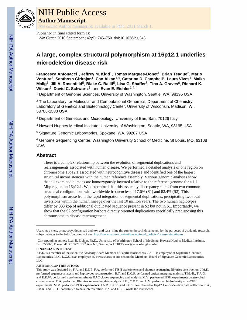
A large, complex structural polymorphism at 16p12.1 underlies microdeletion disease risk Francesca Antonacci 1 , Jeffrey M. Kidd 1 , Tomas Marques-Bonet 1 , Brian Teague 2 , Mario Ventura 3 , Santhosh Girirajan 1 , Can Alkan 1,4 , Catarina D. Campbell 1 , Laura Vives 1 , Maika Malig 1 , Jill A. Rosenfeld 5 , Blake C. Ballif 5 , Lisa G. Shaffer 5 , Tina A. Graves 6 , Richard K. Wilson 6 , David C. Schwartz 3 , and Evan E. Eichler 1,4,† 1 Department of Genome Sciences, University of Washington, Seattle, WA, 98195 USA 2 The Laboratory for Molecular and Computational Genomics, Department of Chemistry, Laboratory of Genetics and Biotechnology Center, University of Wisconsin, Madison, WI, 53706-1580 USA 3 Department of Genetics and Microbiology, University of Bari, Bari, 70126 Italy 4 Howard Hughes Medical Institute, University of Washington, Seattle, WA, 98195 USA 5 Signature Genomic Laboratories, Spokane, WA, 99207 USA 6 Genome Sequencing Center, Washington University School of Medicine, St Louis, MO, 63108 USA Abstract There is a complex relationship between the evolution of segmental duplications and rearrangements associated with human disease. We performed a detailed analysis of one region on chromosome 16p12.1 associated with neurocognitive disease and identified one of the largest structural inconsistencies with the human reference assembly. Various genomic analyses show that all examined humans are homozygously inverted relative to the reference genome for a 1.1- Mbp region on 16p12.1. We determined that this assembly discrepancy stems from two common structural configurations with worldwide frequencies of 17.6% (S1) and 82.4% (S2). This polymorphism arose from the rapid integration of segmental duplications, precipitating two local inversions within the human lineage over the last 10 million years. The two human haplotypes differ by 333 kbp of additional duplicated sequence present in S2 but not in S1. Importantly, we show that the S2 configuration harbors directly oriented duplications specifically predisposing this chromosome to disease rearrangement. Users may view, print, copy, download and text and data- mine the content in such documents, for the purposes of academic research, subject always to the full Conditions of use: http://www.nature.com/authors/editorial_policies/license.html#terms † Corresponding author: Evan E. Eichler, Ph.D., University of Washington School of Medicine, Howard Hughes Medical Institute, Box 355065, Foege S413C, 3720 15 th Ave NE, Seattle, WA 98195, [email protected]. FINANCIAL INTEREST E.E.E. is a member of the Scientific Advisory Board Member of Pacific Biosciences. J.A.R. is employee of Signature Genomic Laboratories, LLC. L.G.S. is an employee of, owns shares in and sits on the Members’ Board of Signature Genomic Laboratories, LLC. AUTHOR CONTRIBUTIONS This study was designed by F.A. and E.E.E. F.A. performed FISH experiments and shotgun sequencing libraries construction. J.M.K. performed sequence analysis and haplotypes reconstruction. B.T. and D.C.S. performed optical mapping analysis. T.M.-B., T.A.G. and R.K.W. performed non-human primate BAC clones sequencing and analysis. M.V. performed FISH experiments on stretched chromosomes. C.A. performed Illumina sequencing data analysis. S.G., C.D.C. and L.V. performed high-density arrayCGH experiments. M.M. performed PCR experiments. J.A.R., B.C.B. and L.G.S. contributed to 16p12.1 microdeletion data collection. F.A., J.M.K. and E.E.E. contributed to data interpretation. F.A. and E.E.E. wrote the manuscript. NIH Public Access Author Manuscript Nat Genet. Author manuscript; available in PMC 2011 March 1. Published in final edited form as: Nat Genet. 2010 September ; 42(9): 745–750. doi:10.1038/ng.643. NIH-PA Author Manuscript NIH-PA Author Manuscript NIH-PA Author Manuscript
-
Upload
independent -
Category
Documents
-
view
1 -
download
0
Transcript of A large and complex structural polymorphism at 16p12.1 underlies microdeletion disease risk
A large, complex structural polymorphism at 16p12.1 underliesmicrodeletion disease risk
Francesca Antonacci1, Jeffrey M. Kidd1, Tomas Marques-Bonet1, Brian Teague2, MarioVentura3, Santhosh Girirajan1, Can Alkan1,4, Catarina D. Campbell1, Laura Vives1, MaikaMalig1, Jill A. Rosenfeld5, Blake C. Ballif5, Lisa G. Shaffer5, Tina A. Graves6, Richard K.Wilson6, David C. Schwartz3, and Evan E. Eichler1,4,†
1 Department of Genome Sciences, University of Washington, Seattle, WA, 98195 USA2 The Laboratory for Molecular and Computational Genomics, Department of Chemistry,Laboratory of Genetics and Biotechnology Center, University of Wisconsin, Madison, WI,53706-1580 USA3 Department of Genetics and Microbiology, University of Bari, Bari, 70126 Italy4 Howard Hughes Medical Institute, University of Washington, Seattle, WA, 98195 USA5 Signature Genomic Laboratories, Spokane, WA, 99207 USA6 Genome Sequencing Center, Washington University School of Medicine, St Louis, MO, 63108USA
AbstractThere is a complex relationship between the evolution of segmental duplications andrearrangements associated with human disease. We performed a detailed analysis of one region onchromosome 16p12.1 associated with neurocognitive disease and identified one of the largeststructural inconsistencies with the human reference assembly. Various genomic analyses showthat all examined humans are homozygously inverted relative to the reference genome for a 1.1-Mbp region on 16p12.1. We determined that this assembly discrepancy stems from two commonstructural configurations with worldwide frequencies of 17.6% (S1) and 82.4% (S2). Thispolymorphism arose from the rapid integration of segmental duplications, precipitating two localinversions within the human lineage over the last 10 million years. The two human haplotypesdiffer by 333 kbp of additional duplicated sequence present in S2 but not in S1. Importantly, weshow that the S2 configuration harbors directly oriented duplications specifically predisposing thischromosome to disease rearrangement.
Users may view, print, copy, download and text and data- mine the content in such documents, for the purposes of academic research,subject always to the full Conditions of use: http://www.nature.com/authors/editorial_policies/license.html#terms
†Corresponding author: Evan E. Eichler, Ph.D., University of Washington School of Medicine, Howard Hughes Medical Institute,Box 355065, Foege S413C, 3720 15th Ave NE, Seattle, WA 98195, [email protected].
FINANCIAL INTERESTE.E.E. is a member of the Scientific Advisory Board Member of Pacific Biosciences. J.A.R. is employee of Signature GenomicLaboratories, LLC. L.G.S. is an employee of, owns shares in and sits on the Members’ Board of Signature Genomic Laboratories,LLC.
AUTHOR CONTRIBUTIONSThis study was designed by F.A. and E.E.E. F.A. performed FISH experiments and shotgun sequencing libraries construction. J.M.K.performed sequence analysis and haplotypes reconstruction. B.T. and D.C.S. performed optical mapping analysis. T.M.-B., T.A.G.and R.K.W. performed non-human primate BAC clones sequencing and analysis. M.V. performed FISH experiments on stretchedchromosomes. C.A. performed Illumina sequencing data analysis. S.G., C.D.C. and L.V. performed high-density arrayCGHexperiments. M.M. performed PCR experiments. J.A.R., B.C.B. and L.G.S. contributed to 16p12.1 microdeletion data collection. F.A.,J.M.K. and E.E.E. contributed to data interpretation. F.A. and E.E.E. wrote the manuscript.
NIH Public AccessAuthor ManuscriptNat Genet. Author manuscript; available in PMC 2011 March 1.
Published in final edited form as:Nat Genet. 2010 September ; 42(9): 745–750. doi:10.1038/ng.643.
NIH
-PA Author Manuscript
NIH
-PA Author Manuscript
NIH
-PA Author Manuscript
INTRODUCTIONNumerous studies have shown that segmental duplications and the flanking unique regionsare sites of both rare and common copy-number polymorphism (CNP) 1–3. Segmentalduplications are blocks of DNA >1 kb in size that occur at more than one site within thegenome and typically share a high level (>90%) of sequence identity 4–6. Duplicated blocksmay be substrates for non-allelic homologous recombination (NAHR) resulting in largestructural polymorphisms and chromosomal rearrangements that directly lead to genomicdisorders 5,7–13. NAHR between directly oriented segmental duplications results indeletions or reciprocal duplications of the genomic segment between them, whereas NAHRbetween inverted segmental duplications leads to an inversion of the intervening sequence.
Recently, a recurrent microdeletion on chromosome 16p12.1 was reported as a risk factorfor childhood intellectual disability and developmental delay 14. The microdeletion wasfound to be inherited in 95.6% of the cases and 24% of the probands carried an additionallarge duplication or deletion elsewhere in the genome. The data suggested a two-hit copy-number variation (CNV) model in which the 16p12.1 microdeletion results in severeneurodevelopmental phenotypes when coupled to an additional genetic, epigenetic, orenvironmental abnormality.
Using high density and targeted array-based comparative genomic hybridization (CGH)experiments, we mapped the 16p12.1 microdeletion breakpoints to large blocks of segmentalduplications, which we posited might mediate the recurrent rearrangement associated withdisease 15. The extensive copy-number variation and inconsistencies between the referencegenome and various genomic analyses, however, complicated breakpoint assessmentsuggesting that large alternative structural configurations might exist within the humanpopulation 16,17. We therefore investigated this region by conducting a detailed analysis byfluorescence in situ hybridization (FISH), arrayCGH, optical mapping, and sequencing oflarge-insert BAC clones in order to understand the extent of human genetic variation, itsorigin, and the impact on disease.
RESULTSResolution of a reference genome assembly error
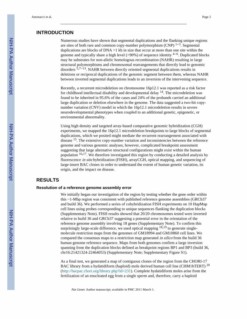
We initially began our investigation of the region by testing whether the gene order withinthis ~1-Mbp region was consistent with published reference genome assemblies (GRCb37and build 36). We performed a series of cohybridization FISH experiments on 10 HapMapcell lines using probes corresponding to unique sequences flanking the duplication blocks(Supplementary Note). FISH results showed that 20/20 chromosomes tested were invertedrelative to build 36 and GRCb37 suggesting a potential error in the orientation of thereference genome assembly involving 18 genes (Supplementary Note). To confirm thissurprisingly large-scale difference, we used optical mapping 18,19 to generate single-molecule restriction maps from the genomes of GM18994 and GM10860 cell lines. Wecompared the consensus maps to a restriction map generated in silico from the build 36human genome reference sequence. Maps from both genomes confirm a large inversionspanning from the duplication blocks defined as breakpoint regions BP1 and BP3 (build 36,chr16:21421324-22464053) (Supplementary Note; Supplementary Figure S1).
As a final test, we generated a map of contiguous clones of the region from the CHORI-17BAC library from a hydatidiform (haploid) mole derived human cell line (CHM1hTERT) 20
(http://bacpac.chori.org/library.php?id=231). Complete hydatidiform moles arise from thefertilization of an enucleated egg from a single sperm and, therefore, carry a haploid
Antonacci et al. Page 2
Nat Genet. Author manuscript; available in PMC 2011 March 1.
NIH
-PA Author Manuscript
NIH
-PA Author Manuscript
NIH
-PA Author Manuscript
complement of the human genome eliminating allelic variation that may confound mappingand assembly. We constructed a contiguous set of 10 BAC clones corresponding to this 1.6-Mbp region on 16p12.1 and then sequenced the inserts using Illumina technology. Wegenerated 406 Mbp of sequence (270-fold coverage) from these clones and aligned it to boththe human reference genome assembly and our reconstructed inverted version of the region(see below). The mapped sequence data from these clones were consistent with the entireregion being inverted within the hydatidiform mole (Supplementary Note). Thus, all threeanalyses indicate that orientation of the sequence between BP1 and BP3 should be flippedwith respect to published versions of the human genome (Figure 1).
Copy number and structural polymorphismOne of the predicted consequences of this inverted orientation of the human genome is thatthe location of previously described segmental duplications and copy-numberpolymorphisms change with respect to disease-associated breakpoints. The deletionbreakpoints associated with intellectual disability now map to BP1 and BP2 based on thecorrect orientation (build 36, chr16:21716331-22464053) (Figure 1A). These variableregions correspond, in part, to two sites of common copy-number polymorphism (CNP2156and CNP2157) identified in the HapMap sample collection by McCarroll and colleagues 2.Both loci have three reported copy-number (CN) states (diploid copy numbers of 2, 3, and4), with the highest copy-number state (CN = 4) having a frequency of 73% in Europeans(CEU), 95% in Yorubans (YRI), and 52% in Asians (CHB/JPT) (Supplementary Note). Weperformed a series of FISH and arrayCGH experiments to determine the absolute copynumber, location and extent of copy-number polymorphism within this region(Supplementary Note).
We analyzed 11 DNA control samples (Supplementary Note) using a customizedoligonucleotide microarray and found good correspondence between predicted CNP2157genotypes and expected signal intensity differences between samples (Figure 2). ArrayCGHdata for CNP2156 was less clear and the data suggested more extensive copy-numbervariation than was originally defined, although the location of this variation could not bedetermined based solely on hybridization data. We therefore designed a series of three-colorFISH experiments to investigate copy number and location. FISH analysis showed that theabsolute copy number of the 68-kbp segment corresponding to the distal region of CNP2157differed by a count of two with respect to previous reports (CN = 4, 5 and 6). Similarly,FISH analysis for the CNP2156 region showed an absolute count that is four copies greaterthan previously reported genotype estimates (Supplementary Note) 2. FISH mappingshowed that the variable sequences corresponding to CNP2156 and CNP2157 map adjacentto one another within the BP1 region (Supplementary Note; Figure 1). Thus, the tworeported CNP regions actually correspond to a single segment of variable sequence that hasbeen duplicatively transposed from BP3 to BP1. In total, these experiments revealed thepresence of two distinct structural configurations for the 16p12.1 region, which we refer toas S1 and S2, with the S2 haplotype showing the greater duplication complexity (Figure 1).
Since our analyses predicted a large, alternate structural polymorphism, we searchedGenBank for additional sequenced BACs from this region. We identified clones anchoredwithin the unique region distal to BP1 and constructed an alternate assembly from four BACclones not included in the human reference genome assembly (Supplementary Note). Weassembled a 433-kbp alternate sequence haplotype corresponding to most of the additionalduplicated sequence in BP1. Detailed comparisons with FISH, optical mapping and fosmidend-sequence pair data all provide strong support for the orientation and location of theadditional duplicated copies on the S2 chromosomal configuration (Supplementary Note).
Antonacci et al. Page 3
Nat Genet. Author manuscript; available in PMC 2011 March 1.
NIH
-PA Author Manuscript
NIH
-PA Author Manuscript
NIH
-PA Author Manuscript

The combined analysis identifies one of the largest, common copy-number polymorphismsin human euchromatin. We identify a total of 333 kbp of duplicated sequence that is specificto S2 when compared to the BP1 region of S1. Since this additional sequence is homologousto BP1 and BP2, this polymorphism creates additional direct and inverted blocks of highsequence identity making S2 prone to rearrangement events mediated by NAHR 15. Only theS2 configuration has segmental duplications in the direct orientation necessary to drive theformation of microdeletions associated with disease. We note that the S2-specific segmentalduplications at BP1 show the highest sequence identity (99.85%) with BP3 when comparedto BP2 (99.47%), consistent with a recent duplicative transposition event from BP3 placinga large inverted duplication within BP1.
Disease riskThe large-scale structural polymorphism between S1 and S2 allows us to make sometestable predictions regarding differences in susceptibility to microdeletion and disease.Since only the S2 configuration possesses directly oriented duplications, we hypothesizedthat the breakpoints would map to this 68-kbp segment and that only carriers of the S2configuration would be predisposed to the 16p12.1 microdeletion. Interestingly, we find thatthe S2 structure is the most common world-wide haplotype with frequencies of 97.5% inAfricans (YRI), 83.1% in Europeans (CEU) and 71.6% in Asian populations (CHB/JPT) 2
(Table 1). This general observation is confirmed by an examination of a larger group ofAfrican samples, which show the almost complete absence of the protective S1 haplotype(Supplementary Note). Thus, we hypothesize that African and European populations shouldbe more at risk for the 16p12.1 microdeletion “syndrome” than Asians.
One way to test if the S2 haplotype predisposes to microdeletion is to determine on whichstructure the microdeletion occurs. However, most of the identified cases are inherited andparental DNA for additional genotyping is not available 14. We therefore determined thestructural genotype present in each of the cases using array comparative genomichybridization. The presence of any S1/S1 homozygotes that also have 16p12.1 microdeletionwould be inconsistent with the proposed rearrangement structures and mechanism. Since theS2 haplotype has a more extended segmental duplication architecture than S1, differences inthe chromosomal configuration can be easily deduced (Figure 2). In particular, the S2-specific duplication block corresponding to the distal segment of CNP2157 (blue empty boxin Figure 2) has a diploid copy number of 2 in S1/S1 individuals, 3 in S1/S2 heterozygotes,and 4 in S2/S2 homozygotes.
We examined 35 microdeletion samples by arrayCGH using two reference samples withknown genotypes (NA15724 = S2/S2 and NA18956 = S1/S2). Self-identified ethnicity wasprovided for 27 of these patients (21 European and 6 African descent). Based on theobserved mean log2 values for the S2 specific duplication block, the genotype of eachsample was determined (Figure 2; Supplementary Figures S2 and S3; Supplementary Note).We found that 97% (34/35) of the cases were homozygous for the S2/S2 haplotype withonly a single heterozygous carrier (S1/S2) being identified in the patient population (Table1). This represents a significant enrichment of the S2 haplotype when matching for ethnicityof the sample collection (p-value = 0.0088, Hardy-Weinberg equilibrium test). Furthermore,arrayCGH data from 15/16 patients were consistent with breakpoints mapping within the 68-kbp S2-specific duplication (Supplementary Note). These combined data strongly suggestthat the S2, and not S1, haplotype predisposes to the 16p12.1 microdeletion associated withintellectual disability and neurocognitive disease (Table 1).
Antonacci et al. Page 4
Nat Genet. Author manuscript; available in PMC 2011 March 1.
NIH
-PA Author Manuscript
NIH
-PA Author Manuscript
NIH
-PA Author Manuscript

Evolutionary originIn order to investigate the ancestral configuration of the 16p12.1 region, we compared theorientation of the region in human with other non-human primate species. Notably, sequencecomparison of the orangutan (WUGSC 2.0.2/ponAbe2) and human sequence at 16p12.1revealed an expansion of the region in human due to the integration of segmentalduplications accompanied by two local inversions of 481 kbp and 142 kbp (SupplementaryNote). We tested for the presence of the larger inversion between BP1 and BP2 (481 kbp) byFISH analysis of cell lines from three chimpanzees (Pan troglodytes), three orangutans(Pongo pygmaeus), two gorillas (Gorilla gorilla) and one macaque (Macaca mulatta)(Supplementary Note). Macaque, orangutan and chimpanzee were found to be invertedwhen compared to the true human genome orientation suggesting that this represents thelikely ancestral state. To resolve the status of the smaller inversion (BP2-BP3) as well asduplications at the boundaries, we identified and sequenced nine large-insert chimpanzee,orangutan and gorilla BAC clones generating 1.8 Mbp of high quality ape sequence from theregion (Supplementary Figure S4). Our results indicated that all African great apes areinverted for the smaller BP2-BP3 interval (142 kbp) when compared to orangutan(ponAbe2) and macaque (rheMac2) genome assemblies. We conclude that the twoinversions occurred in the human-African great ape ancestor and that the region spanningBP1 to BP2 likely flipped back to the ancestral orientation in the chimpanzee lineage(Figure 3). Alternatively, the chimpanzee configuration may represent incomplete lineagesorting of an ancestral state.
Next, we compared the extent of segmental duplications in the 16p12.1 region amonghuman, chimpanzee, gorilla, orangutan, gibbon and macaque using a whole-genome shotgunsequence (WGS) detection method and interspecies arrayCGH 21,22. These analyses showedan expansion of segmental duplications among African great apes (human, chimpanzee,gorilla) with respect to orangutan, gibbon and macaque (Figure 4; Supplementary Note).Sequencing of orangutan BAC clones suggests that this region was largely devoid ofsegmental duplications in orangutan with the exception of BP1 where the composition of theduplication block differs radically from that of human (Figure 3). Sequence analysis of theBAC clones reveals the presence of duplicated sequences that are not present at this locationin human or chimpanzee with the exception of a 20-kbp segment corresponding to the NPIPgene. Overall, we determined that this particular region of 16p12.1 has increased in sizefrom 726 kbp to 1,259 kbp (S1) or 1,671 kbp (S2) during the last 10 million years primarilyas a result of a duplicative transposition of segmental duplications in the region. Our primateanalysis suggests that the region has become increasingly complex in the human-Africangreat ape lineage. The euchromatin has expanded 2.3 fold in size. These changes wereaccompanied by two local inversions of 481 kbp and 142 kbp in length creating the genomicarchitecture that now predisposes this region to microdeletion and neuropsychiatric disease.
DISCUSSIONOur analyses highlight three important properties regarding the organization and evolutionof the human genome. First, the data illustrate that the structure and copy number of evenvery large-scale, euchromatic regions may yet be unresolved in the human referenceassembly. We describe a large 333-kbp polymorphism that has changed in copy, orientationand location over a 1-Mbp portion of chromosome 16p12.1. With estimated frequencies of17.6% and 82.4% for the S1 and S2 configurations respectively, this represents one of thelargest copy-number polymorphisms mapping within human euchromatin.
We show that previous analyses of genome structural variation 2,3,16 have failed toadequately decipher the true structure and copy number of this polymorphism. In particular,CNP analysis using Affymetrix 6.0 microarrays 2 did not accurately determine the extent of
Antonacci et al. Page 5
Nat Genet. Author manuscript; available in PMC 2011 March 1.
NIH
-PA Author Manuscript
NIH
-PA Author Manuscript
NIH
-PA Author Manuscript

the CNP (76 kbp at CNP2156 and 146 kbp at CNP2157) due to the insensitivity of probesmapping within the duplicated regions. Moreover, FISH analyses revealed that the absolutecopy number was incorrect since a baseline copy number of 2 (diploid) was assumed torepresent the population average in previous analyses. This was compounded by the fact thatthe reference genome (GRC37 and build 36) are missing duplicated copies and present anorganization that can not be validated over 1.1 Mbp. We postulate that the presence of theinverted 333-kbp duplication polymorphism led to large-scale misassembly andmisorientation of sequence involving 18 genes (Figure 1). It may be somewhat surprisingthat such a large “error” has been uncovered nearly 10 years after the sequence andassembly of the human genome 23,24; however, it should be pointed out that at least fivedifferent types of molecular, optical mapping and cytogenetic analyses were required toresolve the architecture of this region. We anticipate that other regions of comparablecomplexity and variation will be uncovered and that similar, detailed analyses of large-insertclones will be required to ultimately resolve the true architecture of these regions.
Second, our comparative analyses of human and African great ape genomes reveal theevolutionary rapidity of these complex changes and their intimate association with largerchromosomal rearrangements. The 16p12.1 region has experienced a remarkable “bloating”of euchromatin, doubling the size of this region from 726 kbp to 1.6 Mbp as a result ofduplicative transposition of sequences from other portions of chromosome 16. Most of thesechanges occurred in a ~6 million year window of evolution before the emergence of humansand great apes as distinct lineages (Figure 3) consistent with the burst of duplications in theircommon ancestor 21. In concert with these changes, there have been multiple localinversions specific to humans and African great apes. These findings reinforce the strongassociation between evolutionary inversions and segmental duplications 25–28. It isinteresting that all of the 16p12.1 changes are associated with the spread of the human-greatape gene family morpheus (NPIP) 29. The core duplicon carrying this gene, LCR16a 30,maps to each of the breakpoint regions, including the boundaries of the complex copy-number polymorphism. Sequencing of large-insert ape clones suggests that these sequencesalso demarcate the breakpoints of the evolutionary inversions. Interestingly, the segmentalduplication associated with the NPIP gene family appears to be at the breakpoints of otherrecurrent microdeletions on chromosome 1631–36.
Third, our findings emphasize the impact of this genetic variation with respect to humanhealth and genomic susceptibility to neurocognitive disease. The dramatic changes in the S2chromosome architecture mean that it is the only configuration with homologous segmentalduplications in direct orientation flanking the disease-critical region. Accordingly, we findthat S1 chromosomes are depleted from microdeletion patients (p-value = 0.0088 rejectingHardy-Weinberg equilibrium) and that the breakpoints map specifically to the directlyoriented duplication on S2. Combined, these results suggest that S2 chromosomes are likelyto predispose to 16p12.1 microdeletion while the S1 chromosomes are immune to suchrearrangement. Interestingly, Asian HapMap samples are enriched for S1 chromosomespredicting that this particular cause of intellectual disability may be less common amongthese populations. These results bear striking similarity to another region of the humangenome on 17q21.31 where a largely Mediterranean-European-specific duplication arose indirect orientation predisposing H2 chromosomes to microdeletion associated with 17q21.31syndrome 26,37–40. In both of these cases, changes in disease-causing architecture are alsoassociated with inversions. We posit that this will be the underlying molecular basis forother associations that have been seen with inverted chromosomal haplotypes 41–43. Theseobservations emphasize the importance of correctly defining alternative human genomicconfigurations in order to assess variable risk of subsequent pathogenic rearrangements.Molecular cytogenetics, genomic approaches, and sequencing of long molecules from single
Antonacci et al. Page 6
Nat Genet. Author manuscript; available in PMC 2011 March 1.
NIH
-PA Author Manuscript
NIH
-PA Author Manuscript
NIH
-PA Author Manuscript
haplotypes remain the only way to correctly resolve these complex architectures of thehuman genome.
METHODSFISH analysis
Metaphase spreads were obtained from lymphoblast and fibroblast cell lines from 10 humanHapMap individuals (Coriell Cell Repository, Camden, NJ), three chimpanzees (Douglas;Veronica; Cochise), three orangutans (Susie, ISIS #71; PPY9; PPY6), two gorillas(AG20600; AG05251) and one macaque (MMU2). Stretched chromosomes were preparedaccording to Laan et al. 45. Briefly: cells were resuspended in hypotonic solution (HCM:hepes 100 mM; glycerol 1M; CaCl2 100mM; MgCl2 0.5M) for 15 minutes. The suspensionwas then centrifuged using a cytospin (800–1200 rpm for 5–15 minutes). FISH experimentswere performed using fosmid clones directly labeled by nick-translation with Cy3-dUTP(Perkin-Elmer), Cy5-dUTP (Perkin-Elmer), and fluorescein-dUTP (Enzo) as described byLichter et al. 46 with minor modifications. Briefly: 300 ng of labeled probe were used for theFISH experiments; hybridization was performed at 37°C in 2xSSC, 50% (v/v) formamide,10% (w/v) dextran sulphate, and 3 μg sonicated salmon sperm DNA, in a volume of 10 μL.Posthybridization washing was at 60°C in 0.1xSSC (three times, high stringency). Nucleiwere simultaneously DAPI stained. Digital images were obtained using a Leica DMRXA2epifluorescence microscope equipped with a cooled CCD camera (Princeton Instruments).DAPI, Cy3, Cy5 and fluorescein fluorescence signals, detected with specific filters, wererecorded separately as gray-scale images. Pseudocoloring and merging of images wereperformed using Adobe Photoshop software. A minimum of 50 interphase cells were scoredfor each inversion to statistically determine the orientation of the examined region.
Copy-number variation analysisMicroarray-based comparative genomic hybridization was performed on 35 16p12.1microdeletion cases with intellectual disability/developmental delay and congenitalmalformation 14. ArrayCGH experiments on 16p12.1 microdeletion samples and HapMapsamples were performed with custom, high-density oligonucleotide arrays (12-plexNimbleGen chip with a density of 1 probe per 40 bp within the 16p12.1 region; 4x180KAgilent chip targeted to copy-number polymorphic regions of the human genome (Campbellet al., unpublished), containing 50 probes in the CNP2157 at chr16:22533636-22618896).
The duplication content of human, chimpanzee, gorilla, orangutan, gibbon and macaque wasdetermined using the whole-genome shotgun sequence detection (WSSD) method 21,47. Wealso assessed copy-number differences in shared duplications by interspecific arraycomparative genomic hybridization as previously reported 21 (GEO Accession: GSE13885).We performed cross-species arrayCGH with human, Coriell GM15510 as a reference (GEOaccession number: GSE13884) using chimpanzee (Clint, Coriell S006006), gorilla (Bahati),orangutan (Susie, ISIS #71), and macaque (ID17573) samples.
Optical mappingWe examined the 16p12.1 locus in optical mapping data sets for two genomes, those ofHapMap panel members GM10860 and GM18994. Briefly, optical mapping 18,19,48,49 is awhole-genome, single-molecule system for the discovery and characterization of structuralvariation. Individual genomic DNA molecules are restriction mapped using lightmicroscopy, producing large data sets that are assembled into multi-megabase map contigscovering up to 98% of the euchromatic genome. These map contigs provide a global,detailed assessment of genome structure. We recovered consensus restriction maps matchingthe S1 haplotype from the GM18994 assembly and the S2 haplotype from GM10860; the
Antonacci et al. Page 7
Nat Genet. Author manuscript; available in PMC 2011 March 1.
NIH
-PA Author Manuscript
NIH
-PA Author Manuscript
NIH
-PA Author Manuscript
consensus maps, their alignments back to the build 36 reference sequence (build 36), and amontage of representative single molecule micrographs are depicted in SupplementaryFigure S1.
Illumina sequencingDNA was extracted from 10 BAC clones (CHORI-17) (Supplementary Note) from thegenome of a complete hydatidiform mole (CHM1hTERT) using Roche high pure plasmidisolation kit. 3 μg of DNA from each BAC were used for construction of a shotgunsequencing library as described previously 50,51 using adaptors for paired-end sequencing onan Illumina Genome Analyzer IIX (GAIIX). To allow the simultaneous sequencing ofmultiple BAC clones, we differentially ligated modified adaptors (Supplementary Note) toeach sample during library preparation, enabling the in silico separation of samples post-sequencing 52. We obtained a total of 34,206,404 76-bp reads (17,103,202 pairs) andseparated into 10 pools using 12-bp barcodes, resulting in 20,316,752 reads of length 64 bp.To control for contamination, we first aligned the reads to the E.coli reference genome (K12strain) using mrsFAST (http://mrsfast.sourceforge.net) allowing at most 4-bp mismatches.This experiment resulted in removing 2,363,518 reads (1,181,759 pairs) from considerationdue to contamination. The remaining reads (a total of 406 Mbp generated sequence) werethen mapped to the 16p12 region in build 36 and the S1 and S2 haplotype sequences that weconstructed. We tracked all possible map locations for the concordant pairs and discardedthe discordant mappings. This resulted in reliably mapping of 6,345,136 reads (3,172,568pairs; 406,088,704 bp of sequence) to 16p12, S1 and S2 reference sequences, correspondingto 270.7-fold coverage per BAC sequence on the average (min coverage: 132.5X, maxcoverage: 520.82X). Next, we merged the map locations of the overlapping pairs intocontiguous segments and removed any segment <2 kbp from analysis. We reasoned that thesmaller segments are mapping artifacts due to short repeats in the sequenced BAC clonesand the reference sequences (16p12, S1 and S2). Finally, we visualize the resulting segmentsusing the IGV software (Integrated Genomics Viewer, http://www.broadinstitute.org/igv).
Non-human primate BAC clone sequencingWe selected nine BAC clones from the libraries of chimpanzee (CH251), orangutan(CH276) and gorilla (CH255) genomes mapping to the 16p12.1 segmental duplications inhuman (Supplementary Note). We generated a clone shotgun sequence library andcompletely sequenced the insert of each clone. We aligned the sequence to the humangenome and to the S1 haplotype that we reconstructed with miropeats 53. Final annotationwith common repeats and DupMasker output 44 describing the composition of segmentalduplications was also included with customized Perl scripts.
Supplementary MaterialRefer to Web version on PubMed Central for supplementary material.
AcknowledgmentsWe thank P. Sudmant for useful discussions, G.M. Cooper and T. Brown for critical review of the manuscript, L.Zhou, Y. Fu, R. Shi, J. Wu, S. Shaull, and B.A. Roe for the sequencing of clone AC120780. This work wassupported by a National Science Foundation Graduate Research Fellowship [to J.M.K.], a Marie Curie fellowship[to T.M.-B.], and by the National Institutes of Health [T32 GM07215 and 5T15 LM007359 to B.T.; HG000225 toD.C.S.; HG002385 to E.E.E.]. E.E.E. is an investigator of the Howard Hughes Medical Institute.
Antonacci et al. Page 8
Nat Genet. Author manuscript; available in PMC 2011 March 1.
NIH
-PA Author Manuscript
NIH
-PA Author Manuscript
NIH
-PA Author Manuscript
References1. Itsara A, et al. Population analysis of large copy number variants and hotspots of human genetic
disease. Am J Hum Genet. 2009; 84:148–61. [PubMed: 19166990]
2. McCarroll SA, et al. Integrated detection and population-genetic analysis of SNPs and copy numbervariation. Nat Genet. 2008; 40:1166–74. [PubMed: 18776908]
3. Conrad DF, et al. Origins and functional impact of copy number variation in the human genome.Nature. 2009
4. Cheung VG, et al. Integration of cytogenetic landmarks into the draft sequence of the humangenome. Nature. 2001; 409:953–8. [PubMed: 11237021]
5. Bailey JA, et al. Recent segmental duplications in the human genome. Science (New York, N Y.2002; 297:1003–7.
6. Cheung J, et al. Genome-wide detection of segmental duplications and potential assembly errors inthe human genome sequence. Genome biology. 2003; 4:R25. [PubMed: 12702206]
7. Ji Y, Eichler EE, Schwartz S, Nicholls RD. Structure of chromosomal duplicons and their role inmediating human genomic disorders. Genome research. 2000; 10:597–610. [PubMed: 10810082]
8. Inoue K, Lupski JR. Molecular mechanisms for genomic disorders. Annual review of genomics andhuman genetics. 2002; 3:199–242.
9. Stankiewicz P, Lupski JR. Genome architecture, rearrangements and genomic disorders. Trends ingenetics. 2002; 18:74–82. [PubMed: 11818139]
10. Scherer SW, et al. Human chromosome 7: DNA sequence and biology. Science (New York, N Y.2003; 300:767–72.
11. Eichler EE, Clark RA, She X. An assessment of the sequence gaps: unfinished business in afinished human genome. Nature reviews. 2004; 5:345–54.
12. Shaw CJ, Lupski JR. Implications of human genome architecture for rearrangement-baseddisorders: the genomic basis of disease. Human molecular genetics. 2004; 13(Spec No 1):R57–64.[PubMed: 14764619]
13. Lupski JR, Stankiewicz P. Genomic disorders: molecular mechanisms for rearrangements andconveyed phenotypes. PLoS genetics. 2005; 1:e49. [PubMed: 16444292]
14. Girirajan S, et al. A recurrent 16p12.1 microdeletion supports a two-hit model for severedevelopmental delay. Nat Genet. 42:203–9. [PubMed: 20154674]
15. Lupski JR. Genomic disorders: structural features of the genome can lead to DNA rearrangementsand human disease traits. Trends Genet. 1998; 14:417–22. [PubMed: 9820031]
16. Kidd JM, et al. Mapping and sequencing of structural variation from eight human genomes.Nature. 2008; 453:56–64. [PubMed: 18451855]
17. Tuzun E, et al. Fine-scale structural variation of the human genome. Nat Genet. 2005; 37:727–32.[PubMed: 15895083]
18. Zhou S, et al. A single molecule scaffold for the maize genome. PLoS Genet. 2009; 5:e1000711.[PubMed: 19936062]
19. Teague B, et al. High-resolution human genome structure by single-molecule analysis. Proc NatlAcad Sci U S A. 107:10848–53. [PubMed: 20534489]
20. Fan JB, et al. Paternal origins of complete hydatidiform moles proven by whole genome single-nucleotide polymorphism haplotyping. Genomics. 2002; 79:58–62. [PubMed: 11827458]
21. Marques-Bonet T, et al. A burst of segmental duplications in the genome of the African great apeancestor. Nature. 2009; 457:877–81. [PubMed: 19212409]
22. Bailey JA, et al. Recent segmental duplications in the human genome. Science. 2002; 297:1003–7.[PubMed: 12169732]
23. Lander ES, et al. Initial sequencing and analysis of the human genome. Nature. 2001; 409:860–921. [PubMed: 11237011]
24. Martin J, et al. The sequence and analysis of duplication-rich human chromosome 16. Nature.2004; 432:988–94. [PubMed: 15616553]
25. Caceres M, Sullivan RT, Thomas JW. A recurrent inversion on the eutherian X chromosome. ProcNatl Acad Sci U S A. 2007; 104:18571–6. [PubMed: 18003915]
Antonacci et al. Page 9
Nat Genet. Author manuscript; available in PMC 2011 March 1.
NIH
-PA Author Manuscript
NIH
-PA Author Manuscript
NIH
-PA Author Manuscript

26. Zody MC, et al. Evolutionary toggling of the MAPT 17q21.31 inversion region. Nat Genet. 2008;40:1076–83. [PubMed: 19165922]
27. Kehrer-Sawatzki H, Cooper DN. Molecular mechanisms of chromosomal rearrangement duringprimate evolution. Chromosome Res. 2008; 16:41–56. [PubMed: 18293104]
28. Murphy WJ, et al. A rhesus macaque radiation hybrid map and comparative analysis with thehuman genome. Genomics. 2005; 86:383–95. [PubMed: 16039092]
29. Johnson ME, et al. Positive selection of a gene family during the emergence of humans andAfrican apes. Nature. 2001; 413:514–9. [PubMed: 11586358]
30. Jiang Z, et al. Ancestral reconstruction of segmental duplications reveals punctuated cores ofhuman genome evolution. Nat Genet. 2007; 39:1361–8. [PubMed: 17922013]
31. Weiss LA, et al. Association between microdeletion and microduplication at 16p11.2 and autism.N Engl J Med. 2008; 358:667–75. [PubMed: 18184952]
32. Kumar RA, et al. Recurrent 16p11.2 microdeletions in autism. Hum Mol Genet. 2008; 17:628–38.[PubMed: 18156158]
33. Ullmann R, et al. Array CGH identifies reciprocal 16p13.1 duplications and deletions thatpredispose to autism and/or mental retardation. Hum Mutat. 2007; 28:674–82. [PubMed:17480035]
34. Hannes FD, et al. Recurrent reciprocal deletions and duplications of 16p13.11: the deletion is a riskfactor for MR/MCA while the duplication may be a rare benign variant. J Med Genet. 2009;46:223–32. [PubMed: 18550696]
35. Ballif BC, et al. Discovery of a previously unrecognized microdeletion syndrome of 16p11.2-p12.2. Nat Genet. 2007; 39:1071–3. [PubMed: 17704777]
36. Bochukova EG, et al. Large, rare chromosomal deletions associated with severe early-onsetobesity. Nature. 463:666–70. [PubMed: 19966786]
37. Koolen DA, et al. A new chromosome 17q21.31 microdeletion syndrome associated with acommon inversion polymorphism. Nat Genet. 2006; 38:999–1001. [PubMed: 16906164]
38. Sharp AJ, et al. Discovery of previously unidentified genomic disorders from the duplicationarchitecture of the human genome. Nat Genet. 2006; 38:1038–42. [PubMed: 16906162]
39. Stefansson H, et al. A common inversion under selection in Europeans. Nat Genet. 2005; 37:129–37. [PubMed: 15654335]
40. Shaw-Smith C, et al. Microdeletion encompassing MAPT at chromosome 17q21.3 is associatedwith developmental delay and learning disability. Nat Genet. 2006; 38:1032–7. [PubMed:16906163]
41. Osborne LR, et al. A 1.5 million-base pair inversion polymorphism in families with Williams-Beuren syndrome. Nat Genet. 2001; 29:321–5. [PubMed: 11685205]
42. Giglio S, et al. Olfactory receptor-gene clusters, genomic-inversion polymorphisms, and commonchromosome rearrangements. Am J Hum Genet. 2001; 68:874–83. [PubMed: 11231899]
43. Antonacci F, et al. Characterization of six human disease-associated inversion polymorphisms.Hum Mol Genet. 2009; 18:2555–66. [PubMed: 19383631]
44. Jiang Z, Hubley R, Smit A, Eichler EE. DupMasker: a tool for annotating primate segmentalduplications. Genome Res. 2008; 18:1362–8. [PubMed: 18502942]
45. Laan M, et al. Mechanically stretched chromosomes as targets for high-resolution FISH mapping.Genome Res. 1995; 5:13–20. [PubMed: 8717051]
46. Lichter P, et al. High-resolution mapping of human chromosome 11 by in situ hybridization withcosmid clones. Science. 1990; 247:64–9. [PubMed: 2294592]
47. Bailey JA, Yavor AM, Massa HF, Trask BJ, Eichler EE. Segmental duplications: organization andimpact within the current human genome project assembly. Genome Res. 2001; 11:1005–17.[PubMed: 11381028]
48. Church DM, et al. Lineage-specific biology revealed by a finished genome assembly of the mouse.PLoS Biol. 2009; 7:e1000112. [PubMed: 19468303]
49. Zhou S, et al. Validation of rice genome sequence by optical mapping. BMC Genomics. 2007;8:278. [PubMed: 17697381]
Antonacci et al. Page 10
Nat Genet. Author manuscript; available in PMC 2011 March 1.
NIH
-PA Author Manuscript
NIH
-PA Author Manuscript
NIH
-PA Author Manuscript
50. Ng SB, et al. Targeted capture and massively parallel sequencing of 12 human exomes. Nature.2009; 461:272–6. [PubMed: 19684571]
51. Quail MA, et al. A large genome center’s improvements to the Illumina sequencing system. NatMethods. 2008; 5:1005–10. [PubMed: 19034268]
52. Craig DW, et al. Identification of genetic variants using bar-coded multiplexed sequencing. NatMethods. 2008; 5:887–93. [PubMed: 18794863]
53. Parsons JD. Miropeats: graphical DNA sequence comparisons. Comput Appl Biosci. 1995;11:615–9. [PubMed: 8808577]
Antonacci et al. Page 11
Nat Genet. Author manuscript; available in PMC 2011 March 1.
NIH
-PA Author Manuscript
NIH
-PA Author Manuscript
NIH
-PA Author Manuscript

Figure 1.Alternate structural configurations of the 16p12.1 region. (A) The organization in thereference genome (build 36, top schematic) is compared against two experimentallyvalidated structural configurations (S1 and S2). The locations of the inversion, copy-numberpolymorphisms 2 (CNP2156 and CNP2157), a rare (20/6712) non-pathogenic deletionvariant 1 and segmental duplications (colored rectangles) are indicated. Dashed empty boxesat the S1 structure correspond to regions duplicated in S2 but present in single copy in theS1 haplotype. The S1 and S2 structures differ because of the presence of the distalduplication segment (CNP2156 and CNP2157 at BP1) on the S2 haplotype. Based on thisstructure, the S1 configuration is predicted to be protective against occurrence of the16p12.1 pathogenic microdeletion. The red block corresponds to the 68-kbp segmentalduplication that likely mediates, through NAHR, the recurrent 16p12.1 microdeletion inpatients 14. Segments duplicated in a direct orientation are connected by green lines whilesequences duplicated in an inverted orientation are connected by blue lines. (B) Theorganization of the region was experimentally validated by optical mapping. SwaI single-molecule restriction maps are depicted and summarized for both configurations(Supplementary Note). (C) The large-scale orientation of each block was confirmed by FISHexperiments on interphase nuclei and stretched chromosomes (white rectangles) using
Antonacci et al. Page 12
Nat Genet. Author manuscript; available in PMC 2011 March 1.
NIH
-PA Author Manuscript
NIH
-PA Author Manuscript
NIH
-PA Author Manuscript
probes mapping at the red, blue and green segmental duplications. (D) A contig of 10 BACclones from the genome of the complete hydatidiform mole (CHM1hTERT) along the16p12.1 region was sequenced. All clones mapped against the S2 structure were concordant.
Antonacci et al. Page 13
Nat Genet. Author manuscript; available in PMC 2011 March 1.
NIH
-PA Author Manuscript
NIH
-PA Author Manuscript
NIH
-PA Author Manuscript

Figure 2.ArrayCGH data for 16p12.1 microdeletion patient samples and control HapMap samples(NA15510, NA12004 and NA18555). Probes with log2 ratios above or below a threshold of1.5 standard deviations from the normalized mean log2 ratio are colored green (duplication)or red (deletion), respectively. The positions of copy-number polymorphisms (CNP2156 andCNP2157) and segmental duplications are indicated. Blue empty boxes highlight the S2-specific duplications that have a diploid copy number of 2 in S1/S1 individuals, 3 in S1/S2heterozygotes, and 4 in S2/S2 homozygotes. HapMap sample NA18956 with S1/S2genotype was used as reference.
Antonacci et al. Page 14
Nat Genet. Author manuscript; available in PMC 2011 March 1.
NIH
-PA Author Manuscript
NIH
-PA Author Manuscript
NIH
-PA Author Manuscript
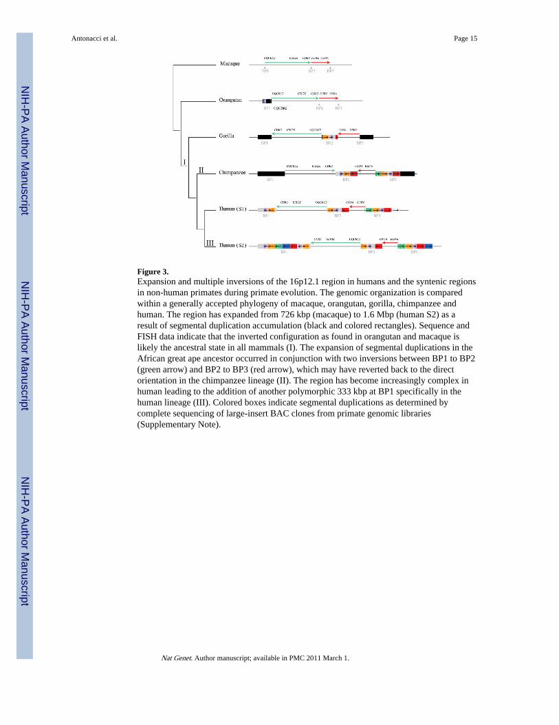
Figure 3.Expansion and multiple inversions of the 16p12.1 region in humans and the syntenic regionsin non-human primates during primate evolution. The genomic organization is comparedwithin a generally accepted phylogeny of macaque, orangutan, gorilla, chimpanzee andhuman. The region has expanded from 726 kbp (macaque) to 1.6 Mbp (human S2) as aresult of segmental duplication accumulation (black and colored rectangles). Sequence andFISH data indicate that the inverted configuration as found in orangutan and macaque islikely the ancestral state in all mammals (I). The expansion of segmental duplications in theAfrican great ape ancestor occurred in conjunction with two inversions between BP1 to BP2(green arrow) and BP2 to BP3 (red arrow), which may have reverted back to the directorientation in the chimpanzee lineage (II). The region has become increasingly complex inhuman leading to the addition of another polymorphic 333 kbp at BP1 specifically in thehuman lineage (III). Colored boxes indicate segmental duplications as determined bycomplete sequencing of large-insert BAC clones from primate genomic libraries(Supplementary Note).
Antonacci et al. Page 15
Nat Genet. Author manuscript; available in PMC 2011 March 1.
NIH
-PA Author Manuscript
NIH
-PA Author Manuscript
NIH
-PA Author Manuscript

Figure 4.Regions of segmental duplication based on read-depth mapping of whole-genome shotgunsequences (WGS) against the human genome. The figure shows an expansion of segmentalduplications in the African great apes (human, chimpanzee, gorilla) with respect toorangutan, gibbon and macaque. Also shown are the segmental duplications in humanannotated using SegDupMasker 44.
Antonacci et al. Page 16
Nat Genet. Author manuscript; available in PMC 2011 March 1.
NIH
-PA Author Manuscript
NIH
-PA Author Manuscript
NIH
-PA Author Manuscript
NIH
-PA Author Manuscript
NIH
-PA Author Manuscript
NIH
-PA Author Manuscript
Antonacci et al. Page 17
Table 1
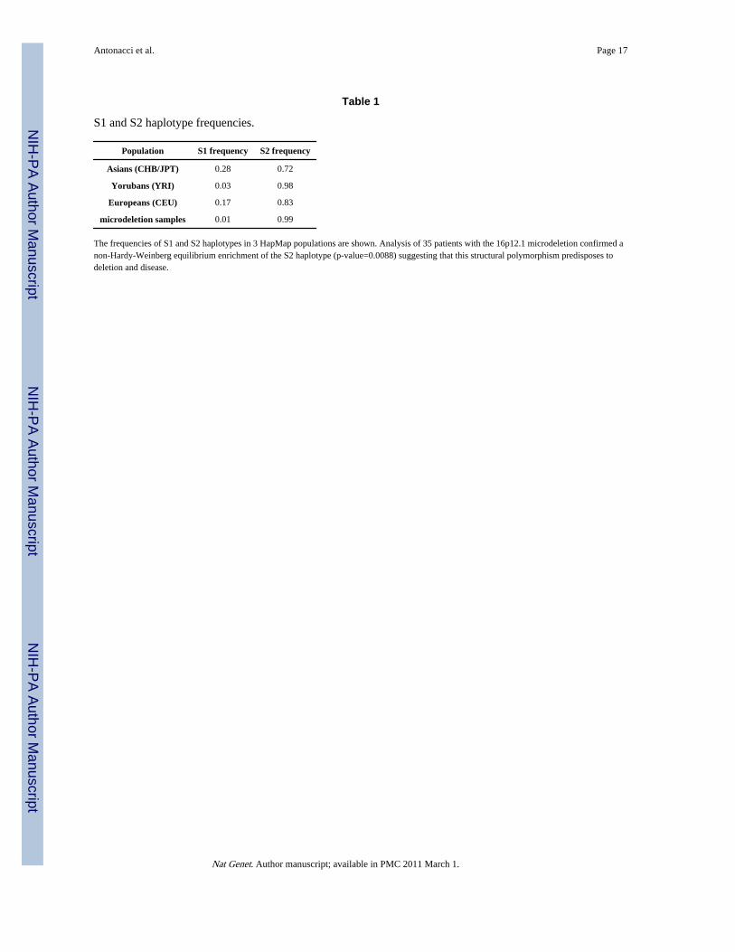
S1 and S2 haplotype frequencies.
Population S1 frequency S2 frequency
Asians (CHB/JPT) 0.28 0.72
Yorubans (YRI) 0.03 0.98
Europeans (CEU) 0.17 0.83
microdeletion samples 0.01 0.99
The frequencies of S1 and S2 haplotypes in 3 HapMap populations are shown. Analysis of 35 patients with the 16p12.1 microdeletion confirmed anon-Hardy-Weinberg equilibrium enrichment of the S2 haplotype (p-value=0.0088) suggesting that this structural polymorphism predisposes todeletion and disease.
Nat Genet. Author manuscript; available in PMC 2011 March 1.