RyR1 S-nitrosylation underlies environmental heat stroke and sudden death in Y522S RyR1 knockin mice
13
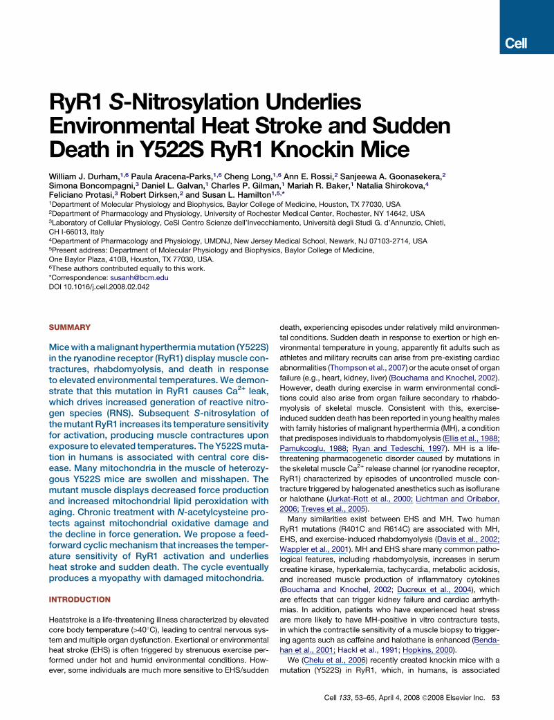
RyR1 S -Nitrosylation Underlies Environmental Heat Stroke and Sudden Death in Y522S RyR1 Knockin Mice William J. Durham, 1,6 Paula Aracena-Parks, 1,6 Cheng Long, 1,6 Ann E. Rossi, 2 Sanjeewa A. Goonasekera, 2 Simona Boncompagni, 3 Daniel L. Galvan, 1 Charles P. Gilman, 1 Mariah R. Baker, 1 Natalia Shirokova, 4 Feliciano Protasi, 3 Robert Dirksen, 2 and Susan L. Hamilton 1,5, * 1 Department of Molecular Physiology and Biophysics, Baylor College of Medicine, Houston, TX 77030, USA 2 Department of Pharmacology and Physiology, University of Rochester Medical Center, Rochester, NY 14642, USA 3 Laboratory of Cellular Physiology, CeSI Centro Scienze dell’Invecchiamento, Universita ` degli Studi G. d’Annunzio, Chieti, CH I-66013, Italy 4 Department of Pharmacology and Physiology, UMDNJ, New Jersey Medical School, Newark, NJ 07103-2714, USA 5 Present address: Department of Molecular Physiology and Biophysics, Baylor College of Medicine, One Baylor Plaza, 410B, Houston, TX 77030, USA. 6 These authors contributed equally to this work. *Correspondence: [email protected] DOI 10.1016/j.cell.2008.02.042 SUMMARY Mice with a malignant hyperthermia mutation (Y522S) in the ryanodine receptor (RyR1) display muscle con- tractures, rhabdomyolysis, and death in response to elevated environmental temperatures. We demon- strate that this mutation in RyR1 causes Ca 2+ leak, which drives increased generation of reactive nitro- gen species (RNS). Subsequent S-nitrosylation of the mutant RyR1 increases its temperature sensitivity for activation, producing muscle contractures upon exposure to elevated temperatures. The Y522S muta- tion in humans is associated with central core dis- ease. Many mitochondria in the muscle of heterozy- gous Y522S mice are swollen and misshapen. The mutant muscle displays decreased force production and increased mitochondrial lipid peroxidation with aging. Chronic treatment with N-acetylcysteine pro- tects against mitochondrial oxidative damage and the decline in force generation. We propose a feed- forward cyclic mechanism that increases the temper- ature sensitivity of RyR1 activation and underlies heat stroke and sudden death. The cycle eventually produces a myopathy with damaged mitochondria. INTRODUCTION Heatstroke is a life-threatening illness characterized by elevated core body temperature (>40 C), leading to central nervous sys- tem and multiple organ dysfunction. Exertional or environmental heat stroke (EHS) is often triggered by strenuous exercise per- formed under hot and humid environmental conditions. How- ever, some individuals are much more sensitive to EHS/sudden death, experiencing episodes under relatively mild environmen- tal conditions. Sudden death in response to exertion or high en- vironmental temperature in young, apparently fit adults such as athletes and military recruits can arise from pre-existing cardiac abnormalities (Thompson et al., 2007) or the acute onset of organ failure (e.g., heart, kidney, liver) (Bouchama and Knochel, 2002). However, death during exercise in warm environmental condi- tions could also arise from organ failure secondary to rhabdo- myolysis of skeletal muscle. Consistent with this, exercise- induced sudden death has been reported in young healthy males with family histories of malignant hyperthermia (MH), a condition that predisposes individuals to rhabdomyolysis (Ellis et al., 1988; Pamukcoglu, 1988; Ryan and Tedeschi, 1997). MH is a life- threatening pharmacogenetic disorder caused by mutations in the skeletal muscle Ca 2+ release channel (or ryanodine receptor, RyR1) characterized by episodes of uncontrolled muscle con- tracture triggered by halogenated anesthetics such as isoflurane or halothane (Jurkat-Rott et al., 2000; Lichtman and Oribabor, 2006; Treves et al., 2005). Many similarities exist between EHS and MH. Two human RyR1 mutations (R401C and R614C) are associated with MH, EHS, and exercise-induced rhabdomyolysis (Davis et al., 2002; Wappler et al., 2001). MH and EHS share many common patho- logical features, including rhabdomyolysis, increases in serum creatine kinase, hyperkalemia, tachycardia, metabolic acidosis, and increased muscle production of inflammatory cytokines (Bouchama and Knochel, 2002; Ducreux et al., 2004), which are effects that can trigger kidney failure and cardiac arrhyth- mias. In addition, patients who have experienced heat stress are more likely to have MH-positive in vitro contracture tests, in which the contractile sensitivity of a muscle biopsy to trigger- ing agents such as caffeine and halothane is enhanced (Benda- han et al., 2001; Hackl et al., 1991; Hopkins, 2000). We (Chelu et al., 2006) recently created knockin mice with a mutation (Y522S) in RyR1, which, in humans, is associated Cell 133, 53–65, April 4, 2008 ª2008 Elsevier Inc. 53
-
Upload
independent -
Category
Documents
-
view
4 -
download
0
Transcript of RyR1 S-nitrosylation underlies environmental heat stroke and sudden death in Y522S RyR1 knockin mice
RyR1 S-Nitrosylation UnderliesEnvironmental Heat Stroke and SuddenDeath in Y522S RyR1 Knockin MiceWilliam J. Durham,1,6 Paula Aracena-Parks,1,6 Cheng Long,1,6 Ann E. Rossi,2 Sanjeewa A. Goonasekera,2
Simona Boncompagni,3 Daniel L. Galvan,1 Charles P. Gilman,1 Mariah R. Baker,1 Natalia Shirokova,4
Feliciano Protasi,3 Robert Dirksen,2 and Susan L. Hamilton1,5,*1Department of Molecular Physiology and Biophysics, Baylor College of Medicine, Houston, TX 77030, USA2Department of Pharmacology and Physiology, University of Rochester Medical Center, Rochester, NY 14642, USA3Laboratory of Cellular Physiology, CeSI Centro Scienze dell’Invecchiamento, Universita degli Studi G. d’Annunzio, Chieti,
CH I-66013, Italy4Department of Pharmacology and Physiology, UMDNJ, New Jersey Medical School, Newark, NJ 07103-2714, USA5Present address: Department of Molecular Physiology and Biophysics, Baylor College of Medicine,
One Baylor Plaza, 410B, Houston, TX 77030, USA.6These authors contributed equally to this work.
*Correspondence: [email protected] 10.1016/j.cell.2008.02.042
SUMMARY
Mice with a malignant hyperthermia mutation (Y522S)in the ryanodine receptor (RyR1) display muscle con-tractures, rhabdomyolysis, and death in responseto elevated environmental temperatures. We demon-strate that this mutation in RyR1 causes Ca2+ leak,which drives increased generation of reactive nitro-gen species (RNS). Subsequent S-nitrosylation ofthe mutant RyR1 increases its temperature sensitivityfor activation, producing muscle contractures uponexposure to elevated temperatures. The Y522S muta-tion in humans is associated with central core dis-ease. Many mitochondria in the muscle of heterozy-gous Y522S mice are swollen and misshapen. Themutant muscle displays decreased force productionand increased mitochondrial lipid peroxidation withaging. Chronic treatment with N-acetylcysteine pro-tects against mitochondrial oxidative damage andthe decline in force generation. We propose a feed-forward cyclic mechanism that increases the temper-ature sensitivity of RyR1 activation and underliesheat stroke and sudden death. The cycle eventuallyproduces a myopathy with damaged mitochondria.
INTRODUCTION
Heatstroke is a life-threatening illness characterized by elevated
core body temperature (>40�C), leading to central nervous sys-
tem and multiple organ dysfunction. Exertional or environmental
heat stroke (EHS) is often triggered by strenuous exercise per-
formed under hot and humid environmental conditions. How-
ever, some individuals are much more sensitive to EHS/sudden
death, experiencing episodes under relatively mild environmen-
tal conditions. Sudden death in response to exertion or high en-
vironmental temperature in young, apparently fit adults such as
athletes and military recruits can arise from pre-existing cardiac
abnormalities (Thompson et al., 2007) or the acute onset of organ
failure (e.g., heart, kidney, liver) (Bouchama and Knochel, 2002).
However, death during exercise in warm environmental condi-
tions could also arise from organ failure secondary to rhabdo-
myolysis of skeletal muscle. Consistent with this, exercise-
induced sudden death has been reported in young healthy males
with family histories of malignant hyperthermia (MH), a condition
that predisposes individuals to rhabdomyolysis (Ellis et al., 1988;
Pamukcoglu, 1988; Ryan and Tedeschi, 1997). MH is a life-
threatening pharmacogenetic disorder caused by mutations in
the skeletal muscle Ca2+ release channel (or ryanodine receptor,
RyR1) characterized by episodes of uncontrolled muscle con-
tracture triggered by halogenated anesthetics such as isoflurane
or halothane (Jurkat-Rott et al., 2000; Lichtman and Oribabor,
2006; Treves et al., 2005).
Many similarities exist between EHS and MH. Two human
RyR1 mutations (R401C and R614C) are associated with MH,
EHS, and exercise-induced rhabdomyolysis (Davis et al., 2002;
Wappler et al., 2001). MH and EHS share many common patho-
logical features, including rhabdomyolysis, increases in serum
creatine kinase, hyperkalemia, tachycardia, metabolic acidosis,
and increased muscle production of inflammatory cytokines
(Bouchama and Knochel, 2002; Ducreux et al., 2004), which
are effects that can trigger kidney failure and cardiac arrhyth-
mias. In addition, patients who have experienced heat stress
are more likely to have MH-positive in vitro contracture tests,
in which the contractile sensitivity of a muscle biopsy to trigger-
ing agents such as caffeine and halothane is enhanced (Benda-
han et al., 2001; Hackl et al., 1991; Hopkins, 2000).
We (Chelu et al., 2006) recently created knockin mice with a
mutation (Y522S) in RyR1, which, in humans, is associated
Cell 133, 53–65, April 4, 2008 ª2008 Elsevier Inc. 53
with MH, a high incidence of central cores, and type I fiber type
predominance (Quane et al., 1994). Heterozygous mice
(RyR1Y522S/wt) are more sensitive to developing skeletal muscle
contractures in response to caffeine treatment in vitro and to iso-
flurane inhalation in vivo, both hallmarks of MH. In addition, heat
alone and/or exercise under warm conditions triggers rhabdo-
myolysis and death in RyR1Y522S/wt mice (Chelu et al., 2006).
Although the mice undergo sustained whole body contractures
upon heat exposure, death also frequently occurs in the absence
of detectable sustained contractures. The molecular and cellular
mechanisms whereby elevated temperatures with or without
exercise leads to death of these mice are unknown. Here we
demonstrate that enhanced Ca2+ leak from mutant RyR1 Ca2+
release channels increases oxidative/nitrosative stress, leading
to S-nitrosylation of RyR1 that further enhances Ca2+ leak and
increases susceptibility to heat-induced sudden death.
RESULTS
Heat Sensitivity of RyR1Y522S/wt MiceRyR1Y522S/wt mice exposed to elevated environmental tempera-
tures undergo an MH-like response characterized by rapid rhab-
domyolysis and death (Chelu et al., 2006). Upon exposure to a
41�C heat challenge, the rectal temperature of anesthetized (non-
triggering anesthetic etomidate) RyR1Y522S/wt mice increases
more rapidly than in RyR1wt/wt mice (Figures 1A and 1B), suggest-
ing that the mutant mice display enhanced metabolism and/or
heat-induced muscle tension rapidly during heat exposure.
Both enhanced metabolism and heat-induced muscle tension
are likely to occur since (1) RyR1Y522S/wt mice exhibit a higher
metabolic rate at 32�C than wild-type mice (Figure 1C), and (2)
solei from RyR1Y522S/wt mice display increased basal stress at
much lower temperatures than solei from wild-type animals (Fig-
ures 1D and 1E). Both enhanced metabolic rate and increased
basal stress are likely to increase skeletal muscle production of
reactive oxygen species (ROS) and reactive nitrogen species
(RNS). To assess the role of ROS and RNS in the response of
RyR1Y522S/wt mice to elevated temperatures, we tested the
effects of treating mice for 3–5 days with either the antioxidant
N-acetylcysteine (NAC, a precursor for glutathione synthesis) or
the nitric oxide synthase (NOS) inhibitor, N (U)-nitro-L-arginine
methyl ester (L-NAME). Both NAC and L-NAME delayed the rapid
rise in core temperature in mutant mice (Figure 1B) and de-
creased the temperature sensitivity of basal tension of isolated
solei (Figure 1E). NAC and L-NAME administered together did
not improve the temperature response above either agent alone,
and therefore combined treatment was not pursued further. NAC
treatment reduces both ROS and RNS, and L-NAME prevents
both RNS and ROS production by NOS (Clark et al., 2004; Pou
et al., 1999), suggesting that either or both may be involved in
the sensitization of RyR1Y522S/wt mice to temperature.
Oxidative/Nitrosative Stress in RyR1Y522S/wt MuscleTo assess oxidative/nitrosative stress in the muscle of the
RyR1Y522S/wt mice, we measured levels of the primary intracellu-
lar antioxidant buffer glutathione (GSH), its oxidized form, gluta-
thione disulfide (GSSG), and the GSH/GSSG ratio in skeletal
muscle homogenates from mice not exposed to elevated tem-
54 Cell 133, 53–65, April 4, 2008 ª2008 Elsevier Inc.
peratures. RyR1Y522S/wt muscle exhibits profound basal oxida-
tive stress, with approximately a 50% reduction in both total
GSH content (Figure S1) and the ratio GSH/GSSG (Figure 1F).
A decreased GSH/GGSG ratio could reflect changes in ROS
and/or RNS levels in muscle. Pretreatment of RyR1Y522S/wt
mice with either NAC or L-NAME partially restores GSH levels
and GSH/GSSG (Figure 1F), suggesting that part of the effect
is due to RNS production.
To assess ROS and RNS production in myotubes derived from
RyR1Y522S/wt mice, we used confocal imaging to compare, re-
spectively, the fluorescence of 5-carboxy-20,70-dichlorodihydro-
fluorescein (DCF) and 4-amino-5-methylamino-20,70-difluoro-
fluorescein (DAF) at room temperature and 37�C. In RyR1Y522S/wt
myotubes, we found a significant temperature dependent
increase in ROS (Figures 1G and 1H) and RNS (Figures 1I and
1J). Both increases in RNS and ROS are blocked by treatment
with ryanodine and GSH ethyl ester (GSHEE), a membrane per-
meant form of glutathione (Figures 1G–1J). The ability of GSHEE
to block temperature dependent increases in DAF fluorescence
suggests that GSHEE treatment also reduces RNS. Importantly,
L-NNA (N[U]-nitro-L-arginine), an inhibitor of nitric oxide syn-
thase (NOS), blocks temperature-dependent increases in DAF
fluorescence but does not significantly alter ROS production
as assessed by DCF fluorescence (Figures 1G–1J). While
L-NNA inhibits superoxide production by NOS (Clark et al.,
2004), the absence of an inhibition of DCF fluorescence by
L-NNA in our experiments suggests that DAF fluorescence in
myotubes specifically reflects RNS production. Overall, these
data demonstrate a temperature-dependent increase in both
ROS and RNS production in RyR1Y522S/wt myotubes. Inhibition
by ryanodine of the temperature-dependent increase in DCF
and DAF fluorescence suggests that both ROS and RNS produc-
tion are stimulated by RyR1-mediated Ca2+ release from the
sarcoplasmic reticulum (SR). Two obvious questions arise from
these studies: (1) Why is there increased ROS/RNS production
with temperature in RyR1Y522S/wt myotubes, and (2) how do
increased ROS/RNS levels relate to the enhanced temperature
sensitivity of these mice?
Temperature-Dependent Increases in RestingCa2+ Levels in Muscle of RyR1Y522S/wt MiceTo define the relationship between the mutation in RyR1 and
increased oxidative/nitrosative stress, we determined the effects
of temperature and antioxidants on resting Ca2+ levels in myo-
tubes from RyR1Y522S/wt and RyR1wt/wt mice. We previously
(Chelu et al., 2006) found enhanced caffeine sensitivity in
RyR1Y522S/wt myotubes in the absence of a change in global rest-
ing Ca2+ levels at room temperature. However, the mutation in
RyR1 is highly likely to produce local changes in Ca2+ that are
rapidly sequestered back into the SR. We re-examined the ef-
fects of the Y522S mutation on Ca2+ homeostasis in myotubes
(Figures 2A–2C) and muscle fibers (Figure 2D) at more physiolog-
ical temperatures. Cytosolic Ca2+ levels increase with tempera-
ture to a much greater extent in RyR1Y522S/wt than in wild-type
myotubes and muscle fibers (Figures 2A–2D). Figure 2C quan-
tifies temperature-dependent changes in the intracellular con-
centration of free Ca2+ (in nM) in RyR1wt/wt and RyR1Y522S/wt
myotubes. The lack of a statistically significant difference in
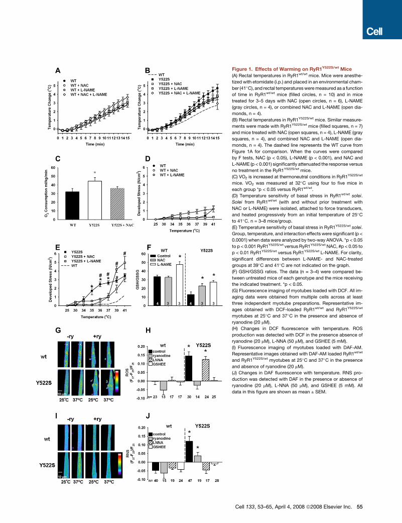
Figure 1. Effects of Warming on RyR1Y522S/wt Mice
(A) Rectal temperatures in RyR1wt/wt mice. Mice were anesthe-
tized with etomidate (i.p.) and placed in an environmental cham-
ber (41�C), and rectal temperatures were measured as a function
of time in RyR1wt/wt mice (filled circles, n = 10) and in mice
treated for 3–5 days with NAC (open circles, n = 6), L-NAME
(gray circles, n = 4), or combined NAC and L-NAME (open dia-
monds, n = 4).
(B) Rectal temperatures in RyR1Y522S/wt mice. Similar measure-
ments were made with RyR1Y522S/wt mice (filled squares, n = 7)
and mice treated with NAC (open squares, n = 4), L-NAME (gray
squares, n = 4), and combined NAC and L-NAME (open dia-
monds, n = 4). The dashed line represents the WT curve from
Figure 1A for comparison. When the curves were compared
by F tests, NAC (p < 0.05), L-NAME (p < 0.001), and NAC and
L-NAME (p < 0.001) significantly attenuated the response versus
no treatment in the RyR1Y522S/wt mice.
(C) VO2 is increased at thermoneutral conditions in RyR1Y522S/wt
mice. VO2 was measured at 32�C using four to five mice in
each group *p < 0.05 versus RyR1wt/wt.
(D) Temperature sensitivity of basal stress in RyR1wt/wt solei.
Solei from RyR1wt/wt (with and without prior treatment with
NAC or L-NAME) were isolated, attached to force transducers,
and heated progressively from an initial temperature of 25�C
to 41�C. n = 3–8 mice/group.
(E) Temperature sensitivity of basal stress in RyR1Y522S/wt solei.
Group, temperature, and interaction effects were significant (p <
0.0001) when data were analyzed by two-way ANOVA. *p < 0.05
to p < 0.001 RyR1Y522S/wt versus RyR1Y522S/wt NAC, #p < 0.05 to
p < 0.01 RyR1Y522S/wt versus RyR1Y522S/wt L-NAME. For clarity,
significant differences between L-NAME- and NAC-treated
groups at 39�C and 41�C are not indicated on the graph.
(F) GSH/GSSG ratios. The data (n = 3–4) were compared be-
tween untreated mice of each genotype and the mice receiving
the indicated treatment. *p < 0.05.
(G) Fluorescence imaging of myotubes loaded with DCF. All im-
aging data were obtained from multiple cells across at least
three independent myotube preparations. Representative im-
ages obtained with DCF-loaded RyR1wt/wt and RyR1Y522S/wt
myotubes at 25�C and 37�C in the presence and absence of
ryanodine (20 mM).
(H) Changes in DCF fluorescence with temperature. ROS
production was detected with DCF in the presence absence of
ryanodine (20 mM), L-NNA (50 mM), and GSHEE (5 mM).
(I) Fluorescence imaging of myotubes loaded with DAF-AM.
Representative images obtained with DAF-AM loaded RyR1wt/wt
and RyR1Y522S/wt myotubes at 25�C and 37�C in the presence
and absence of ryanodine (20 mM).
(J) Changes in DAF fluorescence with temperature. RNS pro-
duction was detected with DAF in the presence or absence of
ryanodine (20 mM), L-NNA (50 mM), and GSHEE (5 mM). All
data in this figure are shown as mean ± SEM.
Cell 133, 53–65, April 4, 2008 ª2008 Elsevier Inc. 55
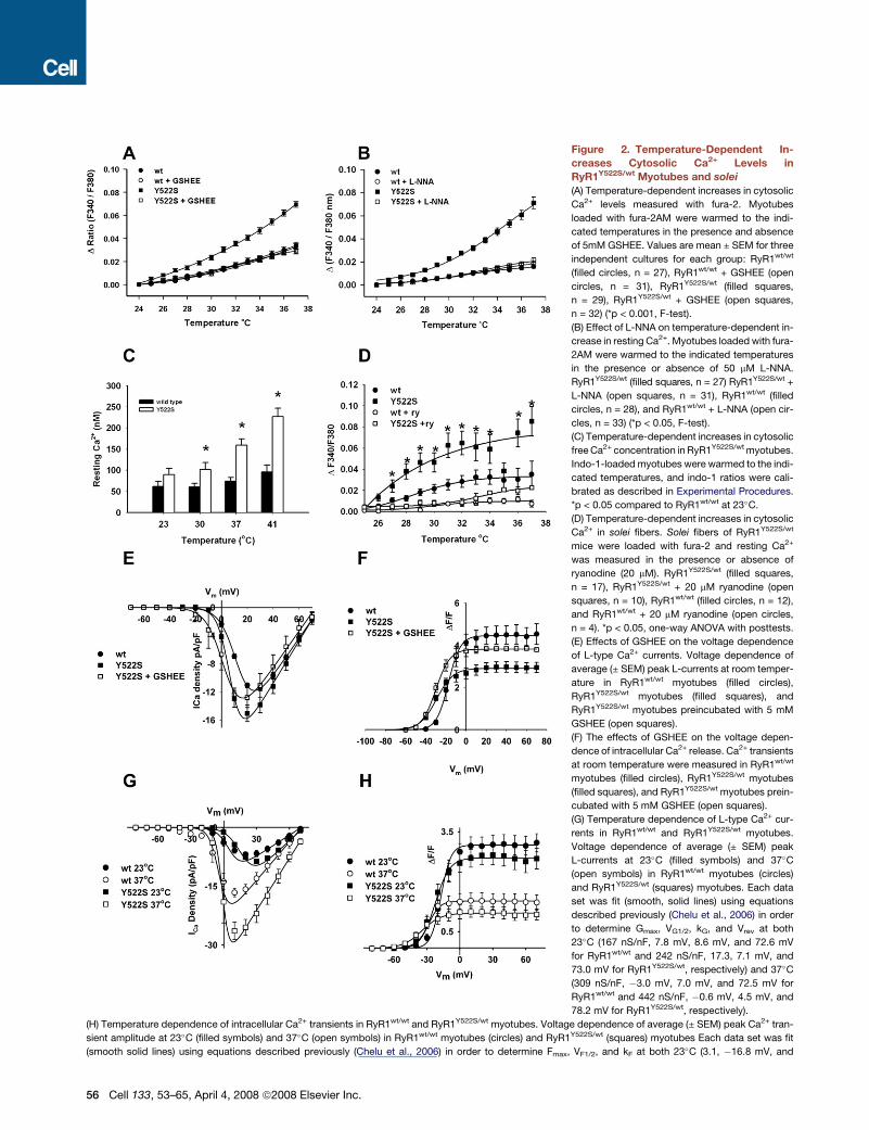
Figure 2. Temperature-Dependent In-
creases Cytosolic Ca2+ Levels in
RyR1Y522S/wt Myotubes and solei
(A) Temperature-dependent increases in cytosolic
Ca2+ levels measured with fura-2. Myotubes
loaded with fura-2AM were warmed to the indi-
cated temperatures in the presence and absence
of 5mM GSHEE. Values are mean ± SEM for three
independent cultures for each group: RyR1wt/wt
(filled circles, n = 27), RyR1wt/wt + GSHEE (open
circles, n = 31), RyR1Y522S/wt (filled squares,
n = 29), RyR1Y522S/wt + GSHEE (open squares,
n = 32) (*p < 0.001, F-test).
(B) Effect of L-NNA on temperature-dependent in-
crease in resting Ca2+. Myotubes loaded with fura-
2AM were warmed to the indicated temperatures
in the presence or absence of 50 mM L-NNA.
RyR1Y522S/wt (filled squares, n = 27) RyR1Y522S/wt +
L-NNA (open squares, n = 31), RyR1wt/wt (filled
circles, n = 28), and RyR1wt/wt + L-NNA (open cir-
cles, n = 33) (*p < 0.05, F-test).
(C) Temperature-dependent increases in cytosolic
free Ca2+ concentration in RyR1Y522S/wt myotubes.
Indo-1-loaded myotubes were warmed to the indi-
cated temperatures, and indo-1 ratios were cali-
brated as described in Experimental Procedures.
*p < 0.05 compared to RyR1wt/wt at 23�C.
(D) Temperature-dependent increases in cytosolic
Ca2+ in solei fibers. Solei fibers of RyR1Y522S/wt
mice were loaded with fura-2 and resting Ca2+
was measured in the presence or absence of
ryanodine (20 mM). RyR1Y522S/wt (filled squares,
n = 17), RyR1Y522S/wt + 20 mM ryanodine (open
squares, n = 10), RyR1wt/wt (filled circles, n = 12),
and RyR1wt/wt + 20 mM ryanodine (open circles,
n = 4). *p < 0.05, one-way ANOVA with posttests.
(E) Effects of GSHEE on the voltage dependence
of L-type Ca2+ currents. Voltage dependence of
average (± SEM) peak L-currents at room temper-
ature in RyR1wt/wt myotubes (filled circles),
RyR1Y522S/wt myotubes (filled squares), and
RyR1Y522S/wt myotubes preincubated with 5 mM
GSHEE (open squares).
(F) The effects of GSHEE on the voltage depen-
dence of intracellular Ca2+ release. Ca2+ transients
at room temperature were measured in RyR1wt/wt
myotubes (filled circles), RyR1Y522S/wt myotubes
(filled squares), and RyR1Y522S/wt myotubes prein-
cubated with 5 mM GSHEE (open squares).
(G) Temperature dependence of L-type Ca2+ cur-
rents in RyR1wt/wt and RyR1Y522S/wt myotubes.
Voltage dependence of average (± SEM) peak
L-currents at 23�C (filled symbols) and 37�C
(open symbols) in RyR1wt/wt myotubes (circles)
and RyR1Y522S/wt (squares) myotubes. Each data
set was fit (smooth, solid lines) using equations
described previously (Chelu et al., 2006) in order
to determine Gmax, VG1/2, kG, and Vrev at both
23�C (167 nS/nF, 7.8 mV, 8.6 mV, and 72.6 mV
for RyR1wt/wt and 242 nS/nF, 17.3, 7.1 mV, and
73.0 mV for RyR1Y522S/wt, respectively) and 37�C
(309 nS/nF, �3.0 mV, 7.0 mV, and 72.5 mV for
RyR1wt/wt and 442 nS/nF, �0.6 mV, 4.5 mV, and
78.2 mV for RyR1Y522S/wt, respectively).
(H) Temperature dependence of intracellular Ca2+ transients in RyR1wt/wt and RyR1Y522S/wt myotubes. Voltage dependence of average (± SEM) peak Ca2+ tran-
sient amplitude at 23�C (filled symbols) and 37�C (open symbols) in RyR1wt/wt myotubes (circles) and RyR1Y522S/wt (squares) myotubes Each data set was fit
(smooth solid lines) using equations described previously (Chelu et al., 2006) in order to determine Fmax, VF1/2, and kF at both 23�C (3.1, �16.8 mV, and
56 Cell 133, 53–65, April 4, 2008 ª2008 Elsevier Inc.
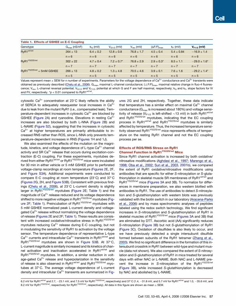
Table 1. Effects of GSHEE on E-C Coupling
Genotype Gmax (nS/nF) kG (mV) VG1/2 (mV) Vrev (mV) (DF/F)max kF (mV) VF1/2 (mV)
RyR1wt/wt 264 ± 13 6.4 ± 0.2 12.8 ± 0.8 76.8 ± 1.7 4.5 ± 0.4 5.4 ± 0.84 �16.9 ± 1.4
n = 6 n = 6 n = 6 n = 6 n = 6 n = 6 n = 6
RyR1Y522S/wt 302 ± 22 4.7 ± 0.4 7.2 ± 0.7* 76.8 ± 2.9 2.9 ± 0.3* 8.5 ± 1.1 �29.0 ± 1.6*
n = 7 n = 7 n = 7 n = 7 n = 7 n = 7 n = 7
RyR1Y522S/wt + 5mM GSHEE 266 ± 13 4.8 ± 0.2 1.3 ± 4.8 70.5 ± 4.6 3.9 ± 0.1 7.6 ± 1.6 �29.2 ± 1.4*
n = 5 n = 5 n = 5 n = 5 n = 5 n = 5 n = 5
Values represent mean ± SEM for n number of experiments. Parameters for the voltage dependence of Ca2+ conductance and Ca2+ transients were
obtained as previously described (Chelu et al., 2006). Gmax, maximal L-channel conductance; (6F/F)max, maximal relative change in fluo-4 fluores-
cence; Vrev, L-channel reversal potential; VG1/2 and VF1/2, potential at which G and F are half maximal, respectively; kG and kF, slope factors for IV
and FV, respectively. *p < 0.01 compared to RyR1wt/wt.
cytosolic Ca2+ concentration at 23�C likely reflects the ability
of SERCA to adequately resequester local increases in Ca2+
due to leak from the mutant RyR1 (i.e., compensated leak). Tem-
perature-dependent increases in cytosolic Ca2+ are blocked by
GSHEE (Figure 2A) and ryanodine. Elevations in resting Ca2+
increases are also blocked by both L-NNA (Figure 2B) and
L-NAME (Figure S2), suggesting that the increases in cytosolic
Ca2+ at higher temperatures are primarily attributable to in-
creased RNS rather than ROS, since L-NNA only prevents tem-
perature-dependent increases in RNS (Figures 1H and 1J).
We also examined the effects of the mutation on the magni-
tude, kinetics, and voltage dependence of L-type Ca2+ channel
activity and SR Ca2+ release that occurs during excitation-con-
traction (E-C) coupling. For these experiments, myotubes de-
rived from either RyR1wt/wt or RyR1Y522S/wt mice were incubated
for 30 min in either vehicle or 5 mM GSHEE prior to perforated
voltage-clamp recordings at room temperature (Figures 2E, 2F,
and Figure S3A). Additional experiments were conducted to
compare E-C coupling at room temperature (23�C) and 37�C
(Figures 2G, 2H, and Figure S3B). Consistent with previous find-
ings (Chelu et al., 2006), at 23�C L-current density is slightly
larger in RyR1Y522S/wt myotubes (Figure 2E; Table 1) and the
magnitude of Ca2+ release reduced and its voltage dependence
shifted to more negative voltages in RyR1Y522S/wt myotubes (Fig-
ure 2F; Table 1). Preincubation of RyR1Y522S/wt myotubes with
5 mM GSHEE normalized peak L-current density and voltage-
gated Ca2+ release without normalizing the voltage dependence
of release (Figures 2E and 2F; Table 1). These results are consis-
tent with increased oxidative/nitrosative stress in RyR1Y522S/wt
myotubes reducing Ca2+ release during E-C coupling, but not
in modulating the sensitivity of RyR1 to activation by the voltage
sensor. The temperature dependence of representative L-type
Ca2+ currents and intracellular Ca2+ transients in RyR1wt/wt and
RyR1Y522S/wt myotubes are shown in Figure S3B. At 37�C,
L-current magnitude is similarly increased and its kinetics of chan-
nel activation and inactivation accelerated in RyR1wt/wt and
RyR1Y522S/wt myotubes. In addition, a similar reduction in volt-
age-gated Ca2+ release and hyperpolarization in the sensitivity
of release is also observed in RyR1wt/wt and RyR1Y522S/wt myo-
tubes at 37�C. The average voltage dependence of L-current
density and intracellular Ca2+ transients are summarized in Fig-
ures 2G and 2H, respectively. Together, these data indicate
that temperature has a similar effect on maximal Ca2+ channel
conductance (Gmax is increased about 180%) and voltage sensi-
tivity of release (VF1/2 is left-shifted �13 mV) in both RyR1wt/wt
and RyR1Y522S/wt myotubes, indicating that the EC coupling
process in RyR1wt/wt and RyR1Y522S/wt myotubes is similarly
affected by temperature. Thus, the increased temperature sensi-
tivity observed RyR1Y522S/wt mice represents effects of temper-
ature on the resting RyR1 channel and not the EC coupling
process per se.
Effects of ROS/RNS Stress on RyR1Channel Function in RyR1Y522S/wt MiceSince RyR1 channel activation is increased by both oxidative/
nitrosative modifications (Aghdasi et al., 1997; Marengo et al.,
1998; Oba et al., 2002; Sun et al., 2003, 2001b), we compared
the extent of RyR1 oxidative/nitrosative modifications using
antibodies that are specific for either S-nitrosylation or S-gluta-
thionylation in skeletal muscle SR membranes of RyR1wt/wt and
RyR1Y522S/wt mice (Figures 3A and 3B). To normalize for differ-
ences in membrane preparation, we also western blotted with
antibodies to RyR1. The use of antibodies to detect S-nitrosyla-
tion and S-glutathionylation with western blots was previously
validated with the biotin switch in our laboratory (Aracena-Parks
et al., 2006) and by mass spectrometric analyses of peptides
labeled using the redox switch technology. We find significant
increases in S-nitrosylation and S-glutathionylation of RyR1 in
skeletal muscles of RyR1Y522S/wt mice (Figures 3A and 3B) that
are eliminated by DTT. Ascorbic acid (AA) significantly reduces
S-nitrosylation (Figure 3B) but not S-glutathionylation of RyR1
(Figure 3C). Oxidation of disulfides is also likely to occur, and
we have previously detected a single intersubunit disulfide
formed between subunits of the RyR1 tetramer (Zhang et al.,
2003). We find no significant difference in the formation of this in-
tersubunit crosslink in RyR1 between wild-type and mutant mus-
cle (data not shown). We also compared the extent of S-nitrosy-
lation and S-glutathionylation of RyR1 in mice treated for several
days with either NAC or L-NAME. Both NAC and L-NAME pre-
vent the increase in S-nitrosylation of the RyR1Y522S/wt
(Figure 3B), while increased S-glutathionylation is decreased
by NAC and abolished by L-NAME.
6.2 mV for RyR1wt/wt and 2.7, �23.1 mV, and 7.5 mV for RyR1Y522S/wt, respectively) and 37�C (1.4, �31.8 mV, and 5.7 mV for RyR1wt/wt and 1.0, �35.6 mV, and
8.2 mV for RyR1Y522S/wt, respectively for RyR1Y522S/wt, respectively). All data in this figure are shown as mean ± SEM.
Cell 133, 53–65, April 4, 2008 ª2008 Elsevier Inc. 57
58 Cell 133, 53–65, April 4, 2008 ª2008 Elsevier Inc.
Ryanodine binds preferentially to the RyR1 open state and is
widely used to assess channel activity (Chu et al., 1990). We
compared [3H] ryanodine binding to membranes derived from
RyR1Y522S/wt and RyR1wt/wt mice (Figures 3D–3H and S4–S6).
The apparent KD for [3H] ryanodine binding is greatly decreased
in SR membranes from RyR1Y522S/wt compared to RyR1wt/wt
mice, and this difference is eliminated by DTT (Figure 3D and
S4). Enhanced caffeine sensitivity, arising from an increased af-
finity of the activating site on RyR1 for Ca2+ (Pessah et al., 1987),
is an inherent property of the mutant channel (Chelu et al., 2006).
Consistent with this, RyR1 from RyR1Y522S/wt mice exhibits
increased affinity for the Ca2+ activation site, and this increase
in affinity is maintained in the presence of DTT (Figures 3E and
S5). Thus, the Y522S mutation alters the intrinsic sensitivity of
the channel to activators (e.g., caffeine, Ca2+, voltage sensor),
while the nitrosative modifications alter the sensitivity of the
channel to temperature with little or no additional effect on its
sensitivity to activators. The modifications also increase the
IC50 for Ca2+ inhibition of [3H] ryanodine binding to membranes
from the muscle of RyR1Y522S/wt mice (Figures 3F and S5), sug-
gesting that the redox-modified mutant channel remains open
at Ca2+ concentrations that normally close the channel, thus
contributing to increased Ca2+ leak.
The previously described binding assays were performed at
room temperature. RyR1 is, however, not stable for extended
periods of time at higher temperatures (Carroll et al., 1991), mak-
ing equilibrium binding studies at physiologic temperatures diffi-
cult. To circumvent this problem, we assessed the rate of asso-
ciation of [3H] ryanodine to skeletal muscle membranes from
RyR1Y522S/wt and RyR1wt/wt mice at different temperatures and
in the presence or absence of either DTT or AA. Representative
association curves are shown in Figure S6, and the kobs values
are shown in Figure 3G. [3H] ryanodine associates much more
rapidly to muscle membranes from RyR1Y522S/wt mice than
from RyR1wt/wt mice at 37�C. This difference is eliminated
by DTT and significantly reduced by AA. Since AA does not sig-
nificantly alter S-glutathionylation but reverses S-nitrosylation
(Figures 3A–3C), these findings indicate that S-nitrosylation en-
hances the temperature sensitivity of RyR1. Our data further sug-
gest that Ca2+ leak from the SR, arising from the Y522S mutation,
increases RNS production that leads to subsequent S-nitrosyla-
tion of RyR1, which, in turn, further enhances Ca2+ leak and
increases RyR1 sensitivity to activation by temperature. This
results in a vicious feed-forward cycle in RyR1Y522S/wt mice,
whereby Ca2+ leak increases RNS production and RNS produc-
tion in turn potentiates increased Ca2+ leak at permissive
temperatures.
To further demonstrate that the temperature-dependent effect
on Ca2+ levels arises from S-nitrosylation of RyR1, we measured
the rates of Ca2+ efflux from SR vesicles from RyR1Y522S/wt and
RyR1wt/wt mice using stopped flow and Ca2+ Green 5N (Donoso
et al., 2000), in the presence and absence of AA (Figure 3H). Fig-
ure S7 shows representative curves using SR vesicles from
RyR1Y522S/wt and RyR1wt/wt membranes in the presence or ab-
sence of AA. Consistent with increased activity of the mutant
channel, we found that the rate of Ca2+-induced Ca2+ release
(in the presence of 1 mM free ATP and 10 mM free Ca2+) from
RyR1Y522S/wt microsomes at 37�C was increased compared to
RyR1wt/wt (Figure S7). The observed rate constants for Ca2+ ef-
flux are shown in Figure 3H. The rate of Ca2+ efflux is significantly
greater for RyR1Y522S/wt membranes at 37�C compared to that of
RyR1wt/wt membranes, and this difference is eliminated by AA,
indicating that the increased rate is due to RyR1 S-nitrosylation.
Ryanodine (100 mM) completely blocks efflux in all conditions
(data not shown).
Effects of the Y522S Mutation on MitochondrialStructure and Muscle FunctionThe human Y522S mutation is associated with a myopathy char-
acterized by central cores devoid of mitochondria. Prolonged
Ca2+ leak combined with increased ROS/RNS production,
such as that observed in RyR1Y522S/wt mice, is likely to impact
the structure and function of closely apposed mitochondria,
which could lead to altered muscle function. Using tetramethyl
rhodamine ethylester (TMRE) to assess mitochondrial mem-
brane potential, we found that TMRE fluorescence increases
with temperature in RyR1Y522S/wt myotubes, but not in RyR1wt/wt
myotubes (Figure S8), indicative of a hyperpolarization of the
mitochondrial membrane potential. We also compared the ultra-
structure of mitochondria in flexor digitorum brevis (FDB) and so-
leus muscle fibers from 2- to 3-month- and 1-year-old RyR1wt/wt
and RyR1Y522S/wt mice. Most mitochondria in FDB fibers of
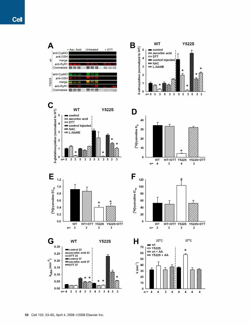
Figure 3. Redox Modifications of RyR1 and Functional Consequences(A) Redox modifications of RyR1. Representative blots obtained with three independent microsomal preparations were obtained from RyR1wt/wt (top) and
RyR1Y522S/wt (bottom) mice. Density of the bands corresponding to the redox modifications of RyR1 were obtained under control conditions (middle) or in the
presence of either ascorbic acid (left) or DTT (right).
(B) Fluorescence signals for S-nitrosylation normalized to the Coomassie stain of each band. Data (mean ± SD, n = 3–5) are presented as the ratio to untreated
microsomes. *p < 0.05 compared to control.
(C) Fluorescence signals for S-glutathionylation normalized to the Coomassie stain of each band. Data (mean ± SD, n = 3–5) are presented as the ratio to untreated
microsomes. *p < 0.05 compared to control.
(D–F) Equilibrium [3H] ryanodine binding. Scatchard plot analysis (see Figure S4) determination of KD values for [3H] ryanodine binding to microsomes from
RyR1wt/wt and RyR1Y522S/wt muscle (D). [3H] ryanodine binding was titrated at different Ca2+ concentrations to calculate EC50 (E) and IC50 (F) values from traces
as those shown in Figure S5. *p < 0.05 compared to RyR1wt/wt or untreated controls.
(G) Temperature dependence of the association kinetics of [3H] ryanodine binding. Microsomes from untreated mice were preincubated in vitro with buffer (un-
treated), AA or DTT as in (A). [3H] ryanodine binding was assessed at different time points (1–90 min), and kobs values (mean ± SD) were determined from three to
four independent experiments. Statistical significance for all panels was obtained by two-way ANOVA. *p < 0.05 compared to RyR1wt/wt or untreated controls.
(H) Rate of Ca2+ efflux from SR vesicles. Ca2+-induced Ca2+ release in the presence of 1 mM free ATP and 9–10 mM free Ca2+ was measured using stopped-flow
spectrofluorometry. Ca2+ release was measured in RyR1wt/wt and RyR1Y522S/wt vesicles using extravesicular calcium green-5N under control conditions or fol-
lowing treatment with AA. Release rate constant values (k) were obtained by peak differential analysis of fluorescence data (representative traces shown in
Figure S7). All data in this figure are shown as mean ± SEM.
Cell 133, 53–65, April 4, 2008 ª2008 Elsevier Inc. 59
wild-type mice are located circumferentially around the myofi-
brils at either side of the Z line in close proximity to the triads
(Rossi et al., 2006). In sections that cut across the intermyofibril-
lar space, the mitochondria in muscle of the RyR1wt/wt mice are
rounded or slightly elongated (Figure 4A, panels 1–3). The inter-
nal matrix is usually dark, and cristae appear well organized and
parallel to one another. A very small percentage of mitochondria
in the wild-type muscle are abnormal with ‘‘myelin figures’’ (Fig-
ure 4A, panel 4) or a somewhat disarranged external membrane
and internal cristae (not shown), but are similar in size to the more
typical mitochondria. In contrast, although typical mitochondria
are also found (Figure 4B, panel 1), in FDB fibers of RyR1Y522S/wt
mice a large number of mitochondria are abnormally shaped,
swollen, and sometimes severely altered (Figure 4B, panels 2–4).
The most noticeable changes include widening and loss of ma-
trix density, an increase in overall size, loss/disorganization of
the internal cristae (Figure 4B, panel 2), disruption of the external
membrane (Figure 4B, panel 3 arrows), and vacuolization (stars
in Figure 4B, panels 2 and 4). The number of severely disrupted
mitochondria (such as those in Figure 4B, panels 2–4) varies sig-
nificantly from fiber to fiber and sample to sample, but is always
much higher in fibers from RyRY522S/wt compared to RyR1wt/wt
mice (9.1 versus 1.1%, Table 2, footnote a). The minimum mito-
chondrial diameter in RyRwt/wt and RyRY522S/wt mice is about
30% larger (p < 0.0001) in fibers from RyRY522S/wt mice, suggest-
ing increased mitochondrial swelling (Table 2, footnote b). The
mitochondria appear to be even more damaged at one year,
but the nature of the damage varies greatly among muscle
groups. In FDB and soleus muscles from 1-year-old RyRY522S/wt
mice, most mitochondria are severely swollen and disrupted
(Figures 4D and 4F, stars). In contrast, in the muscle of RyRwt/wt
mice, mitochondria are identical to those at 2 to 3 months
of age, i.e., dark in appearance and small in size (Figures 4C
and 4E, arrows). To more quantitatively evaluate the mitochon-
drial damage, we isolated samples enriched in mitochondria
from the muscle of both 2- and 12-month-old RyRwt/wt and
RyRY522S/wt mice. Mitochondrial content of the samples was
confirmed as a >100-fold enrichment of succinate dehydroge-
nase (SDH) activity compared to homogenates (not shown).
We measured the level of thiobarbituric acid-reactive sub-
stances (TBARS) as a marker of lipid peroxidation (i.e., oxidative
damage) of mitochondrial membranes. Consistent with the mito-
chondrial damage observed in electron-microscopic (EM) anal-
yses, an increase in lipid peroxidation is observed in mitochon-
dria isolated from 2-month-old RyR1Y522S/wt mice compared to
RyR1wt/wt mitochondria (Figure 4G), and this is substantially
greater at 12 months. To determine if this arises from increased
oxidative/nitrosative stress, we treated mice with NAC in their
water supply for several months prior to sacrifice and mitochon-
drial isolation from pooled skeletal muscles. As can be seen in
Figure 4G, NAC treatment completely reversed the increased
mitochondrial lipid peroxidation observed in RyR1Y522S/wt mice.
To assess the functional consequences of aging under condi-
tions of chronic oxidative/nitrosative stress in muscle of
RyR1Y522S/wt mice, we measured the ability of the soleus muscle
to generate force and found that the muscles from older (R8-
month-old) RyR1Y522S/wt mice display a significant decrease in
maximal developed force, and this decrease is prevented by
60 Cell 133, 53–65, April 4, 2008 ª2008 Elsevier Inc.
chronic administration of NAC in their water supply (Figures 4H
and 4I).
Our findings are consistent with mitochondrial damage result-
ing from prolonged oxidative/nitrosative stress contributing
to contractile dysfunction in aged RyR1Y522S/wt. However, SR
Ca2+ leak and store depletion could also directly impact muscle
function. We find that maximal caffeine induced stress is
reduced in the RyR1Y522S/wt mice, and this is also prevented
by chronic feeding of NAC to the mice in their drinking water (Fig-
ure 4J). The observed reduction in maximal caffeine-induced
contracture could result from a decrease in releasable Ca2+
stores, reduced myofilament Ca2+ sensitivity, or both.
DISCUSSION
Environmental heat stress triggers sudden death in RyR1Y522S/wt
mice. We suggest that this is due to a cycle whereby elevated cy-
tosolic Ca2+, combined with temperature-dependent increases
in RNS, produce nitrosative modifications of the mutant channel
that enhance RyR1 channel activity at elevated temperatures.
The net result is a destructive feed-forward cycle of increased
myoplasmic Ca2+ and RNS with temperature (Figure 5), ulti-
mately producing EHS in RyR1Y522S/wt mice. Over an extended
period, this cycle appears to produce a myopathy characterized
by decreased force generation and damaged mitochondria.
RyR1 blockers, ROS/RNS scavengers, and inhibitors of NOS
abolish temperature-dependent increases in cytosolic Ca2+
and RNS. Since NOS inhibition blocks temperature-dependent
increases in Ca2+ and RNS, but not ROS, these results suggest
that the increase in Ca2+ results from the increase in RNS. We
do not know which NOS isoform(s) is involved, but skeletal mus-
cle is rich in nNOS and this isoform may be activated by
increases in cytosolic Ca2+, since it colocalizes with ryanodine
receptors in cardiac myocytes (Barouch et al., 2002; Hare,
2003). Although most nNOS localizes to the sarcolemma in skel-
etal muscle (Wells et al., 2003), close juxtapositioning of RyR1
and a subpopulation of nNOS in the triad junction might allow
for a very local RyR1-mediated Ca2+ leak from RyR1 to stimulate
NO production by nNOS.
We previously identified seven specific cysteines in one sub-
unit of RyR1 (out of 100) that can be S-nitrosylated (Aracena-
Parks et al., 2006). Of these, four (C315, C811, C906, and
C3635) are endogenously nitrosylated. Stamler and coworkers
(Eu et al., 2000; Sun et al., 2001a, 2003, 2001b) found that
C3635 is the primary RyR1 cysteine that is S-nitrosylated at
low pO2. At high pO2, some cysteines (not C3635) are oxidized,
preventing RyR1 S-nitrosylation (Sun et al., 2003). Thus, C3635
is the best candidate for S-nitrosylation of RyR1Y522S/wt in our
mice, and studies are currently ongoing to determine if nitrosyla-
tion of this residue in necessary and sufficient for enhancing
RyR1 temperature sensitivity.
The Y522S mutation in humans is associated with a myopathy
and central cores in muscle fibers. Although we did not detect
widespread central cores in our mice, young mice displayed ev-
idence of a myopathy in terms of mitochondrial alterations, while
muscle function is not greatly compromised. In contrast, older
RyR1Y522S/wt solei display marked mitochondrial structural dam-
age and a decreased ability to generate force that is prevented
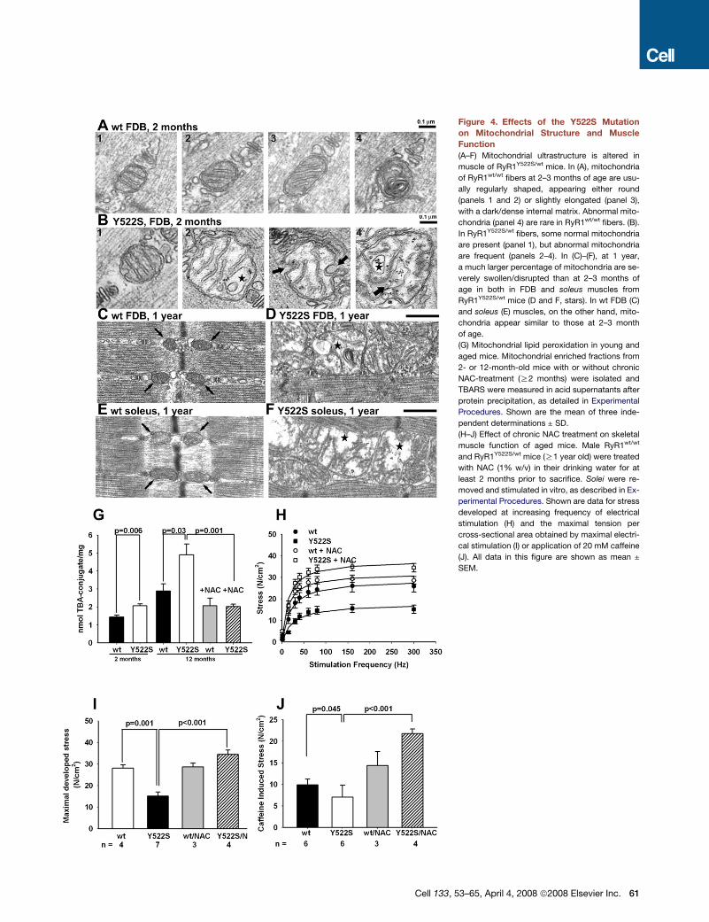
Figure 4. Effects of the Y522S Mutation
on Mitochondrial Structure and Muscle
Function
(A–F) Mitochondrial ultrastructure is altered in
muscle of RyR1Y522S/wt mice. In (A), mitochondria
of RyR1wt/wt fibers at 2–3 months of age are usu-
ally regularly shaped, appearing either round
(panels 1 and 2) or slightly elongated (panel 3),
with a dark/dense internal matrix. Abnormal mito-
chondria (panel 4) are rare in RyR1wt/wt fibers. (B).
In RyR1Y522S/wt fibers, some normal mitochondria
are present (panel 1), but abnormal mitochondria
are frequent (panels 2–4). In (C)–(F), at 1 year,
a much larger percentage of mitochondria are se-
verely swollen/disrupted than at 2–3 months of
age in both in FDB and soleus muscles from
RyR1Y522S/wt mice (D and F, stars). In wt FDB (C)
and soleus (E) muscles, on the other hand, mito-
chondria appear similar to those at 2–3 month
of age.
(G) Mitochondrial lipid peroxidation in young and
aged mice. Mitochondrial enriched fractions from
2- or 12-month-old mice with or without chronic
NAC-treatment (R2 months) were isolated and
TBARS were measured in acid supernatants after
protein precipitation, as detailed in Experimental
Procedures. Shown are the mean of three inde-
pendent determinations ± SD.
(H–J) Effect of chronic NAC treatment on skeletal
muscle function of aged mice. Male RyR1wt/wt
and RyR1Y522S/wt mice (R1 year old) were treated
with NAC (1% w/v) in their drinking water for at
least 2 months prior to sacrifice. Solei were re-
moved and stimulated in vitro, as described in Ex-
perimental Procedures. Shown are data for stress
developed at increasing frequency of electrical
stimulation (H) and the maximal tension per
cross-sectional area obtained by maximal electri-
cal stimulation (I) or application of 20 mM caffeine
(J). All data in this figure are shown as mean ±
SEM.
Cell 133, 53–65, April 4, 2008 ª2008 Elsevier Inc. 61
by treating the mice with NAC. Mitochondrial lipid peroxidation is
greatly increased in the muscle of older RyR1Y522S/wt mice, and
this is also prevented by NAC. Thus, chronic exposure to ele-
Table 2. EM Examination Reveals Frequent Severely Disrupted
and Larger Mitochondria in Y522S Fibers
Genotype/
Age
Percentage of Severely
Disrupted Mitochondriaa
Average Diameter
of Mito, nm ± SDb
RyR1wt/wt
(2–3 months)
1.1 (n = 1019, 2 mice) 187 ± 53
(n = 643, 2 mice)
RyR1Y522S/wt
(2–3 months)
9.1* (n = 1493, 4 mice) 243 ± 72*
(n = 1069, 4 mice)a The relative percentage of severely disrupted mitochondria in FDB fi-
bers from 2- to 3-month-old RyR1wt/wt and RyR1Y522S/wt mice. n, total
number of mitochondria.b Differences in mitochondria mean diameter in FDB fibers from RyR1wt/wt
and RyR1Y522S/wt mice. n, number of measurements. (*p < 0.0001). Data
are mean ± SD.
62 Cell 133, 53–65, April 4, 2008 ª2008 Elsevier Inc.
vated Ca2+ and ROS/RNS leads to progressive mitochondrial
damage and decreased ability to generate force, suggesting
that these pathways contribute to the myopathy observed in
older mice.
The temperature and exercise sensitivity of RyR1Y522S/wt mice
provide new mechanistic insight into environmental and/or exer-
tional heat illness, disorders that have previously been linked in
humans to MH mutations in RyR1 (Davis et al., 2002; Wappler
et al., 2001). Consistent with this linkage, the probability of an
MH response to exercise or volatile anesthetics in RyR1Y522S/wt
mice is decreased by cooling (data not shown). Recent evi-
dence indicates that intensive exercise also promotes RyR1
S-nitrosylation (Bellinger et al., 2008). Our data demonstrate
that exercise, EHS, and heat-induced sudden death can result
from a disease mutation in RyR1 that promotes Ca2+ leak, en-
hances nitrosative stress, and promotes subsequent S-nitrosy-
lation of the mutant RyR1. It remains to be determined if EHS
in humans is caused by RyR1 mutations that promote a similar
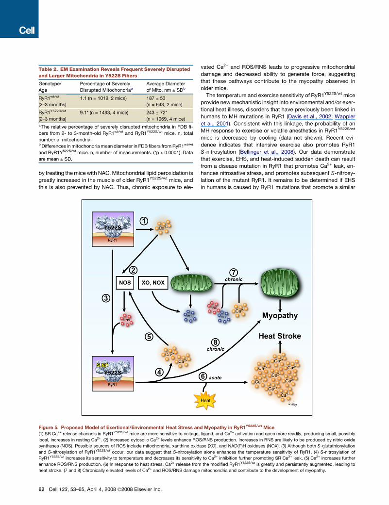
Figure 5. Proposed Model of Exertional/Environmental Heat Stress and Myopathy in RyR1Y522S/wt Mice
(1) SR Ca2+ release channels in RyR1Y522S/wt mice are more sensitive to voltage, ligand, and Ca2+ activation and open more readily, producing small, possibly
local, increases in resting Ca2+. (2) Increased cytosolic Ca2+ levels enhance ROS/RNS production. Increases in RNS are likely to be produced by nitric oxide
synthases (NOS). Possible sources of ROS include mitochondria, xanthine oxidase (XO), and NAD(P)H oxidases (NOX). (3) Although both S-glutathionylation
and S-nitrosylation of RyR1Y522S/wt occur, our data suggest that S-nitrosylation alone enhances the temperature sensitivity of RyR1. (4) S-nitrosylation of
RyR1Y522S/wt increases its sensitivity to temperature and decreases its sensitivity to Ca2+ inhibition further promoting SR Ca2+ leak. (5) Ca2+ increases further
enhance ROS/RNS production. (6) In response to heat stress, Ca2+ release from the modified RyR1Y522S/wt is greatly and persistently augmented, leading to
heat stroke. (7 and 8) Chronically elevated levels of Ca2+ and ROS/RNS damage mitochondria and contribute to the development of myopathy.
feed-forward mechanism of uncontrolled Ca2+ leak and nitrosa-
tive stress.
In summary, our data demonstrate that Ca2+ release channels
in RyR1Y522S/wt mice are leaky, producing elevations in resting
Ca2+, ROS, RNS, and basal stress at physiologically relevant
temperatures. Our data support the involvement of a destructive
feed-forward cycle whereby Ca2+ leak enhances RNS produc-
tion, and subsequent S-nitrosylation of RyR1 further increases
Ca2+ leak, resulting in regenerative Ca2+ release that underlies
uncontrolled contractions during heat stress. We also suggest
that increased Ca2+, RNS, and/or ROS ultimately contribute
to the progressive development of a myopathy characterized
by decreased muscle performance and mitochondrial damage.
An intriguing aspect of this study is the possibility that RyR1
mutations together with nitrosative stress represent a ‘‘double-
hit mechanism’’ that underlies a subset of human cases of
enhanced susceptibility to heat stroke, exertional/environmental
illness, and/or sudden death.
EXPERIMENTAL PROCEDURES
Mice
All procedures were approved by the IACUC at Baylor College of Medicine and
UCAR at the University of Rochester. For in vivo antioxidant treatment, mice
were provided ad libitum access to drinking water containing NAC (1% w/v),
L-NAME (1% w/v), or NAC plus L-NAME (both at 1% w/v).
In Vivo Temperature Sensitivity
Mice were anesthetized with etomidate, which does not trigger MH episodes in
either MH susceptible humans (Robertson, 1992) or RyR1Y522S/wt mice. Two
minutes following etomidate injection, mice were placed in an environmental
chamber at 41�C. The initial core body temperature, as well as the temperature
every minute thereafter, was monitored over the next 15 min of exposure.
Contractile Studies
Muscle collection and contractile studies were performed as previously
described (Chelu et al., 2006). For determination of maximal developed stress,
the data were fitted to a sigmoidal curve with the peak value of this curve
reported as the maximal stress.
Isolation of Subcellular Fractions from Mouse Skeletal Muscle
Muscle from each mouse was quickly collected, snap frozen in liquid nitrogen,
and stored at�80�C for up to 2 weeks. Microsomes or mitochondrial-enriched
samples were obtained from thawed muscle by differential centrifugation (see
Supplemental Data).
Ca2+ Flux Studies
Microsomal vesicles (1mg/ml) from RyR1wt/wt or RyR1Y522S/wt skeletal muscle
were passively loaded with Ca2+, and Ca2+ release kinetics was analyzed at
25�C, as described previously (Donoso et al., 2000), using a KinTek SF-2002
thermoregulated stopped-flow spectrometer (KinTek Corporation). Studies
at 37�C were performed after 5 min equilibration of samples in the equipment.
Release rate constants were obtained from peak differential analysis of raw
fluorescence data.
Lipoperoxidation Assay
Basal lipoperoxidation levels were measured using thiobarbituric acid reactive
substances (TBARS) as described elsewhere (Letelier et al., 2005).
Glutathione Assays
Total GSH and GSSG were assayed using deproteinized muscle homogenates
in a 96-well format according to Tietze (Tietze, 1969), as modified by Griffith
(Griffith, 1980).
Primary Cultures and Fluorescent Microscopy
Primary myotube cultures were grown on Matrigel (BD Biosciences) coated
glass coverslips from 1- to 4-day-old mice as previously described (Pollard
and Walker, 1997). ROS and RNS imaging were performed using the probes
DCF or DAF, as detailed in Supplemental Experimental Procedures. Ca2+ im-
aging was performed as previously described in Long et al. (2007). Perforated
patch clamp recordings of L-type Ca2+ currents (L-currents), and intracellular
Ca2+ transients were recorded as described in Chelu et al. (2006). Details are
described in Supplemental Experimental Procedures.
Western Blotting
Western blotting with mouse anti-glutathione 1:10,000 (Virogen) and rabbit
anti-S-nitrosocysteine 1:10,000 (Sigma) were performed and analyzed as pre-
viously described (Aracena-Parks et al., 2006). Stripping of membranes was
performed with Li-COR Stripping Solution, following the manufacturer’s direc-
tions. Stripped membranes were reprobed with a mouse anti-RyR1 1:10,000
(Affinity Bioreagents), only to confirm the identity of the analyzed band. This
antibody displays different affinities for RyR1 electrophoresed under reducing
or nonreducing conditions. Thus, fluorescent data were normalized to the
Coomasie stain of the blots.
[3H] Ryanodine Binding
Equilibrium binding was performed with microsomes as detailed previously
(Aracena-Parks et al., 2006). Ca2+ titration of binding was performed with
5 nM [3H] ryanodine as described by Rodney et al. (2000). Kinetic assays
with 5 nM [3H] ryanodine at 23�C or 37�C were measured as detailed in
Hawkes et al. (1992). Details of buffers used can be found in Supplemental
Experimental Procedures.
Preparation and Analysis of Samples for EM
EM was performed in FDB and soleus muscles from 2- to 3-month- and 1 year-
old mice as detailed in Paolini et al. (2007). Details are described in Supple-
mental Experimental Procedures.
Statistical Analyses
All analyses were performed in Sigma Plot (Systat Software, Incorporated).
SUPPLEMENTAL DATA
Supplemental data include eight figures, Supplemental Experimental Proce-
dures, and Supplemental References and can be found with this article online
at http://www.cell.com/cgi/content/full/133/1/53/DC1/.
ACKNOWLEDGMENTS
This work was supported by grants from NIH (AR 050503 and AR053349 to
S.L.H., AR44657 to R.T.D., and 5P01AR052354 to S.L.H. and R.T.D.), the Mus-
cular Dystrophy Association to S.L.H, Research Grant GGP030289 from the
Italian Telethon Foundation to F.P, and a NIH Dental and Craniofacial training
grant T32-DE07202 to A.E.R.
Received: July 5, 2007
Revised: October 30, 2007
Accepted: February 29, 2008
Published: April 3, 2008
REFERENCES
Aghdasi, B., Reid, M.B., and Hamilton, S.L. (1997). Nitric oxide protects the
skeletal muscle Ca2+ release channel from oxidation induced activation.
J. Biol. Chem. 272, 25462–25467.
Aracena-Parks, P., Goonasekera, S.A., Gilman, C.P., Dirksen, R.T., Hidalgo,
C., and Hamilton, S.L. (2006). Identification of cysteines involved in S-nitrosy-
lation, S-glutathionylation, and oxidation to disulfides in ryanodine receptor
Type 1. J. Biol. Chem. 281, 40354–40368.
Cell 133, 53–65, April 4, 2008 ª2008 Elsevier Inc. 63
Barouch, L.A., Harrison, R.W., Skaf, M.W., Rosas, G.O., Cappola, T.P.,
Kobeissi, Z.A., Hobai, I.A., Lemmon, C.A., Burnett, A.L., O’Rourke, B., et al.
(2002). Nitric oxide regulates the heart by spatial confinement of nitric oxide
synthase isoforms. Nature 416, 337–339.
Bellinger, A.M., Reiken, S., Dura, M., Murphy, P.W., Deng, S.X., Landry, D.W.,
Nieman, D., Lehnart, S.E., Samaru, M., Lacampagne, A., and Marks, A.R.
(2008). Remodeling of ryanodine receptor complex causes ‘‘leaky’’ channels:
a molecular mechanism for decreased exercise capacity. Proc. Natl. Acad.
Sci. USA 105, 2198–2202.
Bendahan, D., Kozak-Ribbens, G., Confort-Gouny, S., Ghattas, B., Figarella-
Branger, D., Aubert, M., and Cozzone, P.J. (2001). A noninvasive investigation
of muscle energetics supports similarities between exertional heat stroke and
malignant hyperthermia. Anesth. Analg. 93, 683–689.
Bouchama, A., and Knochel, J.P. (2002). Heat stroke. N. Engl. J. Med. 346,
1978–1988.
Carroll, S., Skarmeta, J.G., Yu, X., Collins, K.D., and Inesi, G. (1991). Interde-
pendence of ryanodine binding, oligomeric receptor interactions, and Ca2+ re-
lease regulation in junctional sarcoplasmic reticulum. Arch. Biochem. Biophys.
290, 239–247.
Chelu, M.G., Goonasekera, S.A., Durham, W.J., Tang, W., Lueck, J.D., Riehl,
J., Pessah, I.N., Zhang, P., Bhattacharjee, M.B., Dirksen, R.T., and Hamilton,
S.L. (2006). Heat- and anesthesia-induced malignant hyperthermia in an
RyR1 knock-in mouse. FASEB J. 20, 329–330.
Chu, A., Diaz-Munoz, M., Hawkes, M.J., Brush, K., and Hamilton, S.L. (1990).
Ryanodine as a probe for the functional state of the skeletal muscle sarcoplas-
mic reticulum Ca2+release channel. Mol. Pharmacol. 37, 735–741.
Clark, C.B., Zhang, Y., Martin, S.M., Davies, L.R., Xu, L., Kregel, K.C., Miller,
F.J., Buettner, G.R., and Kerber, R.E. (2004). The nitric oxide synthase inhibitor
N(omega)-nitro-L-arginine decreases defibrillation-induced free radical gener-
ation. Resuscitation 60, 351–357.
Davis, M., Brown, R., Dickson, A., Horton, H., James, D., Laing, N., Marston,
R., Norgate, M., Perlman, D., Pollock, N., and Stonwell, K. (2002). Malignant
hyperthermia associated with exercise-induced rhabdomyolysis or congenital
abnormalities and a novel RYR1 mutation in new zealand and australian
pedigrees. Br. J. Anaesth. 88, 508–515.
Donoso, P., Aracena, P., and Hidalgo, C. (2000). Sulfhydryl oxidation overrides
mg(2+) inhibition of calcium-induced calcium release in skeletal muscle triads.
Biophys. J. 79, 279–286.
Ducreux, S., Zorzato, F., Muller, C., Sewry, C., Muntoni, F., Quinlivan, R.,
Restagno, G., Girard, T., and Treves, S. (2004). Effect of ryanodine receptor
mutations on interleukin-6 release and intracellular calcium homeostasis in
human myotubes from malignant hyperthermia-susceptible individuals and
patients affected by central core disease. J. Biol. Chem. 279, 43838–43846.
Ellis, F.R., Halsall, P.J., and Harriman, D.G. (1988). Malignant hyperpyrexia and
sudden infant death syndrome. Br. J. Anaesth. 60, 28–30.
Eu, J.P., Sun, J., Xu, L., Stamler, J.S., and Meissner, G. (2000). The skeletal
muscle calcium release channel: coupled o2 sensor and no signaling functions.
Cell 102, 499–509.
Griffith, O.W. (1980). Determination of glutathione and glutathione disulfide
using glutathione reductase and 2-vinylpyridine. Anal. Biochem. 106, 207–212.
Hackl, W., Winkler, M., Mauritz, W., Sporn, P., and Steinbereithner, K. (1991).
Muscle biopsy for diagnosis of malignant hyperthermia susceptibility in two
patients with severe exercise-induced myolysis. Br. J. Anaesth. 66, 138–140.
Hare, J.M. (2003). Nitric oxide and excitation-contraction coupling. J. Mol.
Cell. Cardiol. 35, 719–729.
Hawkes, M.J., Nelson, T.E., and Hamilton, S.L. (1992). [3H] ryanodine as
a probe of changes in the functional state of the ca2+ -release channel in
malignant hyperthermia. J. Biol. Chem. 267, 6702–6709.
Hopkins, P.M. (2000). Malignant hyperthermia: advances in clinical manage-
ment and diagnosis. Br. J. Anaesth. 85, 118–128.
Jurkat-Rott, K., McCarthy, T., and Lehmann-Horn, F. (2000). Genetics and
pathogenesis of malignant hyperthermia. Muscle Nerve 23, 4–17.
64 Cell 133, 53–65, April 4, 2008 ª2008 Elsevier Inc.
Letelier, M.E., Lepe, A.M., Faundez, M., Salazar, J., Marin, R., Aracena, P., and
Speisky, H. (2005). Possible mechanisms underlying copper-induced damage
in biological membranes leading to cellular toxicity. Chem. Biol. Interact. 151,
71–82.
Lichtman, A.D., and Oribabor, C. (2006). Malignant hyperthermia following
systemic rewarming after hypothermic cardiopulmonary bypass. Anesth.
Analg. 102, 372–375.
Long, C., Cook, L.G., Hamilton, S.L., Wu, G.Y., and Mitchell, B.M. (2007).
FK506 binding protein 12/12.6 depletion increases endothelial nitric oxide
synthase threonine 495 phosphorylation and blood pressure. Hypertension
49, 569–576.
Marengo, J.J., Hidalgo, C., and Bull, R. (1998). Sulfhydryl oxidation modifies
the calcium dependence of ryanodine-sensitive calcium channels of excitable
cells. Biophys. J. 74, 1263–1277.
Oba, T., Murayama, T., and Ogawa, Y. (2002). Redox states of type 1 ryano-
dine receptor alter Ca(2+) release channel response to modulators. Am.
J. Physiol. Cell Physiol. 282, C684–C692.
Pamukcoglu, T. (1988). Sudden death due to malignant hyperthermia. Am.
J. Forensic Med. Pathol. 9, 161–162.
Paolini, C., Quarta, M., Nori, A., Boncompagni, S., Canato, M., Volpe, P., Allen,
P.D., Reggiani, C., and Protasi, F. (2007). Reorganized stores and impaired
calcium handling in skeletal muscle of mice lacking calsequestrin-1. J. Physiol.
583, 767–784.
Pessah, I.N., Stambuk, R.A., and Casida, J.E. (1987). Ca2+-activated ryano-
dine binding: mechanisms of sensitivity and intensity modulation by Mg2+,
caffeine, and adenine nucleotides. Mol. Pharmacol. 31, 232–238.
Pollard, J.W., and Walker, J.M. (1997). Basic Cell Culture Protocols (Methods
in Molecular Biology), Second Edition (Totowa, NJ: Humana Press).
Pou, S., Keaton, L., Surichamorn, W., and Rosen, G.M. (1999). Mechanism of
superoxide generation by neuronal nitric-oxide synthase. J. Biol. Chem. 274,
9573–9580.
Quane, K.A., Keating, K.E., Healy, J.M., Manning, B.M., Krivosic-Horber, R.,
Krivosic, I., Monnier, N., Lunardi, J., and McCarthy, T.V. (1994). Mutation
screening of the RYR1 gene in malignant hypertherima: detection of a novel
tyr to ser mutation in a pedigree with associated central core. Genomics 23,
236–239.
Robertson, S. (1992). Advantages of etomidate use as an anesthetic agent.
Vet. Clin. North Am. Small Anim. Pract. 22, 277–280.
Rodney, G.G., Williams, B.Y., Strasburg, G.M., Beckingham, K., and Hamilton,
S.L. (2000). Regulation of RYR1 activity by ca(2+) and calmodulin. Biochemis-
try 39, 7807–7812.
Rossi, A.E., Boncompagni, S., Protasi, F., and Dirksen, R.T. (2006). Develop-
mental regulation of mitochondrial triad targeting in skeletal muscle. Biophys.
J. 90, A269.
Ryan, J.F., and Tedeschi, L.G. (1997). Sudden unexplained death in a patient
with a family history of malignant hyperthermia. J. Clin. Anesth. 9, 66–68.
Sun, J., Xin, C., Eu, J.P., Stamler, J.S., and Meissner, G. (2001a). Cysteine-
3635 is responsible for skeletal muscle ryanodine receptor modulation by
no. Proc. Natl. Acad. Sci. USA 98, 11158–11162.
Sun, J., Xu, L., Eu, J.P., Stamler, J.S., and Meissner, G. (2001b). Classes of
thiols that influence the activity of the skeletal muscle calcium release channel.
J. Biol. Chem. 276, 15625–15630.
Sun, J., Xu, L., Eu, J.P., Stamler, J.S., and Meissner, G. (2003). Nitric oxide,
noc-12, and s-nitrosoglutathione modulate the skeletal muscle calcium
release channel/ryanodine receptor by different mechanisms. an allosteric
function for o2 in s-nitrosylation of the channel. J. Biol. Chem. 278, 8184–8189.
Thompson, P.D., Franklin, B.A., Balady, G.J., Blair, S.N., Corrado, D., Estes,
N.A., 3rd, Fulton, J.E., Gordon, N.F., Haskell, W.L., Link, M.S., et al. (2007).
Exercise and acute cardiovascular events placing the risks into perspective:
a scientific statement from the american heart association council on nutrition,
physical activity, and metabolism and the council on clinical cardiology. Circu-
lation 115, 2358–2368.
Tietze, F. (1969). Enzymic method for quantitative determination of nanogram
amounts of total and oxidized glutathione: applications to mammalian blood
and other tissues. Anal. Biochem. 27, 502–522.
Treves, S., Anderson, A.A., Ducreux, S., Divet, A., Bleunven, C., Grasso, C.,
Paesante, S., and Zorzato, F. (2005). Ryanodine receptor 1 mutations, dysre-
gulation of calcium homeostasis and neuromuscular disorders. Neuromuscul.
Disord. 15, 577–587.
Wappler, F., Fiege, M., Steinfath, M., Agarwal, K., Scholz, J., Singh, S.,
Matschke, J., and Schulte Am Esch, J. (2001). Evidence for susceptibility to
malignant hyperthermia in patients with exercise-induced rhabdomyolysis.
Anesthesiology 94, 95–100.
Wells, K.E., Torelli, S., Lu, Q., Brown, S.C., Partridge, T., Muntoni, F., and
Wells, D.J. (2003). Relocalization of neuronal nitric oxide synthase (nNOS) as
a marker for complete restoration of the dystrophin associated protein
complex in skeletal muscle. Neuromuscul. Disord. 13, 21–31.
Zhang, H., Zhang, J.Z., Danila, C.I., and Hamilton, S.L. (2003). A noncontigu-
ous, inter-subunit binding site for calmodulin on the skeletal muscle Ca2+
release channel. J. Biol. Chem. 278, 8348–8355.
Cell 133, 53–65, April 4, 2008 ª2008 Elsevier Inc. 65