Besides adhesion: new perspectives of integrin functions in angiogenesis
Upload
independentCategory
view
0download
0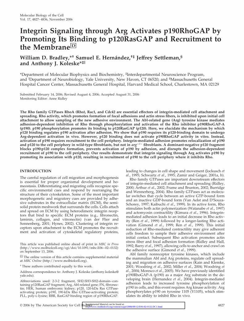
Molecular Biology of the CellVol. 17, 4827–4836, November 2006
Integrin Signaling through Arg Activates p190RhoGAP byPromoting Its Binding to p120RasGAP and Recruitment tothe Membrane□D
William D. Bradley,*† Samuel E. Hernandez,†‡ Jeffrey Settleman,§and Anthony J. Koleske*‡�
*Department of Molecular Biophysics and Biochemistry, ‡Interdepartmental Neuroscience Program,and �Department of Neurobiology, Yale University, New Haven, CT 06520; and §Massachusetts GeneralHospital Cancer Center, Massachusetts General Hospital, Harvard Medical School, Charlestown, MA 02129
Submitted February 14, 2006; Revised August 4, 2006; Accepted August 31, 2006Monitoring Editor: Anne Ridley
The Rho family GTPases RhoA (Rho), Rac1, and Cdc42 are essential effectors of integrin-mediated cell attachment andspreading. Rho activity, which promotes formation of focal adhesions and actin stress fibers, is inhibited upon initial cellattachment to allow sampling of the new adhesive environment. The Abl-related gene (Arg) tyrosine kinase mediatesadhesion-dependent inhibition of Rho through phosphorylation and activation of the Rho inhibitor p190RhoGAP-A(p190). p190 phosphorylation promotes its binding to p120RasGAP (p120). Here, we elucidate the mechanism by whichp120 binding regulates p190 activation after adhesion. We show that p190 requires its p120-binding domain to undergoArg-dependent activation in vivo. However, p120 binding does not activate p190RhoGAP activity in vitro. Instead,activation of p190 requires recruitment to the cell periphery. Integrin-mediated adhesion promotes relocalization of p190and p120 to the cell periphery in wild-type fibroblasts, but not in arg�/� fibroblasts. A dominant-negative p120 fragmentblocks p190:p120 complex formation, prevents activation of p190 by adhesion, and disrupts the adhesion-dependentrecruitment of p190 to the cell periphery. Our results demonstrate that integrin signaling through Arg activates p190 bypromoting its association with p120, resulting in recruitment of p190 to the cell periphery where it inhibits Rho.
INTRODUCTION
The careful regulation of cell migration and morphogenesisis essential for proper organismal development and ho-meostasis. Differentiating and migrating cells recognize spe-cific environmental cues and respond by rearranging thestructure of their cytoskeleton. Some of the most importantmorphogenetic and migratory cues are provided by adhe-sive substrates in the extracellular matrix (ECM), the semi-solid protein meshwork that surrounds the cells. Cells attachand spread on ECM by using heterodimeric integrin recep-tors that bind to specific ECM proteins (e.g., fibronectin,laminin, collagen, and vitronectin) (van der Flier andSonnenberg, 2001; Hynes, 2002). Clustering of integrin re-ceptors upon attachment to the ECM promotes the recruit-ment and activation of cytoskeletal regulatory proteins,
leading to changes in cell shape and movement (Jockusch etal., 1995; Schwartz et al., 1995; Zamir and Geiger, 2001a, b).
Rho family GTPases are important downstream effectorsof integrin-mediated cell attachment and spreading (Ridley,2000; Arthur et al., 2002; Frame and Brunton, 2002; Burridgeand Wennerberg, 2004). Rho family GTPases act as molecu-lar switches that cycle between an active GTP-bound formand an inactive GDP-bound form (Van Aelst and D’Souza-Schorey, 1997; Kaibuchi et al., 1999). In its active form, Rhostimulates both actin polymerization (Watanabe et al., 1997)and actomyosin contractility (Kimura et al., 1996). Integrin-mediated adhesion leads to an initial decrease in Rho activ-ity (Ren et al., 1999) followed by a longer-lasting Rho acti-vation (Gimond et al., 1999; Ren et al., 1999). This initialreduction of Rho-mediated contractility may give adherentcells freedom to sample their adhesive environment afterinitial contact. Subsequent Rho activation promotes actinstress fiber and focal adhesion formation (Ridley and Hall,1992; Barry et al., 1997), allowing cells to anchor and crawl onthe adhesive surface (Gimond et al., 1999).
Abl family nonreceptor tyrosine kinases, which includethe mammalian Abl and Arg proteins, regulate cell spread-ing and migration on adhesive surfaces (Kain and Klemke,2001; Woodring et al., 2002; Miller et al., 2004; Woodring etal., 2004; Moresco et al., 2005). We have previously identifiedp190RhoGAP-A (p190) as a major Arg substrate in the de-veloping brain (Hernandez et al., 2004). Integrin-mediatedadhesion leads to increased tyrosine phosphorylation ofp190 in cells, and this event requires Arg kinase activity. Argphosphorylates p190 on tyrosine 1105 (Y1105), which stim-ulates its ability to inhibit Rho in vivo.
This article was published online ahead of print in MBC in Press(http://www.molbiolcell.org/cgi/doi/10.1091/mbc.E06–02–0132)on September 13, 2006.□D The online version of this article contains supplemental materialat MBC Online (http://www.molbiolcell.org).† These authors contributed equally to this work.
Address correspondence to: Anthony J. Koleske ([email protected]).
Abbreviations used: 2-3-2 fragment, SH2-SH3-SH2-domain–con-taining p120RasGAP fragment; Arg, Abl-related gene; FN, fibronec-tin; HEK, human embryonic kidney; p120, 120-kDa Ras GTPase-activating protein; p190, 190-kDa Rho GTPase-activating protein;PLL, poly-l-lysine; RBR, RasGAP-binding region of p190RhoGAP.
© 2006 by The American Society for Cell Biology 4827 http://www.molbiolcell.org/content/suppl/2006/09/12/E06-02-0132.DC1.htmlSupplemental Material can be found at:
Phosphorylation of Y1105 in the RasGAP-binding region ofp190 promotes formation of a complex between p190 and the120-kDa GTPase-activating protein for Ras (p120RasGAP orp120). Although phosphorylation of Y1105 is essential for com-plex formation, phosphorylation of Y1087 in p190 helps stabi-lize the interaction (Hu and Settleman, 1997), which is medi-ated by the binding of two Src homology (SH)2 domains inp120 to the phosphotyrosines 1087 and 1105 in p190 (Hu andSettleman, 1997; Roof et al., 1998; Hernandez et al., 2004). In-creased p190 phosphorylation and the formation of the p190:p120 complexes correlate with increased disassembly of actinstress fibers in Src-overexpressing cells (Chang et al., 1995),suggesting that p120 binding regulates p190 activation. Despitethis correlative evidence, the mechanism by which p190 be-comes activated in response to adhesion is unclear.
We demonstrate here that Arg is required for the adhe-sion-dependent inhibition of Rho and describe the mecha-nism by which integrin-mediated adhesion activatesp190RhoGAP activity. We show that the p120-binding re-gion of p190 is required for Arg to stimulate its RhoGAPactivity in vivo. However, p120 binding is not sufficient toactivate p190 in vitro. Instead, p190 activation requires itsrecruitment to the cell periphery. We demonstrate that ad-hesion promotes p190 and p120 colocalization at the cellperiphery in wild-type, but not arg�/� cells. A dominant-negative p120 fragment blocks formation of the p190:p120complex and prevents adhesion-dependent p190 activationby blocking p190 recruitment to the cell periphery. Ourresults demonstrate that integrin signaling through Arg ac-tivates p190 by promoting its association with p120 andlocalization to the cell periphery so that it can inhibit Rho.
MATERIALS AND METHODS
PlasmidsFull-length Arg, p190, p120, and Rho constructs were generated as describedpreviously (Hernandez et al., 2004). Hemagglutinin (HA)-tagged p190 frag-ments were generated from a full-length p190 template by using polymerasechain reaction (PCR). Red fluorescent protein (RFP) vector was generated bycutting monomeric RFP from the prSET vector (a generous gift of Roger Tsien,Department of Pharmacology, University of California, San Diego, CA) andligating it into the pEYFP-N1 vector (Clontech, Mountain View, CA). The2-3-2 fragment was generated from a full-length p120 template by using PCRand ligated into the RFP-N1 vector. p120-GFP was generated by ligating p120and humanized Renilla green fluorescent protein (GFP) (Stratagene, La Jolla,CA) into the pK1 vector. The Paxillin, Cortactin, and Src expression constructswere generous gifts of Donna Webb (Department of Biological Sciences,Vanderbilt University, Nashville, TN), Tom Parsons (Department of Micro-biology, University of Virginia, Charlottesville, VA), and John Cooper (De-partment of Cell Biology and Physiology, Washington University in St. Louis,St. Louis, MO), respectively.
FibroblastsWild type and arg�/� fibroblasts were cultured as described previously(Miller et al., 2004). arg�/� fibroblasts were infected with pK1 retrovirusesexpressing yellow fluorescent protein (YFP) or Arg-YFP as described previ-ously (Miller et al., 2004), and they were selected with puromycin (Sigma-Aldrich, St. Louis, MO) at concentrations up to 0.85 �g/ml.
Human Embryonic Kidney (HEK)293 Cell TransfectionHEK293 cells were plated on 10-cm plates with DMEM containing 10% fetalbovine serum and transfected with a minimum of 20 �g of plasmid DNAs perplate by using Lipofectamine 2000 (Invitrogen). More plasmid DNA wastransfected for the HA-tagged 760-C p190 fragment construct to balanceprotein expression levels. When necessary, expression was controlled byusing appropriate empty vectors. After 48 h, transfection efficiency wasmonitored by cotransfection of a GFP expression construct (Stratagene), andcells were used for Rho activity, or p190 or HA immunoprecipitation assays.
Rho Activity AssaysRhotekin-agarose (Upstate Biotechnology, Charlottesville, VA) pull-down as-says were performed on HEK293 cells as reported previously (Hernandez etal., 2004), by using Mg2� lysis buffer (25 mM HEPES, pH 7.5, 150 mM NaCl,
1% NP-40, 10 mM MgCl2, 10% glycerol, protease, and phosphatase inhibi-tors). Active HA-RhoA was detected using an anti-HA monoclonal antibody(mAb) (clone 12CA5; Abcam, Cambridge, MA) and quantified using a den-sitometer and ImageQuant software (GE Healthcare, Little Chalfont, Buck-inghamshire, United Kingdom). Active Rho levels were normalized to totalRho in each sample, and the ratio of activated to total Rho was normalized tothe control ratio for each condition tested.
The enzyme-linked immunosorbent assay (ELISA)-based G-LISA kit (Cy-toskeleton, Denver, CO) was used to determine endogenously active RhoAlevels in fibroblasts according to the manufacturer’s instructions. In brief,wild-type, arg�/�, or arg�/� � Arg-YFP cells were trypsinized and held insuspension for 1 h on 1% agarose-coated plates. Cells were then plated on10-cm dishes coated with 10 �g/ml fibronectin (Sigma-Aldrich) and blockedwith 1% bovine serum albumin (BSA) in phosphate-buffered saline (PBS)(Invitrogen, Carlsbad, CA). After 0, 10, 20, 30, or 60 min, cells were lysed in250 �l of G-LISA lysis buffer (supplemented with protease inhibitors), scrapedinto tubes, and snap-frozen in liquid nitrogen. Cell lysate was subsequentlythawed, clarified for 2 min at 9000 � g, and protein concentration wasnormalized between the various time points. Equal total protein amountswere added to a 96-well dish coated with the Rho binding domain of Rhoeffector proteins (which bind active GTP-bound Rho) in duplicate and incu-bated at 4°C for 30 min with vigorous shaking. Active Rho levels weredetermined by subsequent incubations with anti-Rho antibody and secondaryhorseradish peroxidase-conjugated antibody for 45 min each with vigorousshaking at room temperature. After adding developing solution, active Rhowas determined by measuring absorbance at 490 nm using an ELISA platereader after subtraction from a sample containing only lysis buffer. Equalloading of total RhoA protein at each time point was determined via immu-noblot by using anti-RhoA antibody (Santa Cruz Biotechnology, Santa Cruz,CA). Relative Rho activity was determined by dividing absorbance at eachtime point by the absorbance of the zero time point. Experiments for each celltype were repeated at least three times.
Arg was detected by immunoblot as described previously (Koleske et al.,1998). p190 was detected using either an anti-p190 mAb (clone 30; BD Bio-sciences Transduction Laboratories, Lexington, KY) or anti-HA (Abcam) asindicated. p120 was detected using anti-p120 mAb (clone B4F8; UpstateBiotechnology).
ImmunoprecipitationsHEK293 cells were lysed in modified radioimmunoprecipitation assay (RIPA)buffer (50 mM Tris, pH 7.2, 150 mM NaCl, 0.25% deoxycholate, 1% NP-40,protease, and phosphatase inhibitors). Extracts were precleared with 50 �l ofprotein G Plus/A agarose beads (Calbiochem, San Diego, CA) for 30 min at4°C. The protein concentration of each fraction was determined using theBCA detergent-compatible protein assay kit (Pierce Chemical, Rockford, IL).Two micrograms of anti-p190 mAb, anti-HA polyclonal antibody, anti-FLAGmAb (clone 2EL-1B11; Chemicon International, Temecula, CA), anti-PaxillinmAb (clone 349; BD Biosciences Transduction Laboratories), anti-Src mAb(H-12; Santa Cruz Biotechnology), or anti-Cortactin mAb (Abcam) was incu-bated with 500 �g of extract for 2 h at 4°C with gentle mixing. Then, 50 �l ofprotein G Plus/A agarose beads was added, and the immune complexes wereincubated for 1 h at 4°C. Immunoprecipitates were washed three times with0.5 ml of modified RIPA buffer, mixed with sample buffer, and separated bySDS-PAGE for immunoblot analysis. Coimmunoprecipitating proteins andloading controls were detected by stripping and reprobing the blot withanti-p120 mAb, anti-RFP polyclonal antibody (Chemicon International), andother specified antigens. Where specified, bands were quantified using adensitometer and ImageQuant software (GE Healthcare).
Recombinant Protein Production/PurificationFull-length histidine-tagged (amino terminal) rat p190RhoGAP-A (p190) andfull-length untagged human p120RasGAP (p120) were cloned into the pFast-Bac1 donor plasmid (Invitrogen), which were subsequently used to generatebacmid DNA containing the His-p190 and p120 cDNA. Bacmid DNA wasthen transfected into Sf9 cells for generation of recombinant baculovirusencoding the genes. For protein expression, Hi-5 insect cells were infectedwith recombinant baculovirus(es) at a 0.5 multiplicity of infection and har-vested after 48 h. Cells were lysed in 50 mM HEPES pH 7.25, 5 mM �-mer-captoethanol, 200 mM KCl, 0.01% NP-40, 5% glycerol, and protease inhibitorsby using a French Press. Lysates were centrifuged at 10,000 � g for 10 min topellet cellular debris. The supernatant was then centrifuged at 100,000 � g for1 h, and this second supernatant was incubated with nickel-nitrilotriaceticacid (Ni-NTA) resin (QIAGEN, Valencia, CA) for 2 h. Protein was eluted with20 mM HEPES, pH 7.25, 5 mM �-mercaptoethanol, 200 mM KCl, 0.01% NP-40,5% glycerol, and 100 mM imidazole. Proteins were further purified to 80–90%purity by fast-performance liquid chromatography (FPLC) through an anionexchange Uno Q column (Bio-Rad, Hercules, CA) by using a 1–500 mM KCllinear gradient in FPLC buffer (20 mM HEPES, 0.01% NP-40, 5% glycerol, 0.5mM EDTA, and 0.5 mM dithiothreitol [DTT]). Copurification of p190:p120complex was achieved by simultaneously coinfecting Hi5 insect cells withbaculovirus encoding His-p190 and untagged p120 and Arg. Eluate fromNi-NTA resin columns were then further purified by size exclusion chroma-
W. D. Bradley et al.
Molecular Biology of the Cell4828
tography (Superdex 200 HR-1030; GE Healthcare) as described previously(Tanis et al., 2003).
p190 PhosphorylationThe tyrosine kinase inhibitor PD180970 (Kraker et al., 2000) was included at 15�M in the p190-expressing insect cells to prevent its tyrosine phosphorylationduring expression. p190 was phosphorylated in vitro as described previously(Hernandez et al., 2004). Based on experiments run in parallel with[�-32P]ATP, �60% of the p190 was phosphorylated in these studies.
GAP AssaysThe radioactive filter-binding assay measuring the retention of [�-32P]GTP-bound glutathione S-transferase (GST)-RhoA was carried out as reportedpreviously (Zhang and Zheng, 1998). Briefly, recombinant GST-RhoA waspreloaded with [�-32P]GTP (10 �Ci; 6000 Ci/mmol; PerkinElmer Life andAnalytical Sciences, Boston, MA) in 160 �l of a buffer containing 50 mMHEPES, pH 7.5, 50 mM NaCl, 1 mg/ml BSA, 0.1 mM DTT, 0.1 mM EGTA, and5 mM EDTA for 10 min at 37°C. MgCl2 was then added to a final concentra-tion of 10 mM and incubated for 10 min on ice. The [�-32P]-GTP-loadedGST-RhoA was diluted 25-fold into a buffer containing 25 mM HEPES, pH 7.5,50 mM NaCl, 1 mM MgCl2, 0.1 mM GTP, 0.1 mM DTT, and 1 mg/ml BSA.Fifty microliters of 10 nM p190 or p190:p120 complex (1:1 M ratio at 10 nMeach) was mixed with 950 �l of the loaded and diluted GST-RhoA andincubated at 30°C. At different time points, 100-�l aliquots of the reactionwere terminated by filtering the reaction mixture through nitrocellulose fil-ters, followed by washing with 10 ml of ice-cold buffer containing 50 mMHEPES, pH 7.5, and 10 mM MgCl2. The radioactivity retained on the filterswas measured by scintillation counting.
Immunofluorescence MicroscopyCoverslips were coated with either 0.1% poly-l-lysine (PLL) (Sigma-Aldrich)or 10 �g/ml fibronectin (Sigma-Aldrich) by shaking overnight at 4°C andblocked with a 1% BSA solution (Invitrogen) for 1 h at 37°C. Wild-type,arg�/�, or arg�/� � Arg-YFP fibroblasts were either left untransfected ortransfected with RFP, 2-3-2-RFP, p120-GFP, and/or p190FF-YFP by usingLipofectamine 2000 (Invitrogen), and then allowed to attach to coverslips for10, 20, 30, or 45 min at 37°C. Cells were washed, fixed, and extracted asdescribed previously (Miller et al., 2004). After extraction, cells were blockedovernight at 4°C in a blocking buffer (2% BSA, 0.1% Triton X-100, and 0.1%sodium azide in PBS). Cells were stained with either anti-p190 or anti-p120antibody, Alexa 594-labeled secondary antibody (Invitrogen), and in somecases p190-stained cells were reblocked overnight with blocking buffer, andstained with anti-p120 antibody, Alexa 488-labeled secondary antibody (In-vitrogen), and Alexa 350-phalloidin (Invitrogen). Cells were imaged on aNikon TE2000-S microscope by using 40� Nikon PlanApo 1.0 or 100� NikonPlanFuor 1.3 oil objectives. Images were processed using Adobe Photoshop(Adobe Systems, Mountain View, CA) and NIH ImageJ (http://rsb.info.nih.gov/ij). To ensure specific staining for the protein of interest, wild-type cellsplated on fibronectin were fixed as described above, blocked, stained withanti-p190 antibody, Alexa 594-labeled secondary antibody, then reblocked,and stained only with Alexa 488-labeled secondary antibody, which revealedonly faint fluorescence in the green channel in the thickest part of the cellbody. The same control was performed with anti-p120 antibody. p190 andp120 were considered localized at the cell periphery when staining intensitywas significantly higher at the periphery than within the cell body. Localiza-tion was quantified, where indicated, in 50 or more cells for each condition.
RESULTS
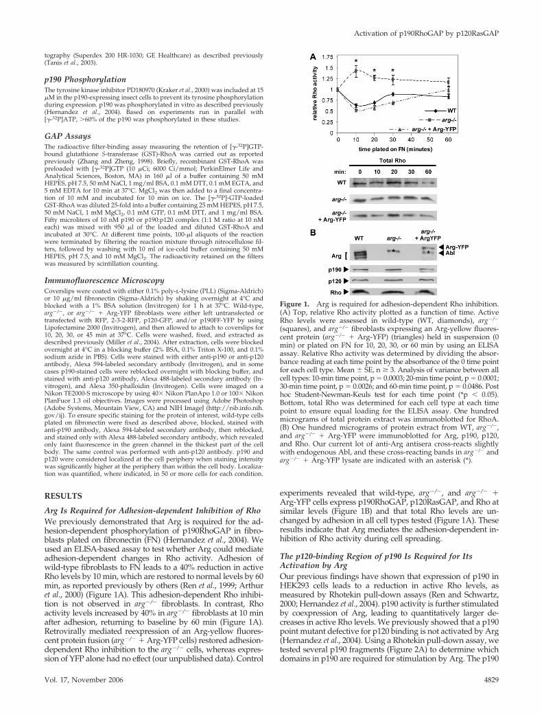
Arg Is Required for Adhesion-dependent Inhibition of RhoWe previously demonstrated that Arg is required for the ad-hesion-dependent phosphorylation of p190RhoGAP in fibro-blasts plated on fibronectin (FN) (Hernandez et al., 2004). Weused an ELISA-based assay to test whether Arg could mediateadhesion-dependent changes in Rho activity. Adhesion ofwild-type fibroblasts to FN leads to a 40% reduction in activeRho levels by 10 min, which are restored to normal levels by 60min, as reported previously by others (Ren et al., 1999; Arthuret al., 2000) (Figure 1A). This adhesion-dependent Rho inhibi-tion is not observed in arg�/� fibroblasts. In contrast, Rhoactivity levels increased by 40% in arg�/� fibroblasts at 10 minafter adhesion, returning to baseline by 60 min (Figure 1A).Retrovirally mediated reexpression of an Arg-yellow fluores-cent protein fusion (arg�/� � Arg-YFP cells) restored adhesion-dependent Rho inhibition to the arg�/� cells, whereas expres-sion of YFP alone had no effect (our unpublished data). Control
experiments revealed that wild-type, arg�/�, and arg�/� �Arg-YFP cells express p190RhoGAP, p120RasGAP, and Rho atsimilar levels (Figure 1B) and that total Rho levels are un-changed by adhesion in all cell types tested (Figure 1A). Theseresults indicate that Arg mediates the adhesion-dependent in-hibition of Rho activity during cell spreading.
The p120-binding Region of p190 Is Required for ItsActivation by ArgOur previous findings have shown that expression of p190 inHEK293 cells leads to a reduction in active Rho levels, asmeasured by Rhotekin pull-down assays (Ren and Schwartz,2000; Hernandez et al., 2004). p190 activity is further stimulatedby coexpression of Arg, leading to quantitatively larger de-creases in active Rho levels. We previously showed that a p190point mutant defective for p120 binding is not activated by Arg(Hernandez et al., 2004). Using a Rhotekin pull-down assay, wetested several p190 fragments (Figure 2A) to determine whichdomains in p190 are required for stimulation by Arg. The p190
Figure 1. Arg is required for adhesion-dependent Rho inhibition.(A) Top, relative Rho activity plotted as a function of time. ActiveRho levels were assessed in wild-type (WT, diamonds), arg�/�
(squares), and arg�/� fibroblasts expressing an Arg-yellow fluores-cent protein (arg�/� � Arg-YFP) (triangles) held in suspension (0min) or plated on FN for 10, 20, 30, or 60 min by using an ELISAassay. Relative Rho activity was determined by dividing the absor-bance reading at each time point by the absorbance of the 0 time pointfor each cell type. Mean � SE, n � 3. Analysis of variance between allcell types: 10-min time point, p � 0.0003; 20-min time point, p � 0.0001;30-min time point, p � 0.0026; and 60-min time point, p � 0.0486. Posthoc Student-Newman-Keuls test for each time point (*p � 0.05).Bottom, total Rho was determined for each cell type at each timepoint to ensure equal loading for the ELISA assay. One hundredmicrograms of total protein extract was immunoblotted for RhoA.(B) One hundred micrograms of protein extract from WT, arg�/�,and arg�/� � Arg-YFP were immunoblotted for Arg, p190, p120,and Rho. Our current lot of anti-Arg antisera cross-reacts slightlywith endogenous Abl, and these cross-reacting bands in arg�/� andarg�/� � Arg-YFP lysate are indicated with an asterisk (*).
Activation of p190RhoGAP by p120RasGAP
Vol. 17, November 2006 4829
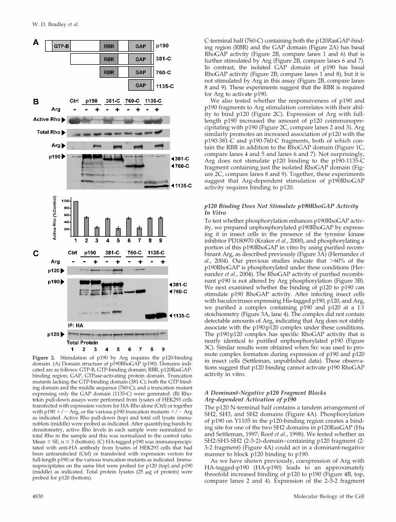
C-terminal half (760-C) containing both the p120RasGAP-bind-ing region (RBR) and the GAP domain (Figure 2A) has basalRhoGAP activity (Figure 2B, compare lanes 1 and 6) that isfurther stimulated by Arg (Figure 2B, compare lanes 6 and 7).In contrast, the isolated GAP domain of p190 has basalRhoGAP activity (Figure 2B, compare lanes 1 and 8), but it isnot stimulated by Arg in this assay (Figure 2B, compare lanes8 and 9). These experiments suggest that the RBR is requiredfor Arg to activate p190.
We also tested whether the responsiveness of p190 andp190 fragments to Arg stimulation correlates with their abil-ity to bind p120 (Figure 2C). Expression of Arg with full-length p190 increased the amount of p120 coimmunopre-cipitating with p190 (Figure 2C, compare lanes 2 and 3). Argsimilarly promotes an increased association of p120 with thep190-381-C and p190-760-C fragments, both of which con-tain the RBR in addition to the RhoGAP domain (Figure 1C,compare lanes 4 and 5 and lanes 6 and 7). Not surprisingly,Arg does not stimulate p120 binding to the p190-1135-Cfragment containing just the isolated RhoGAP domain (Fig-ure 2C, compare lanes 8 and 9). Together, these experimentssuggest that Arg-dependent stimulation of p190RhoGAPactivity requires binding to p120.
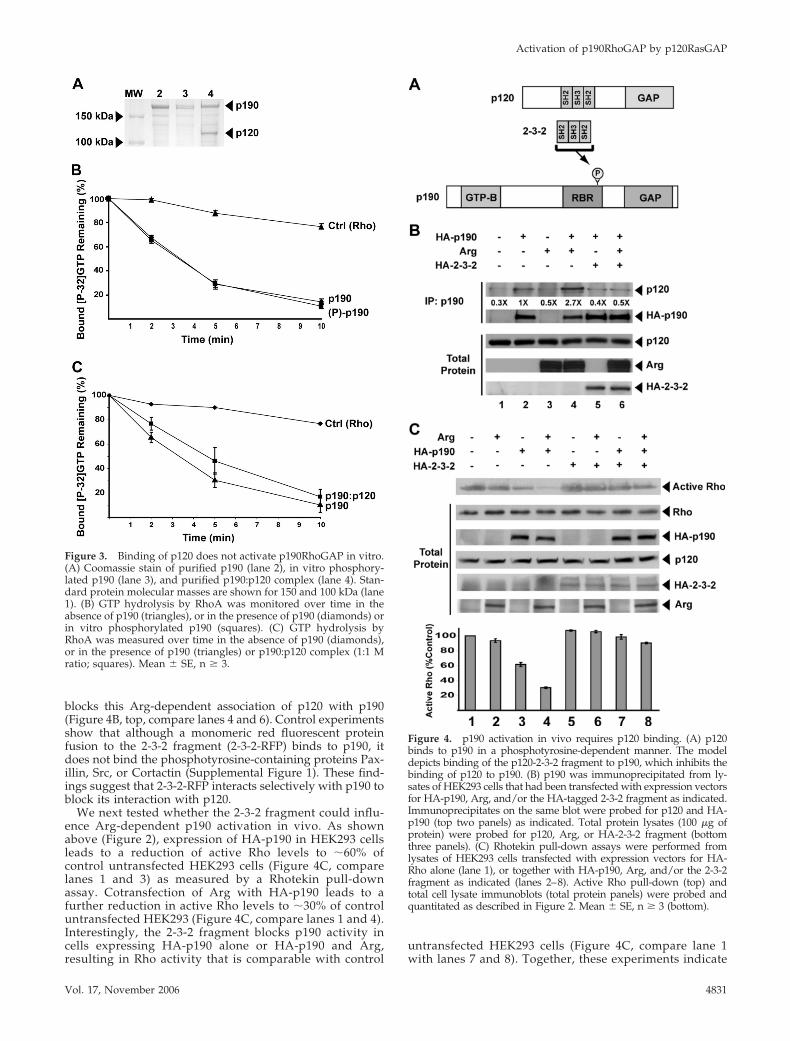
p120 Binding Does Not Stimulate p190RhoGAP ActivityIn VitroTo test whether phosphorylation enhances p190RhoGAP activ-ity, we prepared unphosphorylated p190RhoGAP by express-ing it in insect cells in the presence of the tyrosine kinaseinhibitor PD180970 (Kraker et al., 2000), and phosphorylating aportion of this p190RhoGAP in vitro by using purified recom-binant Arg, as described previously (Figure 3A) (Hernandez etal., 2004). Our previous studies indicate that �60% of thep190RhoGAP is phosphorylated under these conditions (Her-nandez et al., 2004). The RhoGAP activity of purified recombi-nant p190 is not altered by Arg phosphorylation (Figure 3B).We next examined whether the binding of p120 to p190 canstimulate p190 RhoGAP activity. After infecting insect cellswith baculoviruses expressing His-tagged p190, p120, and Arg,we purified a complex containing p190 and p120 at a 1:1stoichiometry (Figure 3A, lane 4). The complex did not containdetectable amounts of Arg, indicating that Arg does not stablyassociate with the p190:p120 complex under these conditions.The p190:p120 complex has specific RhoGAP activity that isnearly identical to purified unphosphorylated p190 (Figure3C). Similar results were obtained when Src was used to pro-mote complex formation during expression of p190 and p120in insect cells (Settleman, unpublished data). These observa-tions suggest that p120 binding cannot activate p190 RhoGAPactivity in vitro.
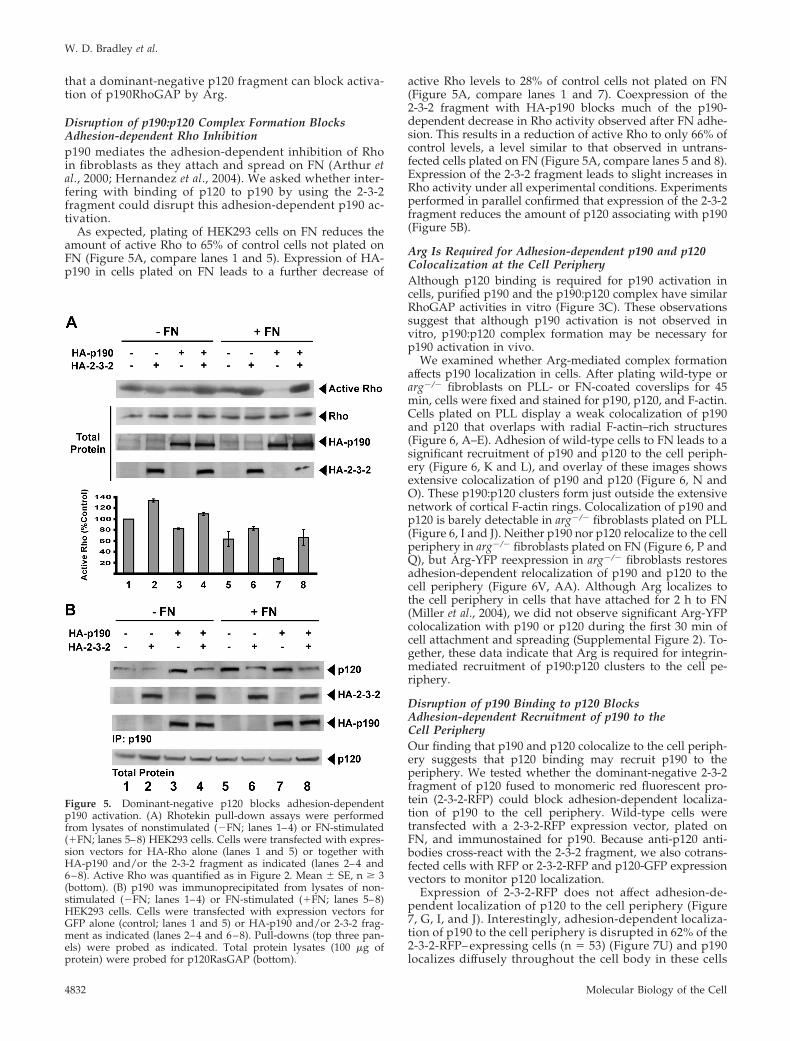
A Dominant-Negative p120 Fragment BlocksArg-dependent Activation of p190The p120 N-terminal half contains a tandem arrangement ofSH2, SH3, and SH2 domains (Figure 4A). Phosphorylationof p190 on Y1105 in the p120-binding region creates a bind-ing site for one of the two SH2 domains in p120RasGAP (Huand Settleman, 1997; Roof et al., 1998). We tested whether anSH2-SH3-SH2 (2-3-2)-domain–containing p120 fragment (2-3-2 fragment) (Figure 4A) could act in a dominant-negativemanner to block p120 binding to p190.
As we have shown previously, coexpression of Arg withHA-tagged-p190 (HA-p190) leads to an approximatelythreefold increased binding of p120 to p190 (Figure 4B, top,compare lanes 2 and 4). Expression of the 2-3-2 fragment
Figure 2. Stimulation of p190 by Arg requires the p120-bindingdomain. (A) Domain structure of p190RhoGAP (p190). Domains indi-cated are as follows: GTP-B, GTP-binding domain; RBR, p120RasGAP-binding region; GAP, GTPase-activating protein domain. Truncationmutants lacking the GTP-binding domain (381-C); both the GTP-bind-ing domain and the middle sequence (760-C); and a truncation mutantexpressing only the GAP domain (1135-C) were generated. (B) Rho-tekin pull-down assays were performed from lysates of HEK293 cellstransfected with expression vectors for HA-Rho alone (Ctrl) or togetherwith p190 �/� Arg, or the various p190 truncation mutants �/� Argas indicated. Active Rho pull-down (top) and total cell lysate immu-noblots (middle) were probed as indicated. After quantifying bands bydensitometry, active Rho levels in each sample were normalized tototal Rho in the sample and this was normalized to the control ratio.Mean � SE, n � 3 (bottom). (C) HA-tagged p190 was immunoprecipi-tated with anti-HA antibody from lysates of HEK293 cells that hadbeen untransfected (Ctrl) or transfected with expression vectors forfull-length p190 or the various truncation mutants as indicated. Immu-noprecipitates on the same blot were probed for p120 (top) and p190(middle) as indicated. Total protein lysates (25 �g of protein) wereprobed for p120 (bottom).
W. D. Bradley et al.
Molecular Biology of the Cell4830
blocks this Arg-dependent association of p120 with p190(Figure 4B, top, compare lanes 4 and 6). Control experimentsshow that although a monomeric red fluorescent proteinfusion to the 2-3-2 fragment (2-3-2-RFP) binds to p190, itdoes not bind the phosphotyrosine-containing proteins Pax-illin, Src, or Cortactin (Supplemental Figure 1). These find-ings suggest that 2-3-2-RFP interacts selectively with p190 toblock its interaction with p120.
We next tested whether the 2-3-2 fragment could influ-ence Arg-dependent p190 activation in vivo. As shownabove (Figure 2), expression of HA-p190 in HEK293 cellsleads to a reduction of active Rho levels to �60% ofcontrol untransfected HEK293 cells (Figure 4C, comparelanes 1 and 3) as measured by a Rhotekin pull-downassay. Cotransfection of Arg with HA-p190 leads to afurther reduction in active Rho levels to �30% of controluntransfected HEK293 (Figure 4C, compare lanes 1 and 4).Interestingly, the 2-3-2 fragment blocks p190 activity incells expressing HA-p190 alone or HA-p190 and Arg,resulting in Rho activity that is comparable with control
untransfected HEK293 cells (Figure 4C, compare lane 1with lanes 7 and 8). Together, these experiments indicate
Figure 3. Binding of p120 does not activate p190RhoGAP in vitro.(A) Coomassie stain of purified p190 (lane 2), in vitro phosphory-lated p190 (lane 3), and purified p190:p120 complex (lane 4). Stan-dard protein molecular masses are shown for 150 and 100 kDa (lane1). (B) GTP hydrolysis by RhoA was monitored over time in theabsence of p190 (triangles), or in the presence of p190 (diamonds) orin vitro phosphorylated p190 (squares). (C) GTP hydrolysis byRhoA was measured over time in the absence of p190 (diamonds),or in the presence of p190 (triangles) or p190:p120 complex (1:1 Mratio; squares). Mean � SE, n � 3.
Figure 4. p190 activation in vivo requires p120 binding. (A) p120binds to p190 in a phosphotyrosine-dependent manner. The modeldepicts binding of the p120-2-3-2 fragment to p190, which inhibits thebinding of p120 to p190. (B) p190 was immunoprecipitated from ly-sates of HEK293 cells that had been transfected with expression vectorsfor HA-p190, Arg, and/or the HA-tagged 2-3-2 fragment as indicated.Immunoprecipitates on the same blot were probed for p120 and HA-p190 (top two panels) as indicated. Total protein lysates (100 �g ofprotein) were probed for p120, Arg, or HA-2-3-2 fragment (bottomthree panels). (C) Rhotekin pull-down assays were performed fromlysates of HEK293 cells transfected with expression vectors for HA-Rho alone (lane 1), or together with HA-p190, Arg, and/or the 2-3-2fragment as indicated (lanes 2–8). Active Rho pull-down (top) andtotal cell lysate immunoblots (total protein panels) were probed andquantitated as described in Figure 2. Mean � SE, n � 3 (bottom).
Activation of p190RhoGAP by p120RasGAP
Vol. 17, November 2006 4831
that a dominant-negative p120 fragment can block activa-tion of p190RhoGAP by Arg.
Disruption of p190:p120 Complex Formation BlocksAdhesion-dependent Rho Inhibitionp190 mediates the adhesion-dependent inhibition of Rhoin fibroblasts as they attach and spread on FN (Arthur etal., 2000; Hernandez et al., 2004). We asked whether inter-fering with binding of p120 to p190 by using the 2-3-2fragment could disrupt this adhesion-dependent p190 ac-tivation.
As expected, plating of HEK293 cells on FN reduces theamount of active Rho to 65% of control cells not plated onFN (Figure 5A, compare lanes 1 and 5). Expression of HA-p190 in cells plated on FN leads to a further decrease of
active Rho levels to 28% of control cells not plated on FN(Figure 5A, compare lanes 1 and 7). Coexpression of the2-3-2 fragment with HA-p190 blocks much of the p190-dependent decrease in Rho activity observed after FN adhe-sion. This results in a reduction of active Rho to only 66% ofcontrol levels, a level similar to that observed in untrans-fected cells plated on FN (Figure 5A, compare lanes 5 and 8).Expression of the 2-3-2 fragment leads to slight increases inRho activity under all experimental conditions. Experimentsperformed in parallel confirmed that expression of the 2-3-2fragment reduces the amount of p120 associating with p190(Figure 5B).
Arg Is Required for Adhesion-dependent p190 and p120Colocalization at the Cell PeripheryAlthough p120 binding is required for p190 activation incells, purified p190 and the p190:p120 complex have similarRhoGAP activities in vitro (Figure 3C). These observationssuggest that although p190 activation is not observed invitro, p190:p120 complex formation may be necessary forp190 activation in vivo.
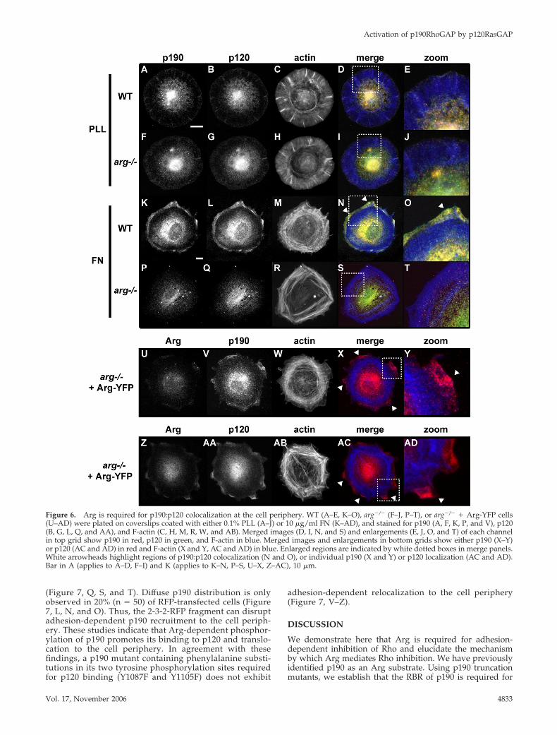
We examined whether Arg-mediated complex formationaffects p190 localization in cells. After plating wild-type orarg�/� fibroblasts on PLL- or FN-coated coverslips for 45min, cells were fixed and stained for p190, p120, and F-actin.Cells plated on PLL display a weak colocalization of p190and p120 that overlaps with radial F-actin–rich structures(Figure 6, A–E). Adhesion of wild-type cells to FN leads to asignificant recruitment of p190 and p120 to the cell periph-ery (Figure 6, K and L), and overlay of these images showsextensive colocalization of p190 and p120 (Figure 6, N andO). These p190:p120 clusters form just outside the extensivenetwork of cortical F-actin rings. Colocalization of p190 andp120 is barely detectable in arg�/� fibroblasts plated on PLL(Figure 6, I and J). Neither p190 nor p120 relocalize to the cellperiphery in arg�/� fibroblasts plated on FN (Figure 6, P andQ), but Arg-YFP reexpression in arg�/� fibroblasts restoresadhesion-dependent relocalization of p190 and p120 to thecell periphery (Figure 6V, AA). Although Arg localizes tothe cell periphery in cells that have attached for 2 h to FN(Miller et al., 2004), we did not observe significant Arg-YFPcolocalization with p190 or p120 during the first 30 min ofcell attachment and spreading (Supplemental Figure 2). To-gether, these data indicate that Arg is required for integrin-mediated recruitment of p190:p120 clusters to the cell pe-riphery.
Disruption of p190 Binding to p120 BlocksAdhesion-dependent Recruitment of p190 to theCell PeripheryOur finding that p190 and p120 colocalize to the cell periph-ery suggests that p120 binding may recruit p190 to theperiphery. We tested whether the dominant-negative 2-3-2fragment of p120 fused to monomeric red fluorescent pro-tein (2-3-2-RFP) could block adhesion-dependent localiza-tion of p190 to the cell periphery. Wild-type cells weretransfected with a 2-3-2-RFP expression vector, plated onFN, and immunostained for p190. Because anti-p120 anti-bodies cross-react with the 2-3-2 fragment, we also cotrans-fected cells with RFP or 2-3-2-RFP and p120-GFP expressionvectors to monitor p120 localization.
Expression of 2-3-2-RFP does not affect adhesion-de-pendent localization of p120 to the cell periphery (Figure7, G, I, and J). Interestingly, adhesion-dependent localiza-tion of p190 to the cell periphery is disrupted in 62% of the2-3-2-RFP– expressing cells (n � 53) (Figure 7U) and p190localizes diffusely throughout the cell body in these cells
Figure 5. Dominant-negative p120 blocks adhesion-dependentp190 activation. (A) Rhotekin pull-down assays were performedfrom lysates of nonstimulated (�FN; lanes 1–4) or FN-stimulated(�FN; lanes 5–8) HEK293 cells. Cells were transfected with expres-sion vectors for HA-Rho alone (lanes 1 and 5) or together withHA-p190 and/or the 2-3-2 fragment as indicated (lanes 2–4 and6–8). Active Rho was quantified as in Figure 2. Mean � SE, n � 3(bottom). (B) p190 was immunoprecipitated from lysates of non-stimulated (�FN; lanes 1–4) or FN-stimulated (�FN; lanes 5–8)HEK293 cells. Cells were transfected with expression vectors forGFP alone (control; lanes 1 and 5) or HA-p190 and/or 2-3-2 frag-ment as indicated (lanes 2–4 and 6–8). Pull-downs (top three pan-els) were probed as indicated. Total protein lysates (100 �g ofprotein) were probed for p120RasGAP (bottom).
W. D. Bradley et al.
Molecular Biology of the Cell4832
(Figure 7, Q, S, and T). Diffuse p190 distribution is onlyobserved in 20% (n � 50) of RFP-transfected cells (Figure7, L, N, and O). Thus, the 2-3-2-RFP fragment can disruptadhesion-dependent p190 recruitment to the cell periph-ery. These studies indicate that Arg-dependent phosphor-ylation of p190 promotes its binding to p120 and translo-cation to the cell periphery. In agreement with thesefindings, a p190 mutant containing phenylalanine substi-tutions in its two tyrosine phosphorylation sites requiredfor p120 binding (Y1087F and Y1105F) does not exhibit
adhesion-dependent relocalization to the cell periphery(Figure 7, V–Z).
DISCUSSION
We demonstrate here that Arg is required for adhesion-dependent inhibition of Rho and elucidate the mechanismby which Arg mediates Rho inhibition. We have previouslyidentified p190 as an Arg substrate. Using p190 truncationmutants, we establish that the RBR of p190 is required for
Figure 6. Arg is required for p190:p120 colocalization at the cell periphery. WT (A–E, K–O), arg�/� (F–J, P–T), or arg�/� � Arg-YFP cells(U–AD) were plated on coverslips coated with either 0.1% PLL (A–J) or 10 �g/ml FN (K–AD), and stained for p190 (A, F, K, P, and V), p120(B, G, L, Q, and AA), and F-actin (C, H, M, R, W, and AB). Merged images (D, I, N, and S) and enlargements (E, J, O, and T) of each channelin top grid show p190 in red, p120 in green, and F-actin in blue. Merged images and enlargements in bottom grids show either p190 (X–Y)or p120 (AC and AD) in red and F-actin (X and Y, AC and AD) in blue. Enlarged regions are indicated by white dotted boxes in merge panels.White arrowheads highlight regions of p190:p120 colocalization (N and O), or individual p190 (X and Y) or p120 localization (AC and AD).Bar in A (applies to A–D, F–I) and K (applies to K–N, P–S, U–X, Z–AC), 10 �m.
Activation of p190RhoGAP by p120RasGAP
Vol. 17, November 2006 4833
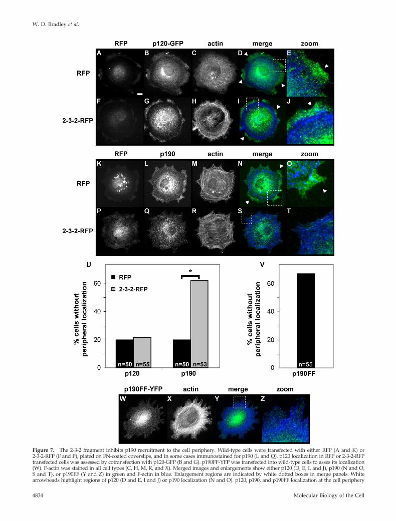
Figure 7. The 2-3-2 fragment inhibits p190 recruitment to the cell periphery. Wild-type cells were transfected with either RFP (A and K) or2-3-2-RFP (F and P), plated on FN-coated coverslips, and in some cases immunostained for p190 (L and Q). p120 localization in RFP or 2-3-2-RFPtransfected cells was assessed by cotransfection with p120-GFP (B and G). p190FF-YFP was transfected into wild-type cells to asses its localization(W). F-actin was stained in all cell types (C, H, M, R, and X). Merged images and enlargements show either p120 (D, E, I, and J), p190 (N and O,S and T), or p190FF (Y and Z) in green and F-actin in blue. Enlargement regions are indicated by white dotted boxes in merge panels. Whitearrowheads highlight regions of p120 (D and E, I and J) or p190 localization (N and O). p120, p190, and p190FF localization at the cell periphery
W. D. Bradley et al.
Molecular Biology of the Cell4834
Arg to stimulate RhoGAP activity. Arg-dependent phos-phorylation of Y1105 in the RBR of p190 promotes p120binding (Hu and Settleman, 1997; Roof et al., 1998; Hernan-dez et al., 2004). Expression of a dominant-negative 2-3-2fragment of p120 blocks binding of p120 to p190 and pre-vents p190 activation by adhesion. We also show that Arg isrequired for p190 and p120 recruitment to the cell peripheryafter adhesion to FN, and p190 localization is disrupted bythe 2-3-2 fragment. Together, our results indicate that p120binding regulates p190 RhoGAP activity in vivo by promot-ing the recruitment of a p190:p120 complex to the cell pe-riphery where it can inhibit Rho.
Phosphorylation of p190 on Y1105 promotes its associa-tion with p120 (Hu and Settleman, 1997; Roof et al., 1998;Hernandez et al., 2004). We have previously reported thatadhesion to FN stimulates p190 phosphorylation in wild-type, but not arg�/� cells, and this phosphorylation is re-stored to these cells by Arg reexpression (Hernandez et al.,2004). Burridge and colleagues have shown that integrinstimulation with RGD-containing peptides stimulates p190in wild-type, but not src�/� yes�/� fyn�/� (SYF) cells (Arthuret al., 2000). Arg phosphorylates p190 directly on Y1105(Hernandez et al., 2004), a site that is also phosphorylated inp190 immunopurified from Src-overexpressing cells (Roof etal., 1998). Together, these studies suggest that both Arg andSrc family kinases are required for integrin-dependent phos-phorylation and activation of p190.
One possible explanation for these findings is that Srcfamily kinases and Arg differentially phosphorylate p190depending on the mode of integrin stimulation (i.e., solublepeptides versus adhesion to FN-coated surfaces). Alterna-tively, Src family kinases may serve as links between inte-grin receptors and Arg kinase activity. Src family kinasescan phosphorylate and activate Abl and Arg in vitro andhave been shown to mediate signaling between the platelet-derived growth factor receptor and Abl family kinases (Plat-tner et al., 1999; Dorey et al., 2001; Tanis et al., 2003). Finally,it is possible that Src family kinases or Arg play a structural,but noncatalytic role in p190 activation. These models can betested by measuring p190 activation in SYF or Arg-deficientcells reconstituted with Src or Arg mutants.
Arg stimulates p190 activity through phosphorylation ofY1105 in the p120-binding region (Hernandez et al., 2004).Because this phosphorylation event promotes binding ofp120 to p190, we examined whether the binding of p120stimulates p190 activity. Our finding that the RhoGAP ac-tivities of p190 and the p190:p120 complex are similar arguesthat complex formation does not activate p190 in vitro.
Active Rho localizes to the plasma membrane where itregulates actomyosin contractility and cytoskeletal rear-rangements (Ridley, 2000; Pertz et al., 2006). Previous studieshave shown that p190 relocalizes to the cell periphery inresponse to adhesion or growth factor stimulation (Chang etal., 1995; Nakahara et al., 1998; Brouns et al., 2000; Tsubouchiet al., 2002). This recruitment of p190 correlates with a re-duction of F-actin stress fibers, which likely reflects de-creased Rho activity (Chang et al., 1995; Nakahara et al.,1998). Our finding that adhesion to FN stimulates p190:p120colocalization at the cell periphery, and that p190 localiza-tion is disrupted by the 2-3-2 fragment, suggests that p120
activates p190 by promoting its recruitment to the mem-brane to inhibit Rho.
p120 has both pleckstrin homology (PH) and Ca2�-depen-dent phospholipid binding (CaLB) domains that may facil-itate interactions with the plasma membrane (Gawler et al.,1995a, b; Drugan et al., 2000; Koehler and Moran, 2001). Wedo not observe significant localization of p120 to the cellperiphery in the absence of integrin-mediated adhesion,suggesting that integrin-mediated adhesion may be requiredto generate phosphatidylinositol species that act as bindingtargets for the PH and CaLB domains in p120. Our findingthat the 2-3-2 fragment blocks p190 localization to the pe-riphery without affecting p120 localization strongly suggeststhat p120 serves as a peripheral binding target for p190.Interestingly, the 2-3-2 fragment did not block p190 relocal-ization in every cell. It is also possible that p190 interacts viaadditional surfaces with other targets at the cell peripherythat facilitate its recruitment. One likely additional target forp190 is RhoE, which is associated with the membrane andcan bind and activate p190 (Wennerberg et al., 2003). It isalso likely that p190 binding stabilizes p120 localization atthe cell periphery, because p120 levels are reduced at theperiphery in arg�/� cells plated on FN (Figure 6Q).
Active Rho promotes cell contractility through the activa-tion of actomyosin contractility (Ridley, 2001). Our findingthat Arg is required for the formation of p190:p120 com-plexes at the cell periphery suggests that Arg activity maylocally regulate Rho activity during cell adhesion andspreading. It is important to note that the experiments pre-sented here were performed during initial cell attachmentand spreading on FN-coated surfaces. The initial globalreduction in Rho activity in wild-type fibroblasts observedby us and others (Ren et al., 1999; Arthur et al., 2000) likelyallows cells to spread and sample their new adhesive envi-ronment. Cells initially spread in all directions, and the p190relocalization we observe along the entire cell periphery isconsistent with a uniform circumferential Rho inhibitionduring this spreading phase. Recently, Hahn and colleaguesobserved that active Rho primarily localizes to protrusionsites in cells exhibiting directed cell migration (Pertz et al.,2006). The localization of active Rho to these protrusive sitesunderscores the need for a dynamic cycling of Rho activityfor efficient directed cell motility. A local inhibitor of Rho,such as p190, is likely critical for localized Rho inactivationduring Rho activity cycling. We have previously shown thatArg promotes lamellipodial protrusion in fibroblasts afterthey adhere and spread on FN (Miller et al., 2004). Wepropose that Arg-dependent formation of p190:p120 clustersmay relax Rho-mediated contractility locally to allow theinitiation or persistence of these cellular protrusions. In sup-port of this, Arthur and Burridge have previously shownthat p190 overexpression promotes cell edge protrusion ascells migrate into wounds induced in a fibroblast monolayer,whereas cells expressing a dominant negative p190 exhibitdeficient membrane protrusion and cell spreading (Arthurand Burridge, 2001). Future imaging studies should revealhow the formation of p190:p120 clusters affect Rho activitylocally and how this affects cell edge protrusion.
ACKNOWLEDGMENTS
We thank Walter Boron for use of the ELISA plate reader, Mark Parker andXianyun Ye for technical assistance, Scott Boyle and Justin Peacock for helpfuldiscussions, and Koleske laboratory members for critical comments on themanuscript. This work was supported by a predoctoral fellowship from theAnna Fuller Fund to W.D.B., a predoctoral National Research Service Award(NS42365) to S.E.H., a grant from the National Institute of General MedicalSciences (CA62142) to J. S., and grants from the National Institute of Neuro-
Figure 7 (cont). was quantified. (U and V) Black bars indicateRFP-transfected cells for p190 and p120 localization, and gray barsindicate 2-3-2-RFP transfected cells, with number of cells (n � 50)indicated in each instance. Student’s t test between p190 localizationin RFP versus 2-3-2-RFP–expressing cells, *p � 0.0001. Bar in A, 10 �m.
Activation of p190RhoGAP by p120RasGAP
Vol. 17, November 2006 4835
logical Disorders and Stroke (NS39475), National Alliance for Research onSchizophrenia and Depression, and the Leukemia and Lymphoma Society ofAmerica to A.J.K.
REFERENCES
Arthur, W. T., and Burridge, K. (2001). RhoA inactivation by p190RhoGAPregulates cell spreading and migration by promoting membrane protrusionand polarity. Mol. Biol. Cell 12, 2711–2720.
Arthur, W. T., Noren, N. K., and Burridge, K. (2002). Regulation of Rho familyGTPases by cell-cell and cell-matrix adhesion. Biol. Res. 35, 239–246.
Arthur, W. T., Petch, L. A., and Burridge, K. (2000). Integrin engagement sup-presses RhoA activity via a c-Src-dependent mechanism. Curr. Biol. 10, 719–722.
Barry, S. T., Flinn, H. M., Humphries, M. J., Critchley, D. R., and Ridley, A. J.(1997). Requirement for Rho in integrin signalling. Cell Adhes. Commun. 4,387–398.
Brouns, M. R., Matheson, S. F., Hu, K. Q., Delalle, I., Caviness, V. S., Silver, J.,Bronson, R. T., and Settleman, J. (2000). The adhesion signaling molecule p190RhoGAP is required for morphogenetic processes in neural development.Development 127, 4891–4903.
Burridge, K., and Wennerberg, K. (2004). Rho and Rac take center stage. Cell116, 167–179.
Chang, J. H., Gill, S., Settleman, J., and Parsons, S. J. (1995). c-Src regulates thesimultaneous rearrangement of actin cytoskeleton, p190RhoGAP, andp120RasGAP following epidermal growth factor stimulation. J. Cell Biol. 130,355–368.
Dorey, K., Engen, J. R., Kretzschmar, J., Wilm, M., Neubauer, G., Schindler, T.,and Superti-Furga, G. (2001). Phosphorylation and structure-based functionalstudies reveal a positive and a negative role for the activation loop of thec-Abl tyrosine kinase. Oncogene 20, 8075–8084.
Drugan, J. K., Rogers-Graham, K., Gilmer, T., Campbell, S., and Clark, G. J.(2000). The Ras/p120 GTPase-activating protein (GAP) interaction is regu-lated by the p120 GAP pleckstrin homology domain. J. Biol. Chem. 275,35021–35027.
Frame, M. C., and Brunton, V. G. (2002). Advances in Rho-dependent actinregulation and oncogenic transformation. Curr. Opin. Genet. Dev. 12, 36–43.
Gawler, D. J., Zhang, L. J., and Moran, M. F. (1995a). Mutation-deletionanalysis of a Ca(2�)-dependent phospholipid binding (CaLB) domain withinp120 GAP, a GTPase-activating protein for p21 ras. Biochem. J. 307, 487–491.
Gawler, D. J., Zhang, L. J., Reedijk, M., Tung, P. S., and Moran, M. F. (1995b).CaLB: a 43 amino acid calcium-dependent membrane/phospholipid bindingdomain in p120 Ras GTPase-activating protein. Oncogene 10, 817–825.
Gimond, C., van Der Flierqq, A., van Delft, S., Brakebusch, C., Kuikman, I.,Collard, J. G., Fassler, R., and Sonnenberg, A. (1999). Induction of cell scat-tering by expression of beta1 integrins in beta1-deficient epithelial cells re-quires activation of members of the rho family of GTPases and downregula-tion of cadherin and catenin function. J. Cell Biol. 147, 1325–1340.
Hernandez, S. E., Settleman, J., and Koleske, A. J. (2004). Adhesion-dependentregulation of p190RhoGAP in the developing brain by the Abl-related genetyrosine kinase. Curr. Biol. 14, 691–696.
Hu, K. Q., and Settleman, J. (1997). Tandem SH2 binding sites mediate theRasGAP-RhoGAP interaction: a conformational mechanism for SH3 domainregulation. EMBO J. 16, 473–483.
Hynes, R. O. (2002). Integrins: bidirectional, allosteric signaling machines.Cell 110, 673–687.
Jockusch, B. M., Bubeck, P., Giehl, K., Kroemker, M., Moschner, J., Rothkegel,M., Rudiger, M., Schluter, K., Stanke, G., and Winkler, J. (1995). The moleculararchitecture of focal adhesions. Annu. Rev. Cell Dev. Biol. 11, 379–416.
Kaibuchi, K., Kuroda, S., and Amano, M. (1999). Regulation of the cytoskel-eton and cell adhesion by the Rho family GTPases in mammalian cells. Annu.Rev. Biochem. 68, 459–486.
Kain, K. H., and Klemke, R. L. (2001). Inhibition of cell migration by Ablfamily tyrosine kinases through uncoupling of Crk-CAS complexes. J. Biol.Chem. 276, 16185–16192.
Kimura, K., Ito, M., Amano, M., Chihara, K., Fukata, Y., Nakafuku, M.,Yamamori, B., Feng, J., Nakano, T., Okawa, K., Iwamatsu, A., and Kaibuchi,K. (1996). Regulation of myosin phosphatase by Rho and Rho-associatedkinase (Rho-kinase). Science 273, 245–248.
Koehler, J. A., and Moran, M. F. (2001). Regulation of extracellular signal-regulated kinase activity by p120 RasGAP does not involve its pleckstrinhomology or calcium-dependent lipid binding domains but does requirethese domains to regulate cell proliferation. Cell Growth Differ. 12, 551–561.
Koleske, A. J., Gifford, A. M., Scott, M. L., Nee, M., Bronson, R. T., Miczek,K. A., and Baltimore, D. (1998). Essential roles for the Abl and Arg tyrosinekinases in neurulation. Neuron 21, 1259–1272.
Kraker, A. J., Hartl, B. G., Amar, A. M., Barvian, M. R., Showalter, H. D., andMoore, C. W. (2000). Biochemical and cellular effects of c-Src kinase-selectivepyrido[2,3-d]pyrimidine tyrosine kinase inhibitors. Biochem. Pharmacol. 60,885–898.
Miller, A. L., Wang, Y., Mooseker, M. S., and Koleske, A. J. (2004). The Abl-relatedgene (Arg) requires its F-actin-microtubule cross-linking activity to regulatelamellipodial dynamics during fibroblast adhesion. J. Cell Biol. 165, 407–419.
Moresco, E. M., Donaldson, S., Williamson, A., and Koleske, A. J. (2005).Integrin-dependent dendrite branch stabilization requires Abl family kinases.J. Neurosci. 25, 6105–6118.
Nakahara, H., Mueller, S. C., Nomizu, M., Yamada, Y., Yeh, Y., and Chen,W. T. (1998). Activation of beta1 integrin signaling stimulates tyrosine phos-phorylation of p190RhoGAP and membrane-protrusive activities at inva-dopodia. J. Biol. Chem. 273, 9–12.
Pertz, O., Hodgson, L., Klemke, R. L., and Hahn, K. M. (2006). Spatiotemporaldynamics of RhoA activity in migrating cells. Nature 440, 1069–1072.
Plattner, R., Kadlec, L., DeMali, K. A., Kazlauskas, A., and Pendergast, A. M.(1999). c-Abl is activated by growth factors and Src family kinases and has arole in the cellular response to PDGF. Genes Dev. 13, 2400–2411.
Ren, X. D., Kiosses, W. B., and Schwartz, M. A. (1999). Regulation of the smallGTP-binding protein Rho by cell adhesion and the cytoskeleton. EMBO J. 18,578–585.
Ren, X. D., and Schwartz, M. A. (2000). Determination of GTP loading on Rho.Methods Enzymol. 325, 264–272.
Ridley, A. (2000). Rho GTPases. Integrating integrin signaling. J. Cell Biol. 150,F107–F109.
Ridley, A. J. (2001). Rho family proteins: coordinating cell responses. TrendsCell Biol. 11, 471–477.
Ridley, A. J., and Hall, A. (1992). The small GTP-binding protein rho regulatesthe assembly of focal adhesions and actin stress fibers in response to growthfactors. Cell 70, 389–399.
Roof, R. W., Haskell, M. D., Dukes, B. D., Sherman, N., Kinter, M., andParsons, S. J. (1998). Phosphotyrosine (p-Tyr)-dependent and -independentmechanisms of p190 RhoGAP-p120 RasGAP interaction: Tyr 1105 of p190, asubstrate for c-Src, is the sole p-Tyr mediator of complex formation. Mol. Cell.Biol. 18, 7052–7063.
Schwartz, M. A., Schaller, M. D., and Ginsberg, M. H. (1995). Integrins:emerging paradigms of signal transduction. Annu. Rev. Cell Dev. Biol. 11,549–599.
Tanis, K. Q., Veach, D., Duewel, H. S., Bornmann, W. G., and Koleske, A. J.(2003). Two distinct phosphorylation pathways have additive effects on Ablfamily kinase activation. Mol. Cell. Biol. 23, 3884–3896.
Tsubouchi, A., Sakakura, J., Yagi, R., Mazaki, Y., Schaefer, E., Yano, H., andSabe, H. (2002). Localized suppression of RhoA activity by Tyr31/118-phos-phorylated paxillin in cell adhesion and migration. J. Cell Biol. 159, 673–683.
Van Aelst, L., and D’Souza-Schorey, C. (1997). Rho GTPases and signalingnetworks. Genes Dev. 11, 2295–2322.
van der Flier, A., and Sonnenberg, A. (2001). Function and interactions ofintegrins. Cell Tissue Res. 305, 285–298.
Watanabe, N., Madaule, P., Reid, T., Ishizaki, T., Watanabe, G., Kakizuka, A.,Saito, Y., Nakao, K., Jockusch, B. M., and Narumiya, S. (1997). p140mDia, amammalian homolog of Drosophila diaphanous, is a target protein for Rhosmall GTPase and is a ligand for profilin. EMBO J. 16, 3044–3056.
Wennerberg, K., Forget, M. A., Ellerbroek, S. M., Arthur, W. T., Burridge, K.,Settleman, J., Der, C. J., and Hansen, S. H. (2003). Rnd proteins function asRhoA antagonists by activating p190 RhoGAP. Curr. Biol. 13, 1106–1115.
Woodring, P. J., Litwack, E. D., O’Leary, D. D., Lucero, G. R., Wang, J. Y., andHunter, T. (2002). Modulation of the F-actin cytoskeleton by c-Abl tyrosinekinase in cell spreading and neurite extension. J. Cell Biol. 156, 879–892.
Woodring, P. J., et al. (2004). c-Abl phosphorylates Dok1 to promote filopodiaduring cell spreading. J. Cell Biol. 165, 493–503.
Zamir, E., and Geiger, B. (2001a). Components of cell-matrix adhesions. J. CellSci. 114, 3577–3579.
Zamir, E., and Geiger, B. (2001b). Molecular complexity and dynamics ofcell-matrix adhesions. J. Cell Sci. 114, 3583–3590.
Zhang, B., and Zheng, Y. (1998). Regulation of RhoA GTP hydrolysis by theGTPase-activating proteins p190, p50RhoGAP, Bcr, and 3BP-1. Biochemistry37, 5249–5257.
W. D. Bradley et al.
Molecular Biology of the Cell4836
Copyright © 2022 FDOKUMEN