Besides adhesion: new perspectives of integrin functions in angiogenesis
10
Review Besides adhesion: new perspectives of integrin functions in angiogenesis Guido Serini, Lucia Napione, Marco Arese, and Federico Bussolino* Department of Oncological Sciences, Institute for Cancer Research and Treatment, University of Torino, Sp 142, Km 3.95, 10060 Candiolo, Torino, Italy Received 4 January 2008; revised 5 February 2008; accepted 13 February 2008; online publish-ahead-of-print 19 February 2008 Time for primary review: 11 days During angiogenic remodelling in embryo and adult life, endothelial cells lining blood vessel walls dynamically modify their integrin-mediated adhesive contacts with the surrounding extracellular matrix. However, besides regulating cell adhesion and migration, integrins dynamically participate in a network with soluble molecules and their receptors. Angiogenesis is characterized by opposing autocrine and paracrine loops of growth factors and semaphorins that regulate the activation of integ- rins on the endothelial surface through tyrosine kinase receptors (TKR) and the neuropilin/plexin system. Moreover, pro- and anti-angiogenic factors can directly bind integrins and regulate endothelial cell behaviour. This review summarizes the recent progress in understanding the reciprocal interactions between integrins, TKR, and semaphorin receptors. KEYWORDS Plexin; Semaphorin; Tyrosine kinase receptors; Endothelial cells 1. Introduction The molecular mechanisms leading to cell–extracellular matrix (ECM) interactions have been crucial for the evol- ution from protozoans to metazoans. Integrins represent the most important family of receptors mediating cell adhesion to ECM. Each integrin is composed of non-homologous transmembrane a and b subunits and they control cell adhesion through complex molecular mechanisms. Outside-in signalling informs the cell about the ECM environment, while inside-out signalling promotes modifications in integrin functional activity. 1,2 Development and remodelling of vascular systems require complex interactions of signals and physical forces orches- trating the activities of endothelial cells (ECs), pericytes, and smooth muscle cells. Besides several redundant soluble factors, which appear to have a relevant role, two classes of molecules have been identified with a high speci- ficity for the vascular system: the family of vascular endo- thelium growth factors (VEGF) and their tyrosine kinase receptors (TKR), VEGFR-1, -2, and -3, and the family of angiopoietins (Ang) and Tie-2 TKR. 3,4 More recently, molecules firstly characterized for their role in axon guidance (e.g. semaphorins, netrins, and slits) have been selectively involved in the remodelling and sprouting phases of angiogenesis. 5 Vascular cells (i.e. ECs, pericytes, and smooth muscle cells) express a wide range of integrins including a1b1, a2b1, a4b1, a5b1, avb1, avb3, avb5, avb8, a6b1, and a6b4. 6 Integrin-mediated cell-to-ECM adhesion plays a deterministic role in vascular development by contributing to cell movement, to protect cells from anoikis and to endow the vasculature with the ability to sense and respond to changes in physical forces. 6,7 In the last 10 years, an increasing body of evidences has demonstrated that integrins are not mere adhesion recep- tors, but influence the biological activity of several other molecular systems within the cell. Here, we reviewed emerging results highlighting new roles of integrins in angiogenesis. 2. Integrins modulate the activation of vascular tyrosine kinase receptors 2.1 Integrins and vascular endothelial growth factor receptors During angiogenesis, ECs adhere to a provisional ECM mainly through avb3 integrin resulting in an increased biological response to VEGF-A dependent on the formation of a complex between the integrin and VEGFR-2. 8–11 The collagen I receptor a2b1 and the laminin receptors a6b1 and a6b4 do not exert a similar effect. The formation of the integrin-TKR complex first requires the activation of VEGFR-2 by its ligand and the ensuing binding of phospha- tidylinositol 3-kinase (PI3K) 9 and c-src, 11 which participate in directional cell migration triggered by VEGF-A. A mono- clonal antibody anti-b3 not only perturbed the complex formation, but it also markedly inhibited VEGFR-2-mediated * Corresponding author. Tel: þ39 0119933347; fax: þ39 09119933524. E-mail address: [email protected] Published on behalf of the European Society of Cardiology. All rights reserved. & The Author 2008. For permissions please email: [email protected]. Cardiovascular Research (2008) 78, 213–222 doi:10.1093/cvr/cvn045 by guest on June 24, 2016 Downloaded from
Transcript of Besides adhesion: new perspectives of integrin functions in angiogenesis

Review
Besides adhesion: new perspectives of integrin functionsin angiogenesis
Guido Serini, Lucia Napione, Marco Arese, and Federico Bussolino*
Department of Oncological Sciences, Institute for Cancer Research and Treatment, University of Torino, Sp 142,Km 3.95, 10060 Candiolo, Torino, Italy
Received 4 January 2008; revised 5 February 2008; accepted 13 February 2008; online publish-ahead-of-print 19 February 2008
Time for primary review: 11 days
During angiogenic remodelling in embryo and adult life, endothelial cells lining blood vessel wallsdynamically modify their integrin-mediated adhesive contacts with the surrounding extracellularmatrix. However, besides regulating cell adhesion and migration, integrins dynamically participatein a network with soluble molecules and their receptors. Angiogenesis is characterized by opposingautocrine and paracrine loops of growth factors and semaphorins that regulate the activation of integ-rins on the endothelial surface through tyrosine kinase receptors (TKR) and the neuropilin/plexinsystem. Moreover, pro- and anti-angiogenic factors can directly bind integrins and regulate endothelialcell behaviour. This review summarizes the recent progress in understanding the reciprocal interactionsbetween integrins, TKR, and semaphorin receptors.
KEYWORDSPlexin;
Semaphorin;
Tyrosine kinase receptors;
Endothelial cells
1. Introduction
The molecular mechanisms leading to cell–extracellularmatrix (ECM) interactions have been crucial for the evol-ution from protozoans to metazoans. Integrins representthe most important family of receptors mediating celladhesion to ECM. Each integrin is composed ofnon-homologous transmembrane a and b subunits andthey control cell adhesion through complex molecularmechanisms. Outside-in signalling informs the cell aboutthe ECM environment, while inside-out signalling promotesmodifications in integrin functional activity.1,2
Development and remodelling of vascular systems requirecomplex interactions of signals and physical forces orches-trating the activities of endothelial cells (ECs), pericytes,and smooth muscle cells. Besides several redundantsoluble factors, which appear to have a relevant role, twoclasses of molecules have been identified with a high speci-ficity for the vascular system: the family of vascular endo-thelium growth factors (VEGF) and their tyrosine kinasereceptors (TKR), VEGFR-1, -2, and -3, and the family ofangiopoietins (Ang) and Tie-2 TKR.3,4 More recently,molecules firstly characterized for their role in axonguidance (e.g. semaphorins, netrins, and slits) have beenselectively involved in the remodelling and sproutingphases of angiogenesis.5
Vascular cells (i.e. ECs, pericytes, and smooth musclecells) express a wide range of integrins including a1b1,
a2b1, a4b1, a5b1, avb1, avb3, avb5, avb8, a6b1, anda6b4.6 Integrin-mediated cell-to-ECM adhesion plays adeterministic role in vascular development by contributingto cell movement, to protect cells from anoikis and toendow the vasculature with the ability to sense andrespond to changes in physical forces.6,7
In the last 10 years, an increasing body of evidences hasdemonstrated that integrins are not mere adhesion recep-tors, but influence the biological activity of several othermolecular systems within the cell. Here, we reviewedemerging results highlighting new roles of integrins inangiogenesis.
2. Integrins modulate the activation ofvascular tyrosine kinase receptors
2.1 Integrins and vascular endothelial growthfactor receptors
During angiogenesis, ECs adhere to a provisional ECM mainlythrough avb3 integrin resulting in an increased biologicalresponse to VEGF-A dependent on the formation of acomplex between the integrin and VEGFR-2.8–11 Thecollagen I receptor a2b1 and the laminin receptors a6b1and a6b4 do not exert a similar effect. The formation ofthe integrin-TKR complex first requires the activation ofVEGFR-2 by its ligand and the ensuing binding of phospha-tidylinositol 3-kinase (PI3K)9 and c-src,11 which participatein directional cell migration triggered by VEGF-A. A mono-clonal antibody anti-b3 not only perturbed the complexformation, but it also markedly inhibited VEGFR-2-mediated
* Corresponding author. Tel: þ39 0119933347; fax: þ39 09119933524.E-mail address: [email protected]
Published on behalf of the European Society of Cardiology. All rights reserved. & The Author 2008.For permissions please email: [email protected].
Cardiovascular Research (2008) 78, 213–222doi:10.1093/cvr/cvn045
by guest on June 24, 2016D
ownloaded from

phosphorylation, PI3K activation, focal adhesion dynamicsas well as EC proliferation and migration triggered byVEGF-A.9 In contrast, avb3 clustering was permissive forVEGFR-2 activation and optimal response of ECs toVEGF-A.9,10,12 Formation of the VEGFR-2/avb3 complexrequires the extracellular domains of both av and b3integrin subunit8 and that of VEGFR-2.13 Recently, a seriesof elegant in vivo and in vitro studies10,11 defined themolecular details by which the b3 cytosolic tail regulatesthe endothelial response to VEGF-A. Indeed, upon VEGF-Astimulation, VEGFR-2 recruits and activates c-src, which in
turn phosphorylates the cytosolic tail of b3 integrin atTyr747 and Tyr759. This c-src-dependent post-translationalmodification is required for the formation of theVEGFR-2/avb3 complex and the conformational activationof the integrin, which enhances its affinity for the ECM11
(Figure 1). Recently, coagulation factor FXIII has beenreported to play a key role in the stabilization and activationof the VEGFR-2/avb3 complex. In such a complex, VEGFR-2is activated in a VEGF-A-independent manner that requiresboth the transglutaminase and tyrosine kinase enzymaticactivities of FXIII and VEGFR-2, respectively.14
Figure 1 Effect of different ECM proteins and integrins on VEGFR-2 response. (A) Effect of avb3/VN pair on VEGFR-2 activation. VEGFR-2 triggering by VEGF-Aactivates a complex of signals involving a direct interaction of c-src to b3 integrin through the sequence YRGT762 114 and to VEGFR-2 through the adaptor T--cell-specific adaptor (TSAd).4 The figure points out the effect of longer VEGF-A isoforms (145, 165, and 189), which bind heparansulphates through a basicsequence; however, the same activity is shared by shortest VEGF-A121. This results in: (i) an association between the b3 integrin and VEGFR-2, whichdepends on VEGFR-2 extracellular domain, av extracellular domain and the b3 subunit; (ii) an increased recruitment of PI3K, (iii) an increased level ofVEGFR-2 phoshorylation, (iv) an increased activity of C-Src associated to b3 integrin, and (v) an enhancement of the biological properties of VEGF-A on EC.(B) Effect of a1b1 or a2b1/collagen I pair on VEGFR-2 activation. The engagement of integrins a1b1 or a2b1 by collagen I promotes the association of SHP-2to Y1174 of VEGFR-2 promoting a fast dephosporylation of the receptor, and enhancement of its internalization and a reduction of receptor responsivenessto VEGF-A. It is possible that SHP-2 is recruited from the cytosolic tail of the integrin to the receptor (see text for details).
G. Serini et al.214
by guest on June 24, 2016D
ownloaded from

All the information obtained from experiments on cul-tured ECs require to be compared with data resulting fromthe analysis of b3 null mice that are alive without grossanatomical defects (reviewed by Hynes6), with the onlyexception of coronary defects.15 Furthermore, b3 null miceshow an increased tumour-associated VEGF-A-dependentangiogenesis, which depends on VEGFR-2 over-expression(reviewed by Hynes6). Hence, in this genetic model, itseems that avb3 acts as a negative regulator of theVEGF-A/VEGF-R2 pathway, a role that is at odds with theanti-angiogenic effects obtained by avb3 antagonists. Inthis case, it has been proposed that antibodies and drugsinteracting with avb3 could act as agonist of negativesignals.6 Alternatively, the increased VEGFR-2 expressionobserved in b3 null mice could represent a molecularcompensation that further put emphasis on the importanceof VEGFR-2/avb3 integrin co-regulation in ECs. The latterhypothesis is supported by the recent finding that inknock-in mice expressing a b3 mutant unable to bephosphorylated in Tyr residues and to form a complex withVEGFR-2, tumour angiogenesis is impaired.11 Indeed, b3integrin could play different or even opposite roles dependingon different critical factors such as: (i) the phases/typesof angiogenesis (e.g. sprouting, intussusception, or fusion);(ii) the ECM ligands and fragments (e.g. tumstatin orcanstatin) engaged; (iii) the association/crosstalk with otherreceptors or extracellular proteins, such as FXIII14 or milkfat globule/EGF factor 8.16
In degranulating platelets, VEGF-A has been reported tointeract with the C-terminal heparin-II domain of fibronectin(FN) and enhance its motility activity on EC.17,18 This effectresults from an association between VEGFR-2 and a5b1,which is exclusively dependent on immobilized VEGF-A/fibronectin (FN) complex.
In contrast to the avb3/vitronectin (VN) pair, collagen I,the ligand of a1b1 and a2b1 integrins, exerts an inhibitoryaction on this TKR.19 EC adhesion to collagen I reducesVEGF-A-induced VEGFR-2 autophosphorylation by recruitingthe tyrosine phosphatase SHP2 to the phosphorylatedTyr1117 of the receptor cytosolic tail. The interaction ofSHP2 with VEGFR-2 is strictly dependent on EC adhesion tocollagen I. The highest VEGFR-2 de-phosphorylation corre-lates with the highest degree of its internalization. Wespeculate that the pro-endocytic and inhibiting activityexerted by SHP2 on VEGFR-2 could be crucial to allow anaccurate response of ECs migrating along VEGF-A gradients,as revealed by studies in Drosophila melanogaster.20 Theeffect of collagen I on VEGFR-2 parallels the effect oftissue inhibitor of metalloprotease (TIMP) -2, which nega-tively regulates VEGFR-2 by activating SHP1 phosphatase.21
TIMP-2/a3b1 integrin signalling, via SHP-1 activation,inhibits cell-cycle and enhances the expression of theanti-migratory membrane protease inhibitor RECK(reversion-inducing-cysteine-rich protein with kazalmotifs), finally resulting in angiogenesis inhibition.22,23
Upon ECs stimulation with TIMP-2, SHP1 shifts from a3b1
integrin to VEGFR-2, which is de-phosphorylated andunable to trigger proliferation. Similarly, a1b1 integrinengaged by collagen I activates the T-cell protein tyrosinephosphatase function that inhibits EGF receptor signal-ling.24,25 It has been reported that in vascular smoothmuscle cells avb3 engagement by VN results in tyrosinephosphorylation of b3 cytosolic domain and recruitment of
SHP2, which modulate the activity of insulin growth factorI receptor.26 Thus, we hypothesize a protective role onVEGFR-2 signalling by VN-engaged avb3, which recruitsSHP2 and preserves the receptor from phosphatase activity.In contrast EC adhesion on collagen I, which is mediated bya1b1 but not by avb3, could allow SHP2 interaction withVEGFR-2 (Figure 1).
The modulatory role of integrins on VEGFRs is notrestricted to the type 2. Stimulation of VEGFR-3 by VEGF-Cor VEGF-D plays a major role in lymphangiogenesis. It hasbeen demonstrated that VEGF-C induces VEGFR-3 to associ-ate with a5b1. Furthermore, integrin a5b1 ligation by FN isrequired for the optimal activation of VEGFR-3 signalling,not only at the receptor level, but also at its downstreamPI3K/Akt pathway.27 Similarly, it has been demonstratedthat b1 integrin engaged by FN or collagen transactivatesVEGFR-3 by promoting the physical association betweenthe integrin and the TKR.28
2.2 a5b1 integrin and the angiopoietinreceptor Tie2
The Ang family and its Tie-2 TKR are required for correctorganization and maturation of the newly formed vessels.In vitro, Tie-2 activation elicits EC adhesion, motility, andsurvival, while Ang-2 has an inhibitory effect.3 We haverecently reported in ECs that Tie2 may be present bothin free-form and associated with a5b1 integrin.29 The acti-vation of a5b1 integrin by FN increases its interaction withTie2 and modulates the time and concentration window ofthe receptor activation. When a5b1 is activated, Tie2 isphosphorylated at lower Ang-1 concentrations than thoserequired on other ECM proteins. Furthermore, a5b1/Tie-2complex allows the prolonged stimulation of Tie2 tyrosinekinase activity up to 1 hour, while free Tie-2 activation isshorter and more transient. Therefore, it seems that a5b1activation could influence Tie2 signal duration and strength.Ang-1 triggers biochemical signals that recruit to thecomplex the p85 regulatory subunit of PI3K and focaladhesion kinase (FAK). It is known that p85 binds activatedTie2,3 whereas FAK is recruited to the cytosolic tail ofclusterized integrins at focal adhesions.7 Thus, Ang-1stimulation triggers both Tie2 and a5b1 signalling, andallows a cross-talk between these pathways by modulatingTie2/a5b1 complex. Our observation that Ang-1/Tie2 pro-motes the a5b1-dependent activation of PI3K signalling,which is known to depend on FAK,30 suggests that FAKrecruitment to Tie2/a5b1 complex could be dependent onactivated Tie2 inside-out signalling (Figure 2).
2.3 Integrins and the hepatocyte growthfactor receptor Met
Besides these relatively specific angiogenic regulators, vas-cularization is under the control of pleiotropic molecules.One of them is hepatocyte growth factor (HGF), whichactivates its TKR Met on EC31 and promotes angiogenesis ina large variety of models. Moreover, HGF has been foundto promote integrin-mediated adhesion.32 When HGF isreleased by platelets forms a complex with FN or VN.These hetero-complexes, but not HGF alone, trigger theassociation between Met and integrins. In particular HGF/FN and HGF/VN, respectively, induce Met to associate
Integrin functions in vascular system 215
by guest on June 24, 2016D
ownloaded from
with a5b1 and avb3 integrins, with subsequent sustainedlevel of auto-phosphorylation when compared with thenon-associated receptor.33 Since it has been found that incancer cells Met can complex with a6b4 integrin,34,35
known to be a regulator of new blood vessel formation incancers,36 it is tempting to speculate about a possibleregulatory role of HGF/Met activity via a6b4 duringtumour angiogenesis.
2.4 Integrins and the fibroblast growthfactor receptors
By using specific neutralizing antibody anti-avb3, Cheresh’sgroup suggested that this integrin cooperates with the TKRsof fibroblast growth factor (FGF)-2 to promote signallingevents necessary for vascular survival and endothelialcell motility, thereby facilitating angiogenesis.37 Specificpeptide antagonists of avb3 integrin inhibit the secondand late wave of FGF-2-mediated activation of mitogen-activated protein kinase (MAPK) resulting in an inhibitionof angiogenesis.38 Further studies allowed a better defi-nition of the signals upstream MAPK and regulated by avb3integrin. The sustained activation of MAPK by FGF-2/avb3depends on p21-activated kinase, which phosphorylatesc-Raf.39 However, the molecular mechanisms of thedescribed cooperation between FGF-2 and avb3 integrinare far to be elucidated. A first hint is the demonstrationin ECs that activated FGR receptor-1 binds this integrinengaged by fibrionogen.40 This result parallels other datashowing an association between FGF receptor-3 and a widenumber of integrins.41
3. Integrins bind angiogenic modulators
3.1 Integrins bind vascular endothelial growthfactors
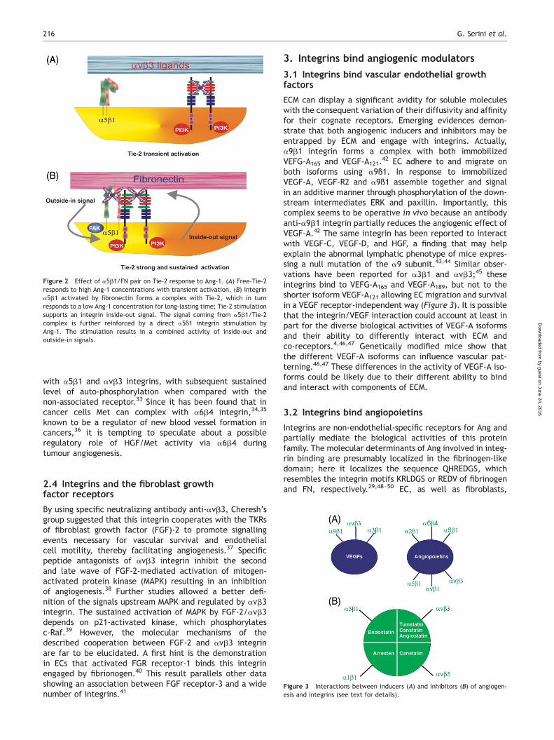
ECM can display a significant avidity for soluble moleculeswith the consequent variation of their diffusivity and affinityfor their cognate receptors. Emerging evidences demon-strate that both angiogenic inducers and inhibitors may beentrapped by ECM and engage with integrins. Actually,a9b1 integrin forms a complex with both immobilizedVEFG-A165 and VEGF-A121.
42 EC adhere to and migrate onboth isoforms using a9ß1. In response to immobilizedVEGF-A, VEGF-R2 and a9ß1 assemble together and signalin an additive manner through phosphorylation of the down-stream intermediates ERK and paxillin. Importantly, thiscomplex seems to be operative in vivo because an antibodyanti-a9b1 integrin partially reduces the angiogenic effect ofVEGF-A.42 The same integrin has been reported to interactwith VEGF-C, VEGF-D, and HGF, a finding that may helpexplain the abnormal lymphatic phenotype of mice expres-sing a null mutation of the a9 subunit.43,44 Similar obser-vations have been reported for a3b1 and avb3;45 theseintegrins bind to VEFG-A165 and VEGF-A189, but not to theshorter isoform VEGF-A121 allowing EC migration and survivalin a VEGF receptor-independent way (Figure 3). It is possiblethat the integrin/VEGF interaction could account at least inpart for the diverse biological activities of VEGF-A isoformsand their ability to differently interact with ECM andco-receptors.4,46,47 Genetically modified mice show thatthe different VEGF-A isoforms can influence vascular pat-terning.46,47 These differences in the activity of VEGF-A iso-forms could be likely due to their different ability to bindand interact with components of ECM.
3.2 Integrins bind angiopoietins
Integrins are non-endothelial-specific receptors for Ang andpartially mediate the biological activities of this proteinfamily. The molecular determinants of Ang involved in integ-rin binding are presumably localized in the fibrinogen-likedomain; here it localizes the sequence QHREDGS, whichresembles the integrin motifs KRLDGS or REDV of fibrinogenand FN, respectively.29,48–50 EC, as well as fibroblasts,
Figure 2 Effect of a5b1/FN pair on Tie-2 response to Ang-1. (A) Free-Tie-2responds to high Ang-1 concentrations with transient activation. (B) Integrina5b1 activated by fibronectin forms a complex with Tie-2, which in turnresponds to a low Ang-1 concentration for long-lasting time; Tie-2 stimulationsupports an integrin inside-out signal. The signal coming from a5b1/Tie-2complex is further reinforced by a direct a5ß1 integrin stimulation byAng-1. The stimulation results in a combined activity of inside-out andoutside-in signals.
Figure 3 Interactions between inducers (A) and inhibitors (B) of angiogen-esis and integrins (see text for details).
G. Serini et al.216
by guest on June 24, 2016D
ownloaded from

adhere to Ang-1 and -2, but only the former is able to inducecell spreading, haptotaxis, and a complete activation ofcytoskeleton dynamics. b1 integrin heterodimers, and inparticular a5b1, seem to be the most efficient adhesivereceptors for Ang-1 independently from the presence ofTie-2.29,50 This observation, together with the observedcooperation between Tie-2 and a5b1 (see above), indicatesthat Ang-1 is capable of triggering both inside-out andoutside-in signals. Indeed different events may occur in vas-cular ECs: (i) when high amounts of Ang-1 are available bothhigh affinity Tie-2 receptor and low affinity/high avidityintegrins may be independently activated; (ii) low Ang-1concentrations, which are unable to sustain prolongedTie-2 and integrin activation, can instead efficiently signalonly through the fraction of Tie-2 that is constitutivelyassociated with a5ß1 and make ECs more sensitive to lowamounts of Ang1.
In other cell types lacking Tie-2, Ang interact only withintegrins. In cardiomyocytes, a wider range of integrins(a2b1, a5b1, a6b1, a6b4, avb1, and avb3) bind Ang-1and Ang-2, resulting in cell survival due to Akt activationand caspase-3 inhibition.48 Glioma cells adhere to Ang-2via a3, a5, av, b1, and b3, but only avb1 engagement byAng-2 sustains efficient signalling that results in matrixmetalloprotease-2 production, cell migration, and inva-sion.49 On breast cancer cells instead adhere and migratetowards Ang-2, by using a5b1.51 Collectively these datasuggest that each cell type binds Ang through a distinctset of integrins, which only partially overlaps among differ-ent cell types (Figure 3).
3.3 Integrins bind angiogenesis inhibitors
A significant number of angiogenesis inhibitors derive fromthe proteolytic cleavage of ECM proteins52 and therefore itis not surprising that they bind integrins (Figure 3). Theinteraction of integrins with their canonical and intactECM ligands results in EC adhesion and activation of anti-apoptotic signals.7 On the contrary, integrins engaged bythese ECM-protein fragments promote apoptosis andreduce proliferation and motility. Altogether these datasuggest that different and opposing outside-in signals aretriggered by integrins depending on whether they areengaged by either intact ECM protein or fragments derivingfrom its cleavage.
Endostatin, the C-terminal non-collagenous domain oftype XVIII collagen, exerts its inhibitory activity on ECmigration mainly by interacting with a5b1. An Arg-richpeptide at the N-terminus of endostatin seems to be import-ant for its interaction with b1,53 supporting previous dataexcluding the role of an Arg-Gly-Asp sequence.54 However,it has been also reported that Arg-Gly-Asp cyclic peptidesinhibit EC binding to immobilized endostatin,55 implyingthat soluble or immobilized endostatin differently interactswith a5b1. It is conceivable that a5b1 conformation isdifferent when it binds an intact ECM ligand or endostatin,thus leading in the latter case to aberrant and perturbed sig-nalling events, such as sustained activation of src, inhibitionof FAK and MAPK.55,56
The non-collagenous domain present in the a chains oftype IV collagen generates three different angiogenic inhibi-tors: tumstatin, arresten, and canstatin. Tumstatin is thenon-collagenous domain of the a3 chain of type IV collagen
that induces apoptosis and inhibits EC proliferation throughits binding to avb3 integrin, leading to suppression ofcap-dependent protein translation. The tumstatin/avb3interaction is independent from the Arg-Gly-Asp bindingsite and therefore it may explain the inhibitory signalstriggered by this interaction. Actually tumstatin inhibitsthe activation of FAK, Akt, and mTOR (target of rapamycin)-mediated phosphorylation of the eukaryotic initiationfactor 4E-binding protein involved in the control of proteinsynthesis.55,57 Arresten corresponds to the non-collagenousdomain of the a1 chain of type IV collagen and blocks ECfunctions by competing with collagen IV binding to a1ß1integrin and inhibiting MAPK-mediated signals.58 Finally,canstatin corresponds to the a2 chain of type IV collagen,binds avb3 and avb5 leading to activation in ECs of anapoptotic program involving both caspase-8 and -9.59
The activation of both caspase-8 and -9 results in theamplification of mitochondrial-dependent apoptotic eventsand in the activation of caspase-3, the central executionerof the apoptotic process. A similar process is activatedby angiostatin, which corresponds to the N-terminal fourkringles of plasminogen and by binding avb3 triggers thenon-mitochondrial caspase-8-dependent apoptotic pathwayin ECs.59,60
4. Semaphorins regulate endothelial integrinfunction and angiogenic remodelling
Semaphorins (Sema) are a family of secreted andmembrane-bound repulsive cues, which have been originallyidentified for their ability to affect axon behaviour in thedeveloping nervous system.61 Semaphorins signal throughfour classes of plexins, named type A-D, a family of mem-brane receptors characterized by the presence in theircytosolic tail of two domains with homology to the R-RasGTPase activating proteins (GAPs), separated by a linkerregion that can bind other small GTPases, such as Rnd-1and Rac1.61 In vertebrates, members of the secreted class3 Sema employ Neuropilin (Nrp)-1 or -2 as co-receptors inassociation with type A or type D plexins.61
4.1 Semaphorins and vascular development
The first evidences for a role of Sema/Nrp/plexin system invascular biology were provided by the groups Klagsbrun andFujisawa, which respectively demonstrated that in ECsNrp-1 acts as VEGFR-2 co-receptor62 and found that Nrp-1is required for mouse cardiovascular development.63 After-wards, several reports confirmed and extended these obser-vations. In zebrafish, knockdown of sema3aa affects themigration of Nrp-1þ angioblasts, finally impairing dorsalaorta formation and normal circulation.64 Additionally,single morpholino knockdown of either sema3aa orsema3ab in TG(fli1:EGFP)y1 embryos results in a lessdramatic phenotype with patterning defects of intersomiticvessels.65 Knockdown of Sema3a gene in outbred CD-1mouse strain and over-expression of dominant negativeSema3 receptor mutants66 or delivery of anti-Sema3A anti-bodies67 in chick embryos were found to cause angiogenicremodelling defects. In a different colony of outbred CD-1mice, Sema3a knock down did not result in vasculardefects.68 These discrepancies could be due to differencesin the genetic background. Indeed, outbred stocks undergo
Integrin functions in vascular system 217
by guest on June 24, 2016D
ownloaded from

genetic heterogeneity depending on colony maintenance;moreover, even within the same outbred colony thegenetic background can change over time.69
The observation that both in vitro and in vivo66 angiogenicECs display autocrine loops of several Sema3 other thanSema3A70–73 together with the partial penetrance of thevascular phenotype in Sema3a2/2 mice suggest that mul-tiple Sema3 could cooperate to regulate angiogenesis.Notably, during angiogenesis and in cultured ECs, opposingautocrine loops of Sema3A66,70,71,73 and VEGF-A72,74–77
have been found and the observed loss of autocrineSema3A in favour of VEGF-A in ECs during malignanttumour progression73 could account at least in part for thestructural and functional abnormalities of tumourvasculature.
In ECs plexinD178 and, albeit to lesser extent, plexinA279
are the most abundant plexins. Both Sema3A and Sema3Cbind with a significantly higher affinity to a receptorcomplex formed by the association of Nrp-1 and/or -2 withplexinD1 than to a complex in which Nrps associates withplexinA1.78 Therefore, the Nrp/plexinD1 complex could rep-resent the most efficient transducer of the chemorepulsiveeffect of Sema3A.72,73,80–82 Different from other Sema3,Sema3E can directly bind to plexinD1.83 Mainly based ondefects in the intersomitic vessel patterning of Sema3Eand plexinD1, Sema3E/plexinD1 has been proposed to bethe major signalling pathway regulating vascular develop-ment.83 However, while Sema3e null mice are viable anddo not show any gross abnormality,83 all plexinD12/2 pupsbecome cyanotic shortly after birth and succumb within24 h because of severe cardiovascular defects.78 Therefore,it is likely that in ECs, plexinD1 transduces signals not onlyfrom Sema3E, but also from other Sema3 likely employingNrp as co-receptors, as originally proposed by Gitler andcolleagues.78 In this respect, it is worth noting that inneurons the simultaneous presence of Nrp1 and plexinD1on the cell surface converts Sema3E/plexinD1 signallingfrom repulsive to attractive.84 Based on the fact that ECsexpress high levels of both Nrp1 and plexinD1 and on obser-vations that Sema3E promotes tumour angiogenesis,85,86 atpresent these findings cannot be easily reconciled with theproposed chemorepulsive effect played by Sema3E viaplexinD1 on ECs of developing mouse embryos.83 Finally,the recent observation that Sema4A upon binding toplexinD1 inhibits EC migration and in vivo angiogenesisfurther indicates that plexinD1 conveys to ECs signals frommultiple Sema.87
The complexity of Sema system in vasculature is furthersupported by the recent data showing that Sema3A is apowerful vasopermeabilizing molecule through a mechanismthat is independent from its effects on integrin functionsand involves VE-cadherin phosphorylation.88
4.2 Semaphorins regulate integrin function
By investigating the cellular and molecular events by whichSema can regulate vascular development, we found thatSema3A impairs EC adhesion and migration by inhibitingintegrin activation.66 Accordingly, after few minutes ofstimulation with Sema3A, adherent ECs lose their focaladhesions and then collapse,82 and Sema3F causes EC retrac-tion as well.89 Moreover, Sema3A and Sema3F have beenshown to inhibit integrin activation and adhesion to the
ECM in several cell types.90–94 Interestingly, Sema3C,which contains an Arg-Gly-Asp motif, has been insteadreported to promote EC adhesion and migration.95
It is well known that integrins undergo conformationalmodifications that regulate their affinity for ECM ligands.2
In the low affinity state, the integrin extracellular domainis bent over the plasmamembrane and the a and b cyto-plasmic tails tightly interact. In ECs chemokines, growthfactors, or fluid shear stress activate signalling pathwaysthat finally favour the transition of integrins towards thehigh affinity state.2,7 Direct interaction of talin trefoilFERM (Protein 4.1, the ezrin, radixin, moesin) domain withthe cytodomain of integrin b subunits results in the unclasp-ing of the cytoplasmic tails of integrin a and b sub-units together with the extension of the extracellulardomain that, by exposing the ECM binding site, accomplishthe transition of integrins to the high affinity state.96 Intalin, the integrin binding site within the trefoil FERMdomain is masked by an intramolecular interaction withthe rod domain. Interaction of membrane phosphatidyl-inositol 4, 5 bisphosphate (PIP2) with talin rod domainlessen this inhibition, thus allowing talin head-to-tail dimer-ization and integrin binding to the FERM domain. Notably,once activated talin can in turn bind and activate theenzyme phosphatidylinositol-4-phosphate 5-kinase (PIPKIg661) that, producing further PIP2, gives rise to a positivefeed-back loop with talin that stabilizes cell adhesion tothe ECM.97 In addition, integrin function is also regulatedby Rap 1 and R-Ras small-GTPases.98 Specifically, activatedR-Ras-GTP localizes at adhesive sites through its C-terminaltail;99 here R-Ras is thought to promote cell adhesion byfavouring the activation of other small GTPases, such asRap1 and Rac1.100 In this regard, it has been recentlyshown that binding of activated R-Ras to RLIP (Ralinteracting protein) 76 leads to Arf (ADP-ribosylationfactor) 6 activation, which promotes adhesion-inducedGTP loading of Rac1.101 In the presence of Nrp-1, thejuxtamembrane basic sequence of class A plexins directlybinds to FARP2, a Rac (ras-related C3 botulinum toxin sub-strate 1) guanosine exchange factor (GEF).94 Sema3Abinding to the Nrp-1/plexinA1 receptor complex triggersthe dissociation of FARP2 from PlexinA1. Afterwards,FARP2 (FERM, RhoGEF, and pleckstrin domain protein 2) isfree to exert its GEF activity leading to a rapid increase ofactive Rac1-GTP that in turn favours the binding of the con-stitutively active small GTPase Rnd1 (Rho family GTPase 1)to the linker region of plexinA1 cytoplasmic (Figure 4).This event finally activates PlexinA1 latent R-Ras GAPactivity that then switches-off R-Ras, thus inhibiting integrinfunction. Importantly, Toyofuku and colleagues also foundthat, similarly to talin, FARP2 contains FERM domain bymeans which it binds plexinA1. Once detached fromplexinA1, the FERM domain of FARP2 competes with talinfor binding to PIPKIg661, finally impairing the PIP2-basedpositive feedback required for the formation of focaladhesions.
EC migration and angiogenesis are also regulated bytransmembrane Sema4D, which, depending on the cellularcontext, can however exert either chemoattractive orchemorepulsive activities. Negishi’s laboratory providedthe first evidence that plexins are endowed with an R-RasGAP enzymatic activity by showing that Sema4D-mediatedplexinB1 stimulation suppresses R-Ras activation102 that
G. Serini et al.218
by guest on June 24, 2016D
ownloaded from

in turn inhibits b1 integrin activation.103 Sema4D-drivenR-Ras inhibition depends on the RasGAP activity triggeredby Rnd1 binding to plexinB1.102,103 However, the inhibitoryactivity of Sema4D is not a general phenomenon. Indeed,as first reported by Gutkind and colleagues104 and thenby us,105 Sema4D is pro-angiogenic and elicits ECmigration.106 Sema4D stimulatory activity on ECs requiresthe formation of a complex between plexinB1 and MetTKR.105 In addition, upon treatment with Sema4DplexinB1 receptor associates with a Rho-GEF capable ofactivating Rho GTPase and its downstream effector Rhokinase, which by phosphorylating myosin light chain couldcontrol the assembly and the contraction of actin stressfibres.106 Altogether these data indicate that Sema4D/plexin B1 mediate different and sometimes oppositeeffects depending on the cellular context. As recentlysuggested, this may be caused by plexinB1 associationwith different TKR receptors; indeed, in carcinoma cellsSema4D can have pro- and anti-migratory effects depend-ing on the interaction, respectively, with either Met orErbB-2 TKRs.107
4.3 Is FAK a converging node for signals triggeredby VEGF-A, integrins, and semaphorins?
Engagement of integrin receptors with their extracellularligands leads to the formation of well-defined structures,termed focal adhesions, linking the ECM, and cytoplasmicactin cytoskeleton. These adhesive structures, which aredynamic and composed by a wide array of transmembraneand cytosolic proteins, serve as sites of force transmissionrequired for cellular movements. Indeed, the coordinatedregulation of formation and turnover of focal adhesions iscentral to cell responses to chemotactic and chemokineticstimuli.108 A primary element of focal adhesion is FAK, akinase that is primarily recruited to sites of integrin cluster-ing via interactions between its C-terminal region andintegrin-associated proteins such as talin and paxillin. Thecytosolic tail of b integrins facilitates FAK activation prob-ably involving FAK clustering and autophosphorylation ofTyr 379 (reviewed by Mitra and Schlaepfer30). Furthermore,FAK is a downstream effector of several TKRs, includingVEGFR-2 and FGF receptors in ECs.39,109,110 VEGF-A via Srcinduces the site-specific tyrosine phosphorylation of FAK onTyr 861, leading to the formation of a complex betweenFAK and avß5, which is essential for the vascular per-meability induced by VEGF-A.111
Recent evidences indicate that the repulsive or attractivefunctions of semamaphorins can involve FAK as well. Indeed,Sema3B can attract neurons by inducing membranere-localization of phosphorylated FAK, which in turnsactivates the cytosolic tyrosine kinase Fyn.112 Similarly,Sema4D elicits EC motility and angiogenesis104,105 by acti-vating proline-rich tyrosine kinase-2, a non-receptor tyro-sine kinase closely related to FAK.106 In contrast, both incultured ECs and in chick chorionallantoic membrane, che-morepulsive Sema3A reduces the basal phosphorylation ofSrc and FAK, and induces a rapid disappearance of focal con-tacts followed by the collapse of the actin cytoskeleton,82,88
thus supporting its role in inhibiting integrin function.66
In addition to FAK and integrins, many other proteinslocalize to focal adhesion, including VEGFR-2.113 Eventhough the membrane topology of semaphorin receptors isstill unknown, it is tempting to speculate that ECM adhesivestructures could represent ‘rendez-vous‘ points, wheremultiple cross-talks between positive and negative regula-tors of integrin function take place.
5. Conclusions
Angiogenesis is a relevant target for the treatment of manydiseases. Inhibition of angiogenesis is the aim of protocolsdeveloped for tumours, chronic inflammatory diseases, andretinopathies, while vascular regeneration inspires thera-peutic angiogenesis in the treatment of ischaemic injuries.Within the molecules investigated as exploitable targetsfor angiogenic therapies, VEGF-A and its receptors are con-sidered the most promising. However, there are tremendousdifferences between pre-clinical and clinical results. Thedata reviewed here clearly indicate that the activity ofevery drug has to be considered in connection with therobust network of regulatory signals that controls angiogen-esis. Indeed, the vast accumulation of experimental andclinical reports on tumour angiogenesis can be revisitedin light of the mechanisms by which cancer modifies
Figure 4 Effect of Sema3A on the inhibition of integrin function. (A) On thecell surface, ab integrin heterodimers exist in a high affinity state, stabilizedby the interaction of talin head (FERM domain) with the b subunit tail. Theinteraction between talin and the actin cytoskeleton occurs. Talin-activatedPIPKIg661 generates a PIP2-based positive feedback loop, which amplifiestalin activation. The small GTPase R-Ras participates in integrin activationthrough a still poorly characterized mechanism. In the absence of SEMA3A,plexinA1 associates with Nrp-1 and the FERM domain-containing guanosineexchange factor FARP2. (B) Upon SEMA3A binding, FARP2 is released fromplexinA1. Free FARP2 then activates Rac and favours Rnd-1 association withand activation of plexinA1 cytoplasmic GAP domain, which in turn inhibitsR-Ras and integrin function. Moreover, released FARP2 inhibits PIPKIg661activity.
Integrin functions in vascular system 219
by guest on June 24, 2016D
ownloaded from

the robustness of regulatory networks leading to theobserved abnormal vascularization, the final aim being topharmacologically restore the robustness of the angiogenicregulatory networks in the everyday clinical practice. Thisinterpretation might explain why some tumours displaylack of sensitivity and others develop resistance toanti-VEGF-A therapy.
Funding
This work has been partially supported by Telethon (grant:GGP04127), Associazione italiana per la ricerca sul cancro (AIRC),European Community (LSHM-CT-2003-503254), Regione Piemonte(Ricerca Finalizzata 2006, Ricerca Scientifica applicata 2004 grantsD10 and A150, Ricerca industriale e competitiva 2006: grantPRESTO), Fondazione Cassa di Risparmio di Torino, Torino, and‘Ministero della Salute’ (Programma Ricerca Oncologica 2006,Ricerca Finalizzata 2006).
Conflict of interest: none declared.
References1. Moissoglu K, Schwartz MA. Integrin signalling in directed cell migration.
Biol Cell 2006;98:547–555.2. Luo BH, Carman CV, Springer TA. Structural basis of integrin regulation
and signaling. Annu Rev Immunol 2007;25:619–647.3. Yancopoulos GD, Davis S, Gale NW, Rudge JS, Wiegand SJ, Holash J.
Vascular-specific growth factors and blood vessel formation. Nature2000;407:242–248.
4. Olsson AK, Dimberg A, Kreuger J, Claesson-Welsh L. VEGF receptorsignalling—in control of vascular function. Nat Rev Mol Cell Biol 2006;7:359–371.
5. Bussolino F, Valdembri D, Caccavari F, Serini G. Semaphoring vascularmorphogenesis. Endothelium 2006;13:81–91.
6. Hynes RO. Cell-matrix adhesion in vascular development. J ThrombHaemost 2007;5(Suppl. 1):32–40.
7. Serini G, Valdembri D, Bussolino F. Integrins and angiogenesis: a stickybusiness. Exp Cell Res 2006;312:651–658.
8. Borges E, Jan Y, Ruoslahti E. PDGF-receptor- and VEGF-receptor-2 bindto the 3 integrin through its extracellular domain. J Biol Chem 2000;275:39867–39873.
9. Soldi R, Mitola S, Strasly S, Defilippi P, Tarone G, Bussolino F. Role ofavb3 integrin in the activation of vascular endothelial growth factorreceptor-2. EMBO J 1999;18:734–740.
10. Mahabeleshwar GH, Feng W, Phillips DR, Byzova TV. Integrin signaling iscritical for pathological angiogenesis. J Exp Med 2006;203:2495–2507.
11. Mahabeleshwar GH, Feng W, Reddy K, Plow EF, Byzova TV. Mechanisms ofintegrin-vascular endothelial growth factor receptor cross-activation inangiogenesis. Circ Res 2007;101:570–580.
12. Masson-Gadais B, Houle F, Laferriere J, Huot J. Integrin avb3requirement for VEGFR2- mediated activation of SAPK2/p38 and forHsp90-dependent phosphorylation of focal adhesion kinase in endo-thelial cells activated by VEGF. Cell Stress Chaperones 2003;8:37–52.
13. Tian F, Zhu CH, Zhang XW, Xie X, Xin XL, Yi YH et al. Philinopside E, anew sulfated saponin from sea cucumber, blocks the interactionbetween kinase insert domain-containing receptor (KDR) and alphav-beta3 integrin via binding to the extracellular domain of KDR. Mol Phar-macol 2007;72:545–552.
14. Dardik R, Inbal A. Complex formation between tissue transglutaminase II(tTG) and vascular endothelial growth factor receptor 2 (VEGFR-2): pro-posed mechanism for modulation of endothelial cell response to VEGF.Exp Cell Res 2006;312:2973–2982.
15. Weis S, Lindquist JN, Barnes LA, Lutu-Fuga KM, Cui J, Wood MR et al.Cooperation between VEGF and b3 integrin during cardiac vasculardevelopment. Blood 2007;109:1962–1970.
16. Silvestre JS, Thery C, Hamard G, Boddaert J, Aguilar B, Delcayre A et al.Lactadherin promotes VEGF-dependent neovascularization. Nat Med2005;11:499–506.
17. Wijelath ES, Murray J, Rahman S, Patel Y, Ishida A, Strand K et al. Novelvascular endothelial growth factor binding domains of fibronectin
enhance vascular endothelial growth factor biological activity. CircRes 2002;91:25–31.
18. Wijelath ES, Rahman S, Namekata M, Murray J, Nishimura T,Mostafavi-Pour Z et al. Heparin-II domain of fibronectin is a vascularendothelial growth factor-binding domain: enhancement of VEGF bio-logical activity by a singular growth factor/matrix protein synergism.Circ Res 2006;99:853–860.
19. Mitola S, Brenchio B, Piccinini M, Tertoolen L, Zammataro L, Breier Get al. Type I collagen limits VEGFR-2 signaling by a SHP2 protein-tyrosine phosphatase-dependent mechanism 1. Circ Res 2006;98:45–54.
20. Jekely G, Sung HH, Luque CM, Rorth P. Regulators of endocytosis main-tain localized receptor tyrosine kinase signaling in guided migration.Dev Cell 2005;9:197–207.
21. Seo DW, Li H, Guedez L, Wingfield PT, Diaz T, Salloum R et al. TIMP-2mediated inhibition of angiogenesis: an MMP-independent mechanism.Cell 2003;114:171–180.
22. Oh J, Seo DW, Diaz T, Wei B, Ward Y, Ray JM et al. Tissue inhibitors ofmetalloproteinase 2 inhibits endothelial cell migration throughincreased expression of RECK. Cancer Res 2004;64:9062–9069.
23. Seo DW, Li H, Qu CK, Oh J, Kim YS, Diaz T et al. Shp-1 mediates theantiproliferative activity of tissue inhibitor of metalloproteinase-2in human microvascular endothelial cells. J Biol Chem 2006;281:3711–3721.
24. Mattila E, Pellinen T, Nevo J, Vuoriluoto K, Arjonen A, Ivaska J. Negativeregulation of EGFR signalling through integrin-alfa1beta1-mediatedactivation of protein tyrosine phosphatase TCPTP. Nat Cell Biol 2005;7:78–85.
25. Chen X, Abair TD, Ibanez MR, Su Y, Frey MR, Dise RS et al. Integrinalpha1beta1 controls reactive oxygen species synthesis by negativelyregulating epidermal growth factor receptor-mediated Rac activation.Mol Cell Biol 2007;27:3313–3326.
26. Ling Y, Maile LA, Clemmons DR. Tyrosine phosphorylation of thebeta3-subunit of the alphaVbeta3 integrin is required for membraneassociation of the tyrosine phosphatase SHP-2 and its further recruit-ment to the insulin-like growth factor I receptor. Mol Endocrinol 2003;17:1824–1833.
27. Zhang X, Groopman JE, Wang JF. Extracellular matrix regulatesendothelial functions through interaction of VEGFR-3 and integrina5b1. J Cell Physiol 2005;202:205–214.
28. Wang JF, Zhang XF, Groopman JE. Stimulation of beta 1 integrin inducestyrosine phosphorylation of vascular endothelial growth factorreceptor-3 and modulates cell migration. J Biol Chem 2001;276:41950–41957.
29. Cascone I, Napione L, Maniero F, Serini G, Bussolino F. Stable interactionbetween alpha5beta1 integrin and Tie2 tyrosine kinase receptor regu-lates endothelial cell response to Ang-1. J Cell Biol 2005;170:993–1004.
30. Mitra SK, Schlaepfer DD. Integrin-regulated FAK-Src signaling in normaland cancer cells. Curr Opin Cell Biol 2006;18:516–523.
31. Bussolino F, Di Renzo MF, Ziche M, Bocchietto E, Olivero M, Naldini Let al. Hepatocyte growth factor is a potent angiogenic factor whichstimulates endothelial cell motility and growth. J Cell Biol 1992;119:629–641.
32. Trusolino L, Serini G, Cecchini G, Besati C, Ambesi-Impiombato FS,Marchisio PC et al. Growth factor-dependent activation of alphavbeta3integrin in normal epithelial cells: implications for tumor invasion.J Cell Biol 1998;142:1145–1156.
33. Rahman S, Patel Y, Murray J, Patel KV, Sumathipala R, Sobel M et al.Novel hepatocyte growth factor (HGF) binding domains on fibronectinand vitronectin coordinate a distinct and amplified Met-integrininduced signalling pathway in endothelial cells. BMC Cell Biol 2005;6:8.
34. Chung J, Yoon SO, Lipscomb EA, Mercurio AM. The Met receptorand alpha 6 beta 4 integrin can function independently to promotecarcinoma invasion. J Biol Chem 2004;279:32287–32293.
35. Trusolino L, Bertotti A, Comoglio PM. A signaling adapter function foralpha6beta4 integrin in the control of HGF-dependent invasivegrowth. Cell 2001;107:643–654.
36. Nikolopoulos SN, Blaikie P, Yoshioka T, Guo W, Giancotti FG. Integrinbeta4 signaling promotes tumor angiogenesis. Cancer Cell 2004;6:471–483.
37. Brooks PC, Montgomery AM, Rosenfeld M, Reisfeld RA, Hu T, Klier G et al.Integrin alpha v beta 3 antagonists promote tumor regression by indu-cing apoptosis of angiogenic blood vessels. Cell 1994;79:1157–1164.
38. Eliceiri BP, Klemke R, Stromblad S, Cheresh DA. Integrin alphavbeta3requirement for sustained mitogen-activated protein kinase activityduring angiogenesis. J Cell Biol 1998;140:1255–1263.
G. Serini et al.220
by guest on June 24, 2016D
ownloaded from

39. Hood JD, Frausto R, Kiosses WB, Schwartz MA, Cheresh DA. Differentialalphav integrin-mediated Ras-ERK signaling during two pathways ofangiogenesis. J Cell Biol 2003;162:933–943.
40. Sahni A, Francis CW. Stimulation of endothelial cell proliferation byFGF-2 in the presence of fibrinogen requires alphavbeta3. Blood 2004;104:3635–3641.
41. Toledo MS, Suzuki E, Handa K, Hakomori S. Effect of ganglioside andtetraspanins in microdomains on interaction of integrins with fibroblastgrowth factor receptor. J Biol Chem 2005;280:16227–16234.
42. Vlahakis NE, Young BA, Atakilit A, Hawkridge AE, Issaka RB, Boudreau Net al. Integrin alpha9beta1 directly binds to vascular endothelial growthfactor (VEGF)-A and contributes to VEGF-A-induced angiogenesis. J BiolChem 2007;282:15187–15196.
43. Vlahakis N, Young BA, Atakilit A, Sheppard D. The lymphangiogenic vas-cular enothelial growth factors VEGF-C and -D are ligand for the integrinalpha9beta1. J Biol Chem 2005;280:4544–4554.
44. Kajia K, Hirakawa S, Ma B, Drinnenberg I, Detmar M. Hepatocyte growthfactor promotes lymphatic vessel formation and function. EMBO J 2005;24:2885–2895.
45. Hutchings H, Ortega N, Plouet J. Extracellular matrix-bound vascularendothelial growth factor promotes endothelial cell adhesion,migration, and survival through integrin ligation. FASEB J 2003;17:1520–1522.
46. Ruhrberg C, Gerhardt H, Golding M, Watson R, Ioannidou S, Fujisawa Het al. Spatially restricted patterning cues provided by heparin-bindingVEGF-A control blood vessel branching morphogenesis. Genes Dev2002;16:2684–2698.
47. Stalmans I, Ng YS, Rohan R, Fruttiger M, Bouche A, Yuce A et al. Arter-iolar and venular patterning in retinas of mice selectively expressingVEGF isoforms. J Clin Invest 2002;109:327–336.
48. Dallabrida SM, Ismail N, Oberle JR, Himes BE, Rupnick MA.Angiopoietin-1 promotes cardiac and skeletal myocyte survivalthrough integrins. Circ Res 2005;96:e8–e24.
49. Hu B, Jarzynka MJ, Guo P, Imanishi Y, Schlaepfer DD, Cheng SY. Angio-poietin 2 induces glioma cell invasion by stimulating matrix metallopro-tease 2 expression through the alphavbeta1 integrin and focal adhesionkinase signaling pathway. Cancer Res 2006;66:775–783.
50. Carlson T, Feng Y, Maisonpierre P, Mrksich M, Morla A. Direct celladhesion to the angiopoietins mediated by integrins. J Biol Chem2001;278:26516–26525.
51. Imanishi Y, Hu B, Jarzynka MJ, Guo P, Elishaev E, Bar-Joseph I et al.Angiopoietin-2 stimulates breast cancer metastasis through thealpha(5)beta(1) integrin-mediated pathway. Cancer Res 2007;67:4254–4263.
52. Kalluri R. Basement membranes: structures, assembly and role in tumorangiogenesis. Nature Rev Cancer 2003;3:422–433.
53. Wickstrom SA, Alitalo K, Keski-Oja J. An endostatin-derived peptideinteracts with integrins and regulates actin cytoskeleton and migrationof endothelial cells. J Biol Chem 2004;279:20178–20185.
54. Rehn M, Veikkola T, Kukk-Valdre E, Nakamura H, Ilmonen M, Lombardo Cet al. Interaction of endostatin with integrins implicated in angiogen-esis. Proc Natl Acad Sci USA 2001;98:1024–1029.
55. Sudhakar A, Sugimoto H, Yang C, Lively J, Zeisberg M, Kalluri R. Humantumstatin and human endostatin exhibit distinct antiangiogenic activi-ties mediated by alpha v beta 3 and alpha 5 beta 1 integrins. ProcNatl Acad Sci USA 2003;100:4766–4771.
56. Wickstrom SA, Alitalo K, Keski-Oja J. Endostatin associates with integrinalpha5beta1 and caveolin-1, and activates Src via a tyrosyl phosphatase-dependent pathway in human endothelial cells. Cancer Res 2002;62:5580–5589.
57. Maeshima Y, Sudhakar A, Lively JC, Ueki K, Kharbanda S, Kahn CR et al.Tumstatin, an endothelial cell-specific inhibitor of protein synthesis.Science 2002;295:140–143.
58. Sudhakar A, Nyberg P, Keshamouni VG, Mannam AP, Li J, Sugimoto Het al. Human alpha1 type IV collagen NC1 domain exhibits distinctantiangiogenic activity mediated by alpha1beta1 integrin. J ClinInvest 2005;115:2801–2810.
59. Magnon C, Galaup A, Mullan B, Rouffiac V, Bouquet C, Bidart JM et al.Canstatin acts on endothelial and tumor cells via mitochondrialdamage initiated through interaction with alphavbeta3 and alphavbeta5integrins. Cancer Res 2005;65:4353–4361.
60. Tarui T, Miles LA, Takada Y. Specific interaction of angiostatin withintegrin alpha(v)beta(3) in endothelial cells. J Biol Chem 2001;276:39562–39568.
61. Kruger RP, Aurandt J, Guan KL. Semaphorins command cells to move.Nat Rev Mol Cell Biol 2005;6:789–800.
62. Soker S, Takashima S, Miao HQ, Neufeld G, Klagsbrun M. Neuropilin-1 isexpressed by endothelial and tumor cells as an isoform-specific receptorfor vascular endothelial growth factor. Cell 1998;92:735–745.
63. Kawasaki T, Kitsukawa T, Bekku Y, Matsuda Y, Sanbo M, Yagi T et al. Arequirement for neuropilin-1 in embryonic vessel formation. Develop-ment 1999;126:4895–4902.
64. Shoji W, Isogai S, Sato-Maeda M, Obinata M, Kuwada JY. Semaphorin3a1regulates angioblast migration and vascular development in zebrafishembryos. Development 2003;130:3227–3236.
65. Torres-Vazquez J, Gitler AD, Fraser SD, Berk JD, Van NP, Fishman MCet al. Semaphorin-plexin signaling guides patterning of the developingvasculature. Dev Cell 2004;7:117–123.
66. Serini G, Valdembri D, Zanivan S, Morterra G, Burkhardt C, Caccavari Fet al. Class 3 semaphorins control vascular morphogenesis by inhibitingintegrin function. Nature 2003;424:391–397.
67. Bates D, Taylor GI, Minichiello J, Farlie P, Cichowitz A, Watson N et al.Neurovascular congruence results from a shared patterning mechanismthat utilizes Semaphorin3A and Neuropilin-1. Dev Biol 2003;255:77–98.
68. Vieira JM, Schwarz Q, Ruhrberg C. Selective requirements for NRP1ligands during neurovascular patterning. Development 2007;134:1833–1843.
69. Chia R, Achilli F, Festing MF, Fisher EM. The origins and uses of mouseoutbred stocks. Nat Genet 2005;37:1181–1186.
70. Ito T, Kagoshima M, Sasaki Y, Li C, Udaka N, Kitsukawa T et al. Repulsiveaxon guidance molecule Sema3A inhibits branching morphogenesis offetal mouse lung. Mech Dev 2000;97:35–45.
71. Damon DH. Vascular endothelial-derived semaphorin 3 inhibitssympathetic axon growth. Am J Physiol Heart Circ Physiol 2006;290:H1220–H1225.
72. Serini G, Ambrosi D, Giraudo E, Gamba A, Preziosi L, Bussolino F.Modeling the early stages of vascular network assembly. EMBO J 2003;22:1771–1779.
73. Vacca A, Scavelli C, Serini G, Di Pietro G, Cirulli T, Merchionne F et al.Loss of inhibitory semaphorin 3A (SEMA3A) autocrine loops in bonemarrow endothelial cells of patients with multiple myeloma. Blood2006;108:1661–1667.
74. Yonekura H, Sakurai S, Liu X, Migita H, Wang H, Yamagishi S et al.Placenta growth factor and vascular endothelial growth factor B and Cexpression in microvascular endothelial cells and pericytes. Implicationin autocrine and paracrine regulation of angiogenesis. J Biol Chem 1999;274:35172–35178.
75. Virgintino D, Errede M, Robertson D, Girolamo F, Masciandaro A,Bertossi M. VEGF expression is developmentally regulated duringhuman brain angiogenesis. Histochem Cell Biol 2003;119:227–232.
76. Tang N, Wang L, Esko J, Giordano FJ, Huang Y, Gerber HP et al. Loss ofHIF-1alpha in endothelial cells disrupts a hypoxia-driven VEGF autocrineloop necessary for tumorigenesis. Cancer Cell 2004;6:485–495.
77. Lee S, Chen TT, Barber CL, Jordan MC, Murdock J, Desai S et al.Autocrine VEGF signaling is required for vascular homeostasis. Cell2007;130:691–703.
78. Gitler AD, Lu MM, Epstein JA. PlexinD1 and semaphorin signaling arerequired in endothelial cells for cardiovascular development. Dev Cell2004;7:107–116.
79. Herzog Y, Guttmann-Raviv N, Neufeld G. Segregation of arterial andvenous markers in subpopulations of blood islands before vesselformation. Dev Dyn 2005;232:1047–1055.
80. Kusy S, Funkelstein L, Bourgais D, Drabkin H, Rougon G, Roche J et al.Redundant functions but temporal and regional regulation of two alter-natively spliced isoforms of semaphorin 3F in the nervous system. MolCell Neurosci 2003;24:409–418.
81. Narazaki M, Tosato G. Ligand-induced internalization selects use ofcommon receptor neuropilin-1 by VEGF165 and semaphorin3A. Blood2006;107:3892–3901.
82. Guttmann-Raviv N, Shraga-Heled N, Varshavsky A, Guimaraes-Sternberg C,Kessler O, Neufeld G. Semaphorin-3A and semaphorin-3F work togetherto repel endothelial cells and to inhibit their survival by induction ofapoptosis. J Biol Chem 2007;282:26294–26305.
83. Gu C, Yoshida Y, Livet J, Reimert DV, Mann F, Merte J et al. Semaphorin3E and plexin-D1 control vascular pattern independently of neuropilins.Science 2005;307:265–268.
84. Chauvet S, Cohen S, Yoshida Y, Fekrane L, Livet J, Gayet O et al.Gating of Sema3E/PlexinD1 signaling by neuropilin-1 switches axonalrepulsion to attraction during brain development. Neuron 2007;56:807–822.
85. Christensen C, Ambartsumian N, Gilestro G, Thomsen B, Comoglio P,Tamagnone L et al. Proteolytic processing converts the repelling signal
Integrin functions in vascular system 221
by guest on June 24, 2016D
ownloaded from

Sema3E into an inducer of invasive growth and lung metastasis. CancerRes 2005;65:6167–6177.
86. Roodink I, Raats J, van der Zwaag B, Verrijp K, Kusters B, vanBokhoven H et al. Plexin D1 expression is induced on tumor vasculatureand tumor cells: a novel target for diagnosis and therapy? Cancer Res2005;65:8317–8323.
87. Toyofuku T, Yabuki M, Kamei J, Kamei M, Makino N, Kumanogoh A et al.Semaphorin-4A, an activator for T-cell-mediated immunity, suppressesangiogenesis via Plexin-D1. EMBO J 2007;26:1373–1384.
88. Acevedo LM, Barillas S, Weis SM, Gothert JR, Cheresh DA. Semaphorin3A suppresses VEGF-mediated angiogenesis yet acts as a vascularpermeability factor. Blood 2008;111:2674–2680.
89. Kessler O, Shraga-Heled N, Lange T, Gutmann-Raviv N, Sabo E, Baruch Let al. Semaphorin-3F is an inhibitor of tumor angiogenesis. Cancer Res2004;64:1008–1015.
90. Potiron VA, Sharma G, Nasarre P, Clarhaut JA, Augustin HG, Gemmill RMet al. Semaphorin SEMA3F affects multiple signaling pathways in lungcancer cells. Cancer Res 2007;67:8708–8715.
91. Bielenberg DR, Hida Y, Shimizu A, Kaipainen A, Kreuter M, Kim CC et al.Semaphorin 3F, a chemorepulsant for endothelial cells, induces a poorlyvascularized, encapsulated, nonmetastatic tumor phenotype. J ClinInvest 2004;114:1260–1271.
92. Kashiwagi H, Shiraga M, Kato H, Kamae T, Yamamoto N, Tadokoro S et al.Negative regulation of platelet function by a secreted cell repulsiveprotein, semaphorin 3A. Blood 2005;106:913–921.
93. Lepelletier Y, Smaniotto S, Hadj-Slimane R, Villa-Verde DM,Nogueira AC, Dardenne M et al. Control of human thymocyte migrationby Neuropilin-1/Semaphorin-3A-mediated interactions. Proc Natl AcadSci USA 2007;104:5545–5550.
94. Toyofuku T, Yoshida J, Sugimoto T, Zhang H, Kumanogoh A, Hori M et al.FARP2 triggers signals for Sema3A-mediated axonal repulsion. NatNeurosci 2005;8:1712–1719.
95. Banu N, Teichman J, Dunlap-Brown M, Villegas G, Tufro A. Semaphorin3C regulates endothelial cell function by increasing integrin activity.FASEB J 2006;20:2150–2152.
96. Campbell ID, Ginsberg MH. The talin–tail interaction places integrinactivation on FERM ground. Trends Biochem Sci 2004;29:429–435.
97. Luo BH, Springer TA. Integrin structures and conformational signaling.Curr Opin Cell Biol 2006;18:579–586.
98. Kinbara K, Goldfinger LE, Hansen M, Chou FL, Ginsberg MH. Ras GTPases:integrins’ friends or foes? Nat Rev Mol Cell Biol 2003;4:767–776.
99. Furuhjelm J, Peranen J. The C-terminal end of R-Ras contains a focaladhesion targeting signal. J Cell Sci 2003;116:3729–3738.
100. Self AJ, Caron E, Paterson HF, Hall A. Analysis of R-Ras signalling path-ways. J Cell Sci 2001;114:1357–1366.
101. Goldfinger LE, Ptak C, Jeffery ED, Shabanowitz J, Hunt DF, Ginsberg MH.RLIP76 (RalBP1) is an R-Ras effector that mediates adhesion-dependentRac activation and cell migration. J Cell Biol 2006;174:877–888.
102. Oinuma I, Ishikawa Y, Katoh H, Negishi M. The Semaphorin 4D receptorPlexin-B1 is aGTPaseactivatingprotein forR-Ras.Science2004;305:862–865.
103. Oinuma I, Katoh H, Negishi M. Semaphorin 4D/Plexin-B1-mediated R-RasGAP activity inhibits cell migration by regulating beta(1) integrinactivity. J Cell Biol 2006;173:601–613.
104. Basile JR, Barac A, Zhu T, Guan KL, Gutkind JS. Class IV semaphorinspromote angiogenesis by stimulating Rho-initiated pathways throughplexin-B. Cancer Res 2004;64:5212–5224.
105. Conrotto P, Valdembri D, Corso S, Serini G, Tamagnone L, Comoglio PMet al. Sema4D induces angiogenesis through Met recruitment by PlexinB1. Blood 2005;105:4321–4329.
106. Basile JR, Gavard J, Gutkind JS. Plexin-B1 utilizes RhoA and Rho kinaseto promote the integrin-dependent activation of Akt and ERK and endo-thelial cell motility. J Biol Chem 2007;282:34888–34895.
107. Swiercz JM, Worzfeld T, Offermanns S. ERBB-2 and met reciprocally regu-late cellular signaling via plexin-B1. J Biol Chem 2008;283:1893–1901.
108. Zamir E, Geiger B. Molecular complexity and dynamics of cell-matrixadhesions. J Cell Sci 2001;114:3583–3590.
109. Avraham HK, Lee TH, Koh Y, Kim TA, Jiang S, Sussman M et al. Vascularendothelial growth factor regulates focal adhesion assembly in humanbrain microvascular endothelial cells through activation of the focaladhesion kinase and related adhesion focal tyrosine kinase. J BiolChem 2003;278:36661–36668.
110. Le Boeuf F, Houle F, Sussman M, Huot J. Phosphorylation of focaladhesion kinase (FAK) on Ser732 is induced by rho-dependent kinaseand is essential for proline-rich tyrosine kinase-2-mediated phosphoryl-ation of FAK on Tyr407 in response to vascular endothelial growth factor.Mol Biol Cell 2006;17:3508–3520.
111. Eliceiri BP, Puente XS, Hood JD, Stupack DG, Schlaepfer DD, Huang XZet al. Src-mediated coupling of focal adhesion kinase to integrinalpha(v)beta5 in vascular endothelial growth factor signaling. J CellBiol 2002;157:149–160.
112. Julien F, Bechara A, Fiore R, Nawabi H, Zhou H, Hoyo-Becerra C et al.Dual functional activity of semaphorin 3B is required for positioningthe anterior commissure. Neuron 2005;48:63–75.
113. Ikeda S, Ushio-Fukai M, Zuo L, Tojo T, Dikalov S, Patrushev NA et al.Novel role of ARF6 in vascular endothelial growth factor-induced signaling and angiogenesis. Circ Res 2005;96:467–475.
114. Arias-Salgado AR, Haj F, Dubois C, Moran B, Kasirer-Friede A, Furie BCet al. PTB-1B is essential positive regulator of platelet integrinsignals. J Cell Biol 2005;170:837–845.
G. Serini et al.222
by guest on June 24, 2016D
ownloaded from