local inhibition of angiogenesis results in an atrophic non ...
Upload
cicbioguneCategory
view
1download
0
New Inhibitors of Angiogenesis with Antitumor Activity in VivoNagore I. Marín-Ramos,†,‡,◆ Dulce Alonso,†,◆ Silvia Ortega-Gutierrez,† Francisco J. Ortega-Nogales,†
Moises Balabasquer,† Henar Vazquez-Villa,† Clara Andradas,§,∥ Sandra Blasco-Benito,§,∥
Eduardo Perez-Gomez,§,∥ Angeles Canales,† Jesus Jimenez-Barbero,⊥,¶ Ana Marquina,#
Jaime Moscoso del Prado,# Cristina Sanchez,§,∥ Mar Martín-Fontecha,*,†
and María L. Lopez-Rodríguez*,†
†Departamento de Química Organica I, Facultad de Ciencias Químicas, §Departamento de Bioquímica y Biología Molecular I,Facultad de Ciencias Biologicas, Universidad Complutense de Madrid, E-28040 Madrid, Spain‡CEI Campus Moncloa, UCM-UPM and CSIC, E-28040 Madrid, Spain∥Instituto de Investigacion Hospital 12 de Octubre, E-28041 Madrid, Spain⊥CIC bioGUNE, Parque Tecnologico de Bizkaia, Edif. 801A, 48160 Derio, Spain¶Ikerbasque, Basque Foundation for Science, 48013 Bilbao, Spain#Departamento de Investigacion, Italfarmaco S.A., Calle de San Rafael, 3, E-28108 Alcobendas, Madrid, Spain
*S Supporting Information
ABSTRACT: Angiogenesis is a requirement for the sustained growth and proliferation of solid tumors, and the development ofnew compounds that induce a sustained inhibition of the proangiogenic signaling generated by tumor hypoxia still remains as animportant unmet need. In this work, we describe a new antiangiogenic compound (22) that inhibits proangiogenic signalingunder hypoxic conditions in breast cancer cells. Compound 22 blocks the MAPK pathway, impairs cellular migration underhypoxic conditions, and regulates a set of genes related to angiogenesis. These responses are mediated by HIF-1α, since theeffects of compound 22 mostly disappear when its expression is knocked-down. Furthermore, administration of compound 22 ina xenograft model of breast cancer produced tumor growth reductions ranging from 46 to 55% in 38% of the treated animalswithout causing any toxic side effects. Importantly, in the responding tumors, a significant reduction in the number of bloodvessels was observed, further supporting the mechanism of action of the compound. These findings provide a rationale for thedevelopment of new antiangiogenic compounds that could eventually lead to new drugs suitable for the treatment of some typesof tumors either alone or in combination with other agents.
■ INTRODUCTION
Angiogenesis, the process of new blood vessel formation, is arequirement for the sustained growth and proliferation of solidtumors. Accordingly, the search for inhibitors of this processhas become a leading line of investigation in anticancerresearch, and it has translated into several drugs in the marketthat have clearly improved outcomes in patients with differenttumor types and metastatic disease. Among these agents,Bevacizumab (Avastin, Genentech Ltd.) was the firstantiangiogenic drug approved by the FDA in 2004.1
Bevacizumab is a monoclonal antibody that targets vascularendothelial growth factor (VEGF, also known as VEGFA) andhinders its from binding to its corresponding receptor, thusblocking its signaling. Initially considered a first-line treatmentfor metastatic colorectal cancer, it is also prescribed for the
treatment of other types of cancer, and its development stillstands out as one of the landmark achievements of anticancerresearch.2,3 Although today other drugs with similar mecha-nisms of action have progressed into the clinic,3−5 severallimitations exist, such as the lack of efficacy in some patients,the appearance of adverse effects, and drug resistance. Amongthem, this last one is perhaps the most important efficacy-limiting factor of current antiangiogenic therapies. It has beensuggested that when VEGF signaling is pharmacologicallyblocked other proangiogenic factors take over its signaling,thereby supporting tumor angiogenesis.3,6,7 Amid thesecompensatory angiogenesis pathways, fibroblast growth factor
Received: December 12, 2014
Article
pubs.acs.org/jmc
© XXXX American Chemical Society A DOI: 10.1021/jm5019252J. Med. Chem. XXXX, XXX, XXX−XXX

(FGF) seems to play an integral role in the resistance to anti-VEGF therapy, and different studies have suggested a criticalrole of FGF signaling in clinical tumor progression.8−10
Although targeting FGF signaling has lagged behind that ofother receptor tyrosine kinases, there is now substantialevidence for the importance of FGF signaling in thepathogenesis of diverse tumor types. Hence, the developmentof compounds that inhibit the FGF pathway is receiving muchattention, although they are still early in development.11−13
Among the different FGFs, FGF-2, also known as basic FGF(bFGF), has been functionally implicated in tumor angio-genesis and it is an important target of antiangiogenictherapies.8,11,13,14 Notwithstanding the importance of blockingangiogenesis for antitumor therapies, it has been shown thatprolonged antiangiogenic treatments eventually lead not onlyto drug resistance but also to enhanced tumor migration andmetastasis.15−17 The main reason for this is that anantiangiogenic compound will eventually generate a hypoxicmicroenvironment, which turns on proangiogenic signaling,increasing the levels of factors that promote the acquisition ofan invasive and metastatic tumor phenotype such as nitric oxide(NO), VEGF, and FGF. In addition, the same cells oftenexpress the cognate membrane receptors for these factors,resulting in autocrine signaling.18 Accordingly, the developmentof new antitumor compounds that simultaneously block
angiogenesis and induce a sustained inhibition of theproangiogenic signaling generated by hypoxia currently remainsan important unmet need, as these agents should be moreeffective drugs than the ones currently in the clinic and shouldlack the associated more aggressive recurrence with metastasisand drug resistance.In this context, we have started a project aimed at the
identification of new small molecules able to block FGFsignaling using our in-house library and a bFGF-induced cellproliferation assay as the primary screen. After identification ofan initial hit, the subsequent hit-to-lead process has allowed usto develop a new inhibitor of angiogenesis (compound 22) thatimpairs proangiogenic signaling under hypoxic conditions inbreast cancer cells, blocks the mitogen-activated protein kinase(MAPK) pathway, inhibits cellular migration, and regulates aset of genes related to angiogenesis. Furthermore, admin-istration of compound 22 in a breast cancer xenograft modelsignificantly decreased the number of blood vessels in tumorsand produced tumor growth reductions from 46 to 55% in 38%of the treated animals without causing any toxic side effects.
■ RESULTS AND DISCUSSIONHit Identification and Hit-to-Lead Process. Selected
representative compounds from our in-house library werescreened in a bFGF-induced cell proliferation assay using
Figure 1. Exploration of the scaffold of the initial hit 1.
Scheme 1a
aReagents and conditions: (a) CSI, CH2Cl2, rt, o/n, 30−34%; (b) H2, Pd(C), EtOH, rt, 3 h, 100%; (c) R2NCO, DIEA, THF, rt, 16 h, 70−71%; (d)
NaH, CH3CN, rt, 3 h, 14−64%; (e) 4-nitrophenylchloroformate, DABCO, CH2Cl2, rt, 5 h, 40%; (f) 1-methylpiperazine, DIEA, CH2Cl2, 0 °C to rt, 3h, 67%; (g) CH3I, CH3CN, rt, 24 h, 36%.
Journal of Medicinal Chemistry Article
DOI: 10.1021/jm5019252J. Med. Chem. XXXX, XXX, XXX−XXX
B
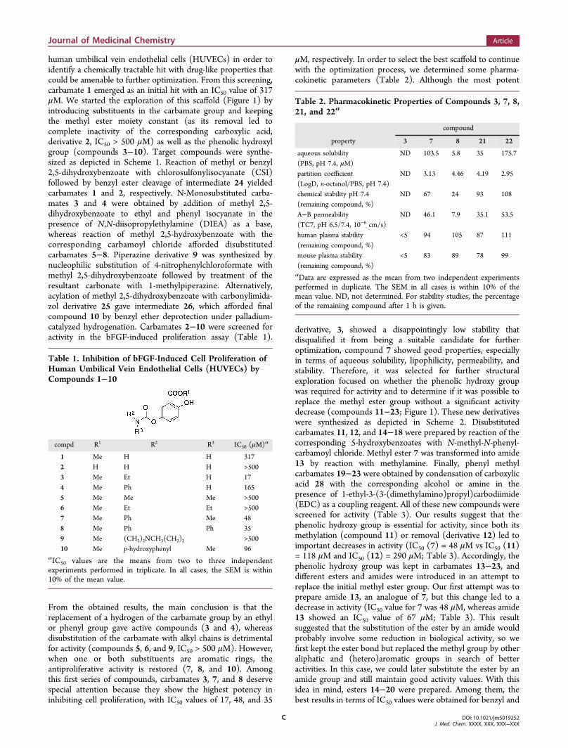
human umbilical vein endothelial cells (HUVECs) in order toidentify a chemically tractable hit with drug-like properties thatcould be amenable to further optimization. From this screening,carbamate 1 emerged as an initial hit with an IC50 value of 317μM. We started the exploration of this scaffold (Figure 1) byintroducing substituents in the carbamate group and keepingthe methyl ester moiety constant (as its removal led tocomplete inactivity of the corresponding carboxylic acid,derivative 2, IC50 > 500 μM) as well as the phenolic hydroxylgroup (compounds 3−10). Target compounds were synthe-sized as depicted in Scheme 1. Reaction of methyl or benzyl2,5-dihydroxybenzoate with chlorosulfonylisocyanate (CSI)followed by benzyl ester cleavage of intermediate 24 yieldedcarbamates 1 and 2, respectively. N-Monosubstituted carba-mates 3 and 4 were obtained by addition of methyl 2,5-dihydroxybenzoate to ethyl and phenyl isocyanate in thepresence of N,N-diisopropylethylamine (DIEA) as a base,whereas reaction of methyl 2,5-hydroxybenzoate with thecorresponding carbamoyl chloride afforded disubstitutedcarbamates 5−8. Piperazine derivative 9 was synthesized bynucleophilic substitution of 4-nitrophenylchloroformate withmethyl 2,5-dihydroxybenzoate followed by treatment of theresultant carbonate with 1-methylpiperazine. Alternatively,acylation of methyl 2,5-dihydroxybenzoate with carbonylimida-zol derivative 25 gave intermediate 26, which afforded finalcompound 10 by benzyl ether deprotection under palladium-catalyzed hydrogenation. Carbamates 2−10 were screened foractivity in the bFGF-induced proliferation assay (Table 1).
From the obtained results, the main conclusion is that thereplacement of a hydrogen of the carbamate group by an ethylor phenyl group gave active compounds (3 and 4), whereasdisubstitution of the carbamate with alkyl chains is detrimentalfor activity (compounds 5, 6, and 9, IC50 > 500 μM). However,when one or both substituents are aromatic rings, theantiproliferative activity is restored (7, 8, and 10). Amongthis first series of compounds, carbamates 3, 7, and 8 deservespecial attention because they show the highest potency ininhibiting cell proliferation, with IC50 values of 17, 48, and 35
μM, respectively. In order to select the best scaffold to continuewith the optimization process, we determined some pharma-cokinetic parameters (Table 2). Although the most potent
derivative, 3, showed a disappointingly low stability thatdisqualified it from being a suitable candidate for furtheroptimization, compound 7 showed good properties, especiallyin terms of aqueous solubility, lipophilicity, permeability, andstability. Therefore, it was selected for further structuralexploration focused on whether the phenolic hydroxy groupwas required for activity and to determine if it was possible toreplace the methyl ester group without a significant activitydecrease (compounds 11−23; Figure 1). These new derivativeswere synthesized as depicted in Scheme 2. Disubstitutedcarbamates 11, 12, and 14−18 were prepared by reaction of thecorresponding 5-hydroxybenzoates with N-methyl-N-phenyl-carbamoyl chloride. Methyl ester 7 was transformed into amide13 by reaction with methylamine. Finally, phenyl methylcarbamates 19−23 were obtained by condensation of carboxylicacid 28 with the corresponding alcohol or amine in thepresence of 1-ethyl-3-(3-(dimethylamino)propyl)carbodiimide(EDC) as a coupling reagent. All of these new compounds werescreened for activity (Table 3). Our results suggest that thephenolic hydroxy group is essential for activity, since both itsmethylation (compound 11) or removal (derivative 12) led toimportant decreases in activity (IC50 (7) = 48 μM vs IC50 (11)= 118 μM and IC50 (12) = 290 μM; Table 3). Accordingly, thephenolic hydroxy group was kept in carbamates 13−23, anddifferent esters and amides were introduced in an attempt toreplace the initial methyl ester group. Our first attempt was toprepare amide 13, an analogue of 7, but this change led to adecrease in activity (IC50 value for 7 was 48 μM, whereas amide13 showed an IC50 value of 67 μM; Table 3). This resultsuggested that the substitution of the ester by an amide wouldprobably involve some reduction in biological activity, so wefirst kept the ester bond but replaced the methyl group by otheraliphatic and (hetero)aromatic groups in search of betteractivities. In this case, we could later substitute the ester by anamide group and still maintain good activity values. With thisidea in mind, esters 14−20 were prepared. Among them, thebest results in terms of IC50 values were obtained for benzyl and
Table 1. Inhibition of bFGF-Induced Cell Proliferation ofHuman Umbilical Vein Endothelial Cells (HUVECs) byCompounds 1−10
compd R1 R2 R3 IC50 (μM)a
1 Me H H 3172 H H H >5003 Me Et H 174 Me Ph H 1655 Me Me Me >5006 Me Et Et >5007 Me Ph Me 488 Me Ph Ph 359 Me (CH2)2NCH3(CH2)2 >50010 Me p-hydroxyphenyl Me 96
aIC50 values are the means from two to three independentexperiments performed in triplicate. In all cases, the SEM is within10% of the mean value.
Table 2. Pharmacokinetic Properties of Compounds 3, 7, 8,21, and 22a
compound
property 3 7 8 21 22
aqueous solubility ND 103.5 5.8 35 175.7(PBS, pH 7.4, μM)partition coefficient ND 3.13 4.46 4.19 2.95(LogD, n-octanol/PBS, pH 7.4)chemical stability pH 7.4 ND 67 24 93 108(remaining compound, %)A−B permeability ND 46.1 7.9 35.1 53.5(TC7, pH 6.5/7.4, 10−6 cm/s)human plasma stability <5 94 105 87 111(remaining compound, %)mouse plasma stability <5 83 89 78 99(remaining compound, %)aData are expressed as the mean from two independent experimentsperformed in duplicate. The SEM in all cases is within 10% of themean value. ND, not determined. For stability studies, the percentageof the remaining compound after 1 h is given.
Journal of Medicinal Chemistry Article
DOI: 10.1021/jm5019252J. Med. Chem. XXXX, XXX, XXX−XXX
C

3-methylpyridinyl groups as R1 substituents, compounds 14and 18, with IC50 values of 17 and 16 μM, respectively (Table3). Hence, these two R1 groups were selected, and the analogueamides 21−23 were synthesized. The biological activity of theseamides was similar to that of the corresponding esters, asshown, for example, by the IC50 values of esters 14 and 18 (17and 16 μM, respectively) when compared with the IC50 valuesof amides 21 and 22 (22 and 14 μM, respectively). Hence, wedetermined their pharmacokinetic properties in order to selectthe best candidate to continue with the biological studies.Taking into account all of these data (Table 2), amide 22showed the best overall profile with the highest solubility(175.7 μM), stability (around 100% under the three assayedconditions), and permeability values. Accordingly, this com-pound was selected for in-depth characterization.Compound 22 Inhibits Proangiogenic Signaling in
MCF7 Breast Cancer Cells under Hypoxic Conditions.Tumor hypoxia, a common feature of many solid tumors, hasbeen identified as a key driver for angiogenic regulationmechanisms. Hence, we first explored whether compound 22 isable to inhibit the proangiogenic signaling generated by hypoxiain the MCF7 human breast adenocarcinoma cell line that waschosen as a model. Our results show that compound 22decreases the levels of important proangiogenic factors VEGFand bFGF in hypoxic MCF7 cells (Figure 2A,B). In addition,this derivative also induces a decrease in the NO levels, whichruns parallel to a strong inhibition of iNOS expression (Figure2C,F).Considering the importance of the enhancement of autocrine
signaling under hypoxic conditions, especially in terms of
activation of the corresponding receptors, VEGFR and FGFR,we also assessed whether compound 22 affected theiractivation. Remarkably, this derivative inhibits the activationof these two receptors, as it decreases their phosphorylated(active) forms (Figure 2D,E).The main effects of the activation of the FGFR pathway
include the induction of proliferation, migration, andantiapoptotic signals. Proliferation enhancement is mainlyachieved through activation of the MAPK cascade, whereasthe induction of antiapoptotic signals is mediated by activationof the PI3K/AKT pathway.8,11 This latter cell survival pathwayis also reinforced by VEGFR activation.3 Hence, we exploredwhether compound 22 was able to suppress the phosphor-ylation of downstream kinases AKT, MEK, and ERK. Asexpected, hypoxia activated the AKT and the MEK and ERKsignaling pathways, as demonstrated by the increasedphosphorylation of these kinases, and, remarkably, incubationof the cells with compound 22 prevented this activation (Figure3A). Importantly, inhibition of these signaling pathways bycompound 22 was accompanied by an impairment in hypoxia-stimulated cell migration (Figure 3B). In order to determinewhether the decrease in migration was due to generalcytotoxicity, we carried out a similar set of experiments inwhich cells were incubated with compound 22 for 48 h, afterwhich the compound was removed and then the cells were
Scheme 2a
aReagents and conditions: (a) NaH, CH3CN, rt, 3 h, 14−64%; (b)CH3NH2, CH3OH, 0 °C to rt, 3 h, 76%; (c) H2, Pd(C), EtOH, rt, 3 h,100%; (d) R1OH, R1NH2, or R
1NHMe, EDC, DMAP, DMF, 0 °C tort, 16 h, 27−64%.
Table 3. Inhibition of bFGF-Induced Cell Proliferation ofHuman Umbilical Vein Endothelial Cells (HUVECs) byCompounds 11−23a
aIC50 values are the means from two to three independentexperiments performed in triplicate. In all cases, the SEM is within10% of the mean value.
Journal of Medicinal Chemistry Article
DOI: 10.1021/jm5019252J. Med. Chem. XXXX, XXX, XXX−XXX
D

Figure 2. Compound 22 decreases the production of hypoxia-induced proangiogenic factors VEGF, bFGF, and NO and inhibits the activation oftheir corresponding receptors. Incubation of MCF7 cells with compound 22 (50 μM) under hypoxic conditions significantly reduces the levels of(A) VEGF, (B) bFGF, and (C) NO, decreases the activation of the (D) VEGF and (E) FGF receptors, and decreases (F) iNOS expression (131kDa band). β-Actin (42 kDa) is shown as a loading control. Data correspond to the average ± SEM of at least three independent experiments, andrepresentative gels are shown. The bar graphs in panels D and E represent the optical density of the immunoreactive phosphorylated proteinnormalized to the total corresponding protein, which is expressed as the percentage relative to normoxia. The bar graph in panel F represents theoptical density of the immunoreactive protein (iNOS) expressed as the percentage relative to normoxia. *, P < 0.05; **, P < 0.01; ***, P < 0.001 (vshypoxic vehicle-treated cells) (Student’s t test).
Figure 3. Compound 22 inhibits hypoxia-activated signaling pathways and suppresses cell migration. (A) Representative western blots ofphosphorylated (pAKT) and total AKT (T-AKT), phosphorylated MEK1/2 (pMEK1/2) and total MEK1/2 (T-MEK1/2), and phosphorylatedERK1/2 (pERK1/2) and total ERK1/2 (T-ERK1/2). Lysates were obtained from MCF7 cells treated with compound 22 (50 μM) under hypoxicconditions. Data correspond to the average ± SEM of at least three independent experiments. The bar graphs in panel A represent the optical densityof the immunoreactive phosphorylated protein normalized to the total corresponding protein, which is expressed as the percentage relative tonormoxia. *, P < 0.05; **, P < 0.01 (Student’s t test). (B) In vitro scratches (wounds) were made by scraping confluent cell monolayers with a sterilepipet tip and were visualized by phase contrast microscopy. After 48 h under hypoxic conditions, the remaining wound area was quantified. The bargraph represents the average ± SEM of at least three independent experiments and three different fields. ***, P < 0.001 (vs DMSO-treated cells)(Student’s t test). Bar, 250 μm.
Journal of Medicinal Chemistry Article
DOI: 10.1021/jm5019252J. Med. Chem. XXXX, XXX, XXX−XXX
E

incubated for an additional 48 h. The obtained results showthat cells recover their ability to migrate after removal of thecompound (Supporting Information Figure S1). In addition,the number of viable cells remains similar to that in the vehicle-treated cells (Supporting Information Figure S1E). Takentogether, these data strongly suggest that compound 22 ismainly affecting cell migration and not inducing generalcytotoxicity.Compound 22 Inhibits the Production of Hypoxia-
Induced Proangiogenic Signals via Hypoxia-InducibleFactor-1α (HIF-1α). Intratumoral hypoxia is one of the majorfactors that drive tumor angiogenesis, and hypoxia-drivenangiogenesis is primarily mediated by HIF-1α, often consideredto be a master regulator of angiogenesis under hypoxia.19 Inaddition, in MCF7 breast cancer cells, HIF-1α is the factor thatmainly contributes to the expression of genes under hypoxicconditions.20 Therefore, we analyzed whether HIF-1α wasinvolved in the antiangiogenic response elicited by compound22. To this end, we knocked-down HIF-1α using selective smallinterfering RNAs (siRNAs) (Figure 4A). As shown in Figure4B,C, hypoxia induced an increase in bFGF and VEGF levels inMCF7 cells transfected with a nontargeted (control) siRNA (CsiRNA), and this effect was prevented by compound 22.Conversely, genetic silencing of HIF-1α abrogated the increasein these two proangiogenic factors upon hypoxia stimulus, andcompound 22 did not enhance this effect. These results suggestthat the effect of compound 22 on bFGF and VEGF levels ismediated via HIF-1α. To further ascribe the effects ofcompound 22 to HIF-1α modulation and not to othermembers of its family, mainly HIF-2α, we selected twoproteins, BNip3 and Ang-2, which have been described to bemainly regulated by HIF-1α and HIF-2α, respectively.21 Asexpected, and consistent with the literature, hypoxia increasedthe levels of both proteins, BNip3 and Ang-2. Remarkably,
compound 22 decreased only the levels of BNip3 (Figure 4D)without affecting the expression of Ang-2 (Figure 4E). Theseresults provide further support for the specific involvement ofHIF-1α in the effects induced by compound 22. In addition,and to discard potential effects of this derivative upstream ofHIFs, we verified that compound 22 did not affect theexpression levels either of HIF-1α or of HIF-2α (Figure 4F).
Antiangiogenic Gene Profile of Hypoxic MCF7 CellsTreated with Compound 22. To further confirm theantiangiogenic profile of compound 22, we analyzed theexpression of 84 key genes involved in angiogenesis in hypoxicMCF-7 cells treated with this compound. We identified 12genes that were significantly affected by compound 22 (foldchange ≥ 2; Figure 5). As expected, several proangiogenicgenes were downregulated in the presence of compound 22.Among them are several cytokines such as CCL11, IL-1β, orthe chemokine-like PROK2 that have been linked to angio-genesis in solid tumors22−24 as well as other known
Figure 4. Compound 22 inhibits the production of the hypoxia-induced proangiogenic factors via HIF-1α. (A) HIF-1α mRNA levels after transienttransfection of MCF7 cells with a siRNA selectively targeting HIF-1α (HIF-1α siRNA) or with a nontargeted siRNA (C siRNA). Results areexpressed in arbitrary units (au). bFGF (B) and VEGF (C) levels in MCF7 cells transiently transfected with the indicated siRNAs under normoxicand hypoxic conditions and in the presence/absence of compound 22. Representative western blots of (D) BNip3 (22 kDa), (E) Ang2 (65 kDa),and (F) HIF-1α (132 kDa) and HIF-2α (115 kDa). In all cases, actin (42 kDa), marked with an arrowhead, is used as a loading control. Lysates wereobtained from MCF7 cells treated with compound 22 (50 μM) under normoxic or hypoxic conditions as indicated. Data correspond to the average± SEM of at least three independent experiments. The bar graphs in panels D and E represent the optical density of the immunoreactive protein(BNip3 or Ang2, respectively) expressed as the percentage relative to normoxia. *, P < 0.05; ***, P < 0.001 (vs hypoxic vehicle-treated cells)(Student’s t test).
Figure 5. Compound 22 regulates the expression of angiogenesis-related genes. An angiogenesis PCR array was performed in hypoxicMCF7 cells challenged with compound 22 or the correspondingvehicle. The graph shows the 12 genes that were modulated (threshold= 2-fold increase/decrease) in compound 22-treated cells vs controlcells. Results are expressed as fold regulation.
Journal of Medicinal Chemistry Article
DOI: 10.1021/jm5019252J. Med. Chem. XXXX, XXX, XXX−XXX
F

proangiogenic factors such as the vascular endothelial cadherinCDH5 and the receptors VEGFR-2 (also known as KDR) andNotch4.25 On the other hand, upregulation of several genes inresponse to compound 22 was also observed, including thechemokine CXCL9, which has been described as attenuatingangiogenesis in some situations.26 Surprisingly, we observed anincrease in the transcript levels of certain proangiogenic factorssuch as the cell adhesion molecules integrin ITGB3 andPECAM1, the angiopoietin receptor TIE1, and the proangio-genic factors FGF1 and FGF2. These apparently contradictoryresults may be due to differential regulation at the transcrip-tional and translational levels. In this regard, for example, it isworth noting that although some increase is observed at thetranscriptional level (Figure 5) compound 22 reduces theprotein levels of FGF2 (bFGF), as shown in Figure 2B.Antitumor Effect of Compound 22 in Vivo. In order to
assess the in vivo efficacy of compound 22, we used a breastcancer xenograft model. Tumor-bearing mice were injectedintraperitoneally with compound 22 (25 mg/kg) once a day for28 days, and tumor volumes were routinely measured (Figure6A). In vehicle-treated animals, tumors grew in an exponentialmanner. Treatment of mice with compound 22 produced noeffect in 62% of them (5 out of 8), but we observed a significantreduction in tumor growth (ranging from 46 to 55%) in theremaining 38% (3 out of 8) (Figure 6B).
To analyze the in vivo inhibition of angiogenesis, wequantified the number of blood vessels within the tumors byimmunofluorescence staining of CD31 (a marker of endothelialcells) in vehicle-treated animals as well as in responding andnot-responding individuals (Figure 6C). Significant inhibitionof angiogenesis was not detected in nonresponding animals. Incontrast, in the tumors of compound-responding mice, amarked reduction in the number of blood vessels was observed.Remarkably, this result correlates with the expression level ofVEGF (Figure 6D). Importantly, the inhibition of angiogenesisand tumor growth induced by compound 22 was notaccompanied by any sign of toxicity, as assessed byhistopathological analysis of liver, lungs, spleen, and heart ofcompound-treated animals (data not shown). The degree ofinterindividual variability in the response to 22 might be relatedto a different bioavailability of the compound caused by thedistinct growth and size of each individual tumor or by theexistence of clonal variability of xenograft cells, something thathas been previously observed for other antitumor targets27 andalso in the clinic after treatment with other angiogenesisinhibitors. In this case, it is possible that increasing the numberof individuals would also augment the number of positive cases.In addition, it is important to note that a tumor is aheterogeneous entity, with hypoxic portions but also withother zones, near the blood vessel, which are not hypoxic, andeach may have different signaling factors. In this context, Figure
Figure 6. Antitumor effects of compound 22 in a breast cancer xenograft model. (A) Tumor growth in vehicle-treated (represented as mean ± SEM,gray dashed line, n = 8) and compound 22-treated animals (represented individually, n = 8, solid gray lines). (B) Tumor weight at the end of thetreatment for vehicle-treated animals (white bar), compound 22-responding animals (black bar), and compound 22-treated not-responding animals(gray bar). (C) Compound 22 significantly reduces angiogenesis in responding animals (22-R), whereas it does not affect the number of bloodvessels in treated but not-responding animals (22-NR). Images correspond to representative immunofluorescence stainings of tumor sections of eachexperimental group. Blood vessels are stained with an antibody against CD31 (in green), and nuclei are shown in blue. Scale bar, 100 μm. The bargraph represents the number of blood vessels (mean ± SEM, 3 tumors/experimental group and 4 sections/tumor) for vehicle-treated animals (whitebar), compound 22-responding animals (black bar), and not-responding animals (gray bar). *, P < 0.05; ***, P < 0.001 (vs compound 22-treatednonresponding mice) (Student’s t test). (D) Compound 22 significantly reduces VEGF mRNA levels in responding animals (22-R) compared tovehicle-treated mice or mice treated with compound 22 that are not responding (22-NR). Images correspond to representative data obtained fromindependent samples of tumor sections from each experimental group. Controls include lack of RNA (right lane, labeled −) and GAPDH ashousekeeping gene.
Journal of Medicinal Chemistry Article
DOI: 10.1021/jm5019252J. Med. Chem. XXXX, XXX, XXX−XXX
G

5 suggests upregulation of some proangiogenic genes even inthe presence of compound 22. Hence, it is possible that in themice in which the drug decreased tumor size the effects of thedownregulated proangiogenic genes predominated, whereas theincrease in tumor size observed in the other mice wasdominated by effects of the proangiogenic genes that remainedupregulated even in the presence of the compound.In conclusion, in this work, we describe a new series of
antiangiogenic compounds. Among them, the optimal com-pound (22) inhibits proangiogenic signaling under hypoxicconditions in breast cancer cells. Specifically, administration of22 decreases the levels of the proangiogenic molecules VEGF,bFGF, and NO. Also, this compound inhibits the active formsof the corresponding receptors of these factors (phosphorylatedforms of VEGFR and bFGFR) and the levels of the iNOSenzyme. These effects correlate with a blockade of the MAPKpathway and the inhibition of cellular migration, and they aremediated by HIF-1α, since the effects of compound 22 mostlydisappear when its expression is knocked-down. Additionally,gene profiling identified a set of genes related to angiogenesiswhose expression is altered by compound 22 and that mightcontribute to the antiangiogenic effects. Furthermore, admin-istration of compound 22 in a xenograft model produced tumorgrowth reductions ranging from 46 to 55% in the 38% of thetreated animals. Importantly, in the responding tumors, asignificant reduction in the number of blood vessels and in thelevels of VEGF was observed, further supporting themechanism of action of the compound. Although betterefficacy would be desirable, the fact that compound 22 didnot induce any toxic effects in vivo and that it was able toeffectively block angiogenesis in the tumors of respondinganimals strongly support the potential of this compound as alead for the development of new antiangiogenic agents suitablefor the treatment of cancer either alone or in combination withother benchmark drugs.
■ EXPERIMENTAL PROCEDURESCompound Synthesis. Unless stated otherwise, starting materials,
reagents, and solvents were purchased as high-grade commercialproducts from Sigma-Aldrich, Acros, ABCR, Bachem, Fluorochem,Scharlab, or Panreac and were used without further purification.Dichloromethane was freshly distilled from calcium hydride.Tetrahydrofuran and diethyl ether were freshly distilled from sodiumand benzophenone. All nonaqueous reactions were performed underan argon atmosphere in oven-dried glassware. Full details regarding thesynthetic procedures and characterization data of all compounds aregiven in the Supporting Information. Spectroscopic data of alldescribed compounds were consistent with the proposed structures.Satisfactory HPLC chromatograms and elemental analyses (C, H, N)were obtained for the final compounds, confirming a purity of at least95% for all tested compounds. Pharmacokinetic properties of selectedcompounds 3, 7, 8, 21, and 22 were determined at CEREP (www.cerep.fr).The synthesis and structural characterization of derivative 22 is
detailed below. Detailed procedures and structural data for the rest ofthe compounds can be found in the Supporting Information.Synthesis of 2-Hydroxy-5-({[methyl(phenyl)amino]-
carbonyl}oxy)benzoic Acid (28). To a solution of benzyl ester 14(120 mg, 0.3 mmol) in absolute EtOH (20 mL) was added 10% Pd/C(50 mg), and the mixture was hydrogenated at room temperature for 4h, with an initial hydrogen pressure of 30 psi. The reaction mixture wasfiltered through a pad of Celite and washed with EtOH. The solventwas evaporated to afford the title pure compound as a solid inquantitative yield. mp 157−158 °C. Rf (dichloromethane/EtOH, 95:5)0.20. IR (KBr, cm−1) 3071, 1699, 1596, 1489. 1H NMR (CDCl3) δ3.44 (s, 3H), 6.95 (d, 1H, J = 8.9), 7.22−7.45 (m, 6H), 7.61 (m, 1H).
13C NMR (CDCl3) δ 38.4 (CH3), 114.2 (C), 118.8, 119.1, 126.1,127.0, 127.3 (5CH), 129.3 (3CH), 142.5, 142.7, 154.8, 159.2, 169.8(5C).
Synthesis of 4-Hydroxy-3-{[(pyridin-3-ylmethyl)amino]-carbonyl}phenyl methyl(phenyl)carbamate (22). To a solutionof benzoic acid 28 (228 mg, 0.8 mmol) in anhydrous DMF (10 mL)were added EDC (230 mg, 1.2 mmol) and DMAP (23 mg, 0.3 mmol),and the mixture was stirred at room temperature for 15 min. Then, asolution of pyridin-3-ylmethylamine (87 mg, 0.8 mmol) in DMF (5mL) was added at 0 °C, and the reaction mixture was stirred for 2 h atthis temperature and at room temperature for 14 additional hours. Themixture was evaporated, and the residue was purified by columnchromatography (dichloromethane/EtOH, 95:5) to give compound22 in 37% yield. mp 110−112 °C. Rf (dichloromethane/EtOH, 9:1)0.32. IR (KBr, cm−1) 3348, 1719, 1646, 1599, 1545, 1492. 1H NMR(CDCl3) δ 3.35 (s, 3H), 4.40 (d, 2H, J = 5.7), 6.87 (d, 1H, J = 9.0),7.04−7.06 (m, 1H), 7.19−7.36 (m, 6H), 7.41 (m, 1H), 7.60 (d, 1H, J= 7.9), 8.12 (m, 1H), 8.44 (br s, 2H). 13C NMR (CDCl3) δ 38.7(CH3), 41.3 (CH2), 115.0 (C), 119.1, 120.2, 124.1, 126.3, 127.2, 127.8(6CH), 129.5 (3CH), 134.3 (C), 136.5 (CH), 142.9, 143.0 (2C),148.8, 149.3 (2CH), 154.9, 159.0, 169.4 (3C).
Inhibition of bFGF-Induced Cell Proliferation of HumanUmbilical Vein Endothelial Cells (HUVECs). HUVECs, obtainedfrom American Type Culture Collection, were cultured in a humidifiedatmosphere of 95% air and 5% CO2 at 37 °C in M199 mediumcontaining 10% fetal bovine serum (FBS) and 10 μg/mL heparin.Cells were incubated in the presence of bFGF (1 μg/mL) and theappropriate concentration of compound or vehicle (0.4% DMSO) for2 days, and cell proliferation was quantified spectrofluorimetrically.IC50 values are the mean from at least two independent experimentscarried out in triplicate. In all cases, the standard error of the mean(SEM) is within 10% of the mean value.
Determination of VEGF and bFGF Levels. Cells were seeded in12-well plates at a density of 5 × 10⁴ cells per well and were grown for24 h to obtain a 70−80% confluent monolayer. Then, medium wasreplaced with fresh DMEM with or without 150 μM CoCl2. After 5 h,compound 22 or vehicle (DMSO) was added to the culture medium,and cells were incubated for 4 h more. Supernatants were thencollected and used straightaway or stored at −80 °C for further use.Concentrations of VEGF and bFGF in the culture medium weremeasured using an enzyme-linked immunosorbent assay (ELISA),according to the manufacturer’s instructions (VEGF human ELISA kitand FGF-basic human ELISA kit, Invitrogen). Absorbance wasmeasured at 450 nm using an Asys UVM 340 (Biochrom Ltd.,Cambridge, UK) microplate reader, and data were normalized to thekit controls and the number of producing cells. Data from three to fiveindependent experiments carried out in triplicate were represented asmean fold ± SEM with bar graphs.
Nitric Oxide (NO) Quantification. Nitric oxide production wasmeasured through determination of nitrite concentration in the culturemedium using the Griess test. Briefly, cells were seeded in 96-wellplates at a density of 1 × 10⁴ cells per well in DMEM with 10% fetalbovine serum (FBS) and incubated for 24 h prior to treatments. Themedium was then replaced with fresh DMEM with or without 150 μMCoCl2; after 5 h of incubation, compound 22 or vehicle was added,and incubation was continued for another 4 h. Then, 100 μL ofsupernatant from each condition was mixed with 100 μL of Griessreagent (1% sulphanilamide, 0.1% N-(1-naphthyl)ethylendiaminedihydrochloride, 2.5% phosphoric acid). After 15 min at roomtemperature in the dark, absorbance was measured at 548 nm in anAsys UVM 340 (Biochrom Ltd., Cambridge, UK) microplate reader.The concentration of nitrite, a stable oxidized derivative of NO incultured cells, was determined from a sodium nitrite (NaNO2, Sigma-Aldrich) standard curve. Data from three independent experimentsperformed in triplicate were presented as mean ± SEM.
Western Blot Analysis. MCF-7 cells were plated at a density of 2× 10⁶ cells in 15 cm dishes and allowed to grow for 24 h in DMEMwith 1% FBS to obtain an 80% confluent monolayer. The medium wasthen replaced by fresh DMEM with or without 150 μM CoCl2, andcells were incubated for 5 h to allow a hypoxic response to be
Journal of Medicinal Chemistry Article
DOI: 10.1021/jm5019252J. Med. Chem. XXXX, XXX, XXX−XXX
H
generated. After that, compound 22 or vehicle was added, and cellswere incubated for 4 h. Cells were washed with PBS and lysed withice-cold RIPA buffer (50 mM Tris-HCl, pH 7.4, 150 mM NaCl, 1%Igepal) containing protease and phosphatase inhibitors (Roche andSigma-Aldrich, respectively). Lysates were clarified by centrifugation at10 000g for 10 min at 4 °C and used straightaway or stored at −80 °Cuntil use. Protein concentration was measured (DC protein assay kit,Bio-Rad), and samples with equal amounts of total protein werediluted into Laemmli reducing sample buffer (Bio-Rad) and denaturedat 95 °C for 5 min. Samples were then resolved on 4−20% SDS-PAGEgels (Bio-Rad), and proteins were transferred to nitrocellulosemembranes (GE Healthcare, Amersham). After 1 h of incubation inblocking buffer [10 mM Tris-HCl, pH 8.0, 150 mM NaCl, 0.05%Tween-20 (TBS-T) with 1% BSA], membranes were incubatedovernight at 4 °C with the corresponding primary antibody. Then,membranes were washed three times (5 min each) with TBS-T andincubated with the corresponding secondary antibody for 1 h at roomtemperature. Protein bands were visualized using enhanced chem-iluminescence detection reagents (GE Healthcare, Amersham) in aFujifilm LAS-3000 developer (Tokyo, Japan) and quantified bydensitometry using ImageJ software (NIH).Primary antibodies were from Cell Signaling and used at a 1:1000
dilution (rabbit anti-phospho-AKT (pS473), rabbit anti-AKT, rabbitanti-phospho-ERK1/2, rabbit anti-ERK1/2, rabbit anti-phospho-MEK1/2, rabbit anti-MEK1/2, rabbit anti-VEGFR, rabbit anti-phospho-VEGFR, rabbit anti-FGFR, and rabbit anti-phospho-FGFR)or from Santa Cruz Biotechnology and used at a 1:200 dilution(mouse anti-HIF-1α, mouse anti-HIF-2α, mouse anti-iNOS, and rabbitanti-β actin). Secondary antibodies used were goat anti-mouse or goatanti-rabbit IgG HRP conjugates (1:5000, Sigma-Aldrich) accordingly.Relative phosphorylation levels from three independent experimentswere presented as the mean ± SEM with bar graphs.Migration or Wound Healing Assay. Cells were seeded in 96-
well plates at a density of 1.5 × 10⁵ cells per well in DMEM with 10%FBS and grown for 24 h at 37 °C and 5% of CO2 to obtain a 90−100%confluent monolayer. Wounds were made with a sterile p20 pipet tip,and each well was washed twice with PBS to eliminate nonadherentcells and cell debris. Fresh DMEM with or without 150 μM CoCl2 wasthen added, and after 5 h of incubation, compound 22 (50 μM) orvehicle was added. At this time (0 h) and after 48 h, cells werephotographed under phase contrast with an Olympus FW1200microscope. Empty area in each wound was quantified using ImageJsoftware (NIH) and compared with the corresponding area of theinitial wound. The percentage of area from three independentexperiments performed in triplicate was presented as the mean ±SEM with bar graphs.RNA Interference-Mediated Silencing of the HIF-1α Gene.
Cells were transfected with specific siRNA duplexes usingDharmaFECT 1 as the transfection reagent according to themanufacturer’s instructions (Dharmacon-Thermo Scientific, Lafayette,CO). Selective siRNA against human HIF-1α was a smart pool fromDharmacon-Thermo Scientific, and the sequences were as follows: 5′-GAACAAAUACAUGGGAUUA-3′; 5′-AGAAUGAAGUGUACC-CUAA-3′; 5′-GAUGGAAGCACUAGACAAA-3′; and 5′-CAAGU-AGCCUCUUUGACAA-3′. The nontargeted control sequence, 5′-UUCUCCGAACGUGUCACGU-3′, was from Applied Biosystems-Ambion (Austin, TX). Twenty-four hours after transfection, cells wereseeded for ELISA assays, which were performed as described below.Quantitive Polymerase Chain Reaction (qPCR). RNA from cell
cultures or tumor tissues was isolated with TRIzol reagent (Sigma).cDNA was subsequently obtained with Transcriptor reverse tran-scriptase (Roche). Real-time quantitative PCR assays were performedusing the FastStart master mix with Rox (Roche), and probes wereobtained from the Universal Probe Library (Roche). The primers usedfor human HIF-1α were as follows: sense, 5′-GATAGCAAGA-CTTTCCTCAGTCG-3′; and antisense, 5′-TGGCTCATATCCCA-TCAATTC-3′. Amplifications were run in a 7900 HT-fast real-timePCR system (Applied Biosystems). Each value was normalized tohuman β-actin RNA levels as an internal control: sense, 5′-
CCAACCGCGAGAAGATGA-3′; and antisense, 5′-CCAGAGGC-GTACAGGGATAG-3′.
Gene Expresssion Analysis. The RT2 profiler PCR array ofhuman angiogenesis (Qiagen, Valencia, CA), which analyzes theexpression of 84 key genes involved in modulating the biologicalprocesses of angiogenesis, was used. RNA from cell cultures wasisolated with TRIzol reagent (Sigma, St. Louis, MO), including a DNAdigestion step with genomic DNA elimination mix (Qiagen). cDNAwas subsequently obtained with a RT2
first strand kit according to themanufacturer’s instructions (Qiagen). Real-time PCR assay wasperformed using the RT2 profiler PCR array of human angiogenesisin combination with RT2 SYBR Green master mix (Qiagen).Amplifications were run in a 7900 HT-fast real-time PCR system(Applied Biosystems, Carlsbard, CA), and data were analyzed usingthe SABiosciences PCR array data analysis template Excel (Qiagen).
VEGF Expression Analysis. RNA was isolated from tumors withTRIzol reagent (Invitrogen, Barcelona, Spain) with the real star kit(Durviz, Valencia, Spain), and cDNA was obtained with Transcriptorreverse transcriptase (Roche Applied Science, Barcelona, Spain). Theprimers used for VEGF-A amplification were as follows: sense, 5′-GTCCTGTGTGCCGCTGAT-3′; antisense, 5′-AGGTTTGAT-CCGCATGATCT-3′. GAPDH was used as reference (sense, 5′-GGGAAGCTCACTGGCATGGCCTTCC-3′; antisense, 5′-CATGTGGGCCATGAGGTCCACCAC-3′).
Subcutaneous Xenografts. All procedures involving animalswere performed with the approval of the Complutense UniversityAnimal Experimentation Committee in compliance with Europeanofficial regulations. Five million MDA-MB-231 breast cancer cells in100 μL of PBS were subcutaneously injected into the flank of 6-week-old athymic mice (Harlan Interfauna Iberica, Barcelona, Spain).Tumors were routinely measured with an external caliper, and volumewas calculated as (4π/3) × (width/2)2 × (length/2). When tumorsreached ca. 200 mm3, the mice were treated intraperitoneally threetimes a week with compound 22 (25 mg/kg) or vehicle (DMSO 0.2mg/μL in PBS) for 4 weeks. After treatment, animals were sacrificed,and tumors and organs were collected. Tumors were divided intodifferent portions for preparation of tissue sections for immuno-fluorescent staining [frozen in Tissue-Tek (Sakura Finetek Europe,Zoeterwoude, The Netherlands)] or snap frozen for RNA extraction(and stored at −80 °C until use). Organs collected were fixed informaldehyde and stained with hematoxylin−eosin for analysis.
For immunofluorescence analysis, Tissue-Tek frozen sections werefixed in PFA 4% and were subjected to heat-induced antigen retrievalin citrate buffer. Then, sections were blocked with PBS containing0.25% TritonX-100 and 10% goat serum and incubated with anti-CD31 (Pharmingen/BD Biosciences, San Jose, CA). Secondary anti-mouse antibodies conjugated with Alexa Flour 488 were fromInvitrogen (Carlsbad, CA). Cell nuclei were stained with DAPI(Invitrogen). Images were acquired using a Leica DM400B microscope(Leica, Wetzlar, Germany).
■ ASSOCIATED CONTENT
*S Supporting InformationCell migration impairment of compound 22 (Figure S1),detailed synthetic procedures, characterization data of finalcompounds 1−21 and 23, and intermediates 24−26 and 27d,e,and elemental and HPLC-MS purity analyses of finalcompounds 1−23. The Supporting Information is availablefree of charge on the ACS Publications website at DOI:10.1021/jm5019252.
■ AUTHOR INFORMATION
Corresponding Authors*(M.M.-F.) Phone: 34-91-3944894; Fax: 34-91-3944103; E-mail: [email protected].*(M.L.L.-R.) Phone: 34-91-3944239; Fax: 34-91-3944103; E-mail: [email protected].
Journal of Medicinal Chemistry Article
DOI: 10.1021/jm5019252J. Med. Chem. XXXX, XXX, XXX−XXX
I

Author Contributions◆N.I.M.-R. and D.A. contributed equally to this work.NotesThe authors declare no competing financial interest.
■ ACKNOWLEDGMENTSThis work was supported by Italfarmaco and grants from theSpanish Ministerio de Economıa y Competitividad (MINECO,SAF2010-22198, SAF2013-48271, and predoctoral fellowshipsto F.J.O.-N., M.B., and S.B.-B.), Instituto de Salud Carlos III(PI11/00295 and PI14/01101), and Comunidad de Madrid(S2010/BMD-2353). E.P.-G. is a recipient of a postdoctoralresearch contract of the Asociacion Espanola Contra el Cancer.The authors thank Campus de Excelencia CEI Moncloa for apredoctoral fellowship to N.I.M.-R and UCM’s CAI Cytometryand Fluorescence Microscopy facility for their assistance.
■ ABBREVIATIONS USEDbFGF, basic FGF; CSI, chlorosulfonylisocyanate; DIEA, N,N-diisopropylethylamine; EDC, ethyl-3-(3-(dimethylamino)-propyl)carbodiimide; FBS, fetal bovine serum; FGFR, FGFreceptor; HIF-1α, hypoxia-inducible factor-1α; HUVECs,human umbilical vein endothelial cells; SEM, standard errorof the mean; siRNA, small interfering RNA; VEGF, vascularendothelial growth factor
■ REFERENCES(1) Ferrara, N.; Hillan, K. J.; Gerber, H. P.; Novotny, W. Discoveryand development of bevacizumab, an anti-VEGF antibody for treatingcancer. Nat. Rev. Drug Discovery 2004, 3, 391−400.(2) Folkman, J. Angiogenesis: an organizing principle for drugdiscovery? Nat. Rev. Drug Discovery 2007, 6, 273−286.(3) Gacche, R. N.; Meshram, R. J. Angiogenic factors as potentialdrug target: efficacy and limitations of anti-angiogenic therapy.Biochim. Biophys. Acta 2014, 1846, 161−179.(4) Wu, J. M.; Staton, C. A. Anti-angiogenic drug discovery: lessonsfrom the past and thoughts for the future. Expert Opin. Drug Discovery2012, 7, 723−743.(5) Bellou, S.; Pentheroudakis, G.; Murphy, C.; Fotsis, T. Anti-angiogenesis in cancer therapy: hercules and hydra. Cancer Lett. 2013,338, 219−228.(6) Helfrich, I.; Scheffrahn, I.; Bartling, S.; Weis, J.; von Felbert, V.;Middleton, M.; Kato, M.; Ergun, S.; Augustin, H. G.; Schadendorf, D.Resistance to antiangiogenic therapy is directed by vascular phenotype,vessel stabilization, and maturation in malignant melanoma. J. Exp.Med. 2010, 207, 491−503.(7) Petrillo, M.; Scambia, G.; Ferrandina, G. Novel targets for VEGF-independent anti-angiogenic drugs. Expert Opin. Invest. Drugs 2012,21, 451−472.(8) Turner, N.; Grose, R. Fibroblast growth factor signalling: fromdevelopment to cancer. Nat. Rev. Cancer 2010, 10, 116−129.(9) Lieu, C.; Heymach, J.; Overman, M.; Tran, H.; Kopetz, S. BeyondVEGF: inhibition of the fibroblast growth factor pathway andantiangiogenesis. Clin. Cancer Res. 2011, 17, 6130−6139.(10) Liang, G.; Chen, G.; Wei, X.; Zhao, Y.; Li, X. Small moleculeinhibition of fibroblast growth factor receptors in cancer. CytokineGrowth Factor Rev. 2013, 24, 467−475.(11) Dieci, M. V.; Arnedos, M.; Andre, F.; Soria, J. C. Fibroblastgrowth factor receptor inhibitors as a cancer treatment: from a biologicrationale to medical perspectives. Cancer Discovery 2013, 3, 264−279.(12) Bono, F.; De Smet, F.; Herbert, C.; De Bock, K.; Georgiadou,M.; Fons, P.; Tjwa, M.; Alcouffe, C.; Ny, A.; Bianciotto, M.; Jonckx, B.;Murakami, M.; Lanahan, A. A.; Michielsen, C.; Sibrac, D.; Dol-Gleizes,F.; Mazzone, M.; Zacchigna, S.; Herault, J. P.; Fischer, C.; Rigon, P.;Ruiz de Almodovar, C.; Claes, F.; Blanc, I.; Poesen, K.; Zhang, J.;Segura, I.; Gueguen, G.; Bordes, M. F.; Lambrechts, D.; Broussy, R.;
van de Wouwer, M.; Michaux, C.; Shimada, T.; Jean, I.; Blacher, S.;Noel, A.; Motte, P.; Rom, E.; Rakic, J. M.; Katsuma, S.; Schaeffer, P.;Yayon, A.; Van Schepdael, A.; Schwalbe, H.; Gervasio, F. L.; Carmeliet,G.; Rozensky, J.; Dewerchin, M.; Simons, M.; Christopoulos, A.;Herbert, J. M.; Carmeliet, P. Inhibition of tumor angiogenesis andgrowth by a small-molecule multi-FGF receptor blocker with allostericproperties. Cancer Cell 2013, 23, 477−488.(13) Li, D.; Wei, X.; Xie, K.; Chen, K.; Li, J.; Fang, J. A novel decoyreceptor fusion protein for FGF-2 potently inhibits tumour growth. Br.J. Cancer 2014, 111, 68−77.(14) Wang, Y.; Becker, D. Antisense targeting of basic fibroblastgrowth factor and fibroblast growth factor receptor-1 in humanmelanomas blocks intratumoral angiogenesis and tumor growth. Nat.Med. 1997, 3, 887−893.(15) Ebos, J. M.; Lee, C. R.; Cruz-Munoz, W.; Bjarnason, G. A.;Christensen, J. G.; Kerbel, R. S. Accelerated metastasis after short-termtreatment with a potent inhibitor of tumor angiogenesis. Cancer Cell2009, 15, 232−239.(16) Loges, S.; Mazzone, M.; Hohensinner, P.; Carmeliet, P.Silencing or fueling metastasis with VEGF inhibitors: antiangiogenesisrevisited. Cancer Cell 2009, 15, 167−170.(17) Paez-Ribes, M.; Allen, E.; Hudock, J.; Takeda, T.; Okuyama, H.;Vinals, F.; Inoue, M.; Bergers, G.; Hanahan, D.; Casanovas, O.Antiangiogenic therapy elicits malignant progression of tumors toincreased local invasion and distant metastasis. Cancer Cell 2009, 15,220−231.(18) Semenza, G. L. Hypoxia-inducible factors: mediators of cancerprogression and targets for cancer therapy. Trends Pharmacol. Sci.2012, 33, 207−214.(19) Philip, B.; Ito, K.; Moreno-Sanchez, R.; Ralph, S. J. HIFexpression and the role of hypoxic microenvironments within primarytumours as protective sites driving cancer stem cell renewal andmetastatic progression. Carcinogenesis 2013, 34, 1699−1707.(20) Mole, D. R.; Blancher, C.; Copley, R. R.; Pollard, P. J.; Gleadle,J. M.; Ragoussis, J.; Ratcliffe, P. J. Genome-wide association ofhypoxia-inducible factor (HIF)-1alpha and HIF-2alpha DNA bindingwith expression profiling of hypoxia-inducible transcripts. J. Biol. Chem.2009, 284, 16767−16775.(21) Keith, B.; Johnson, R. S.; Simon, M. C. HIF-1α and HIF-2α:sibling rivalry in hypoxic tumour growth and progression. Nat. Rev.Cancer 2012, 12, 9−22.(22) Levina, V.; Nolen, B. M.; Marrangoni, A. M.; Cheng, P.; Marks,J. R.; Szczepanski, M. J.; Szajnik, M. E.; Gorelik, E.; Lokshin, A. E. Roleof eotaxin-1 signaling in ovarian cancer. Clin. Cancer Res. 2009, 15,2647−2656.(23) Naldini, A.; Filippi, I.; Miglietta, D.; Moschetta, M.; Giavazzi, R.;Carraro, F. Interleukin-1beta regulates the migratory potential ofMDAMB231 breast cancer cells through the hypoxia-inducible factor-1alpha. Eur. J. Cancer 2010, 46, 3400−3408.(24) Curtis, V. F.; Wang, H.; Yang, P.; McLendon, R. E.; Li, X.;Zhou, Q. Y.; Wang, X. F. A PK2/Bv8/PROK2 antagonist suppressestumorigenic processes by inhibiting angiogenesis in glioma andblocking myeloid cell infiltration in pancreatic cancer. PLoS One 2013,8, e54916.(25) Leong, K. G.; Karsan, A. Recent insights into the role of Notchsignaling in tumorigenesis. Blood 2006, 107, 2223−2233.(26) Sahin, H.; Borkham-Kamphorst, E.; Kuppe, C.; Zaldivar, M. M.;Grouls, C.; Al-samman, M.; Nellen, A.; Schmitz, P.; Heinrichs, D.;Berres, M. L.; Doleschel, D.; Scholten, D.; Weiskirchen, R.; Moeller,M. J.; Kiessling, F.; Trautwein, C.; Wasmuth, H. E. Chemokine Cxcl9attenuates liver fibrosis-associated angiogenesis in mice. Hepatology2012, 55, 1610−1619.(27) Puig, T.; Aguilar, H.; Cufi, S.; Oliveras, G.; Turrado, C.; Ortega-Gutierrez, S.; Benhamu, B.; Lopez-Rodriguez, M. L.; Urruticoechea,A.; Colomer, R. A novel inhibitor of fatty acid synthase shows activityagainst HER2+ breast cancer xenografts and is active in anti-HER2drug-resistant cell lines. Breast Cancer Res. 2011, 13, R131.
Journal of Medicinal Chemistry Article
DOI: 10.1021/jm5019252J. Med. Chem. XXXX, XXX, XXX−XXX
J
Copyright © 2022 FDOKUMEN