An Arg-Gly-Asp sequence within thrombin promotes endothelial cell adhesion
10
An Arg-Gly-Asp Sequence Within Thrombin Promotes Endothelial Cell Adhesion Rachel Bar-Shavit, * Valerie Sabbah,* Maria Grazia Lampugnani,* Pier Carlo Marchisio, § John W. Fenton II,II1 Israel Vlodavsky,* and Elisabetta Dejana~ • Department of Oncology, Hadassah University Hospital, Jerusalem 91120, Israel; ¢Instituto di Ricerche Farmacologiche, Mafio Negri, 20157, Milano, Italy; § Dipartimento di Scienze Biomediche ed Oncologia, Universita di Torino, 10126, Torino, Italy; IIWadsworth Center for Laboratories and Research, New York State Department of Health, Albany, New York 12201; and ¶Department of Physiology, Cell Biology and Biochemistry, Albany Medical College, Union University, Albany, New York 12008 Abstract. Thrombin, in addition to its central role in hemostasis, possesses diverse cellular bioregulatory functions implicated in wound healing, inflammation, and atherosclerosis. In the present study we demon- strate that thrombin molecules modified either at the procoagulant or catalytic sites induce endothelial cell (EC) adhesion, spreading, and cytoskeletal reorganiza- tion. The most potent adhesive thrombin analogue (NO2-o~-thrombin) was obtained by nitration of tyro- sine residues. The cell adhesion promoting activity of NO2-o~-thrombin was blocked upon the formation of thrombin-antithrombin III (ATIII) complexes and by antiprothrombin antibodies, but was unaffected by hirudin. Arg-Gly-Asp-containing peptides, fully in- hibited EC adhesion to NO2-o~-thrombin, while syn- thetic peptides corresponding to thrombin "Loop B" mitogenic site and the thrombin-derived chemotactic fragment "CB67-129", were uneffective. Im- munofluorescence studies indicated that EC adhesion to NO2-ct-thrombin was followed by cell spreading, ac- tin microfilament assembly, and formation of focal contacts. By the use of specific antibodies, the vitronectin (vn) receptor (¢v#3) was found to be local- ized in clusters upon cell adhesion to NO2-o~- thrombin. An anti otv/~3antibody blocked EC adhesion and spreading while antifibronectin (fn) receptor (ol5~) antibodies were uneffective. While native throm- bin exhibited a very low cell attachment activity, thrombin that was incubated at 37°C before coating of plastic surfaces induced EC attachment and spreading. We propose that under certain conditions the naturally hindered RGD domain within thrombin is exposed for interaction with c~433on EC. This in turn promotes cell adhesion, spreading, and reorganization of cytoskeletal elements, which may altogether contribute to repair mechanisms in the disturbed vessel wall. This study defines a new biological role of thrombin and characterizes a new recognition mechanism on EC for this molecule. T rIE integrity of the vascular system is maintained by factors that mediate the attachment and growth of en- dothelial cells (ECs). ~ The luminal surface of blood vessels is composed of closely apposed ECs providing a non- thrombogenic surface designed to maintain blood fluidity (31). Upon vascular injury, exposure of blood to thrombo- genie subendothelial structures stimulate rapid generation of a hemostatic plug consisting of platelets and fibrin (2, 33, 41). Thrombin (E.C. 3.4.21.5), the final activation product of the clotting cascade, is responsible for converting fibrinogen to fibrin monomers that polymerize spontaneously to form a typical clot mesh. In addition to its major role in hemosta- 1. Abbreviations used in this paper: ABAE, adult bovine aortic endothelial cells; ATIII, antithrombin III; BAEC, bovine aortic endothelial cells; EC, endothelial cells; ECM, extracellular matrix; HUVEC, human umbilical vein endothelial cells; MeSO2, methysulfonyl fluoride; TLCK, N-u-tosyl- L-lysylchloromethyl-ketone. sis, thrombin, a multifunctional serine protease, degrades various constituents of the extracellular matrix (ECM) (28) and induces cellular responses such as proliferation and chemotaxis (3, 4, 6, 9, 36). We have recently demonstrated that thrombin binds to the subendothelial ECM through a short anchorage binding site, leaving the majority of the mol- ecule functional and available for cellular interactions (5). ECM-immobilized thrombin was found to be protected from inactivation by the circulating inhibitor antithxombin II1 (ATIII). Thus, thrombin when sequestered by the ECM may exhibit a localized, long-acting stimulation of surrounding tissues (6). Amino acid analysis of thrombin B-chain reveals the presence of an Arg-Gly-Asp-Ala sequence at residues 187-190 (26). Because thrombin is ubiquitous to injury sites, we wondered whether it can participate in EC adhesion and thus contribute to repair mechanisms of vascular lesions. In the present study we demonstrate that thrombin preparations modified at either the procoagulant or catalytic sites induces © The Rockefeller University Press, 0021-9525/91/01/335/10 $2.00 The Journal of Cen Biology, Volume 112, Number 2, January 1991 335-344 335
-
Upload
independent -
Category
Documents
-
view
0 -
download
0
Transcript of An Arg-Gly-Asp sequence within thrombin promotes endothelial cell adhesion

An Arg-Gly-Asp Sequence Within Thrombin Promotes Endothelial Cell Adhesion Rachel Bar-Shavit , * Valerie Sabbah,* Mar ia Grazia Lampugnani ,* Pier Car lo Marchisio, § John W. Fenton II,II1 Israel Vlodavsky,* and Elisabet ta Dejana~
• Department of Oncology, Hadassah University Hospital, Jerusalem 91120, Israel; ¢Instituto di Ricerche Farmacologiche, Mafio Negri, 20157, Milano, Italy; § Dipartimento di Scienze Biomediche ed Oncologia, Universita di Torino, 10126, Torino, Italy; IIWadsworth Center for Laboratories and Research, New York State Department of Health, Albany, New York 12201; and ¶ Department of Physiology, Cell Biology and Biochemistry, Albany Medical College, Union University, Albany, New York 12008
Abstract. Thrombin, in addition to its central role in hemostasis, possesses diverse cellular bioregulatory functions implicated in wound healing, inflammation, and atherosclerosis. In the present study we demon- strate that thrombin molecules modified either at the procoagulant or catalytic sites induce endothelial cell (EC) adhesion, spreading, and cytoskeletal reorganiza- tion. The most potent adhesive thrombin analogue (NO2-o~-thrombin) was obtained by nitration of tyro- sine residues. The cell adhesion promoting activity of NO2-o~-thrombin was blocked upon the formation of thrombin-antithrombin III (ATIII) complexes and by antiprothrombin antibodies, but was unaffected by hirudin. Arg-Gly-Asp-containing peptides, fully in- hibited EC adhesion to NO2-o~-thrombin, while syn- thetic peptides corresponding to thrombin "Loop B" mitogenic site and the thrombin-derived chemotactic fragment "CB67-129", were uneffective. Im- munofluorescence studies indicated that EC adhesion to NO2-ct-thrombin was followed by cell spreading, ac-
tin microfilament assembly, and formation of focal contacts. By the use of specific antibodies, the vitronectin (vn) receptor (¢v#3) was found to be local- ized in clusters upon cell adhesion to NO2-o~- thrombin. An anti otv/~3 antibody blocked EC adhesion and spreading while antifibronectin (fn) receptor (ol5~) antibodies were uneffective. While native throm- bin exhibited a very low cell attachment activity, thrombin that was incubated at 37°C before coating of plastic surfaces induced EC attachment and spreading. We propose that under certain conditions the naturally hindered RGD domain within thrombin is exposed for interaction with c~433 on EC. This in turn promotes cell adhesion, spreading, and reorganization of cytoskeletal elements, which may altogether contribute to repair mechanisms in the disturbed vessel wall. This study defines a new biological role of thrombin and characterizes a new recognition mechanism on EC for this molecule.
T rIE integrity of the vascular system is maintained by factors that mediate the attachment and growth of en- dothelial cells (ECs). ~ The luminal surface of blood
vessels is composed of closely apposed ECs providing a non- thrombogenic surface designed to maintain blood fluidity (31). Upon vascular injury, exposure of blood to thrombo- genie subendothelial structures stimulate rapid generation of a hemostatic plug consisting of platelets and fibrin (2, 33, 41).
Thrombin (E.C. 3.4.21.5), the final activation product of the clotting cascade, is responsible for converting fibrinogen to fibrin monomers that polymerize spontaneously to form a typical clot mesh. In addition to its major role in hemosta-
1. Abbreviations used in this paper: ABAE, adult bovine aortic endothelial cells; ATIII, antithrombin III; BAEC, bovine aortic endothelial cells; EC, endothelial cells; ECM, extracellular matrix; HUVEC, human umbilical vein endothelial cells; MeSO2, methysulfonyl fluoride; TLCK, N-u-tosyl- L-lysylchloromethyl-ketone.
sis, thrombin, a multifunctional serine protease, degrades various constituents of the extracellular matrix (ECM) (28) and induces cellular responses such as proliferation and chemotaxis (3, 4, 6, 9, 36). We have recently demonstrated that thrombin binds to the subendothelial ECM through a short anchorage binding site, leaving the majority of the mol- ecule functional and available for cellular interactions (5). ECM-immobilized thrombin was found to be protected from inactivation by the circulating inhibitor antithxombin II1 (ATIII). Thus, thrombin when sequestered by the ECM may exhibit a localized, long-acting stimulation of surrounding tissues (6). Amino acid analysis of thrombin B-chain reveals the presence of an Arg-Gly-Asp-Ala sequence at residues 187-190 (26). Because thrombin is ubiquitous to injury sites, we wondered whether it can participate in EC adhesion and thus contribute to repair mechanisms of vascular lesions. In the present study we demonstrate that thrombin preparations modified at either the procoagulant or catalytic sites induces
© The Rockefeller University Press, 0021-9525/91/01/335/10 $2.00 The Journal of Cen Biology, Volume 112, Number 2, January 1991 335-344 335

EC adhesion, spreading and reorganization of thick microfil- ament bundles. This EC adhesion was inhibited by RGD containing peptides, was accompanied by clustering of vitro- nectin receptors in focal contacts, and was inhibited by anti- bodies directed against the vitronectin receptor. We propose that upon certain modifications of thrombin, the naturally hindered RGD domain is exposed for interactions with the vitronectin receptor (c~v/$3) on the EC surface.
Materials and Methods
Ce//s
Cloned populations of adult bovine aortic ECs (ABAE) were isolated as de- scribed (30). Ceils were cultured in DME (1 g glucose/liter) containing 10% bovine calf serum, penicillin (50 U/rni), and streptomycin (50 #g/ml) (Gibco Laboratories, Grand Island, NY) at 37°C in 10% CO2 humidified incubators. Partially purified, brain-derived bFGF (100 ng/ml) was added every other day during the phase of active cell growth. Human umbilical vein ECs (HUVEC) were obtained and cultured as described (15). The cells were grown to confluence in plastic flasks (Falcon Labware, Becton Dickin- son & Co., Oxnard, CA) in medium 199 (M199) supplemented with 20% FCS and antibiotics. Cells were cultured at 37°C in 5% CO2 humidified incubators and the medium replaced three times a week. Experiments with HUVEC were carded out between the first and fifth passage and with ABAE at passage 5-30. Cells were dissociated with 0.05% trypsin/0.02% EDTA in PBS and subcultured at a split ratio of 1:5. ECs were characterized by indirect immunofluorescence using rabbit anti human factor VIII antigen (Behringwerke Ag, Mailburg, Federal Republic of Germany).
Human Thrombin Preparations Highly purified human a-thrombin was prepared from fraction IV paste, evaluated for purity, and characterized as described previously (22, 23). Nitration of tyrosine residues to yield NO2-a-thrombin was performed using tetranitromethane at pH 7.8 as described (36). Specific activity was reduced from 3,975 to 14 clotting U/rag protein and the p-nitropbenyl-p- guanidinobenzoate-titratable active sites were reduced from 84.4 to 66%. The derivatives ct-thrombin, methylsulfonyl fluoride (MeSO2)-c~-throm- bin, TLCK-t~-thrombin and exosite afffinity-labeled thrombin were prepared as described previously (6). Modified thrombin preparations were dialyzed extensively against 0.75 M NaC1, and stored frozen at -70°C. Thrombin chemotactic fragment ("CB67-129") and the synthetic peptides correspond- ing to residues 36%380, 371-380, and 375-380 of thrombin B-chain, as well as various constructs of this region, were kindly provided by Dr.' George D. Wilner (American Red Cross, Albany, NY).
Preparation of Coated Surfaces Thrombin and various modified thrombin preparations Were diluted to 10-50/~l/ml in PBS containing 0.1% BSA and adsorbed onto the surface of 16-ram wells of 4-well plates (Nunc, Roskilde, Denmark) by incubation at 4°C overnight. Unbound thrombin was removed and the dishes washed three times with PBS. To determine the amount of thrombin adsorbed to the surface of the dish, thrombin was iodinated as described (27) and the amount of protein adsorbed to the tissue culture plastic determined by 3,-count- ing of radioactive material solubilized with 1 N NaOH. In few experiments human plasma vitronectin, purified as described (42), and human plasma fibronectin, purified by affinity chromatography on gelatin-Sepharose (20) were used to coat culture dishes and glass coverslips, as described. Glass coverslips (10 mm diameter) were cleaned by sonication for 10 rain, im- mersed in ethanol/ether (l:l[vol/vol]) for 2 h and dried before placing in 4-well tissue culture plates (Falcon Labware, Oxnard CA). The coverslips were coated with 0.3 ml of either vitronectin (7 #g/ml), ot-thrombin, or NO2- ~-thrombin (100 #g/ml) in PBS containing Ca 2+ and Mg 2+, pH 7.4, for 2 h at 370C. Residual protein binding sites on coverslips were saturated by further incubation (30 min, 370C) with 1% BSA (fatty acid free; Sigma Chemical Co., St. Louis, MO) in Ca2+,Mg 2+ containing PBS. Coverslips were then washed twice with PBS before cell seeding.
Preparation of ECM Corneal endothelial cells were plated at an initial density of 4 × 10 a ceils per 35-mm dish (Falcon Labware) and maintained as described for aortic
EC except that 5% dextran T-40 (Pharmacia, Uppsala, Sweden) was in- cluded in the growth medium to obtain a thicker ECM layer. 6-8 d after the cells reached confluency the subendothelial ECM was exposed by dis- solving (3 min, 22°C) the cell layer with a solution containing 0.5% Triton X-100 and 20 mM NI-hOH in PBS (25). The ECM remained intact, firmly attached to the entire area of the tissue culture dish and free of nuclear debris.
Antibodies Antibodies recognizing the vitronectin receptor (~v~s) and the fibronectin receptor (~5/31) were used. ctv~3 antibodies were the following. (a) A purified human platelet GplIIa rabbit antiserum that ~cognizes integrin /~3 (24, 37) was prepared in our laboratory as previously described (15). (b) mAb LM142 directed against uv chain. (c) mAb LM609 directed to t~vB3 complex. These mAbs were kindly donated by Dr. D. Cheresh (Department of Immunology, Scripps Clinic and Research Foundation, La Jolla, CA) (11). The anti-t~5/~l antibodies were: (a) a goat antiserum directed to purified a5/31 prepared in our laboratory and characterized elsewhere (12); (b) a rabbit antiserum to the ct5 cytoplasmic domain, kindly donated by Dr. J. A. McDonald (Washington University, School of Medicine, St. Louis, MO); and mAb (B1E5) directed to c~5 chain was obtained from Dr. C. Damsky (Department of Anatomy, University of California San Francisco, CA). Preimmune goat and rabbit sera and a mAb (10E3) to human platelet GplIb-IIIa, which does not recognize endothelial cells (10) (kindly donated by Dr. B. S. Coller, State University of New York at Stony Brook, NY) were used as negative controls. The localization of vinculin was assayed with a mouse mAb to chicken gizzard vinculin cross reacting with the mammalian vinculin (clone V1N-11-5; code 6501; Bio Makor, Rehovot, Israel). Anti- prothrombin antibodies were purchased from DAKOPATTS (Copenhagen, Denmark).
Attachment Assay Confluent endothelial cells were dissociated with trypsin-EDTA solution washed once in growth medium, and resuspended in DME containing 0.2 % BSA. Cells (1.3 × 10⁵ cells/well) were added to each protein-coated well and incubated at 37°C for 2 h. The plates were washed three times with PBS and the firmly attached cells were fixed with 3 % paraformaldehyde. Fixed cells were rinsed with 0.1 M borate buffer (pH 8.5), stained (10 rain at 220C) with 0.1 ml methylene blue (1% in 0.1 M borate buffer, pH 8.5) per well, and washed four times in borate buffer. This procedure removed practically all noncell-bound dye. Specific cell incorporated methylene blue was dissolved with 0.1 N HCI (0.2 nil/well, 40 min, 37°C) and determined by its absor- bance at 600 nm. Uptake of methylene blue is linearly correlated to the number of viable cells (29).
To determine whether protein synthesis and/or secretion participate in thrombin induced cell attachment, ECs were plated with or without 5 ng/ml emetine (Sigma Chemical Co.) or 0.7 ng/ml monensin (Sigma Chemical Co.) to inhibit protein synthesis and secretion, respectively (14, 17, 41). Cells were treated with the drugs 1 h before plating on specific substrates and kept in the presence of the drugs during the adhesion assay. Each point is the average of triplicate wells and standard error did not exceed +5% of the mean. Each experiment was performed at least three times yielding comparable results and the variation between different determinants did not exceed 15 %.
RGD-containing Peptides The synthetic tetra-, penta-, and hexapeptides (GRGD, GRGDS, GRGDSP, GRGESP) were obtained from Peninsula Laboratories, Inc. (Belmont, CA). In some experiments ECs were plated in the presence of these synthetic pep- tides and analyzed for the extent of cell attachment.
Fluorescence Studies
For immunofluorescence experiments, detached cells were washed once in growth medium resuspended in serum-free M199 medium at a concentra- tion of 1.5-2 × 10⁵ cells/ml, and 0.3 ml of the cell suspension was seeded on each coated coverslip. After the indicated time of incubation at 37°C, the wells were washed three times with 1 ml PBS containing Ca +2 and Mg +2. The coverslip-attached cells were fixed in 3% formaldehyde (from paraformaldehyde) in PBS, pH 7.6, containing 2 % sucrose, for 5 min at room temperature. After rinsing in PBS, cells were permeabilized by soak- ing the coverslips for 3-5 rain at 0°C in Hepes-Triton X-100 buffer (20 mM Hepes, pH 7.4, 300 mM sucrose, 50 mM NaC1, 3 mM MgCI2, and 0.5% Triton X-100). This procedure of fixation and permeabilization has been
The Journal of Cell Biology, Volume 112, 1991 336
100 A _.------e
oo _ _ _ _ . ,
<
0 5 1 0 1 5 2 0 2 5
Time, h
c m E e- ¢J (0
,<
Q (J
~e
100 '
80 "
6 0 '
4 0 '
2 0 '
0 0
B
• 2 '0 " 4 ' 0 " 6 ' 0 " 8 '0 " 1 0 0 " I 2 0
NO2.-(x-thrombin, i~glml
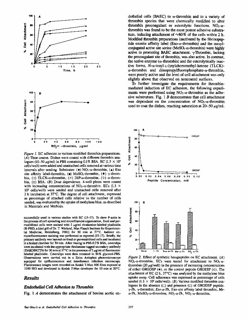
Figure 1. EC adhesion to various modified thrombin preparations. (A) Time course. Dishes were coated with different thrombin ana- logues (10-50/~g/ml) in PBS containing 0.1% BSA. EC (1.3 x l0 s cells/well) were added and unattached cells removed at various time intervals after seeding. Substrates: (e) NO2-ot-thrombin, (*) Exo site affinity label-thrombin, (-) MeSO2-thrombin, (*) ~-throm- bin, (n) TLCK-ot-thrombin, (x) DIP-u-thrombin, (<>) u-throm- bin, (o) BSA. (B) Dose dependence. 4-well plates were coated with increasing concentrations of NO2-c~-thrombin. ECs (1.3 x 10⁵ cells/well) were seeded and unattached ceils removed after 2 h incubation at 37°C. The degree of cell attachment, expressed as percentage of attached cells relative to the number of cells seeded, was evaluated by the uptake of methylene blue, as described in Materials and Methods.
successfully used in various studies with EC (15-17). To show F-actin in the process of cell spreading and microfilament organization, fixed and per- meabilized cells were stained with 2/~g/ml rhodamine-labeled phailoidin (R-PHD, a kind gift of Dr. T. Wieland, Max Planck Institute for Experimen- tal Medicine, Heidelberg, FRG) for 30 min at 37°C. Indirect im- munofluorescence staining was performed as reported (15-17). Briefly, the primary antibody was layered on fixed an permeabilized cells and incubated in a humid chamber for 30 rain. After riming in PBS-0.2% BSA, coverslips were incubated with the appropriate rhodamine-tagged secondary antibody (DAKOPATTS) for 30 pain at 37°C in the presence of 2/zg/ml of fluorescein- labeled phalloidin. Coverslips were then mounted in 50% glycerol-PBS. Observations were carried out in a Zeiss Axiophot photomicroscope equipped for epifluorescence and interference refection microscopy. Fluorescence images were recorded on Kodak T-Max 400 films exposed at 1000 ISO and developed in Kodak T-Max developer for 10 min at 200C.
Results
Endothelial Cell Adhesion to Thrombin
Fig. 1 A demonstrates the attachment of bovine aortic en-
dothelial cells (BAEC) to t~-thrombin and to a variety of thrombin species that were chemically modified to alter thrombin procoagulant or esterolytic functions. NO2-t~- thrombin was found to be the most potent adhesive substra- tum, inducing attachment of ,',,80% of the cells within 2 h. Modified thrombin preparations inactivated by the fibrinopep- tide exosite affinity label (Exo-a-thrombin) and the mesyl- conjugated active site serine (MeSO2-ot-thrombin) were highly active in promoting BAEC attachment. T-Thrombin, lacking the procoagulant site of thrombin, was also active. In contrast, the native enzyme (t~-thrombin) and the esterolytically inac- tive forms, N-t~-tosyl-L-lysylchoromethyl-ketone (TLCK)- u-thrombin and diisopropylfluorophosphate-ot-thrombin, were poorly active and the level of cell attachment was only slightly above that observed on noncoated surfaces.
To further investigate the requirements for thrombin- mediated induction of EC adhesion, the following experi- ments were performed using NO2-t~-thrombin as the adhe- sive substratum. Fig. 1 B demonstrates that cell attachment was dependent on the concentration of NO2-c~-thrombin used to coat the dishes, reaching saturation at 20-50/~g/ml.
100
" 80 E .1=
<
= 4 0
0
N 20
A
~ o - ~c~ GqGESP
Got~DSP 0 & • i • i
0 . 0 0 " 0 . ( ) 2 ' 0 . ( ) 4 • 0 . 0 6 • 0 , ( ) 8 0 . 1 0 0 . 1 2
Peptide Concentration, mM
100 -
c • 80. E
<
4 0 - ¢j
20"
oN None ¥ -Th IExo-cI-Th Me-~ -Th NO -~-Th
Figure 2. Effect of synthetic hexapeptides on EC attachment. (A) NO2-a-thrombin. ECs were tested for attachment to NO2-u- thrombin (20/~g/well) in the presence of increasing concentrations of either GRGDSP (o), or the control peptide GRGESP (o). The attachment of EC (2 h, 37°C) was analyzed by the methylene blue uptake assay. Cell adherence was expressed as percentage of cells seeded (1.3 x 1@ cells/well). (B) Various modified thrombin ana- logues in the absence (D) and presence (o) of GRGDSP peptide. T-Th, T-thrombin; Exo-o~-Th, Exo-site affinity label thrombin; Me- o~-Th, MeSO2-0t-thrombin; NO2-o~-Th, NO2-a-thrombin.
Bar-Shavit et al. Endothelial Cell Adhesion to Thrombin 337

~-S--S] I 42 48 16
-.. Fs--s7
169 182 185 190
" Loop B "
O CHO
, o ) 65
s I s I i
2,45 219
Figure 3. Schematic presentation of the human thrombin B-chain. Residues are aligned with residues 16-245 of bovine chymotryp- sinogen. Individual residues are identified by the single letter code, Tyr (Y) in hexagons. The carbohydrate (CHO) attachment at Asn 60E is assumed from the bovine enzyme (26). The disulfide bonds (S-S) are shown above the sequence line.
Effect of Synthetic Peptides on EC Attachment to NOz-~-thrombin
We next investigated the involvement of RGD in EC attach- ment to thrombin. For this purpose increasing concentra- tions (0.001-0.1 mM) of the synthetic hexapeptides GRGDSP or GRGESP were tested for their ability to inhibit EC attach- ment to NO2-o~-thrombin. While >95 % inhibition was ob- tained in the presence of 0.01 mlVl GRGDSP, no significant inhibition was observed with GRGESP (Fig. 2 A). Similarly, GRGDSP inhibited completely the attachment of EC to vari-
120
140
120 E J¢ 100 o ¢II
< 8o
60 o
4o
2o
NO~ i b c d A B C D
NO:~- : H i t NO~- : ATI I I
E F
Figure 5. Effect of thrombin inhibitors on EC attachment to NO2- ~x-thrombin. Dishes were coated with NO2-~x-thrombin (20/~g/ml, 0.57 nmol). Before coating, NO2-~-thrombin was incubated with increasing amounts of hirudin or ATIII (0.57-2.85 nmol) plus hepa- rin (0.3 U/ml), at a ratio of NO2-tx-thrombin:inhibitors of 1:1 (a, A), 1:2 (b, B), or 1:5 (c, C) for NO2-:Hir and NO2-:ATIII, re- spectively. Dishes were also coated with NO2-~x-thrombin (NO2-; 20/~g/ml, 0.57 nmol), hirudin (d; 20 ~g/ml, 2.85), ATIII (D; 171 /~g/ml, 2.85 nmol), fibronectin (E; 30 t*g/ml, 0.57 nmol), or fibronectin/ATIII (F; 30:171 #g/ml; 0.57:2.85 nmol). Attachment of EC was analyzed by the methylene blue uptake assay. Adhesion to NO2-ct-thrombin was regarded as 100%.
ous thrombin preparations (Fig. 2 B). These results indicate that the attachment activity of various modified thrombin preparations can be attributed to an RGD sequence in the protein. As demonstrated in Fig. 3, RGDA sequence in thrombin is located downstream at residues 187-190 of hu- man thrombin B-chain, when residues are aligned with residues 16-245 of bovine chymotrypsin (26). Other de- scribed functional domains in thrombin, outside the proteo-
,oo0
''0 0 !! ,o _ '. ' ° to ,o NO£ I I I I I I
-" -Thrombln
Figure 4. Effect of various thrombin fragments on EC attachment to NO2-u-thrombin. Attachment (2 h, 37°C) of EC to NO2-tx- thrombin coated dishes in the absence (NO2-) and presence of syn- thetic peptides (20/zg/ml) corresponding to the loop B mitogenic site (I, 14 amino acids; H, 10 amino acids; and III, 6 amino acids), Loop B (B), the chemotactic peptide CB67-120 (CB), and increas- ing concentrations of soluble tx-thrombin (a, 20/zg/ml; b, 10/zg/ml; c, 2/zg/ml; d, 0.2/zg/ml). The number of firmly attached ECs was determined by the methylene blue uptake assay. Attachment in the presence of each protein is expressed as percentage of the number of cells attached to NO2-tx-thrombin alone (= 100%).
NRS
1 : 2 0
0 None NO=- 1:100 1:75 1:50 1:25 1:20
Ab, D i lu t ion
Figure 6. Effect of antiprothrombin antibodies on EC attachment to NO2-oe-thrombin. Increasing amounts of antiprothrombin anti- bodies or normal rabbit serum (NRS) were incubated (2 h, 37°C) with ECs (1.3 x 10⁵ cells/well) in contact with NO2-ot-thrombin coated wells. Cells were also incubated in wells coated with NOT cx-thrombin alone (NO2-). Attachment of ECs was determined by the methylene blue uptake assay. 100% = adhesion to NO2-tx-
thrombin alone.
The Journal of Cell Biology, Volume 112, 1991 338

lyric pocket, are located upstream to the RGDA region in dis- tinct and separate regions. The Loop-B domain located between residues 60 and 61 within the variable region 2 (VR2) of thrombin B-chain, while the chemotactic pepride CB67-129 is located at residues 33-84 according to this num- bering system. When the synthetic peptide corresponding to Loop B mitogenic site, as well as various constructs of this region were tested for their ability to compete with EC at- tachment to NO2-o~-thrombin, only 10-20% inhibition was observed. Similarly, the thrombin-derived chemotacric frag- ment CB67-129 inhibited EC attachment to thrombin by ,~,15 %. In contrast, soluble cx-thrombin inhibited the attach- ment of EC to NO2-c~-thrombin in a dose-dependent man- ner (55 % inhibition at 20 #g/ml) (Fig. 4). This may indicate that the RGD domain in thrombin is only partially cryptic, and that additional neighboring sequences may assist in rec- ognition of this RGD sequence by the appropriate cell sur- face receptors. The low attachment-inducing activity of na- tive ot-thrombin (Fig. 1) does not appear to be due to inactivation as a result of coating on a solid support, since all the other modified thrombin preparations retained their cell attachment activity both when present in solution (com- petition experiments) or when adsorbed onto plastic or glass surfaces. Moreover, plasric-bound cx-thrombin exhibited other functional properties such as esterolytic activity, in- duction of platelet activation, and smooth muscle cell proliferation (data not shown).
Effect of Specific Thrombin Inhibitors on EC Attachment
The question arose as to whether thrombin when forming a complex with either its physiological inhibitor ATIII or with the leech-derived high affinity inhibitor hirudin, retains at- tachment activity. To test this possibility, equimolar amounts of thrombin and ATIII in the presence of 0.3 U/ml heparin as a catalyst were used to coat the dishes. As shown in Fig. 5, 45% inhibition of EC attachment to NO2-ot-thrombin was observed. This inhibition increased as the amount of ATIII increased, reaching 75 and 93 % inhibition at a ratio of 1:2 and 1:5 thrombin to ATIII, respectively. Under these conditions the amount of surface-adsorbed thrombin was not affected by the levels of ATm, as indicated by the similar amounts of bound ~zq-o~-thrombin (data not shown). The specificity of this ATIII-induced inhibition of EC attachment to NOz-ct-thrombin was demonstrated by a lack of inhibi- tion of EC adhesion to fibronectin, even at a ratio of 1:5 fibronectin/ATIII (Fig. 5). On the other hand, hirudin, the leech-derived high affinity inhibitor of thrombin, did not in- hibit EC attachment to NO2-ct-thrombin even at a ratio of 1:5 thrombin/hirudin (Fig. 5).
Anriprothrombin antibodies, which also recognize throm- bin, inhibited in a dose-dependent manner EC attachment to thrombin. As demonstrated in Fig. 6, complete inhibition was obtained at a 1:25 dilution of the antiserum, while there was no significant effect in the presence of preimmune se- rum. Th~s inhibition further demonstrates the specificity of EC attachment to thrombin.
Effect of NOz-a-Thrombin on EC Cytoskeletal Organization
As demonstrated in Fig. 7, NO2-ot-thrombin induced spread- ing of EC in a manner comparable to the subendothelial ex-
tracellular matrix. The distribution of the microfilamentous cytoskeleton and the localization of vinculin were studied by fluorescence microscopy on fixed and permeab'flized EC (Fig. 8). F-acrin was visualized by fluorochrome-labeled phal- loidin and vinculin was identified with a specific monoclonal antibody that recognizes the mammalian form of vinculin. The extent of adhesion was evaluated by IRM, a technique that records the distance between the adhesion substratum and the cell ventral membrane. The smaller the distance the darker the signal intensity, black indicating tight adhesion. As shown in Fig. 8, upon plating of EC on NO2-a-thrombin the cells spread and organized a network of thick microfilamentous bundles. Formation of stress fibers oc- curred with appearance of vinculin streaks at their endings in correspondence with focal contacts. These cells also showed multiple IRM black streaks that corresponded to the endings of stress fibers and distinct arrowhead-shaped vincu- lin speckles. Cell spreading and cytoskeletal organization on NO2-c~-thrombin was comparable to that observed upon seeding of EC on a vitronectin substratum (15). Immuno- fluorescence staining using anti-a, and anri-/~3 antibodies (Fig. 9) gave a peculiar pattern of oval and arrowhead-shaped spots usually located at stress fiber endings. The amount of B3 clusters was, however, reduced in comparison to EC ad- bering to vitronectin (Fig. 9). A comparable distribution was observed in studies with HUVEC using anti-a, (LM142) mAb (Fig. 9), or an anti-c~,/33 complex mAb (LM609), or anti-/~3 monoclonal and polyclonal antibodies (data not shown). On BAEC only the polyclonal anti-/~3 antibodies gave a good staining at adhesion plaques, while staining with the monoclonal antibodies used in this study was less easily detectable, possibly due to lack of cross-reactivity.
Anti-et,/~3 antibodies (LM609) blocked EC adhesion to NO2-c~-thrombin (Fig. 10) and subsequent ceil spreading (not shown). In contrast, the mAb BIE5 and an anti-o~sB~ goat antiserum were ineffective at concentrations that mark- edly inhibited EC adhesion to fibronectin. These results in- dicate that the ot,/~3 integrin is involved in EC adhesion to NO2-ot-thrombin.
We investigated the possible involvement of ECM proteins synthesized and secreted by EC during the process of cell adhesion to NOra-thrombin. For this purpose EC were treated with either emerine to block protein synthesis, or with monensin to prevent glycoprotein secretion. No sig- nificant effect on the extent of cell adhesion was observed with both types of treatments (data not shown).
Attachment of EC to Native a-Thrombin
To explore the possibility that under certain conditions (i.e., limited autoproteolysis) the RGD region of native ot-throm- bin, can be exposed and function in promoting cell adhesion, the enzyme was preincubated at 37°C before coating of plas- tic surfaces. Fig. 11 demonstrated that t~-thrombin that was first incubated at 37°C was a potent inducer of EC attach- ment as compared to lack of such an activity in the absence of preincubation. This effect was rather rapid since a sig- nificant attachment activity was observed even after a 15- min preincubation of o~-thrombin at 37°C. When the protease inhibitor TLCK was present during preincubation of throm- bin at 37°C, the ability of thrombin to induce EC attachment subsequent to coating of plastic surfaces was reduced by >65 % (data not shown). These data suggests that preincuba-
Bar-Shavit et al. Endothelial Cell Adhesion to Thrombin 339

Figure 7. Attachment of endothelial cells to NO2-c~-thrombin. Culture dishes were coated with 20 /Lg/ml of (a) NO2-ct-thrombin or (b) BSA. Dishes were also coated with (c) natu- rally produced subendothelial ECM. Phase micrographs were taken after 2-h incubation at 37°C with the cells. Bar, 100/~m.
The Journal of Cell Biology, Volume 112, 1991 340

Figure 8. HUVECs attach, spread, and organize focal contacts when plated on NO2-a-thrombin. Cell attachment was associated with (a) stress fiber formation, and (b) vinculin organization at stress fiber endings. These endings were found to correspond to dark spots in interference reflection microscopy (c). The arrow- heads in a, b, and c correspond to the position of a single focal con- tact. Bar, 10 #m.
tion at 37°C and the associated autoproteolysis of the throm- bin molecule may result in surface exposure of the RGD do- main in thrombin, which may hence mediate EC adhesion after adsorbance to the tissue culture plastic.
Discuss ion
Thrombin, the central bioregulatory enzyme in hemostasis, is actively engaged in governing thrombus formation. Dur- ing the initial phases of the hemostatic response after vessel injury, thrombin is generated to produce a fibrin network, promoting further fibrin dependent aggregation of platelets adherent to the exposed subendothelium (7, 33, 35, 41). These processes contribute to the formation of thrombus that initially seals the vessel to prevent excessive blood loss. Dur- ing subsequent wound healing, after vessel injury, ECs initi- ate local repair mechanisms involving cell attachment, migration, and proliferation to renew the damaged vessel. The adhesive phenotype of endothelial cells appears to be a critical factor regulating this repair process. Therefore, to understand the molecular events involved in thrombus for-
marion and wound healing, it is necessary to delineate struc- tural interactions between cells and molecules participating in these processes.
In this report, we present data showing that the RGD se- quence within thrombin promotes EC adhesion, spreading, and cytoskeletal reorganization. Modifications of the throm- bin molecule either at the procoagulant or catalytic site gave rise to species of thrombin that were highly adhesive for en- dothelial cells. This was found for thrombin that was catalyt- ically blocked by MeSO2 at the active site serine or by the fibrinopeptide exosite affinity label. ECs attached also to 3,-thrombin, exhibiting no procoagulant activity and a low proteolytic function. Furthermore, nitration of tyrosine resi- dues which yielded a reduced procoagulant activity, resulted in a maximal induction of EC attachment. Indeed, amino acid analysis shows the presence of a tyrosine residue located at position 185 of thrombin B-chain, preceding the only RGDA sequence in the protein (residues 186-189). This RGD sequence in thrombin is apparently hindered, located inwards towards the core of the enzyme and therefore is not exposed in the native molecule, which exhibits a very low at- tachment activity. In support of these findings are recent crystallographic studies indicating that the RGD domain is not accessible on the surface of the thrombin molecule (8). Upon specific modifications, however, this region may be ex- posed, for example, by nitration of the tyrosine residue adja- cent to the RGD sequence. This tyrosine (Tyr-185) appar- ently plays a significant role in altering the conformation of ct-thrombin, resulting in exposure of RGD sequence at an exosite domain. The degree of nitration (eight or three nitrotyrosines per mole) did not affect significantly the at- tachment-promoting activity of the modified thrombin (data not shown).
EC attachment was completely abolished in the presence of ATIZI, forming a complex with NO2-ot-thrombin, suggest- ing that ATIH masks the RGD region in thrombin. In contrast, hirudin, a leech-derived protein (Mr •7,000) with high affin- ity to thrombin, did not inhibit its attachment promoting ac- tivity. Hirudin has a negatively charged tall (1, 19, 34), thought to bind to a region neighboring the catalytic site located up- stream to the RGD sequence at the frontal side of thrombin. The RGD domain is therefore an unlikely candidate for inter- action with hirudin (21). Complexes of NO2-ot-thrombin and hirudin served as potent attachment substrates for EC, in correlation with the described thrombin-hirudin interac- tion regions (thrombin chemotactic and mitogenic domains) and the distinct RGD domain responsible for EC attachment. When anti-prothrombin antibodies were present during the adhesion assay, cell attachment was completely blocked, fur- ther reflecting the specific nature of EC-thrombin interac- tions.
EC spreading on NO2-o~-thrombin was followed by actin microfilament assembly, and appearance of interference reflective microscopy black streaks and vinculin speckles at stress fiber endings. Qualitatively, this type of cellular re- sponses were comparable to those observed after seeding the cells on a series of characteristic matrix proteins such as vitronectin, fibronectin, or von Willebrand factor (13, 15, 17, 18). This phenomenon is of particular relevance because it indicates that the plasma membrane has interacted with the substratum via specific receptors and that this interaction has triggered a cascade of events leading to the organization of
Bar-Shavit et al. Endothelial Cell Adhesion to Thrombin 341

Figure 9. Distribution of av receptors,/~3 receptors, arid F-actin HUVECs (a-f) and BAECs (g-j) were incubated (1.3 × 10 5 cells/well) for 4 h on coverslips coated with vitronectin (c, d, i, and j ) , NO2-.-thrombin (a, b, g, and h), or thrombin (e and f ) . The cells were permeabilized and stained with anti-c~vmAb (LM142) (b, d, and f ) , rabbit anti-GpIIIa antiserum (MAR-2) (h and j ) , or F-PHD (a, c, e, g, and 0. Plating of HUVECs on NO2-ct-thrombin supports spreading and stress fiber formation to a lesser extent than on vitronectin (compare a and c and b and d, respectively); txv clusters were fewer and more weakly stained, see arrowheads (b and d). In HUVECs the organization of/33 was comparable to that of uv (not shown). Plating on ~x-thrombin did not result in stress fiber formation or integrin receptor organization (e and f ) . BAECs plated on NO2-tx-thrombin (g and h) adhered but exhibited a bit less spreading and stress fiber formation than on vitronectin (i and j ) . Such reduced flattening was reflected in a less elaborate pattern of 133 receptor clustering and focal contact formation on NO2-tx-thrombin than on vitronectin (compare h and j ) . Bar, 10/~m.
The Journal of~Cell Biology, Volume 112, 1991 342

2oo 1 Figure 10. Effect of integfin re-
2~15°] ]l~! ~ ~ ~ ceptor antibodies on H U V E C f i b r o n e c t i n adhesion to different substrata. l oo 1 ~ r r -~ HUVEC were seeded on NO2-
a-thrombin (100 #g/ml; A); so~ (3 #g/ml; B); or o . vitronectin (3 #g/ml; C). Be-
A B c fore the adhesion assay HUVEC were incubated for
30 min at 37°C with: buffer (z~); otvB3 antibody, LM609 mAb; (a); a5 antibody, B1E5 mAb, (ta); asBl goat antiserum (t~). When goat nonimmune serum or mAb 10E3 were used, no change in cell adhesion was observed. Adhesion to BSA (1% in coating) was 7 + 1. Data are means + SEM of five replicates from one experi- ment out of four performed with comparable results.
adhesion plaques in which vinculin and other proteins are as- sociated. It is therefore likely that the effect of NO2-ot- thrombin on cell spreading and cytoskeletal organization is mediated by a specific recognition mechanism.
Inhibition studies revealed that an anti-txvl$3 mAb was able to block adhesion of EC to NO2-a-thrombin, while anti-asBl antibodies were ineffective. Vitronectin receptor (av~3) was also found to be clustered in focal contacts upon EC adhesion to NO2-a-thrombin. Altogether these data strongly suggest that a,B3 plays a leading role in mediating EC interaction with NO2-ot-thrombin. The vitronectin re- ceptor (39) is very promiscuous and recognizes, besides vitro- nectin, also other substrata including yon Willebrand factor, and fibrinogen (11, 32). The data reported here provide evi- dence that this receptor binds modified thrombin as well, thus identifying a novel ligand for this integrin. Seeding of EC on NO2-a-thrombin resulted in deposition of fibronec- tin that could influence cell adhesion. However, blocking en- dogenous matrix formation by treatment with inhibitors of protein synthesis and secretion (14, 40), had no effect on EC adhesion and spreading onto NO2-a-thrombin (data not shown). This indicates that NO2-a-thrombin itself could directly induce EC adhesion and cytoskeletal organization.
120 '
f" 100 ' o E .1=
o o
20'
None ~ - T h a b c
Prelncubated ~. -Th
Figure 11. Adhesion of EC to native a-thrombin. Culture dishes were coated with 20 #g/ml of a-thrombin (aTh), or with 20 #g/ml of thrombin that was first incubated in solution at 370C for 60 min (a); 30 rain (b); or 15 min (c). Cells were also incubated with dishes coated with BSA alone (None). The number of attached ECs was determined by the methylene blue uptake assay. 100% attach- ment represents the number of cells attached to thrombin that was preincubated at 37°C for 60 min. This value was similar to that ob- tained on NO2-a-thrombin coated dishes under the same condi- tions.
The biological relevance of the adhesive properties of modified thrombin molecules remains to be fully elucidated. The native enzyme has very little adhesive properties, de- spite having an RGD domain. However, it may still com- pete for adhesion to modified NO2-ot-thrombin (Fig. 4) and with other adhesive proteins, for example fibronectin (data not shown). The fact that thrombin molecule is composed of various functional domains some of which are exosite ex- posed regions and others like the RGD domain, are hin- dered, is intriguing. We have demonstrated that preincuba- tion of ot-thrombin at 37°C for a period of 15-60 min before coating of the plastic surface resulted in potent adhesive properties (Fig. 11). This could in part be due to autoproteol- ysis of the enzyme (as was indeed indicated by the inhibitory effect of TLCK) and/or to other structural modifications of the molecule occurring during incubation at 37°C. We postu- late that the RGD site can be surface exposed to response to specific modifications and signals which may be triggered under stress or pathological conditions. In would healing and inflammatory processes, extensive recruitment of leukocytes takes place. Moreover, it has been demonstrated that throm- bin that is ubiquitous to vascular injury, is a potent chemo- taxin for mononuclear phagocytes (3). Therefore, we suggest that during tissue repair when excess of mononuclear- phagocytic cells (highly rich in proteolytic enzymes) are present, these cells may act on thrombin in a manner that will modify the molecule and expose the RGD domain for direct participation in cellular attachment. The effect of various proteolytic enzymes (i.e., elastase, plasmin, cathepsin G, etc.) on thrombin attachment activity is under a current in- vestigation.
In conclusion, we present evidence that certain modifica- tions of thrombin may result in "pointing out" of its RGD re- gion that may then be actively engaged in supporting EC adhesion and cytoskeletal reorganization. We suggest that thrombin, in addition to playing a pivotal role in hemostasis, participates in repair mechanisms and maintenance of the integrity of the internal vessel lining by acting as a matrix protein for EC. Our data on the involvement of the otvB3 integrin in promoting EC adhesion to modified thrombin identify a new binding site for this molecule on EC.
This work was supported by GIF, The German-Israeli Foundation for Scientific Research and Development, the Israel Academy of Sciences (to R. Bar-Shavit), Public Health Service grant CA32089 (to I. Vlodavsky), and the National Research Council (Progetto FinalizTato =Biotecnologie e Biostrumentazione" and "Tecnologie Biomedicihe e Sanitarie"), and by the Italian National Council and the Israeli National Council for Research and Development (to E. Dejana).
Received for publication 23 July 1990 and in revised form 5 October 1990.
References
1. Bagdy, D., E. Barbas, L. Graft, T. E. Petersen, and S. Magnusson. 1976. Hirudin. Methods Enzymol. 45:669-678.
2. Barbieri, B., G. Balconi, E. Dejana, and M. B. Donati. 1981. Evidence that viLscular endothelial cell can induce the retraction of fibrin clots. Proc. Soc. Exp. Biol. Med. 168:204-207.
3. Bar-Shavit, R., A. Kahn, G. D. Wilner, and J. W. Fenton II. 1983. Mono- cyte chemotaxis: stimulation by specific exosite region in thrombin. Science (Wash. DC). 220:728-731.
4. Bar-Shavit, R., A. J. Kahn, K. G. Mann, and G. D. Wilner. 1986. Identification of a thrombin sequence with growth factor activity on mac- rophages. Proc. Natl. Acad. Sci. USA. 83:976-980.
5. Bar-Shavit, R., A. Eldor, and I. Vlodavsky. 1989. Binding of thrombin to subendothelial extracellular matrix: protection and expression of func-
Bar-Shavit et al. Endothelial Cell Adhesion to Thrombin 343

tional properties. J. Clin. Invest. 84:1096-1104. 6. Bar-Shavit, R., M. Benezra, A. Eldor, E. Hy-Am, J. W. Fenton II, G. D.
Wilner, and I. Vlodavsky. 1990. Thrombin-immobilized to extracellular matrix is a potent mitogen for vascular smooth muscle cells: non- enzymatic mode of action. Cell. Reg. 1:453--463.
7. Bennet, J. S., and G. Vilaire. 1979. Exposure of platelet fibrinogen recep- tors by ADP and epinephrine. J. Clin. Invest. 64:1393-1401.
8. Bode, W., I. Mayer, U. Baumaqn, R. Huber, S. R. Stone, and J. Hof- stcenge. 1989. The refined 1.9 A crystal structure of human ot-thrombin: interaction with D-Phe-Pro-Arg chloromethylketone and significance of the Tyr-Pro-Pro-Trp insertion segment. EMBO (Eur. Mol. Biol. Organ.) J. 8:3467-3475.
9. Carney, D. H., and D, D. Cunningham. 1978. Cell surface action ofthrom- bin is sufficient to initiate division of chick cells. Cell. 14:811-823.
10. Charo, I. F., L. S. Bekeart, and D. R. Phillips. 1987. Platelet glycoprotein Ilb-llIa-like proteins mediate endothefial cell attachment to adhesive pro- reins and extracellular matrix. J. Biol. Chem. 262:9935-9938.
11. Cheresh, D. A. 1987. Human endothelial cells synthesize and express an Arg-Gly-Asp directed adhesion receptor involved in attachment to fibrinogen and vun Willebrnnd factor. Proe. Natl. Acad. Sci. USA. 84:6471-6475.
12. Conforti, G., A. Zanetti, S. Colella, M. Abbadini, P. C. Marchisio, R. Pytela, P. G. Giancotti, G. Tarone, L. R. Langnino, and E. Dejana. 1990. Interaction of fibronectin with cultured human endothelial cells: characterization of the specific receptor. Blood. 73:1576-1585.
13. Dejana, E., S. Colella, L. R. Langnino, G. Balconi, G. C. Corbascio, and P. C. Marchisio. 1987. Fibrinogen induces adhesion, spreading, and microfilament organization of human endothelial cells in vitro. J. Cell Biol. 104:1403-1411.
14. Dejana, E., F. Bertocchi, M. C. Bortolami, A. Regonesi, A. Tontq, F. Breviario, and R. Giavazzi. 1988. Interleukin-1 promotes tumor cell adhesion to cultured human endothelial cells. J. Clin. Invest. 92:1466- 1470.
15. Dejana, E., S. Colella, G. Conforti, M. Abbadini, M. Gagoli, and P. C. Marchisio. 1988. Fibronectin and vitronectin regulate the organization of their respective .~rg-Gly-Asp adhesion receptors in cultured human en- dothelial cells. J. Cell Biol. 107:1215-1223.
16. Dejana, E., L. R. Langnino, S. Colella, G. C. Corbascio, E. Plow, M. Ginsberg, and P. C. Marchisio. 1988. The localization of platelet Gpl lb- IHa-related protein in endothelial cell adhesion structures. Blood. 71:566-572.
17. Dejana, E., M. G. Lampugnani, M. Giorgi, M. Gaboli, A. B. Federici, Z. M. Ruggeri, and P. C. Marchisio. 1989. Von Willehrand factor pro- motes endothelial cell adhesion via an Arg-Gly-Asp-dependent mecha- nism. J. Cell Biol. 109:365-375.
18. Dejana, E., M. G. Lampugnani, M. Giorgi, M. Gaboli, and P. C. Marchi- sio. 1990. Fibrinogen indices endothelial cell adhesion and spreading via the release of endogenous matrix proteins and the recruitment of more than one integrin receptor. Blood. 75:1509-1517.
19. Dodt, J., H.-P., Muller, V. Scemuller, and J.-Y. Chang. 1984. The com- plete amino acid sequence of hirudin, a thrombin specific inhibitor. FEBS (Fed. Fur. Biochem. Soc.) Lett. 165:180-183.
20. Engvall, E., and E. Ruoslahti. 1977. Binding of soluble form of fibroblast surface protein, fibronectin, to collagen. Int. J. Cancer. 20:1-5.
21. Fenton, J. W. II, and D. H. Bing. 1986. Thrombin active-site regions. Sere. Thromb. Haemostasis. 12:200-208.
22. Fenton, J. W. 1I, M. J. Fasco, A. B. Stackrow, A. L. Aronson, A. M. Young, and J. W. Finlayson. 1977. Human thrombins: production, evaluation and properties of c~-thrombin. J. Biol. Chem. 252:3587-3598.
23. Fenton, J. W. II, B. M. Landis, D. A. Walz, and J. S. Finlayson. 1977. Human thrombins. In Chemistry and Biology of Thrombin. R. L. Lund- bland, J. W. Fentun II, and K. G. Mann, editors. Ann Arbor Science, Ann Arbor, MI. 43-70.
24. Fitzgerald, L. A., I. F. Charo, and D. R. Phillips. 1985. Human and bovine endothelial cells synthesize membrane proteins similar to human platelets giycoproteins Fib and Ilia. Y. Biol. Chem. 260:10893-10896.
25. Pridman, R., Y. Alon, R. Doljansky, Z. Fnks, and I. Vlodavsky. 1985. Cell interaction with the extracellufar matrices produced by endothelial cells and fibroblasts. Exp. Cell Res. 158:462-476.
26. Furie, B., D. H. Bing, R. J. Feldmann, D.J. Robinson, J. P. Burnier, and B. C. Furie. 1982. Computer-generated models of blood coagulation factor Xa, factor IXa, and thrombin based upon structural homology with other serine proteases. J. Biol. Chem. 257:3875-3882.
27. Glenn, K. C., D. H. Carney, J. W. Fentun II, and D. D. Cunningham. 1980. Thrombin active site regions required for fibroblast receptor bind- ing and initiation of cell division. J. Biol. Chem. 252:6609-6616.
28. Goldfarb, R. H., and L. A. Liotta. 1986. Thrombin cleavage of extraceilu- lar matrix proteins. Bioregulatory functions of thrombin. Ann. NYAcad. Sci. 485:288-292.
29. Goldman, R., and Z. Bar-Shavit. 1979. Dual effect of normal and stimu- lated macrophages and their conditioned media on target cell prolifera- tion. J. Natl. Cancer Inst. 63:1004-1016.
30. Gospodarowicz, D., J. Moran, D. Braun, andC. R. Birdwell. 1976. Clonal growth of bovine vascular endothelial cells in tissue culture: fibroblast growth factor as a survival agent. Proc. Natl. Acad. Sci. USA. 73: 4120-4124.
31. Kaplan, K. 1982. Interactions of platelets with endothelial cells. In Pathobi- ology of the Endothelial Cell. H. L. Nossel and H. J. Vogel, editors. Aca- demic Press, Inc. New York. 337-349.
32. Lawler, J., R. Weinstein, and O. R. Hynes. 1988. Cell attachment to thrombosvondin: the role of Arg-Gly-Asp. calcium and integrin recep- tors. J. Cell Biol. 107:351-361.
33. Marguerie, G. A., E. F. Plow, and T. S. Edington. 1979. Human platelets possess an inducible and saturable receptor specific for fibrinogen. J. Biol. Chem. 254:5357-5363.
34. Markwardt, F. 1970. Himdin as an inhibitor of thrombin. Methods En- zymol. 19:924-932.
35. McLean, J. R., R. E. Maxwell, and D. Heftier. 1964. Fibrinogen and aden- osine diphosphate: induced aggregation of platelets. Nature (Lond.). 202: 605-606.
36. Perdue, J. F., W. Lubensky, E. Kivity, S. A. Sootier, and J. W. Penton II. 1981. Protease mitogenic response of chick embryo fibroblasts and receptor binding/processing of human c~-thrombin. J. Biol. Chem. 256:2767-2776.
37. Plow, E. F., E. G. Loftus, D. S. Levin, D. Dixon, J. Forsyth, and M. H. Ginsberg. 1985. Immunologic relationship between platelet membrane giycoprotein Gpllb-IIIa and cell surface molecules expressed by a variety of cells. Proc. Natl. Acad. Sci. USA. 83:6002--6006.
38. Romain, J., R. M. La Chance, T .J . Broekelmann, C. J. R. Kennedy, E. A. Wayner, W. G. Carter, andJ. A. McDonald. 1989. The fibronectin receptor is organized by extracellular matrix fibronectin: implications for oncogenic transformation and for cell recognition of fibronectin matrices. J. Cell Biol. 108:2529-2543.
39. Suzuki, S., W. S. Argraves, H. Arai, L. R. Languino, M. D. Piersch- bacher, and E. Ruoslahti. 1987. Amino acid sequence of vitronectin receptor subunits and comparative expression of adhesive receptor mRNA. J. Biol. Chem. 262:14080-14085.
40. Uchida, N., H. Smilowitz, and M. L. Tanzer. 1979. Monovalant iono- phores inhibit secretion of procollagen and fibronectin from cultured hu- man fihroblasts. Proc. Natl. Acad. Sci. USA. 76:1868-1872.
41. Weiss, H. J., and J. Rogers. 1971. Fibrinogen and platelets in the primary arrest of bleeding-studies in two patients with congenital afibrinogene- mia. N. Engl. J. Med. 285:369-374.
42. Yatohgo, T., M. Izumi, H. Kashiwagi, and M. Hayashi. 1988. Novel purification of vitronectin from human plasma by heparin affinity chroma- tography. Cell Struct. Funct. 13:281-292.
The Journal of Cell Biology, Volume 112, 1991 344