Potentiation of in vivo neuroprotection by BclXL and GDNF co-expression depends on post-lesion time...
10
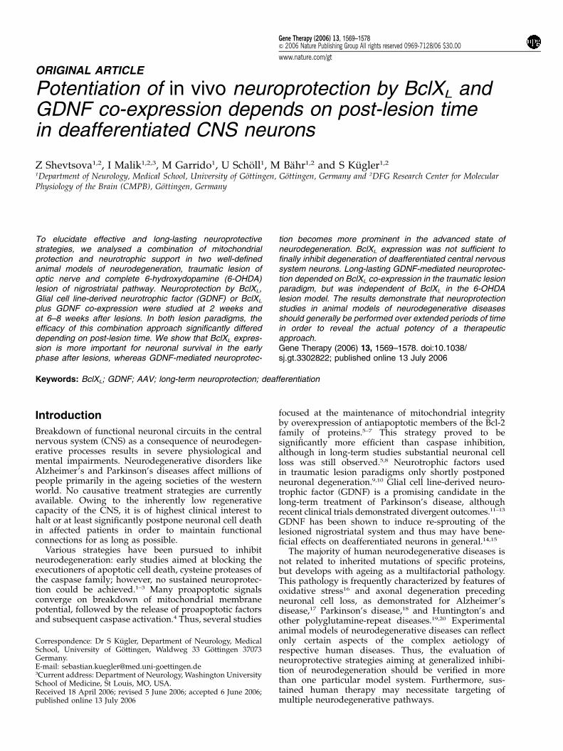
ORIGINAL ARTICLE Potentiation of in vivo neuroprotection by BclX L and GDNF co-expression depends on post-lesion time in deafferentiated CNS neurons Z Shevtsova 1,2 , I Malik 1,2,3 , M Garrido 1 , U Scho ¨ll 1 , M Ba ¨hr 1,2 and S Ku ¨ gler 1,2 1 Department of Neurology, Medical School, University of Go ¨ttingen, Go ¨ttingen, Germany and 2 DFG Research Center for Molecular Physiology of the Brain (CMPB), Go ¨ttingen, Germany To elucidate effective and long-lasting neuroprotective strategies, we analysed a combination of mitochondrial protection and neurotrophic support in two well-defined animal models of neurodegeneration, traumatic lesion of optic nerve and complete 6-hydroxydopamine (6-OHDA) lesion of nigrostriatal pathway. Neuroprotection by BclX L , Glial cell line-derived neurotrophic factor (GDNF) or BclX L plus GDNF co-expression were studied at 2 weeks and at 6–8 weeks after lesions. In both lesion paradigms, the efficacy of this combination approach significantly differed depending on post-lesion time. We show that BclX L expres- sion is more important for neuronal survival in the early phase after lesions, whereas GDNF-mediated neuroprotec- tion becomes more prominent in the advanced state of neurodegeneration. BclX L expression was not sufficient to finally inhibit degeneration of deafferentiated central nervous system neurons. Long-lasting GDNF-mediated neuroprotec- tion depended on BclX L co-expression in the traumatic lesion paradigm, but was independent of BclX L in the 6-OHDA lesion model. The results demonstrate that neuroprotection studies in animal models of neurodegenerative diseases should generally be performed over extended periods of time in order to reveal the actual potency of a therapeutic approach. Gene Therapy (2006) 13, 1569–1578. doi:10.1038/ sj.gt.3302822; published online 13 July 2006 Keywords: BclX L ; GDNF; AAV; long-term neuroprotection; deafferentiation Introduction Breakdown of functional neuronal circuits in the central nervous system (CNS) as a consequence of neurodegen- erative processes results in severe physiological and mental impairments. Neurodegenerative disorders like Alzheimer’s and Parkinson’s diseases affect millions of people primarily in the ageing societies of the western world. No causative treatment strategies are currently available. Owing to the inherently low regenerative capacity of the CNS, it is of highest clinical interest to halt or at least significantly postpone neuronal cell death in affected patients in order to maintain functional connections for as long as possible. Various strategies have been pursued to inhibit neurodegeneration: early studies aimed at blocking the executioners of apoptotic cell death, cysteine proteases of the caspase family; however, no sustained neuroprotec- tion could be achieved. 1–3 Many proapoptotic signals converge on breakdown of mitochondrial membrane potential, followed by the release of proapoptotic factors and subsequent caspase activation. 4 Thus, several studies focused at the maintenance of mitochondrial integrity by overexpression of antiapoptotic members of the Bcl-2 family of proteins. 5–7 This strategy proved to be significantly more efficient than caspase inhibition, although in long-term studies substantial neuronal cell loss was still observed. 5,8 Neurotrophic factors used in traumatic lesion paradigms only shortly postponed neuronal degeneration. 9,10 Glial cell line-derived neuro- trophic factor (GDNF) is a promising candidate in the long-term treatment of Parkinson’s disease, although recent clinical trials demonstrated divergent outcomes. 11–13 GDNF has been shown to induce re-sprouting of the lesioned nigrostriatal system and thus may have bene- ficial effects on deafferentiated neurons in general. 14,15 The majority of human neurodegenerative diseases is not related to inherited mutations of specific proteins, but develops with ageing as a multifactorial pathology. This pathology is frequently characterized by features of oxidative stress 16 and axonal degeneration preceding neuronal cell loss, as demonstrated for Alzheimer’s disease, 17 Parkinson’s disease, 18 and Huntington’s and other polyglutamine-repeat diseases. 19,20 Experimental animal models of neurodegenerative diseases can reflect only certain aspects of the complex aetiology of respective human diseases. Thus, the evaluation of neuroprotective strategies aiming at generalized inhibi- tion of neurodegeneration should be verified in more than one particular model system. Furthermore, sus- tained human therapy may necessitate targeting of multiple neurodegenerative pathways. Received 18 April 2006; revised 5 June 2006; accepted 6 June 2006; published online 13 July 2006 Correspondence: Dr S Ku ¨gler, Department of Neurology, Medical School, University of Go ¨ttingen, Waldweg 33 Go ¨ ttingen 37073 Germany. E-mail: [email protected] 3 Current address: Department of Neurology, Washington University School of Medicine, St Louis, MO, USA. Gene Therapy (2006) 13, 1569–1578 & 2006 Nature Publishing Group All rights reserved 0969-7128/06 $30.00 www.nature.com/gt
-
Upload
independent -
Category
Documents
-
view
0 -
download
0
Transcript of Potentiation of in vivo neuroprotection by BclXL and GDNF co-expression depends on post-lesion time...
ORIGINAL ARTICLE
Potentiation of in vivo neuroprotection by BclXL andGDNF co-expression depends on post-lesion timein deafferentiated CNS neurons
Z Shevtsova1,2, I Malik1,2,3, M Garrido1, U Scholl1, M Bahr1,2 and S Kugler1,2
1Department of Neurology, Medical School, University of Gottingen, Gottingen, Germany and 2DFG Research Center for MolecularPhysiology of the Brain (CMPB), Gottingen, Germany
To elucidate effective and long-lasting neuroprotectivestrategies, we analysed a combination of mitochondrialprotection and neurotrophic support in two well-definedanimal models of neurodegeneration, traumatic lesion ofoptic nerve and complete 6-hydroxydopamine (6-OHDA)lesion of nigrostriatal pathway. Neuroprotection by BclXL,Glial cell line-derived neurotrophic factor (GDNF) or BclXL
plus GDNF co-expression were studied at 2 weeks andat 6–8 weeks after lesions. In both lesion paradigms, theefficacy of this combination approach significantly differeddepending on post-lesion time. We show that BclXL expres-sion is more important for neuronal survival in the earlyphase after lesions, whereas GDNF-mediated neuroprotec-
tion becomes more prominent in the advanced state ofneurodegeneration. BclXL expression was not sufficient tofinally inhibit degeneration of deafferentiated central nervoussystem neurons. Long-lasting GDNF-mediated neuroprotec-tion depended on BclXL co-expression in the traumatic lesionparadigm, but was independent of BclXL in the 6-OHDAlesion model. The results demonstrate that neuroprotectionstudies in animal models of neurodegenerative diseasesshould generally be performed over extended periods of timein order to reveal the actual potency of a therapeuticapproach.Gene Therapy (2006) 13, 1569–1578. doi:10.1038/sj.gt.3302822; published online 13 July 2006
Keywords: BclXL; GDNF; AAV; long-term neuroprotection; deafferentiation
Introduction
Breakdown of functional neuronal circuits in the centralnervous system (CNS) as a consequence of neurodegen-erative processes results in severe physiological andmental impairments. Neurodegenerative disorders likeAlzheimer’s and Parkinson’s diseases affect millions ofpeople primarily in the ageing societies of the westernworld. No causative treatment strategies are currentlyavailable. Owing to the inherently low regenerativecapacity of the CNS, it is of highest clinical interest tohalt or at least significantly postpone neuronal cell deathin affected patients in order to maintain functionalconnections for as long as possible.
Various strategies have been pursued to inhibitneurodegeneration: early studies aimed at blocking theexecutioners of apoptotic cell death, cysteine proteases ofthe caspase family; however, no sustained neuroprotec-tion could be achieved.1–3 Many proapoptotic signalsconverge on breakdown of mitochondrial membranepotential, followed by the release of proapoptotic factorsand subsequent caspase activation.4 Thus, several studies
focused at the maintenance of mitochondrial integrityby overexpression of antiapoptotic members of the Bcl-2family of proteins.5–7 This strategy proved to besignificantly more efficient than caspase inhibition,although in long-term studies substantial neuronal cellloss was still observed.5,8 Neurotrophic factors usedin traumatic lesion paradigms only shortly postponedneuronal degeneration.9,10 Glial cell line-derived neuro-trophic factor (GDNF) is a promising candidate in thelong-term treatment of Parkinson’s disease, althoughrecent clinical trials demonstrated divergent outcomes.11–13
GDNF has been shown to induce re-sprouting of thelesioned nigrostriatal system and thus may have bene-ficial effects on deafferentiated neurons in general.14,15
The majority of human neurodegenerative diseases isnot related to inherited mutations of specific proteins,but develops with ageing as a multifactorial pathology.This pathology is frequently characterized by features ofoxidative stress16 and axonal degeneration precedingneuronal cell loss, as demonstrated for Alzheimer’sdisease,17 Parkinson’s disease,18 and Huntington’s andother polyglutamine-repeat diseases.19,20 Experimentalanimal models of neurodegenerative diseases can reflectonly certain aspects of the complex aetiology ofrespective human diseases. Thus, the evaluation ofneuroprotective strategies aiming at generalized inhibi-tion of neurodegeneration should be verified in morethan one particular model system. Furthermore, sus-tained human therapy may necessitate targeting ofmultiple neurodegenerative pathways.
Received 18 April 2006; revised 5 June 2006; accepted 6 June 2006;published online 13 July 2006
Correspondence: Dr S Kugler, Department of Neurology, MedicalSchool, University of Gottingen, Waldweg 33 Gottingen 37073Germany.E-mail: [email protected] address: Department of Neurology, Washington UniversitySchool of Medicine, St Louis, MO, USA.
Gene Therapy (2006) 13, 1569–1578& 2006 Nature Publishing Group All rights reserved 0969-7128/06 $30.00
www.nature.com/gt

We have recently demonstrated that overexpressionof BclXL efficiently prevented neurodegeneration in amodel of traumatic axonal lesion.5 While at 2 weeks afterlesion this neuroprotection appeared to be almostcomplete, there was still substantial neuronal loss at 8weeks after lesion, indicating that mitochondrial integ-rity alone was not sufficient for sustained inhibition ofdegeneration in deafferentiated CNS neurons. In thepresent study, we aimed at the inhibition of this ongoingdegeneration by the concomitant expression of theneurotrophin GDNF together with BclXL. We show thatalthough GDNF has little protective effect on axotomizedretinal ganglion cells on its own, it potentiates theprotective effect of BclXL. This synergistic effect wasdetectable only at 8 weeks after lesion. Attempting togeneralize these results in a different lesion model, weexpressed BclXL and GDNF in dopaminergic (DA)neurons of the substantia nigra. Severe oxidative stressvia injection of 6-hydroxydopamine (6-OHDA) into themedial forebrain bundle (MFB) was used to inducedegeneration of the nigrostriatal projections and of DAnigral neurons. In this paradigm, we detected additiveneuroprotective effects of BclXL and GDNF only at 2weeks, whereas at 6 weeks after lesion, BclXL no longercontributed to the effective and stable neuroprotectionachieved by GDNF.
Our results emphasize that combined application ofneuroprotective molecules enhances the potency ofneuroprotective gene transfer strategies. However, inboth lesion experimental paradigms investigations per-formed at later time points after lesion (6–8 weeks)revealed substantially different outcomes than thoseobtained at 2 weeks after lesion. We conclude thatstudies lasting for extended periods of time are neces-sary to verify the actual potency of neuroprotectiveapproaches.
Results
Efficient co-transduction of target neurons in retina andsubstantia nigra by two AAV-2 vectorsIn order to evaluate efficient and long-lasting neuropro-tective strategies, we aimed to express BclXL or GDNF, orBclXL plus GDNF in ganglion cells of the retina (RGCs)and in DA neurons of the substantia nigra pars compacta(SNpc). We constructed adeno-associated virus serotype2 (AAV-2) viral vectors expressing either BclXL or GDNFplus a fluorescent reporter protein (Enhanced greenfluorescence protein (EGFP) or DsRed2; Figure 1a).Fluorescent reporters simplified tracing of transducedneuronal populations. After intravitreal co-injection of1�107 transducing units (t.u.) of each AAV-BclXL-DsRedand AAV-GDNF-EGFP, we found efficient co-transduc-tion of RGCs in an area of 20–25% of the retina (Figures2a–c and 3a, b), with the vast majority of transduced cellsexpressing both fluorescent reporters at similar levels.Limiting transduction of RGCs to a relatively smallregion of the retina allowed us to quantify RGC numbersin vector-transduced and in non-transduced areas ofthe same retina (Figure 3a and b). After stereotaxicco-injection of 1.5� 108 t.u. of both vectors into thesubstantia nigra, we also detected highly efficient co-expression from both vectors (Figure 2d–f). Efficienttransduction of DA neurons was documented bystaining transduced sections for tyrosine-hydroxylaseimmunoreactivity, demonstrating almost complete trans-duction of DA neurons (Figure 2g–i).
GDNF expression is only marginally effective inprotecting retinal ganglion cells from axotomy-induceddegenerationRGCs are the only retinal population of neurons affectedimmediately by optic nerve axotomy. They specifically
Figure 1 AAV2 vectors and experimental schedules used in this study. (a) Schematic depiction of the AAV-2 vector genomes. TR, invertedterminal repeats; hSyn1, human synapsin 1 gene promoter; BclXL, rat BclXL; GDNF, human glial cell line-derived neurotrophic factor; EGFP,enhanced green fluorescent protein; DsRed, red fluorescent protein 2; Int, intron; SV40, SV40 polyadenylation site; TB, synthetic transcriptionblocker; WPRE, Woodchuck hepatitis virus post-transcriptional regulatory element; bGH, bovine growth hormone polyadenylation site. (b)Experimental schedule for axotomy studies given in weeks. FG, fluorogold; RGC, retinal ganglion cells. (c) Schematic schedule for 6-OHDAlesion experiments given in weeks. TH+, tyrosine hydroxylase-positive neurons in SNpc.
Synergistic neuroprotection by Bcl-XL and GDNFZ Shevtsova et al
1570
Gene Therapy

degenerate after this traumatic lesion, with only 10–15%of RGCs surviving the lesion after 14 days. In aprevious study, we have shown that BclXL expressionalmost totally inhibited this neuronal cell death up to14 days after lesion, but at later time points protectsonly a subset of RGCs, that is, 42% at 8 weeks afterlesion.5 In the present study, we aimed to improveneuroprotection by using a combination of mitochon-drial integrity protection through BclXL and neuro-trophic support through GDNF. To test for the efficacyof GDNF alone to protect RGCs from axotomy-induceddegeneration, we expressed GDNF in RGCs andquantified surviving RGCs at 2 and 8 weeks afteraxonal lesion (see Figure 1b for details of experimentalschedule and next paragraph for detailed quantificationprocedure). At 2 weeks after lesion, we counted 33%surviving RGCs upon GDNF expression, as comparedto 18% in controls, which were either counted in thenon-transduced areas of the same retina or in AAV-EGFP-transduced and axotomized retinas (Figure 4,Table 1). At 8 weeks after lesion, we detected onlymarginal neuroprotection of RGCs by GDNF expression(10% surviving RGCs after GDNF expression, 5% incontrols). Although its neuroprotective effect was signi-ficant, GDNF alone did not mediate efficient protectionfrom axotomy-induced neurodegeneration. However,as demonstrated for BclXL-expressing RGCs before,5
surviving RGCs in GDNF-transduced retinas main-tained their dendritic and axonal protrusions (Figure 3eand f).
Figure 2 Transduction properties of AAV-2 vectors in rat retina (a–c) and SNpc (d–i). Co-transduction of retina at 3 weeks after theinjection of 107 t.u. of each AAV-GDNF-EGFP (a) and AAV-BclXL-DsRed (b) is shown as an overlay in (c). Neurons in SNpc co-transduced by AAV-GDNF-EGFP (d) and AAV-BclXL-DsRed (e) areshown as a merged image in (f). Representative transduction ofSNpc DA neurons after injection of 1.5� 108 t.u. of AAV vector asdemarcated by EGFP fluorescence (g), where DA neurons arevisualized by TH immunoreactivity (h), is shown as an overlay in(i). Scale bar: 500 mm in (c), 100 mm in (f) and 400 mm in (i).
Figure 3 Retinal transduction by intravitreal application of 1�107 transducing AAV-2 transducing units. The retinal area typicallytransduced by 107 t.u. is shown in (a) as an overlay of EGFP fluorescence and phase contrast. (b) Schematic depiction of the areas in whichsurviving RGCs were counted (rectangles in either fully transduced or untransduced part of the same retina). Magnified images of the whole-mounted BclXL and GDNF-transduced retina at 8 weeks after axotomy show FG-labelled cells in (c) and vector-transduced cells detected byEGFP fluorescence in (d). Arrow points at a microglial cell that has taken up FG label by phagocytosis of RGCs. Black circle demarcates anEGFP-expressing amacrine cell not labelled by the RGC-specific FG fluorescence. FG-labelled RGCs display the typical punctate perinuclearfluorescence pattern. Only RGCs as seen in (c) or in inset of (h, k) were quantified for survival. A typical high-magnification image of an RGCtransduced by AAV-GDNF, as demarcated by EGFP fluorescence (e) co-localized with FG-labelling (f), showing preserved neurites at 2 weeksafter axotomy (white arrowheads). Representative FG-labelled areas of whole-mounted retinal specimens are shown at 2 weeks (g, h) and at 8weeks (j, k) after axotomy. FG fluorescence of the untransduced retinal regions is shown in (g) at 2 weeks and in (j) at 8 weeks after axotomy.In the areas of retina where RGCs are co-transduced with AAV-BclXL and AAV-GDNF, surviving RGCs are detected by FG-labelling at 2weeks (h) and at 8 weeks (k) after axotomy. The insets in (g–k) represent magnified images of the corresponding panel. Arrowheads point atRGCs, whereas arrows point at microglial cells distinguished by characteristic sigmoidal and ramified shape and uniform FG labelling. Scalebar: 500 mm in (a) and 100 mm in (g) – (k).
Synergistic neuroprotection by Bcl-XL and GDNFZ Shevtsova et al
1571
Gene Therapy

GDNF and BclXL act synergisticaly in long-termneuroprotection of axotomized retinal ganglion cellsDespite the low efficacy of the neurotrophin GDNF inpreserving RGCs from axotomy-induced degeneration,we reasoned that GDNF nonetheless may have atherapeutic effect in RGCs, which are basically pro-tected by BclXL expression. Co-expression of BclXL andGDNF resulted in a 93% protection of RGCs fromaxotomy-induced degeneration at 2 weeks after lesion(Figure 3h, Table 1). RGCs were unequivocally identi-fied by their punctuate perinuclear fluorogold (FG)labelling (Figure 3c and d). In whole-mounted retinas,
almost no infiltration of microglial cells was observedin transduced regions. In contrast, in non-transducedareas, the majority of FG has been incorporated intomicroglial cells, which have phagocytosed the degene-rating RGCs (Figure 3g). In microglial cells, the FG labelis uniformly distributed, and these cells are also clearlyidentified by their sigmoid shape and ramifications. At8 weeks after axotomy, numerous fluorescently labelledmicroglial cells were still obvious in the non-transducedcontrol regions of the retinas (Figure 3j), whereas onlyfew such cells were detectable in the retinal area inwhich RGCs expressed BclXL plus GDNF. Here, themajority of FG-labelled cells were clearly identified asRGCs (Figure 3k). Owing to the potent neuroprotectionmediated by BclXL alone (89% protected RGCs),neuroprotection achieved by BclXL plus GDNF co-expression could not be considered as a statisticallysignificant additive neuroprotective effect at 2 weeksafter lesion. However, at 8 weeks after lesion, we couldunequivocally document that BclXL and GDNF co-expression had synergistic (i.e., more than additive,Po0.001) neuroprotective effects. While BclXL or GDNFexpression alone rescued 42 or 10% of RGCs, respec-tively, co-expression prevented neurodegeneration in67% of RGCs (Figures 3k and 4, Table 1). Thus, BclXL-mediated neuroprotection decreased over time but waspotentiated by GDNF, although in a BclXL-dependentmanner.
Evaluation of BclXL plus GDNF co-expression in the6-OHDA-induced lesion of the nigrostriatal projectionHaving confirmed the efficacy of BclXL and GDNF co-expression in the axotomy model, we sought to genera-lize these results in a different lesion paradigm. 6-OHDAis rapidly metabolized to toxic oxidation products and iswidely used to induce degeneration of the nigrostriatalprojection followed by degeneration of nigral DAneurons. The most severe neurotoxic effects are obtainedby injection of 6-OHDA into the projection tract fromsubstantia nigra to basal ganglia, the MFB. This appli-cation mode results in a complete eradication of DAterminals in the striatum and in almost completedegeneration of the nigrostriatal projections and of nigralDA cell bodies and dendrites.
The schedule of the experimental outline is givenschematically in Figure 1c. Vectors were injected into the
Figure 4 Quantification of RGC survival after axotomy. RGCswere counted in retinas transduced by control vector AAV-EGFP(white bars), AAV-BclXL (light grey bars), AAV-GDNF (dark greybars) or AAV-BclXL plus AAV-GDNF (hatched bars) at 2 and 8weeks after optic nerve transection. Quantification of survivingRGCs is shown as RGCs/mm2 in the vector-transduced retinal area.RGC counts obtained for control vector AAV-EGFP in the vector-transduced retinal area after axotomy did not differ significantlyfrom RGC counts obtained in the non-transduced retinal areas. Starsabove each bar represent the significance level of respective vector-mediated protection as compared to the untransduced region of thesame retina. Note that RGC counts obtained in BclXL and GDNF-transduced retinas were significantly higher than the addition ofRGC counts obtained in BclXL-or GDNF-transduced retinas alone,as demarcated by the (+) symbol. Black bar represents RGC countsfrom unlesioned retinas. Bars presented as mean values7s.d.’s(***Po0.001, *Po0.05, NS, non significant).
Table 1 Quantification of retinal ganglion cells protected from axotomy (RGCs/mm2) and TH+ neurons protected from 6-OHDA toxicity(absolute numbers per SNpc)
AAV-BclXL Ctrl. area AAA-GDNF Ctrl. area AAV-Bcl-XL plus AAV-GDNF Ctrl. area
(a) RGCs/mm2 protected from axotomy (unlesioned¼ 2200780 RGCs/mm2)2 weeks 1963779 36879 720746 402746 2052774 3957598 weeks 931772 129732 226738 9779 1479781 73713
AAV-EGFP AAV-BclXL AAV-GDNF AAV-BclXL+AAV-GDNF Unlesioned
(b) Total number of TH+ neurons in SNpc protected from 6-OHDA toxicity2 weeks 27927301 69477783 86907998 108767392 1135675696 weeks 8417249 34967780 85987528 88407262 n.d.
Abbreviations: ctrl.area, RGC counts from untransduced parts of retinae; s.d.’s, standard deviations.Numbers presented as means7s.d.’s.
Synergistic neuroprotection by Bcl-XL and GDNFZ Shevtsova et al
1572
Gene Therapy

SNpc 3 weeks before stereotaxic application of 6-OHDAinto the MFB. At 2 weeks after 6-OHDA application,apomorphine-induced rotations were counted and it wasconfirmed by more than six contralateral rotations perminute that all operated animals had developed a
complete lesion of the nigrostriatal projections. Ratswere killed at either 2 or 6 weeks after lesion and showeda complete loss of DA terminals in the striatum asconfirmed by tyrosine-hydroxylase (TH)-immunostain-ing (Figure 5a and b).
Figure 5 TH immunoreactivity in rat brain after unilateral 6-OHDA lesion. Representative CPu (caudate–putamen) regions show the TH-IRprojections of the DA neurons (a) at 3 weeks after 6-OHDA injection into MFB and (b) on contralateral (unlesioned) side. The insets in (a) and(b) depict the magnified views on the TH+ fibre density in the corresponding areas. Immunohistochemical visualisation of surviving DA-neurons by TH-immunostaining in AAV-transduced SNpc (c, d, f, g, i, j, l, m) at 2 and 6 weeks after the MFB lesion compared to theunlesioned side of the same animal (e, h, k, n) at 6 weeks after the lesion. The AAV-EGFP-transduced SNpc is shown at 2 and 6 weeks after 6-OHDA lesion in (c) and (d). TH+ neurons transduced by AAV-BclXL are shown in (f), at 2 weeks after lesion, and in (g), at 6 weeks after lesion.The inset in (g) shows the predominant morphology of surviving DA neurons transduced by AAV-BclXL at 6 weeks after the lesion. Note thatmost cell bodies of the remaining TH+ neurons are devoid of processes. The AAV-GDNF-transduced nigrae are shown at 2 and 6 weeks afterthe 6-OHDA lesion in (i) and (j); the nigrae co-transduced with AAV-GDNF and AAV-BclXL at 2 and 6 weeks after the MFB lesion are shownin (l) and (m). The inset in (j) shows the morphology of the majority of surviving DA neurons in AAV-GDNF-transduced SNpc at 6 weeksafter the 6-OHDA lesion, whereas the inset in (k) depicts the native morphology of neurons in untreated SNpc. Note that the fibre density ofthe DA neurons protected by GDNF at 6 weeks after the 6-OHDA lesion (inset in (j)) is indistinguishable from that of unlesioned side (inset in(k)). Scale bar: 200 mm in (b) and 400 mm in (n).
Synergistic neuroprotection by Bcl-XL and GDNFZ Shevtsova et al
1573
Gene Therapy

BclXL temporarily protects DA cell bodies from6-OHDA toxicityIn animals injected with a control virus expressing onlyEGFP, 25% of nigral DA neurons survived the lesionfor 2 weeks, whereas only 7% survived for 6 weeks(Figures 5c, d and 6a, Table 1). Care was taken to excludeTH-stained neurons of the ventral tegmental area (VTA)from counting, as these neurons were not directlyaffected by the lesion. BclXL overexpression promotedsurvival of DA neurons to 61% at 2 weeks after lesionand to 31% at 6 weeks after lesion (Table 1). Importantly,the protective effect of BclXL was mainly restricted to thecell bodies of DA neurons, as the dense neuritic networkof these neurons present under normal conditions waslost to a large extent (Figure 5f and g). It should be notedthat the decrease in numbers of TH-positive neurons wasaccompanied by a respective decrease in numbers ofneurons showing EGFP fluorescence in both EGFP-andBclXL plus EGFP-transduced SNpc. These results indi-
cated that not only a downregulation of TH expressionbut a real loss of DA neurons occurred after 6-OHDAlesion in our paradigm. The rate of decrease in TH-positive neuron numbers in either BclXL plus EGFP-or in EGFP-transduced nigrae was similar, indicatingthat BclXL expression did not protect against the slowlyprogressive neurodegeneration at later stages after6-OHDA intoxication (Figure 6a, Table 1). These resultsshowed that no sustained neuroprotection could beachieved by Bcl-XL overexpression in this experimentalparadigm.
GDNF protects DA cell bodies and dendriticprotrusions from 6-OHDA toxicityAt 2 weeks after 6-OHDA lesion, DA neurons in GDNF-transduced SNpc were substantially protected fromdegeneration in that 77% survived at this time (Figure5i, Table 1). In contrast to BclXL-mediated neuroprotec-tion, surviving DA neurons showed a dense network of
Figure 6 Quantification of DA neuron survival in SNpc after 6-OHDA lesion. In (a) the number of surviving TH+ neurons is shown aftercontrol vector injection (AAV-EGFP) and after injection of AAV-BclXL, or AAV-GDNF, or AAV-BclXL plus AAV-GDNF. Dark-grey barsrepresent the mean values of surviving TH+ neurons in SNpc at 2 weeks after 6-OHDA lesion, and light-grey bars represent mean values ofsurviving TH+ neurons at 6 weeks after 6-OHDA lesion. The mean number of TH+ neurons in unlesioned SNpc is shown by black bar. Barsrepresent mean values7s.d.’s (***Po0.001, NS, non-significant). Immunohistochemical staining for BclXL in SNpc neurons transduced byAAV- BclXL confirmed that BclXL expression is not downregulated by 6-OHDA application (b–d). Immunohistochemical staining for BclXL
was also used to demonstrate that GDNF expression did not result in BclXL upregulation in SNpc neurons after AAV-GDNF transduction (e–g). Scale bar: 50 mm.
Synergistic neuroprotection by Bcl-XL and GDNFZ Shevtsova et al
1574
Gene Therapy

dendritic arborizations throughout the SNpc, indicatingthat GDNF not only protects DA cell bodies but also theirprocesses from 6-OHDA toxicity. At 6 weeks after6-OHDA lesion, no significant decrease in TH-positivecell numbers has taken place (76% surviving DAneurons; Figure 5j, Table 1), indicating that GDNF caninhibit and not only postpone 6-OHDA-induced neuro-degeneration.
As it was demonstrated earlier, high levels of GDNFexpression may result in disturbances of DA metabolismand aberrant sprouting of DA fibres.21 Therefore, theGDNF expression cassette used in our vector constructdid not contain transcriptional control elements, whichstrongly enhance transgene expression, such as theWPRE. GDNF expression levels in AAV-EGFP- and inAAV-GDNF-transduced SNpc as quantified by enzyme-linked immunosorbent assay (ELISA) were 11 and190 pg/mg, respectively. Thus, GDNF levels obtainedfrom AAV-GDNF in this study were approximately 10times lower than the GDNF levels shown to induceabberant sprouting of DA fibres in a recent study.21
BclXL and GDNF co-expression has additiveneuroprotective effects at 2 weeks after6-OHDA lesionCo-expression of BclXL and GDNF resulted in almostcomplete protection of SNpc DA neurons from 6-OHDAtoxicity at 2 weeks after lesion: 96% of DA neuronssurvived at this time (Figures 5l and 6a, Table 1). Whencompared to 77% surviving neurons through theexpression of GDNF alone and 61% surviving neuronsthrough the expression of BclXL alone, these datarepresent a statistically significant additive neuroprotec-tive effect (Po0.001; Figure 6a). Moreover, no obviousmorphological differences were detected between unle-sioned control DA neurons and DA neurons expressingBclXL plus GDNF (Figure 5l–n).
Co-expression of BclXL with GDNF has no beneficialeffect on DA neuron survival at 6 weeks after6-OHDA lesionAlthough BclXL expression alone still resulted insignificant neuroprotection from 6-OHDA toxicity at6 weeks after lesion, its co-expression with GDNF at thistime did not result in any additive neuroprotective effect.It rather appeared that neuroprotection at this time wasexclusively mediated by GDNF, as the numbers of TH-positive neurons in the GDNF group were identical tothe numbers of TH-positive neurons in the GDNF plusBclXL group (Figures 5m and 6a, Table 1). In order toexclude any effect of 6-OHDA on BclXL expression, AAV-BclXL-transduced nigrae were immunohistochemicallystained for BclXL expression. In AAV-Bcl-XL-transducedDA-neurons surviving for up to 6 weeks after 6-OHDAintoxication, we detected the same level of cellularimmunoreactivity for BclXL as in unlesioned controlsthat received AAV-BclXL but were not subjected to6-OHDA lesion (Figure 6b–d). In contrast, we could notdetect any obvious upregulation of BclXL (Figure 6e–g)or Bcl-2 expression (not shown) in AAV-GDNF-transduced DA-neurons. These latter results demon-strate that the long-lasting neuroprotective effect ofGDNF on survival of DA neurons was independentfrom BclXL.
Discussion
Neurodegenerative diseases like Alzheimer’s andParkinson’s diseases are characterized by slow andmultifactorial progression, and potential therapeuticintervention is difficult owing to diagnosis at relativelylate stages of life. After diagnosis, patients have one ormore decades of life expectancy, which would necessitatea long-lasting therapeutical approach in order to halt orat least significantly postpone neurodegeneration. Anypotential therapeutic benefit should thus be confirmed tobe effective over extended periods of time. Furthermore,the multifactorial nature of neurodegeneration in itslater stages suggests that therapeutic approaches target-ing multiple neurodegenerative pathways might benecessary.
In many neurodegenerative diseases, axonal damageprecedes the loss of neuronal cell bodies.17–20 Thisprinciple is at least partially recapitulated in the twoanimal models used in this study: both traumaticallylesioned RGCs and 6-OHDA lesioned nigrostriatalneurons of SNpc are primarily affected through axonaldamage, resulting in complete deafferentiation. Thus, the6-OHDA lesion as used in this study is consideredprimarily a model of neuronal deafferentiation ratherthan a model of Parkinson’s disease. Both lesion para-digms affected specific CNS neuron populations, whichwere selectively transduced by recombinant AAV vectorsin order to express therapeutically active proteins.
Our data demonstrate that:
(i) In both lesion paradigms, BclXL overexpression wasnot sufficient to finally preserve neurons fromdegeneration, with a 50% decrease in numbers ofsurviving neurons at 6–8 weeks after lesions ascompared to 2 weeks after lesions. Thus, main-tenance of mitochondrial integrity may be essentialfor neuronal survival early after lesion, butappeared not to be sufficient for long-term survivalof deafferentiated CNS neurons.
(ii) When used in combination, BclXL and GDNFprovided almost complete neuroprotection in bothlesion paradigms at 2 weeks after lesion. This effectwas clearly additive in the 6-OHDA model, indicat-ing the important role for targeting multiple path-ways in neurodegenerative diseases.
(iii) Only at later time points after lesions (at 6–8 weeks)it was possible to demonstrate that BclXL andGDNF exerted synergistic neuroprotective effectsin the traumatic lesion paradigm, but that no suchpotentiative effect was detectable in the oxidativestress lesion paradigm. These data highlight thefact that the actual potency of a neuroprotectiveapproach may be overlooked if the study designdoes not allow for long-term analysis.
Our results obtained in short-term experiments (i.e.analysis of neuronal survival at 2 weeks post-lesion time)correspond to previous studies demonstrating that acombination of expression of GDNF with the antiapop-totic Bcl-2 or X-linked inhibitor of apoptosis (XIAP)proteins may have additive neuroprotective effects.22–25
However, Natsume et al.22,23 overexpressed GDNF andBcl-2 in lesion paradigms, which result in significantlyless severe impact on the affected neuronal populations.Furthermore, these investigations were not extended to
Synergistic neuroprotection by Bcl-XL and GDNFZ Shevtsova et al
1575
Gene Therapy

long-lasting post-lesion times than 2 weeks, and thuscould not demonstrate whether Bcl-2 and GDNF expres-sion may have any potentiating neuroprotective effect inlater stages of neurodegeneration. The same issue holdstrue for the co-expression of GDNF with the caspaseinhibitor XIAP,24,25 which was analysed at 2 weeks afterlesions only. In addition, in these latter studies adeno-viral vectors have been used, which could not restricttransgene expression to the target cell populations.
In our present study, we have evaluated the neuro-protective properties of BclXL instead of Bcl-2 for tworeasons: (i) BclXL has been shown to be more potent ininhibiting mitochondrial membrane potential break-down than Bcl-2,26 probably owing to its efficienttargeting to mitochondria.27 (ii) BclXL but not Bcl2 caninhibit tumour necrosis factora-activated TRAIL-mediated apoptotic and non-apoptotic cell death (necro-sis and autophagy),28,29 which play important roles inseveral neurodegenerative paradigms.30–32 Accordingly,in both model systems evaluated in this study, BclXL plusGDNF co-expression resulted in the most effectiveprevention of neurodegeneration reported so far.
While the mode of action of BclXL is well defined,26
this does not hold true for GDNF. GDNF may exert itsbeneficial effects through several receptor systems,including its prototype receptors GDNF family receptor1 (GFRa) and Ret, but also neural cell adhesion molecule(NCAM) and heparansulphate-proteoglycans like syn-decan-3.33 The low neuroprotective efficacy of GDNFexpression alone on axotomized RGCs may be explainedby the fact that only a small subset of RGCs expressesGFRa and Ret in the adult retina.34 After axonal lesion,an upregulation of GFRa on RGCs has been reported.34
Thus, RGCs initially protected from degeneration byBclXL expression may become more responsive to GDNFat later time points after lesion, resulting in synergisticneuroprotection depending on receptor upregulation inadvanced stages of neurodegeneration. DA neurons ofthe SNpc express high levels of GDNF receptors,35 andthus may respond to the neurotrophin without a delay.The stable and sustained protection of DA neurons andthe potentiation of neuroprotection in axotomized RGCsby GDNF suggest that this molecule may serve as apotent neuroprotective agent in different paradigms ofneurodegeneration, provided that appropriate receptorsystems are present in affected neurons.
Taken together, we demonstrate that a combinationapproach of targeting different pathways of neurodegen-eration simultaneously was highly effective over longterm in the traumatic axonal lesion model, but onlyfor limited time in the oxidative stress lesion model.Importantly, the actual potency of the therapeuticapproach could only be revealed at late time points afterlesions in both experimental paradigms, suggesting thatstudies aiming to define neuroprotective strategiesshould be generally performed over extended periodsof time. These results should have important implicationsfor the design of therapeutic neuroprotective studies.
Materials and methods
AAV-2 vectorsViral vectors based on the AAV-2 were constructed withbi-cistronic expression cassettes as described.36 Vector
genomes contained the rat cDNA for either BclXL or forGDNF pre–pro-peptide as functional transgenes and inaddition a fluorescent reporter gene (EGFP or DsRed2,Clontech, Heidelberg, Germany). All transgenes wereexpressed from independent human synapsin 1 genepromoters (Figure 1a). Vectors were propagated in 293cells using pDG as helper plasmid,37 and purified byiodixanol step gradient centrifugation and subsequentfast protein liquid chromatography (FPLC) on an Akta-FPLC system equipped with heparin-affinity columns(Amersham, Freiburg, Germany). After dialysis, genometitres were determined by quantitative PCR, purity bysodium dodecyl sulphate-gel electrophoresis andinfectious titres by transduction of cultured primaryhippocampal neurons. Genome particles to t.u. ratio was25:1–35:1.
AnimalsAdult female Wistar rats (250–280 g) were housed at221C, five per cage, under standard conditions witha 12:12-h dark and light cycle. For all invasive proce-dures, rats were anaesthetized with chloral hydrate(0.34 mg/kg body weight). For transcardial perfusions,animal were deeply anaesthetized with a lethal overdoseof chloral hydrate (0.9 mg/kg body weight). All animalprocedures were performed in accordance to Germanguidelines for care and use of laboratory animals and tothe respective European Community Council Directive86/609/EEC.
Each experimental group consisted of five animals.AAV-EGFP-injected animals were used as controls for allexperimental groups. In the retina, non-transducedregions were additionally used as an internal control.RGC counts obtained for the control vector-AAV-EGFP inthe vector transduced retinal area after axotomy didnot differ significantly from RGC counts obtained in thenon-transduced retinal areas.
Surgical proceduresIntravitreal vector injections, unilateral axotomy of theoptic nerve, and retrograde FG labelling of retinalganglion cells (RGCs) was performed essentially asdescribed.5 Transduction of 20–25% of the retinal areawas achieved by injection of a small volume of vectorat low titre (2 ml containing 1�107 t.u.) without priorremoval of fluid from the intravitreal space. Thisapplication mode resulted in reduced spread of the virusin the intravitreal space and RGC transduction in alimited area only. It thus allowed for an internal controlof neuroprotection in the same retinal specimen (RGCcounts in transduced versus non-transduced areas; seeFigure 3b) and for clearcut visualization of the axons oftransduced RGCs projecting towards the optic disc.
Vector injections into SNpc were performed usinga Kopf stereotaxic device by a computer controlledmicroinjector pump (World Precision Instruments, Ber-lin, Germany). Coordinates were AP: �0.53; ML: +0.22;DV: �0.78, relative to bregma. A fine bore glass capillary(30 mm tip diameter) was used to deliver the viralsuspension of 1.5� 108 t.u. of each vector in a 2 mlvolume at 400 nl/min. The capillary was kept at theinjection site for 2 min before and 4 min after theinjection. No alterations in numbers of TH-positiveneurons were observed after application of the variousvectors as compared to the contralateral non-injected
Synergistic neuroprotection by Bcl-XL and GDNFZ Shevtsova et al
1576
Gene Therapy
nigra. 6-OHDA lesion was performed at 3 weeks aftervector inoculations by stereotaxically injecting the toxin(3 ml of freshly prepared 5 mg/ml in 0.2% ascorbic acid)into the ipsilateral MFB at coordinates AP: �0.2; ML:+0.2; DV: �0.85, relative to bregma. In order to confirmthat the lesion was complete, we used apomorphineinduced rotational behaviour test in all animal groups at2 weeks after 6-OHDA application. Apomorphine wasinjected intraperitoneally at 0.2 mg/kg of body weight.Number of clockwise vs counter-clockwise rotationswas counted at a rotational behaviour monitor (Stoelting)for 45 min.
GDNF – ELISAQuantification of GDNF tissue levels in transduced andcontralateral SNpc was determined by ELISA (GDNFEmax Immunoassay System, Promega, Mannheim,Germany) according to the manufacturer’s instructions.Tissue samples of SNpc were obtained from 2-mm -thicktissue slices by means of small biopsy needles with innerdiameter of 2 mm.
Quantification of neuroprotection in axotomy lesionAt the respective time points after axotomy, rats (n¼ 5per condition) were killed by an overdose of chloralhydrate and transcardially perfused with 4% parafor-maldehyde (PFA). Eyes were removed, post-fixed for 1 hin 4% PFA, and then retinas were whole-mounted onobject slides embedded in Moviol. In the centre of thevector-transduced retinal area (as identified by EGFP-and/or DsRed-expressing RGCs) at 2/3 retinal radiusRGCs were counted in three non-overlapping 1 mm2
areas essentially as described.5
Quantification of neuroprotection in 6-OHDA lesionAt the respective time points after 6-OHDA injectioninto MFB, rats (n¼ 5 per condition) were killed by anoverdose of chloral hydrate and transcardially perfusedwith 4% PFA. Brains were cut on a cryostat to 18-mm-thick coronal sections, which were stored at �801C untilprocessing. After antigen retrieval (incubation in TBS(tris-buffered saline; 20 mM Tris, pH 9.0, 136 mM NaCl;at 601C for 6 h), immunofluorescent visualization ofDA neurons in SNpc was performed with primaryrabbit antibody against tyrosine hydroxylase (anti-TH,1:500, Advanced ImmunoChemicals Inc., Long Beach,CA, USA; overnight incubation at 41C) followed by1 h incubation at room temperature with secondaryCy3-conjugated goat anti-rabbit antibody (Dianova,Hamburg, Germany). TH-immunoreactive (TH-IR) cellswere counted in every eighth brain section in the regionanatomically correspondent to SNpc. For the unlesionedcontrol, TH-IR cells in SNpc were counted on thecontralateral to the lesion side. TH-IR cells of the VTAarea were excluded from counting. To obtain thecorresponding total DA cell numbers, the counted valueswere multiplied by 8, assuming that the averagediameter of the adult rat DA-neuron is 16–18 mm.38
StatisticsRGC and TH-IR cell counts are presented asmeans7s.d.’s. For each time point and experimentalcondition, n¼ 5 were used. For retinas, three indepen-dent fields per retina were counted, and for TH-IR cellsof SNpc region at least 13 sections were counted per
animal. Statistical significance was assessed by split-plotanalysis of variance and alpha-adjusting for multiplicity(Bonferroni–Holm sequentially rejective procedure) and,at the 1% level, revealed Po0.001 for either BclXL-or GDNF-transduced areas vs untransduced and vs thecontrol AAV-EGFP-transduced SNpc at all time points.Analysis of variance (factor 1 – BclXL; factor 2 – GDNF;interaction 1/2¼BclXL+GDNF) revealed that interactionwas not additive (Po0.001), but more than additive(¼ synergistic) for retina, but not for the SNpc, where itwas additive at 2 weeks, but not significant at 6 weeksafter lesion when compared to the GDNF-treated group.Pairwise comparisons of values were performed bypaired, one-tailed Student’s t-test.
Acknowledgements
This work was funded by Deutsche Forschungsge-meinschaft through the DFG-Research Center ‘MolecularPhysiology of the Brain’. MG holds a studentship fromEC Marie Curie Actions RTN-NSR #504636.
References
1 Kermer P, Klocker N, Bahr M. Long-term effect of inhibition ofced 3-like caspases on the survival of axotomized retinalganglion cells in vivo. Exp Neurol 1999; 158: 202–205.
2 Perrelet D, Ferri A, MacKenzie AE, Smith GM, Korneluk RG,Liston P et al. IAP family proteins delay motoneuron cell deathin vivo. Eur J Neurosci 2000; 12: 2059–2067.
3 Rideout HJ, Stefanis L. Caspase inhibition: a potential therapeu-tic strategy in neurological diseases. Histol Histopathol 2001; 16:895–908.
4 Chang LK, Putcha GV, Deshmukh M, Johnson Jr EM. Mitochon-drial involvement in the point of no return in neuronalapoptosis. Biochimie 2002; 84: 223–231.
5 Malik JM, Shevtsova Z, Bahr M, Kugler S. Long-term in vivoinhibition of CNS neurodegeneration by Bcl-XL gene transfer.Mol Ther 2005; 11: 373–381.
6 Wong LF, Ralph GS, Walmsley LE, Bienemann AS, Parham S,Kingsman SM et al. Lentiviral-mediated delivery of Bcl-2 orGDNF protects against excitotoxicity in the rat hippocampus.Mol Ther 2005; 11: 89–95.
7 Azzouz M, Hottinger A, Paterna JC, Zurn AD, Aebischer P,Bueler H. Increased motorneuron survival and improvedneuromuscular function in transgenic ALS mice after intraspinalinjection of an adeno-associated virus encoding Bcl-2. Hum MolGenet 2000; 9: 803–811.
8 Kim R, Emi M, Tanabe K. Role of mitochondria as the gardens ofcell death. Cancer Chemother Pharmacol 2005: 1–9.
9 Cheng L, Sapieha P, Kittlerova P, Hauswirth WW, Di Polo A.TrkB gene transfer protects retinal ganglion cells from axotomy-induced death in vivo. J Neurosci 2002; 22: 3977–3986.
10 van Adel BA, Kostic C, Deglon N, Ball AK, Arsenijevic Y.Delivery of ciliary neurotrophic factor via lentiviral-mediatedtransfer protects axotomized retinal ganglion cells for anextended period of time. Hum Gene Ther 2003; 14: 103–115.
11 Gill SS, Patel NK, Hotton GR, O’Sullivan K, McCarter R,Bunnage M et al. Direct brain infusion of glial cell line-derivedneurotrophic factor in Parkinson disease. Nat Med 2003; 9:589–595.
12 Nutt JG, Burchiel KJ, Comella CL, Jankovic J, Lang AE, Laws ERet al. Randomized, double-blind trial of glial cell line-derivedneurotrophic factor (GDNF) in PD. Neurology 2003; 60: 69–73.
Synergistic neuroprotection by Bcl-XL and GDNFZ Shevtsova et al
1577
Gene Therapy

13 Patel NK, Bunnage M, Plaha P, Svendsen CN, Heywood P, GillSS. Intraputamenal infusion of glial cell line-derived neuro-trophic factor in PD: a two-year outcome study. Ann Neurol 2005;57: 298–302.
14 Love S, Plaha P, Patel NK, Hotton GR, Brooks DJ, Gill SS. Glialcell line-derived neurotrophic factor induces neuronal sproutingin human brain. Nat Med 2005; 11: 703–704.
15 Bjorklund A, Rosenblad C, Winkler C, Kirik D. Studies onneuroprotective and regenerative effects of GDNF in a partiallesion model of Parkinson’s disease. Neurobiol Dis 1997; 4:186–200.
16 Tretter L, Sipos I, Adam-Vizi V. Initiation of neuronal damage bycomplex I deficiency and oxidative stress in Parkinson’s disease.Neurochem Res 2004; 29: 569–577.
17 Stokin GB, Lillo C, Falzone TL, Brusch RG, Rockenstein E,Mount SL et al. Axonopathy and transport deficits early inthe pathogenesis of Alzheimer’s disease. Science 2005; 307:1282–1288.
18 Dauer W, Przedborski S. Parkinson’s disease: mechanisms andmodels. Neuron 2003; 39: 889–909.
19 Gunawardena S, Goldstein LS. Polyglutamine diseases andtransport problems: deadly traffic jams on neuronal highways.Arch Neurol 2005; 62: 46–51.
20 Li H, Li SH, Yu ZX, Shelbourne P, Li XJ. Huntingtin aggregate-associated axonal degeneration is an early pathological event inHuntington’s disease mice. J Neurosci 2001; 21: 8473–8481.
21 Georgievska B, Kirik D, Bjorklund A. Aberrant sprouting anddownregulation of tyrosine hydroxylase in lesioned nigrostriataldopamine neurons induced by long-lasting overexpression ofglial cell line derived neurotrophic factor in the striatum bylentiviral gene transfer. Exp Neurol 2002; 177: 461–474.
22 Natsume A, Mata M, Wolfe D, Oligino T, Goss J, Huang S et al.Bcl-2 and GDNF delivered by HSV-mediated gene transfer afterspinal root avulsion provide a synergistic effect. J Neurotrauma2002; 19: 61–68.
23 Natsume A, Mata M, Goss J, Huang S, Wolfe D, Oligino T et al.Bcl-2 and GDNF delivered by HSV-mediated gene transferact additively to protect dopaminergic neurons from 6-OHDA-induced degeneration. Exp Neurol 2001; 169: 231–238.
24 Eberhardt O, Coelln RV, Kugler S, Lindenau J, Rathke-Hartlieb S,Gerhardt E et al. Protection by synergistic effects of adeno-virus-mediated X-chromosome-linked inhibitor of apoptosisand glial cell line-derived neurotrophic factor gene transferin the 1-methyl-4-phenyl-1,2,3,6-tetrahydropyridine model ofParkinson’s disease. J Neurosci 2000; 20: 9126–9134.
25 Straten G, Schmeer C, Kretz A, Gerhardt E, Kugler S, Schulz JBet al. Potential synergistic protection of retinal ganglion cellsfrom axotomy-induced apoptosis by adenoviral administrationof glial cell line-derived neurotrophic factor and X-chromosome-linked inhibitor of apoptosis. Neurobiol Dis 2002; 11: 123–133.
26 Kim R. Unknotting the roles of Bcl-2 and Bcl-xL in cell death.Biochem Biophys Res Commun 2005; 333: 336–343.
27 Kaufmann T, Schlipf S, Sanz J, Neubert K, Stein R, Borner C.Characterization of the signal that directs Bcl-x(L), but notBcl-2, to the mitochondrial outer membrane. J Cell Biol 2003;160: 53–64.
28 Keogh SA, Walczak H, Bouchier-Hayes L, Martin SJ. Failureof Bcl-2 to block cytochrome c redistribution during TRAIL-induced apoptosis. FEBS Lett 2000; 471: 93–98.
29 Kim IK, Jung YK, Noh DY, Song YS, Choi CH, Oh BH et al.Functional screening of genes suppressing TRAIL-inducedapoptosis: distinct inhibitory activities of Bcl-XL and Bcl-2.Br J Cancer 2003; 88: 910–917.
30 Anglade P, Vyas S, Javoy-Agid F, Herrero MT, Michel PP,Marquez JM et al. Apoptosis and autophagy in nigral neuronsof patients with Parkinson’s disease. Histol Histopathol 1997; 12:25–31.
31 Cacquevel M, Lebeurrier N, Cheenne S, Vivien D. Cytokines inneuroinflammation and Alzheimer’s disease. Curr Drug Targets2004; 5: 529–534.
32 Hirsch EC, Breidert T, Rousselet E, Hunot S, Hartmann A,Michel PP. The role of glial reaction and inflammation inParkinson’s disease. Ann NY Acad Sci 2003; 991: 214–228.
33 Sariola H, Saarma M. Novel functions and signalling pathwaysfor GDNF. J Cell Sci 2003; 116: 3855–3862.
34 Lindqvist N, Peinado-Ramonn P, Vidal-Sanz M, Hallbook F..GDNF, Ret, GFRalpha1 and 2 in the adult rat retino-tectal systemafter optic nerve transection. Exp Neurol 2004; 187: 487–499.
35 Trupp M, Arenas E, Fainzilber M, Nilsson AS, Sieber BA,Grigoriou M et al. Functional receptor for GDNF encoded by thec-ret proto-oncogene. Nature 1996; 381: 785–789.
36 Kugler S, Lingor P, Scholl U, Zolotukhin S, Bahr M. Differentialtransgene expression in brain cells in vivo and in vitro fromAAV-2 vectors with small transcriptional control units. Virology2003; 311: 89–95.
37 Grimm D, Kern A, Rittner K, Kleinschmidt JA. Novel tools forproduction and purification of recombinant adenoassociatedvirus vectors. Hum Gene Ther 1998; 9: 2745–2760.
38 Juraska JM, Wilson CJ, Groves PM. The substantia nigra of therat: a Golgi study. J Comp Neurol 1977; 172: 585–600.
Synergistic neuroprotection by Bcl-XL and GDNFZ Shevtsova et al
1578
Gene Therapy