A study of the mechanisms involved in the neurotoxic action of 3,4-methylenedioxymethamphetamine...
13
A study of the mechanisms involved in the neurotoxic action of 3,4-methylenedioxymethamphetamine (MDMA, ‘ecstasy’) on dopamine neurones in mouse brain 1 M. Isabel Colado, 1 Jorge Camarero, 2 Annis O. Mechan, 1 Veronica Sanchez, 1,2 Blanca Esteban, 2 J. Martin Elliott & * ,2,3 A. Richard Green 1 Departamento de Farmacologı´a, Facultad de Medicina, Universidad Complutense, Madrid 28040, Spain; 2 Pharmacology Research Group, School of Pharmacy, De Montfort University, Leicester LE1 9RH and 3 AstraZeneca R&D Charnwood, Bakewell Road, Loughborough LE11 5RH 1 Administration of 3,4-methylenedioxymethamphetamine (MDMA, ‘ecstasy’) to mice produces acute hyperthermia and long-term degeneration of striatal dopamine nerve terminals. Attenuation of the hyperthermia decreases the neurodegeneration. We have investigated the mechanisms involved in producing the neurotoxic loss of striatal dopamine. 2 MDMA produced a dose-dependent loss in striatal dopamine concentration 7 days later with 3 doses of 25 mg kg 71 (3 h apart) producing a 70% loss. 3 Pretreatment 30 min before each MDMA dose with either of the N-methyl-D-aspartate antagonists AR-R15896AR (20, 5, 5 mg kg 71 ) or MK-801 (0.5 mg kg 71 63) failed to provide neuroprotection. 4 Pretreatment with clomethiazole (50 mg kg 71 63) was similarly ineective in protecting against MDMA-induced dopamine loss. 5 The free radical trapping compound PBN (150 mg kg 71 63) was neuroprotective, but it proved impossible to separate neuroprotection from a hypothermic eect on body temperature. 6 Pretreatment with the nitric oxide synthase (NOS) inhibitor 7-NI (50 mg kg 71 63) produced neuroprotection, but also significant hypothermia. Two other NOS inhibitors, S-methyl-L- thiocitrulline (10 mg kg 71 63) and AR-R17477AR (5 mg kg 71 63), provided significant neuropro- tection and had little eect on MDMA-induced hyperthermia. 7 MDMA (20 mg kg 71 ) increased 2,3-dihydroxybenzoic acid formation from salicylic acid perfused through a microdialysis tube implanted in the striatum, indicating increased free radical formation. This increase was prevented by AR-R17477AR administration. Since AR-R17477AR was also found to have no radical trapping activity this result suggests that MDMA-induced neurotoxicity results from MDMA or dopamine metabolites producing radicals that combine with NO to form tissue- damaging peroxynitrites. British Journal of Pharmacology (2001) 134, 1711 – 1723 Keywords: 3,4-methylenedioxymethamphetamine; MDMA; ecstasy; NMDA antagonists; nitric oxide synthase inhibitors; clomethiazole; PBN; free radicals; dopamine; neuroprotection Abbreviations: aCSF, artificial cerebrospinal fluid; ANOVA, analysis of variance; AR-R15896AR, S-(+)-a-phenyl-2-pyridine ethanamide dihydrochloride; AR-R17477AR, N-(4-(2-((3-chlorophenylmethyl) amino)-ethyl)phenyl) 2-thiophene carboxamidine hydrochloride; BHT, butylated hydroxytoluene; 2,3-DHBA, 2,3-dihydroxybenzoic acid; DOPAC, 3,4-dihydroxyphenylacetic acid; GABA, g-aminobutyric acid; h.p.l.c., high performance liquid chromatography; 5-HT, 5-hydroxytryptamine HVA, homovanillic acid; L-NAME, N G -nitro-L-arginine methyl ester; L-NOARG, N o -nitro-L-arginine; MDMA, (+) 3,4-methylenedioxymethamphetamine HCl; MK-801, dizocilpine; 7-NI, 7-nitroindazole; NMDA, N-methyl-D-aspartate; NOS, nitric oxide synthase; eNOS, endothelial nitric oxide synthase; nNOS neuronal nitric oxide synthase; PBN, a-phenyl-N-tert-butyl nitrone; S-MTC, S-methyl-L-thiocitrulline Introduction Administration of 3,4-methylenedioxymethamphetamine (MDMA, ecstasy) to rats produces a rapid and marked release of both 5-hydroxytryptamine (5-HT) (Schmidt et al., 1987; Gough et al., 1991; Gudelsky & Nash, 1996) and dopamine (Koch & Galloway, 1997; Sabol & Seiden, 1998; Colado et al., 1999) in the brain. There is general agreement that MDMA also produces a substantial and sustained long term neurotoxic loss of 5-HT nerve terminals in several brain regions of rats, guinea-pigs and several species of non-human primates (Stone et al., 1986; Schmidt, 1987; Schmidt & Kehne, 1990; Steele et al., 1994; Green et al., 1995; White et al., 1996; Colado et al., 1997; 1999; Huether et al., 1997). There is also a general consensus that MDMA, given at a dose that produces a major neurotoxic loss of 5-HT, produces no long term neurotoxic loss of cerebral dopamine content in these species British Journal of Pharmacology (2001) 134, 1711 – 1723 ª 2001 Nature Publishing Group All rights reserved 0007 – 1188/01 $15.00 www.nature.com/bjp *Author for correspondence; E-mail: [email protected]
-
Upload
independent -
Category
Documents
-
view
4 -
download
0
Transcript of A study of the mechanisms involved in the neurotoxic action of 3,4-methylenedioxymethamphetamine...

A study of the mechanisms involved in the neurotoxic action of3,4-methylenedioxymethamphetamine (MDMA, `ecstasy') ondopamine neurones in mouse brain
1M. Isabel Colado, 1Jorge Camarero, 2Annis O. Mechan, 1Veronica Sanchez, 1,2Blanca Esteban,2J. Martin Elliott & *,2,3A. Richard Green
1Departamento de FarmacologõÂ a, Facultad de Medicina, Universidad Complutense, Madrid 28040, Spain; 2PharmacologyResearch Group, School of Pharmacy, De Montfort University, Leicester LE1 9RH and 3AstraZeneca R&D Charnwood,Bakewell Road, Loughborough LE11 5RH
1 Administration of 3,4-methylenedioxymethamphetamine (MDMA, `ecstasy') to mice producesacute hyperthermia and long-term degeneration of striatal dopamine nerve terminals. Attenuation ofthe hyperthermia decreases the neurodegeneration. We have investigated the mechanisms involved inproducing the neurotoxic loss of striatal dopamine.
2 MDMA produced a dose-dependent loss in striatal dopamine concentration 7 days later with 3doses of 25 mg kg71 (3 h apart) producing a 70% loss.
3 Pretreatment 30 min before each MDMA dose with either of the N-methyl-D-aspartate antagonistsAR-R15896AR (20, 5, 5 mg kg71) or MK-801 (0.5 mg kg7163) failed to provide neuroprotection.
4 Pretreatment with clomethiazole (50 mg kg7163) was similarly ine�ective in protecting againstMDMA-induced dopamine loss.
5 The free radical trapping compound PBN (150 mg kg7163) was neuroprotective, but it provedimpossible to separate neuroprotection from a hypothermic e�ect on body temperature.
6 Pretreatment with the nitric oxide synthase (NOS) inhibitor 7-NI (50 mg kg7163) producedneuroprotection, but also signi®cant hypothermia. Two other NOS inhibitors, S-methyl-L-thiocitrulline (10 mg kg7163) and AR-R17477AR (5 mg kg7163), provided signi®cant neuropro-tection and had little e�ect on MDMA-induced hyperthermia.
7 MDMA (20 mg kg71) increased 2,3-dihydroxybenzoic acid formation from salicylic acid perfusedthrough a microdialysis tube implanted in the striatum, indicating increased free radical formation.This increase was prevented by AR-R17477AR administration. Since AR-R17477AR was also foundto have no radical trapping activity this result suggests that MDMA-induced neurotoxicity resultsfrom MDMA or dopamine metabolites producing radicals that combine with NO to form tissue-damaging peroxynitrites.British Journal of Pharmacology (2001) 134, 1711 ± 1723
Keywords: 3,4-methylenedioxymethamphetamine; MDMA; ecstasy; NMDA antagonists; nitric oxide synthase inhibitors;clomethiazole; PBN; free radicals; dopamine; neuroprotection
Abbreviations: aCSF, arti®cial cerebrospinal ¯uid; ANOVA, analysis of variance; AR-R15896AR, S-(+)-a-phenyl-2-pyridineethanamide dihydrochloride; AR-R17477AR, N-(4-(2-((3-chlorophenylmethyl) amino)-ethyl)phenyl) 2-thiophenecarboxamidine hydrochloride; BHT, butylated hydroxytoluene; 2,3-DHBA, 2,3-dihydroxybenzoic acid;DOPAC, 3,4-dihydroxyphenylacetic acid; GABA, g-aminobutyric acid; h.p.l.c., high performance liquidchromatography; 5-HT, 5-hydroxytryptamine HVA, homovanillic acid; L-NAME, NG-nitro-L-arginine methylester; L-NOARG, No-nitro-L-arginine; MDMA, (+) 3,4-methylenedioxymethamphetamine HCl; MK-801,dizocilpine; 7-NI, 7-nitroindazole; NMDA, N-methyl-D-aspartate; NOS, nitric oxide synthase; eNOS,endothelial nitric oxide synthase; nNOS neuronal nitric oxide synthase; PBN, a-phenyl-N-tert-butyl nitrone;S-MTC, S-methyl-L-thiocitrulline
Introduction
Administration of 3,4-methylenedioxymethamphetamine(MDMA, ecstasy) to rats produces a rapid and markedrelease of both 5-hydroxytryptamine (5-HT) (Schmidt et al.,1987; Gough et al., 1991; Gudelsky & Nash, 1996) and
dopamine (Koch & Galloway, 1997; Sabol & Seiden, 1998;Colado et al., 1999) in the brain.
There is general agreement that MDMA also produces asubstantial and sustained long term neurotoxic loss of 5-HTnerve terminals in several brain regions of rats, guinea-pigsand several species of non-human primates (Stone et al.,
1986; Schmidt, 1987; Schmidt & Kehne, 1990; Steele et al.,1994; Green et al., 1995; White et al., 1996; Colado et al.,1997; 1999; Huether et al., 1997). There is also a general
consensus that MDMA, given at a dose that produces amajor neurotoxic loss of 5-HT, produces no long termneurotoxic loss of cerebral dopamine content in these species
British Journal of Pharmacology (2001) 134, 1711 ± 1723 ã 2001 Nature Publishing Group All rights reserved 0007 ± 1188/01 $15.00
www.nature.com/bjp
*Author for correspondence;E-mail: [email protected]

(Stone et al., 1986; Battaglia et al., 1987; Schmidt & Kehne,1990; Lew et al., 1996; Sabol et al., 1996; Colado et al., 1997;1999). In contrast, MDMA is generally accepted to behave as
a relatively selective dopaminergic neurotoxin in mice, havinglittle e�ect on 5-HT containing neurones, but producing asustained loss in the concentration of dopamine and its
metabolites in the striatum (Stone et al., 1987; Logan et al.,1988; O'Callaghan & Miller, 1994; O'Shea et al., 2001). TheMDMA-induced loss in the striatal catechol content in mice
presumably re¯ects a neurotoxic degeneration of dopaminenerve terminals similar to that seen following methampheta-mine administration to both rats and mice (Koda & Gibb,1973; Sonsalla et al., 1989; Green et al., 1992; Baldwin et al.,
1993; Bowyer et al., 1994; Itzhak & Ali, 1996).We recently reported on a study that examined the e�ect of
monoamine uptake inhibitors on their ability to prevent
MDMA-induced neurodegeneration in mouse striatum(O'Shea et al., 2001). We have now extended theseobservations to try to obtain a greater understanding of the
mechanisms involved in the neurodegenerative e�ect ofMDMA on dopamine neurones in mouse brain by examiningthe e�ect of a range of putative neuroprotective agents. We
have examined the e�ect of N-methyl-D-aspartate (NMDA)antagonists, the free radical trapping nitrone PBN andseveral nitric oxide synthase (NOS) inhibitors. The e�ect of
clomethiazole, a GABAmimetic agent which has been shownto protect against MDMA-induced neurotoxicity to 5-HTneurones in rat brain (Colado et al., 1993; 1998), has also
been investigated. In addition, the e�ect of MDMA on freeradical formation in the striatum was examined by use of invivo microdialysis and measurement of the conversion ofsalicylic acid to 2,3-dihydroxy benzoic acid (2,3-DHBA) as
previously described (Colado et al., 1997). In all experimentsrectal temperature of the mice was monitored as there issubstantial evidence in both rats (Malberg et al., 1996; Farfel
& Seiden, 1995a, b; Colado et al., 1998) and mice (Miller &O'Callaghan, 1994; Albers & Sonsalla, 1995) that preventingthe normal MDMA-induced hyperthermia, or production of
Figure 1 E�ect of di�erent doses of MDMA on striatal dopamine and dopamine metabolite concentrations 7 days later. Micewere administered MDMA (25 mg kg71 i.p.) or saline three times at 3 h intervals. Results shown as mean+s.e. mean (n=5±6). (a)Striatal dopamine concentrations. MDMA treatment resulted in a signi®cant reduction in dopamine levels in both MDMA (63)and MDMA (62) groups, compared to saline-treated control mice (***P50.001) and compared to the MDMA (61) group(DDDP50.001), while there was no signi®cant di�erence between MDMA (63) and MDMA (62) treatment groups. (b) Dopamineloss measured 1 week following administration of 1, 2 or 3 doses of MDMA, calculated as a % of control values. (c) StriatalDOPAC concentrations. MDMA treatment resulted in a signi®cant reduction in DOPAC levels in both MDMA (63) and MDMA(62) groups, compared to saline-treated control mice (**P50.01) and compared to the MDMA (61) group (DP50.05,(DDP50.01). There was no signi®cant di�erence between MDMA (63) and MDMA (62) treatment groups. (d) Striatal HVAconcentrations. MDMA treatment resulted in a signi®cant reduction in HVA levels in the MDMA (62) treatment group, comparedto saline-treated control mice (*P50.05), while there was no signi®cant di�erence between MDMA (63) and MDMA (62)treatment groups.
British Journal of Pharmacology vol 134 (8)
MDMA-induced neurotoxicity in miceM.I. Colado et al1712
frank hypothermia, is an e�ective way of producing aneuroprotective e�ect.
Methods
Animals
Adult male NIH/Swiss mice (Harlan UK Ltd., Bicester,Oxon, U.K. and Harlan Iberica, Barcelona, Spain), weighing
25 ± 30 g, were used in almost all studies. They were housedin groups of 5 ± 7, at an ambient temperature of 20+28C anda 12 h light/dark cycle (lights on: 0730 h). Food and water
were freely provided.In the study on the e�ect of 7-NI, adult male Swiss-
Webster mice (CFW1, Harlan Iberica, Barcelona, Spain)
weighing 30 ± 35 g were used. They were housed in groups of10, in conditions of constant temperature (21+28C) and a12 h light/dark cycle (lights on: 0700 h) and given free accessto food and water.
Drugs and drug administration
Pretreatment compounds were administered 30 min prior toeach dose of MDMA (25 mg kg71 i.p.) or saline, which wasrepeated for a total of 3 times, at 3 h intervals. This dose
regime for MDMA was used because it produces a signi®cantloss of dopaminergic nerve terminals in mice (O'Shea et al.,2001). In the studies on 7-NI and S-MTC and the
microdialysis experiments a dose of MDMA of 20 mg kg71
was used because the higher dose was causing somemortality. When this dose was used a hyperthermic responseand striatal dopamine loss were produced similar to those
seen at the higher dose of MDMA, suggesting that the maine�ects of the drug were similar to that seen in other parts ofthe study. Both control (saline injected) and MDMA-treated
groups were always studied in each experiment to control forany variation in response.Drugs were administered intraperitoneally (i.p.) unless
otherwise speci®ed. All drugs were administered in normalsaline (0.9% NaCl71) at a volume of 10 ml kg w v71, unlessotherwise stated and doses are quoted as the base weight.Drugs were obtained from the following sources: (+)3,4-
methylenedioxymethamphetamine HCl (MDMA) (Sigma-Aldrich Company Ltd., Poole, Dorset, U.K.; Dr P. Guiry,University College, Dublin and NIDA, Research Triangle
Park, NC, U.S.A.), S-(+)-a-phenyl-2-pyridine ethanamidedihydrochloride (AR-R15896AR) and N-(4-(2-((3-chlorophe-nylmethyl) amino)-ethyl)phenyl) 2-thiophene carboxamidine
(AR-R17477AR) (AstraZeneca R&D Wilmington, Wilming-ton, DE, U.S.A.), (+)-MK 801 maleate (Tocris CooksonLtd., Avonmouth, Bristol, U.K.), a-phenyl-N-tert-butyl
nitrone (PBN) and 7-nitroindazole (7-NI) (Sigma), S-methyl-L-thiocitrulline (S-MTC) (Calbiochem), clomethia-zole edisilate (AstraZeneca R&D SoÈ dertaÈ lje, SoÈ dertaÈ lje,Sweden).
Measurement of rectal temperature
Temperature measurement was performed using an MC 8700thermometer, with digital readout (EXACON A/S, Roskilde,Denmark) and a lubricated H-SF1 rectal temperature probe
(Cranbrook Electronics, Ascot, Berkshire, U.K.). Eachmouse was lightly restrained by hand, for approximately10 s, whilst the probe was inserted approximately 2 cm into
its rectum and a steady reading was obtained.
Effect of different doses of MDMA on striatal dopamineconcentration
In order to assess the e�ect of di�erent total doses ofMDMA on striatal dopamine concentration, the protocol
employed for all other experiments was modi®ed. In thisexperiment, groups of mice (n=6 in each case) wereadministered either MDMA (25 mg kg71) or saline 3 times
at 3 h intervals (0, 3, 6 h). The ®rst group were administeredMDMA at each time point, the second group wereadministered MDMA at 0 and 3 h and saline at 6 h, the
third group were administered MDMA at 0 h and saline at 3and 6 h and the fourth group were administered saline ateach of the three time-points.
Measurement of monoamines and their metabolites incerebral tissue
One week following MDMA, mice were killed by cervicaldislocation and decapitation, the brains rapidly removed andthe striatum dissected out on ice. Tissue was homogenized
and dopamine, homovanillic acid (HVA) and 3,4-dihydrox-yphenylacetic acid (DOPAC) were measured by highperformance liquid chromatography (h.p.l.c.). Brie¯y, the
mobile phase consisted of KH2PO4 (0.05 M), octanesulphonicacid (0.16 mM), EDTA (0.1 mM) and methanol (16%), andwas adjusted to pH 3 with phosphoric acid, ®ltered anddegassed. The ¯ow rate was 1 ml min71 and the working
electrode potential was set at +800 mV.The h.p.l.c. system consisted of a pump (Waters 510)
linked to an automatic sample injector (Loop 200 ml, Waters
712 WISP), a stainless steel reversed-phase column (Spher-isorb ODS2, 5 mm, 15064.6 mm) with a precolumn and anamperometric detector (Waters M460). The current produced
was monitored via an integrator (Waters M745).
Implantation of microdialysis probe in the striatum
Mice were anaesthetized with sodium pentobarbitone (Euta-Lender, 40 mg kg71, i.p.) and secured in a Kopf stereotaxicframe (model 900) coupled to a Kopf mouse adaptor (model
921). The dialysis probe (2.5 mm6240 mm; Cuprophan) wasimplanted in the right striatum +0.7 mm anteroposterior,72.0 mm mediolateral from bregma and 76 mm dorsoven-
tral from the surface of the brain (Franklin & Paxinos, 1997).Probes were secured to the skull as described by Baldwin etal. (1994).
Measurement of free radical formation using in vivomicrodialysis
Free radical formation in the brain was measured in vivo bythe method described in detail by Colado et al. (1997) withsome modi®cations. The method relies on the fact that
hydroxyl free radicals react with salicylic acid to generate 2,3-and 2,5-dihydroxybenzoic acids (2,3-DHBA and 2,5-DHBA).This reaction is utilized by measuring the formation of 2,3-
British Journal of Pharmacology vol 134 (8)
MDMA-induced neurotoxicity in miceM.I. Colado et al 1713
and 2,5-DHBA in the dialysate of a microdialysis probeimplanted in the striatum (see above), which is being perfusedwith salicylic acid (see Chiueh et al., 1992; Giovanni et al.,
1995). As 2,5-DHBA is also formed peripherally throughsalicylate hydroxylation by cytochrome P450, it has beenconsidered that 2,3-DHBA concentration in the dialysate isthe only reliable marker of hydroxyl radical formation in the
striatum.Twenty-four hours after implantation, probes were
perfused with arti®cial cerebrospinal ¯uid (mM): KCl 2.5;
NaCl 125; MgCl2.6H2O 1.18; CaCl2.2H2O 1.26; NaH2-
PO4.H2O 0.5; Na2HPO4.2H2O 5; pH=6.55 containingsalicylic acid (2.5 mM) at a rate of 1 ml min71 and samples
collected from the freely moving animals at 30 minintervals. The ®rst 60 min sample was discarded and thenext two 30 min baseline samples collected. The mobile
phase consisted of KH2PO4 (0.025 M) acetonitrile (20%)and methanol (10%) and was adjusted to pH 3.25 withphosphoric acid, ®ltered and degassed. The ¯ow rate was1 ml min71.
The h.p.l.c. system consisted of a pump (Waters 510)linked to manual sample injector (Loop 20 ml Rheodyne), astainless steel reversed-phase column (25064.6 mm, 5 mm C8
Ultracarb, Phenomenex) with a precolumn (3064.6 mm,5 mm C8 Ultracarb, Phenomenex) and a coulometric detector(Coulochem 5100A) with a 5011 analytical cell. The working
electrode potential was set at +400 mV with 1 mA gain. Thecurrent produced was monitored via a computer datahandling system (AXXIOM 747).
FeCl2+ascorbic acid-induced lipid peroxidation
Rat cortical synaptosomes (P2 fraction) were prepared as
described by Lynch & Voss (1990) and resuspended in Trisbu�er (50 mM; pH 7.4) containing NaCl (120 mM) andKCl (5 mM) to produce a protein concentration of
approximately 400 mg ml71. Membranes (400 ml) wereadded to 50 ml FeCl2 (30 mM), 50 ml ascorbic acid (1 mM)and 50 ml AR-R17477AR (1 ± 100 mM ®nal concentration)
and incubated at 378C for 30 min. The free radicalscavenger butylated hydroxytoluene (BHT; 0.1 ± 100 mM®nal concentration) was added to the incubation as apositive control. Lipid peroxidation was measured by a
modi®cation of the method described by Das & Ratty(1987), whereby the thiobarbituric acid-reacting substances(TBARS), predominantly malondialdehyde, produced as a
secondary product were quanti®ed by use of the 2-
thiobarbituric acid colour reaction. Brie¯y, 150 ml HCl(0.5 N) and 300 ml trichloroacetic acid (40% v v71) wereadded to the incubation mixture followed by 300 ml 2-
thiobarbituric acid (2% w v71). Samples were heated for15 min at 908C, then centrifuged at 15006g at 48C for15 min. The pink colour resulting from the reaction wasmeasured by recording the optical density at 532 nm and
the malondialdehyde concentration was thus calculated bythe use of a standard curve prepared with malondialde-hyde tetrabutylammonium salt. The experiments were
performed at least three times for each compound andassays were performed in triplicate.
Statistics
Comparison of MDMA-treated and saline-treated groups
with respect to striatal monoamine concentrations wasperformed using an unpaired t-test. All other monoaminedata were analysed using one-way analysis of variance(ANOVA) followed by the Tukey multiple comparison test
when a signi®cant F value was obtained. Statisticalanalyses of the temperature measurements and microdia-lysis studies were performed using the statistical computer
package BMDP/386 Dynamic (BMDP Statistical Solutions,Cork, Eire). Data were analysed by ANOVA withrepeated measures (program 2V) or, where missing values
occurred, an unbalanced repeated measures model (pro-gram 5V) was used. Both used treatment as the betweensubjects factor and time as the repeated measure. ANOVA
was performed on both pre-treatment and post-treatmentdata.
Results
Effect of repeated doses of MDMA on rectal temperature
The ®rst dose of MDMA (25 mg kg71 i.p.) produced a rapidrise in rectal temperature lasting over 2 h. The rectal
temperature also increased rapidly following both the secondand third doses of MDMA (25 mg kg71), which were injectedat 3 and 6 h after the ®rst administration (see for exampleFigure 2b).
Effect of repeated doses of MDMA on the concentrationof dopamine and 5-HT in the striatum 7 days later
A single dose of MDMA (25 mg kg71, at t=0 h) produced a3% non-signi®cant decrease in striatal dopamine concentra-
tion, compared to saline-treated control animals (Figure 1a) 7days later. Mice administered 2 doses of MDMA(25 mg kg71, at t=0 and 3 h) had a 57% decrease in striatal
dopamine concentration, while mice administered 3 doses ofMDMA (25 mg kg71, at t=0, 3 and 6 h) had a 71% declinein striatal dopamine levels (Figure 1a). Thus MDMA-induceddopamine loss was dose-dependent (Figure 1b). The con-
centration of the two dopamine metabolites HVA andDOPAC decreased in a similar way (Figure 1c,d).
In contrast, administration of a single dose or repeated
doses of MDMA (25 mg kg71) was without e�ect on thestriatal concentration of 5-HT or its major metabolite 5-HIAA (Table 1).
Table 1 The e�ect of number of doses of MDMA(25 mgkg71 i.p.) on the concentration of 5-HT and 5-HIAAin mouse striatum 7 days later
Indole concentration (ng g71)Injected No of doses 5-HT 5-HIAA
Saline 3 220+10 (6) 208+21 (6)MDMA 1 230+23 (6) 219+34 (5)MDMA 2 193+25 (6) 199+41 (6)MDMA 3 210+14 (5) 241+13 (5)
The concentration of 5-HT and 5-HIAA in mouse striatumfollowing MDMA (25 mg kg
71
) injected at 3 h intervals (1 ± 3doses). Results shown as mean+s.e.mean (n). There were nostatistically signi®cant di�erences between the groups.
British Journal of Pharmacology vol 134 (8)
MDMA-induced neurotoxicity in miceM.I. Colado et al1714
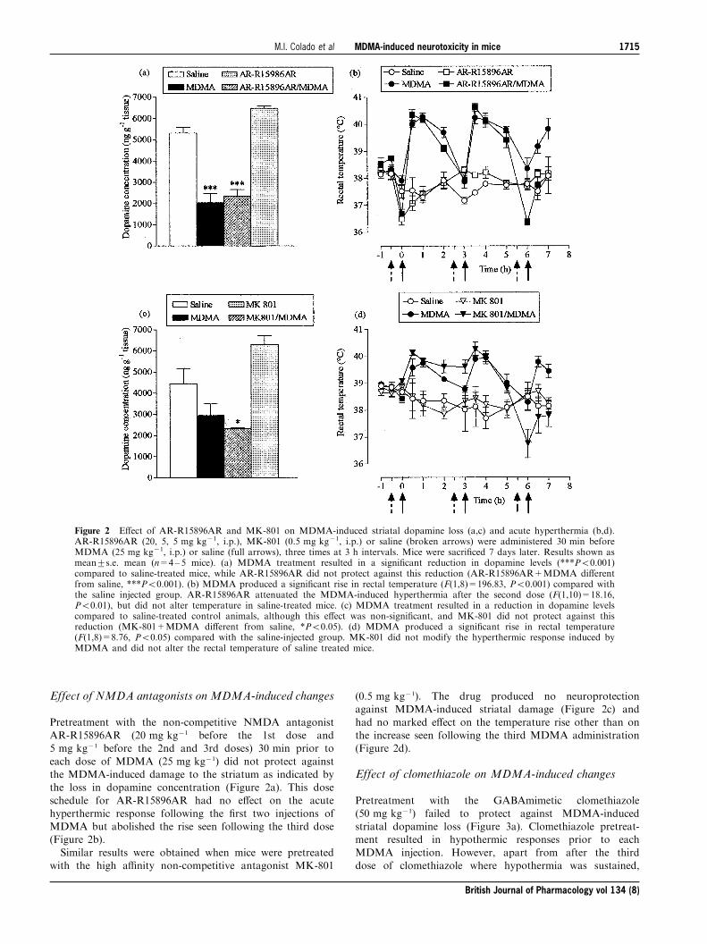
Effect of NMDA antagonists on MDMA-induced changes
Pretreatment with the non-competitive NMDA antagonistAR-R15896AR (20 mg kg71 before the 1st dose and5 mg kg71 before the 2nd and 3rd doses) 30 min prior to
each dose of MDMA (25 mg kg71) did not protect againstthe MDMA-induced damage to the striatum as indicated bythe loss in dopamine concentration (Figure 2a). This dose
schedule for AR-R15896AR had no e�ect on the acutehyperthermic response following the ®rst two injections ofMDMA but abolished the rise seen following the third dose(Figure 2b).
Similar results were obtained when mice were pretreatedwith the high a�nity non-competitive antagonist MK-801
(0.5 mg kg71). The drug produced no neuroprotectionagainst MDMA-induced striatal damage (Figure 2c) and
had no marked e�ect on the temperature rise other than onthe increase seen following the third MDMA administration(Figure 2d).
Effect of clomethiazole on MDMA-induced changes
Pretreatment with the GABAmimetic clomethiazole(50 mg kg71) failed to protect against MDMA-inducedstriatal dopamine loss (Figure 3a). Clomethiazole pretreat-ment resulted in hypothermic responses prior to each
MDMA injection. However, apart from after the thirddose of clomethiazole where hypothermia was sustained,
Figure 2 E�ect of AR-R15896AR and MK-801 on MDMA-induced striatal dopamine loss (a,c) and acute hyperthermia (b,d).AR-R15896AR (20, 5, 5 mg kg71, i.p.), MK-801 (0.5 mg kg71, i.p.) or saline (broken arrows) were administered 30 min beforeMDMA (25 mg kg71, i.p.) or saline (full arrows), three times at 3 h intervals. Mice were sacri®ced 7 days later. Results shown asmean+s.e. mean (n=4±5 mice). (a) MDMA treatment resulted in a signi®cant reduction in dopamine levels (***P50.001)compared to saline-treated mice, while AR-R15896AR did not protect against this reduction (AR-R15896AR+MDMA di�erentfrom saline, ***P50.001). (b) MDMA produced a signi®cant rise in rectal temperature (F(1,8)=196.83, P50.001) compared withthe saline injected group. AR-R15896AR attenuated the MDMA-induced hyperthermia after the second dose (F(1,10)=18.16,P50.01), but did not alter temperature in saline-treated mice. (c) MDMA treatment resulted in a reduction in dopamine levelscompared to saline-treated control animals, although this e�ect was non-signi®cant, and MK-801 did not protect against thisreduction (MK-801+MDMA di�erent from saline, *P50.05). (d) MDMA produced a signi®cant rise in rectal temperature(F(1,8)=8.76, P50.05) compared with the saline-injected group. MK-801 did not modify the hyperthermic response induced byMDMA and did not alter the rectal temperature of saline treated mice.
British Journal of Pharmacology vol 134 (8)
MDMA-induced neurotoxicity in miceM.I. Colado et al 1715
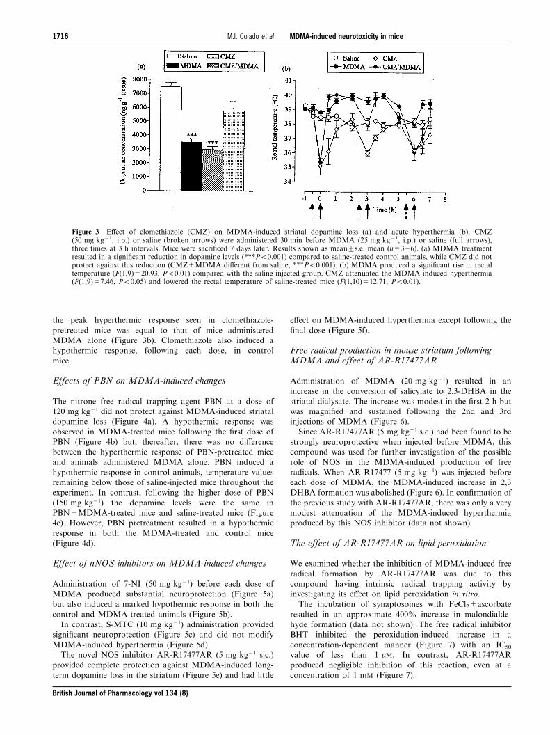
the peak hyperthermic response seen in clomethiazole-
pretreated mice was equal to that of mice administeredMDMA alone (Figure 3b). Clomethiazole also induced ahypothermic response, following each dose, in control
mice.
Effects of PBN on MDMA-induced changes
The nitrone free radical trapping agent PBN at a dose of120 mg kg71 did not protect against MDMA-induced striataldopamine loss (Figure 4a). A hypothermic response was
observed in MDMA-treated mice following the ®rst dose ofPBN (Figure 4b) but, thereafter, there was no di�erencebetween the hyperthermic response of PBN-pretreated mice
and animals administered MDMA alone. PBN induced ahypothermic response in control animals, temperature valuesremaining below those of saline-injected mice throughout the
experiment. In contrast, following the higher dose of PBN(150 mg kg71) the dopamine levels were the same inPBN+MDMA-treated mice and saline-treated mice (Figure4c). However, PBN pretreatment resulted in a hypothermic
response in both the MDMA-treated and control mice(Figure 4d).
Effect of nNOS inhibitors on MDMA-induced changes
Administration of 7-NI (50 mg kg71) before each dose of
MDMA produced substantial neuroprotection (Figure 5a)but also induced a marked hypothermic response in both thecontrol and MDMA-treated animals (Figure 5b).
In contrast, S-MTC (10 mg kg71) administration providedsigni®cant neuroprotection (Figure 5c) and did not modifyMDMA-induced hyperthermia (Figure 5d).The novel NOS inhibitor AR-R17477AR (5 mg kg71 s.c.)
provided complete protection against MDMA-induced long-term dopamine loss in the striatum (Figure 5e) and had little
e�ect on MDMA-induced hyperthermia except following the
®nal dose (Figure 5f).
Free radical production in mouse striatum followingMDMA and effect of AR-R17477AR
Administration of MDMA (20 mg kg71) resulted in an
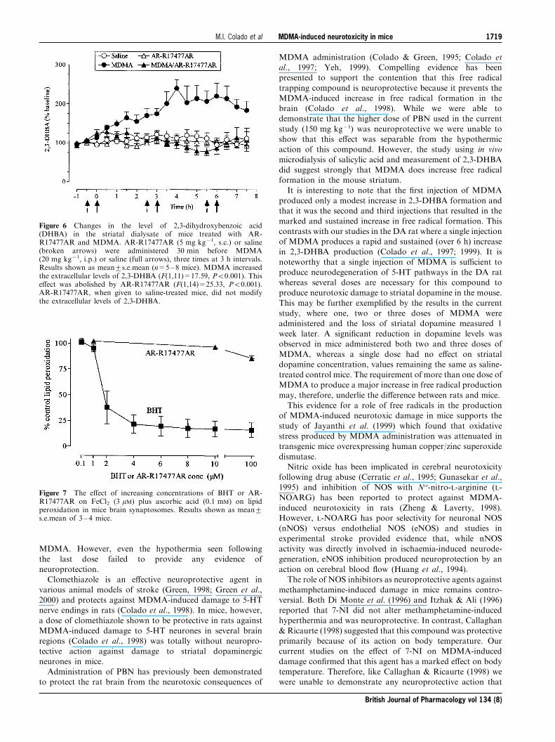
increase in the conversion of salicylate to 2,3-DHBA in thestriatal dialysate. The increase was modest in the ®rst 2 h butwas magni®ed and sustained following the 2nd and 3rdinjections of MDMA (Figure 6).
Since AR-R17477AR (5 mg kg71 s.c.) had been found to bestrongly neuroprotective when injected before MDMA, thiscompound was used for further investigation of the possible
role of NOS in the MDMA-induced production of freeradicals. When AR-R17477 (5 mg kg71) was injected beforeeach dose of MDMA, the MDMA-induced increase in 2,3
DHBA formation was abolished (Figure 6). In con®rmation ofthe previous study with AR-R17477AR, there was only a verymodest attenuation of the MDMA-induced hyperthermiaproduced by this NOS inhibitor (data not shown).
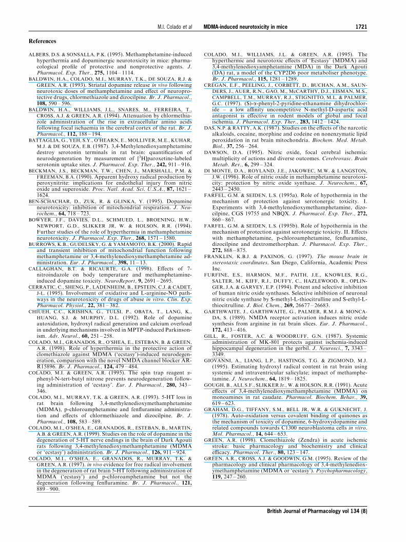
The effect of AR-R17477AR on lipid peroxidation
We examined whether the inhibition of MDMA-induced freeradical formation by AR-R17477AR was due to thiscompound having intrinsic radical trapping activity by
investigating its e�ect on lipid peroxidation in vitro.The incubation of synaptosomes with FeCl2+ascorbate
resulted in an approximate 400% increase in malondialde-
hyde formation (data not shown). The free radical inhibitorBHT inhibited the peroxidation-induced increase in aconcentration-dependent manner (Figure 7) with an IC50
value of less than 1 mM. In contrast, AR-R17477AR
produced negligible inhibition of this reaction, even at aconcentration of 1 mM (Figure 7).
Figure 3 E�ect of clomethiazole (CMZ) on MDMA-induced striatal dopamine loss (a) and acute hyperthermia (b). CMZ(50 mg kg71, i.p.) or saline (broken arrows) were administered 30 min before MDMA (25 mg kg71, i.p.) or saline (full arrows),three times at 3 h intervals. Mice were sacri®ced 7 days later. Results shown as mean+s.e. mean (n=3±6). (a) MDMA treatmentresulted in a signi®cant reduction in dopamine levels (***P50.001) compared to saline-treated control animals, while CMZ did notprotect against this reduction (CMZ+MDMA di�erent from saline, ***P50.001). (b) MDMA produced a signi®cant rise in rectaltemperature (F(1,9)=20.93, P50.01) compared with the saline injected group. CMZ attenuated the MDMA-induced hyperthermia(F(1,9)=7.46, P50.05) and lowered the rectal temperature of saline-treated mice (F(1,10)=12.71, P50.01).
British Journal of Pharmacology vol 134 (8)
MDMA-induced neurotoxicity in miceM.I. Colado et al1716

Discussion
The current study has emphasized both the similarities and
di�erences in the long-term neurotoxic e�ect of MDMA inrats and mice. As reviewed in the introduction the keydi�erence is that, in mice, this amphetamine derivative
damages dopaminergic rather than serotonergic nerveendings. In addition we have now shown that the pro®leof activity of putative neuroprotective compounds also
di�ers.Early studies demonstrated the e�cacy of MK-801 in
protecting against the neurodegenerative action of metham-phetamine in both mice (Sonsalla et al., 1989; Green et al.,
1992) and rats (Green et al., 1992) and also against MDMA-induced damage to 5-HT nerve endings in rats (Colado et al.,1993). However, it was subsequently shown that this
protective action resulted from MK-801 lowering bodytemperature in both species thereby producing neuroprotec-tion (Albers & Sonsalla, 1995; Malberg et al., 1996; Farfel &
Seiden, 1995a; 1995b; Colado et al., 1998). Evidence suggeststhat merely preventing amphetamine-induced hyperthermia,rather than producing frank hypothermia, is enough to
produce signi®cant neuroprotection (Colado et al., 1998;1999).In the current study we investigated whether NMDA
antagonists protected against MDMA-induced damage tostriatal dopamine in mice. Neither MK-801 nor AR-R15896AR produced any neuroprotection, even thoughboth were given at doses previously demonstrated to be
neuroprotective in animal models of stroke (Cregan et al.,1997; Gill et al., 1987). The doses chosen had little e�ecton MDMA-induced hyperthermia until the third dose of
Figure 4 E�ect of a-phenyl-N-tert-butyl nitrone (PBN) on MDMA-induced striatal dopamine loss (a,c) and acute hyperthermia(b,d). PBN (120 or 150 mg kg71, i.p.) or saline (broken arrows) were administered 30 min before MDMA (25 mg kg71, i.p.) orsaline (full arrows), three times at 3 h intervals. Mice were sacri®ced 7 days later. Results shown as mean+s.e. mean (n=3±6 mice).(a) MDMA treatment resulted in a signi®cant reduction in dopamine levels (***P50.001) compared to saline-treated animals, whilePBN did not protect against this reduction (PBN+MDMA di�erent from saline, ***P50.001). (b) MDMA produced a signi®cantrise in rectal temperature (F(1,8)=208.37, P50.001) compared with the saline-injected group. PBN (120 mg kg71) induced ahypothermic response in MDMA-treated mice, following the ®rst dose (F(1,9)=15.28, P50.01), and reduced body temperature insaline-treated mice F(1,7=77.39, P50.001). (c) MDMA treatment resulted in a reduction in dopamine levels compared to saline-treated control animals, although this e�ect was non-signi®cant, and there was no di�erence between PBN+MDMA-treated miceand saline-treated mice. (d) MDMA produced a signi®cant rise in rectal temperature (F(1,8)=8.76, P50.05) compared with thesaline-injected group. PBN (150 mg kg71) abolished the hyperthermic response induced by MDMA (F(1,9)=34.22, P50.001) andproduced hypothermia in saline treated mice (F(1,6)=14.69, P50.01).
British Journal of Pharmacology vol 134 (8)
MDMA-induced neurotoxicity in miceM.I. Colado et al 1717

Figure 5 E�ect of 7-nitroindazole (7-NI), S-methyl-L-thiocitrulline (S-MTC) and AR-R17477AR MDMA-induced striataldopamine loss (a,c,e) and acute hyperthermia (b,d,f). 7-NI (50 mg kg71, i.p.), S-MTC (10 mg kg71, i.p.), AR-R17477AR(5 mg kg71, s.c.), or saline (broken arrows) were administered 30 min before MDMA (20 mg kg71, i.p.) or saline (full arrows),three times at 3 h intervals. Mice were sacri®ced 7 days later. Results shown as mean+s.e.mean (n=6 10 mice). (a) MDMAtreatment resulted in a signi®cant reduction in dopamine levels (***P50.001) compared to saline-treated mice. 7-NI pretreatmentsigni®cantly, although not completely, protected against this reduction (7-NI+MDMA di�erent from MDMA, DDDP50.001; 7-NI+MDMA di�erent from saline, *P50.05). (b) MDMA produced a signi®cant rise in body temperature (F(1,22)=7.55, P50.01)compared with the saline-injected group. 7-NI abolished the MDMA-induced hyperthermia (F(1,25)=24.33, P50.001) and induceda frank hypothermia in saline-treated mice (F(1,16)=169.02, P50.001). (c) MDMA treatment resulted in a signi®cant reduction indopamine levels (***P50.001) compared to saline-treated mice. S-MTC pretreatment signi®cantly, although not completely,protected against this reduction (S-MTC+MDMA di�erent from MDMA, DDP50.01; S-MTC+MDMA di�erent from saline,**P50.01). (d) MDMA produced a signi®cant rise in body temperature (F(1,16)=17.23, P50.001) compared with the salineinjected group. S-MTC did not modify the hyperthermic response induced by MDMA and did not alter the body temperature ofsaline-treated mice. (e) MDMA treatment resulted in a signi®cant reduction in dopamine levels (***P50.001) compared to saline-treated control mice. AR-R17477AR provided complete protection against this loss (AR-R17477AR+MDMA di�erent fromMDMA, DDP50.01). (f) MDMA produced a signi®cant rise in body temperature (F(1,8)=18.03, P50.01) compared with the salineinjected group. AR-R17477AR attenuated the MDMA-induced hyperthermic response following the second dose (F(1,7)=68.60,P50.001), but did not modify the rectal temperature of saline-treated mice.
British Journal of Pharmacology vol 134 (8)
MDMA-induced neurotoxicity in miceM.I. Colado et al1718
MDMA. However, even the hypothermia seen following
the last dose failed to provide any evidence ofneuroprotection.Clomethiazole is an e�ective neuroprotective agent in
various animal models of stroke (Green, 1998; Green et al.,2000) and protects against MDMA-induced damage to 5-HTnerve endings in rats (Colado et al., 1998). In mice, however,a dose of clomethiazole shown to be protective in rats against
MDMA-induced damage to 5-HT neurones in several brainregions (Colado et al., 1998) was totally without neuropro-tective action against damage to striatal dopaminergic
neurones in mice.Administration of PBN has previously been demonstrated
to protect the rat brain from the neurotoxic consequences of
MDMA administration (Colado & Green, 1995; Colado etal., 1997; Yeh, 1999). Compelling evidence has beenpresented to support the contention that this free radical
trapping compound is neuroprotective because it prevents theMDMA-induced increase in free radical formation in thebrain (Colado et al., 1998). While we were able todemonstrate that the higher dose of PBN used in the current
study (150 mg kg71) was neuroprotective we were unable toshow that this e�ect was separable from the hypothermicaction of this compound. However, the study using in vivo
microdialysis of salicylic acid and measurement of 2,3-DHBAdid suggest strongly that MDMA does increase free radicalformation in the mouse striatum.
It is interesting to note that the ®rst injection of MDMAproduced only a modest increase in 2,3-DHBA formation andthat it was the second and third injections that resulted in the
marked and sustained increase in free radical formation. Thiscontrasts with our studies in the DA rat where a single injectionof MDMA produces a rapid and sustained (over 6 h) increasein 2,3-DHBA production (Colado et al., 1997; 1999). It is
noteworthy that a single injection of MDMA is su�cient toproduce neurodegeneration of 5-HT pathways in the DA ratwhereas several doses are necessary for this compound to
produce neurotoxic damage to striatal dopamine in the mouse.This may be further exempli®ed by the results in the currentstudy, where one, two or three doses of MDMA were
administered and the loss of striatal dopamine measured 1week later. A signi®cant reduction in dopamine levels wasobserved in mice administered both two and three doses of
MDMA, whereas a single dose had no e�ect on striataldopamine concentration, values remaining the same as saline-treated control mice. The requirement of more than one dose ofMDMA to produce a major increase in free radical production
may, therefore, underlie the di�erence between rats and mice.This evidence for a role of free radicals in the production
of MDMA-induced neurotoxic damage in mice supports the
study of Jayanthi et al. (1999) which found that oxidativestress produced by MDMA administration was attenuated intransgenic mice overexpressing human copper/zinc superoxide
dismutase.Nitric oxide has been implicated in cerebral neurotoxicity
following drug abuse (Cerratic et al., 1995; Gunasekar et al.,1995) and inhibition of NOS with No-nitro-L-arginine (L-
NOARG) has been reported to protect against MDMA-induced neurotoxicity in rats (Zheng & Laverty, 1998).However, L-NOARG has poor selectivity for neuronal NOS
(nNOS) versus endothelial NOS (eNOS) and studies inexperimental stroke provided evidence that, while nNOSactivity was directly involved in ischaemia-induced neurode-
generation, eNOS inhibition produced neuroprotection by anaction on cerebral blood ¯ow (Huang et al., 1994).The role of NOS inhibitors as neuroprotective agents against
methamphetamine-induced damage in mice remains contro-versial. Both Di Monte et al. (1996) and Itzhak & Ali (1996)reported that 7-NI did not alter methamphetamine-inducedhyperthermia and was neuroprotective. In contrast, Callaghan
& Ricaurte (1998) suggested that this compound was protectiveprimarily because of its action on body temperature. Ourcurrent studies on the e�ect of 7-NI on MDMA-induced
damage con®rmed that this agent has a marked e�ect on bodytemperature. Therefore, like Callaghan & Ricaurte (1998) wewere unable to demonstrate any neuroprotective action that
Figure 6 Changes in the level of 2,3-dihydroxybenzoic acid(DHBA) in the striatal dialysate of mice treated with AR-R17477AR and MDMA. AR-R17477AR (5 mg kg71, s.c.) or saline(broken arrows) were administered 30 min before MDMA(20 mg kg71, i.p.) or saline (full arrows), three times at 3 h intervals.Results shown as mean+s.e.mean (n=5±8 mice). MDMA increasedthe extracellular levels of 2,3-DHBA (F(1,11)=17.59, P50.001). Thise�ect was abolished by AR-R17477AR (F(1,14)=25.33, P50.001).AR-R17477AR, when given to saline-treated mice, did not modifythe extracellular levels of 2,3-DHBA.
Figure 7 The e�ect of increasing concentrations of BHT or AR-R17477AR on FeCl2 (3 mM) plus ascorbic acid (0.1 mM) on lipidperoxidation in mice brain synaptosomes. Results shown as mean+s.e.mean of 3 ± 4 mice.
British Journal of Pharmacology vol 134 (8)
MDMA-induced neurotoxicity in miceM.I. Colado et al 1719

was separable from a hypothermic action. Taraska & Finnegan(1997) were similarly unable to separate the neuroprotectiveaction of the NOS inhibitor L-NAME from its hypothermic
action, concluding that nitric oxide played little role inMDMA-induced neurodegeneration.However, in our studies, administration of S-MTC, given at
a dose previously shown to protect against MPTP-induced
damage to dopamine neurones (Matthews et al., 1997), had noe�ect on the MDMA-induced hyperthermia and provided asigni®cant neuroprotective e�ect. Similarly, AR-R17477AR,
when given at a dose that has no overt behavioural e�ects oractions on blood pressure (Johansson et al., 1999), had littlee�ect on the hyperthermia following the ®rst two doses of
MDMA but was strongly neuroprotective. It is acknowledgedthat it did produce hypothermia following the third MDMAdose, but our study with the NMDA antagonists indicated that
this action alone is not enough to produce neuroprotection.The data, therefore, do suggest a role for NOS inhibitors in
producing neuroprotection against MDMA-induced damageto dopamine nerve endings in mouse striatum and demon-
strate that this e�ect is not primarily involved with any actionon body temperature. The question that follows, however,concerns the selectivity of the inhibitors used. L-NOARG and
7-NI show little selectivity for nNOS versus eNOS (Dawson,1995; O'Neill et al., 2000). In contrast, both S-MTC and AR-R17477AR have been reported to display good selectivity
towards nNOS using in vitro studies (Fur®ne et al., 1994;O'Neill et al., 2000). While the selective action of AR-R17477AR is supported by evidence that this compound had
no e�ect on blood pressure or heart rate when given to ratsat twice the dose used in the present investigation (Johanssonet al., 1999) it has to be pointed out that selectivity againstrodent NOS isoforms was not examined by O'Neill et al.
(2000) so caution should be exercised in proposing that it isnNOS that is involved in the mechanisms whereby NOSinhibition protects against MDMA-induced neurotoxicity.
It, therefore, seems reasonable to propose that NOS isinvolved in the neurodegeneration of dopamine nerve endingsthat follows administration of MDMA to mice. The question
that remains, however, concerns the speci®c mechanismsinvolved. It is generally accepted in ischaemic stroke researchthat ischaemia-induced glutamate release produces excessivestimulation of NMDA receptors leading to an increase in
intracellular calcium, activation of NOS and excess produc-tion of NO. (Garthwaite et al., 1989; Moncada et al., 1991).In turn NO. interacts with the superoxide anion, O2
7 and
forms the highly toxic peroxynitrite ion (ONOO7) (Beckmanet al., 1990). While this cascade of events has previously beenproposed to explain methamphetamine-induced neurotoxicity
in mice (for example, Itzhak et al., 1998), such a mechanismcannot explain the current observations. In the case ofMDMA-induced neurotoxic degeneration our data provide
clear evidence that an increase in excitatory amino acidneurotransmission does not underlie the neurotoxic e�ect ofMDMA in mice since neither of the two NMDA antagonistsexamined (MK-801 and AR-R15896AR) provided any
protection against MDMA-induced damage to dopamineneurones in the striatum. Nevertheless, administration ofNOS inhibitors did result in substantial neuroprotection and
mechanisms not involving raised extracellular glutamate mustbe evoked to explain the involvement of NOS in theneurodegenerative cascade.
It was recently shown that MDMA administrationdecreased cytochrome oxidase complex IV of the electrontransport chain in dopamine rich areas (Burrows et al., 2000).
It may be that the MDMA-induced release of dopaminecompromised mitochondrial function because of auto-oxida-tion of dopamine metabolites to form quinones and hydroxyland reactive oxygen species (Graham et al., 1978). Quinones
and reactive oxygen species have been shown to inhibitmitochondrial enzymes (Zhang et al., 1990; Ben-Schachar etal., 1995). Since there is also evidence that MDMA is
metabolized to catechol and quinone compounds, anotherpossibility is that further metabolism of these compounds alsoleads to free radical formation (Colado et al., 1995; 1997;
1999; Sprague & Nichols, 1995; Yeh, 1999) and disruption ofmitochondrial function. This would lead to an increase inintracellular calcium levels (Khodorov et al., 1999) and
activation of both nNOS and eNOS (Stuehr & Gri�th, 1992).Of particular interest is the observation that administration
of the NOS inhibitor AR-R17477AR prevented the MDMA-induced rise in free radical formation. Since we were able to
demonstrate that AR-R17477AR is not a radical trappingagent, by its lack of e�ect on FeCl2/ascorbate-induced lipidperoxidation, these data indicate that nitric oxide is probably
interacting in some way with MDMA and/or dopaminemetabolite auto-oxidation products to produce a radical thatcan be detected by the salicylate/2,3-DHBA reaction path-
way. The most likely candidate is the peroxynitrite ion, aradical which has already been implicated in the neurotoxicdamage to dopamine neurones that follows methampheta-
mine administration (Imam & Ali, 2000; Imam et al., 1999;2001) and is detected by the salicylate/2,3-DHBA assay(Narayan et al., 1997; Halliwell & Kaur, 1997).
In turn, this proposal goes some way to explaining why
dopamine neurones are damaged following MDMA admin-istration to mice, whereas 5-HT neurones are damagedfollowing MDMA administration to rats. In our previous
studies we have been unable to demonstrate that administra-tion of NOS inhibitors prevented MDMA-induced neurode-generation in rats (Colado et al., unpublished observations).
Based on the results of the current study, therefore, it appearsthat the free radical entity produced in rats is notperoxynitrite. Thus, the neurotoxic damage produced indi�erent monoaminergic systems in rats and mice may relate
to the particular free radical ion being produced in these twospecies. This may, in turn, be related to a di�erent pattern ofmetabolites being formed following administration of
MDMA to rats and mice.In conclusion, our data do demonstrate that free radicals
are involved in MDMA-induced damage in mice, as they are
in rats (Colado et al., 1997; 1999; Yeh, 1999). It seemsprobable that in mice MDMA metabolites produce speci®cradicals which in turn react with nitrite ions. By inhibiting
NOS, this interaction is decreased su�ciently to abolish orattenuate the formation of the highly toxic peroxynitrite ions.This proposal is supported by our study using in vivomicrodialysis.
M.I. Colado thanks Plan Nacional sobre Drogas (Ministerio delInterior), CICYT (SAF98-0074) and AstraZeneca R&D SoÈ dertaÈ ljefor ®nancial support.
British Journal of Pharmacology vol 134 (8)
MDMA-induced neurotoxicity in miceM.I. Colado et al1720
References
ALBERS, D.S. & SONSALLA, P.K. (1995). Methamphetamine-inducedhyperthermia and dopaminergic neurotoxicity in mice: pharma-cological pro®le of protective and nonprotective agents. J.Pharmacol. Exp. Ther., 275, 1104 ± 1114.
BALDWIN, H.A., COLADO, M.I., MURRAY, T.K., DE SOUZA, R.J. &
GREEN, A.R. (1993). Striatal dopamine release in vivo followingneurotoxic doses of methamphetamine and e�ect of neuropro-tective drugs, chlormethiazole and dizocilpine. Br. J. Pharmacol.,108, 590 ± 596.
BALDWIN, H.A., WILLIAMS, J.L., SNARES, M., FERREIRA, T.,
CROSS, A.J. & GREEN, A.R. (1994). Attenuation by chlormethia-zole administration of the rise in extracellular amino acidsfollowing focal ischaemia in the cerebral cortex of the rat. Br. J.Pharmacol., 112, 188 ± 194.
BATTAGLIA, G., YEH, S.Y., O'HEARN, E., MOLLIVER, M.E., KUHAR,
M.J. & DE SOUZA, E.B. (1987). 3,4-Methylenedioxyamphetaminedestroy serotonin terminals in rat brain: quanti®cation ofneurodegeneration by measurement of [3H]paroxetine-labeledserotonin uptake sites. J. Pharmacol. Exp. Ther., 242, 911 ± 916.
BECKMAN, J.S., BECKMAN, T.W., CHEN, J., MARSHALL, P.M. &
FREEMAN, B.A. (1990). Apparent hydroxy radical production byperoxynitrite: implications for endothelial injury from nitricoxide and superoxide. Proc. Natl. Acad. Sci. U.S.A., 87, 1621 ±1624.
BEN-SCHACHAR, D., ZUK, R. & GLINKA, Y. (1995). Dopamineneurotoxicity: inhibition of mitochondrial respiration. J. Neu-rochem., 64, 718 ± 723.
BOWYER, J.F., DAVIES, D.L., SCHMUED, L., BROENING, H.W.,
NEWPORT, G.D., SLIKKER JR, W. & HOLSON, R.R. (1994).Further studies of the role of hyperthermia in methamphetamineneurotoxicity. J. Pharmacol. Exp. Ther., 268, 1571 ± 1580.
BURROWS, K.B., GUDELSKY, G. & YAMAMOTO, B.K. (2000). Rapidand transient inhibition of mitochondrial function followingmethamphetamine or 3,4-methylenedioxymethamphetamine ad-ministration. Eur. J. Pharmacol., 398, 11 ± 13.
CALLAGHAN, B.T. & RICAURTE, G.A. (1998). E�ects of 7-nitroindazole on body temperature and methamphetamine-induced dopamine toxicity. NeuroReport, 9, 2691 ± 2695.
CERRATIC, C., SHENG, P., LADENHEIM, B., EPSTEIN, C.J. & CADET,
J-L. (1995). Involvement of oxidative and L-arginine-NO path-ways in the neurotoxicity of drugs of abuse in vitro. Clin. Exp.Pharmacol. Physiol., 22, 381 ± 382.
CHIUEH, C.C., KRISHNA, G., TULSI, P., OBATA, T., LANG, K.,
HUANG, S.J. & MURPHY, D.L. (1992). Role of dopamineautoxidation, hydroxyl radical generation and calcium overloadin underlying mechanisms involved in MPTP-induced Parkinson-ism. Adv. Neurol., 60, 251 ± 258.
COLADO, M.I., GRANADOS, R., O'SHEA, E., ESTEBAN, B. & GREEN,
A.R. (1998). Role of hyperthermia in the protective action ofclomethiazole against MDMA (`ecstasy')-induced neurodegen-eration, comparison with the novel NMDA channel blocker AR-R15896. Br. J. Pharmacol., 124, 479 ± 484.
COLADO, M.I. & GREEN, A.R. (1995). The spin trap reagent a-phenyl-N-tert-butyl nitrone prevents neurodegeneration follow-ing administration of `ecstasy'. Eur. J. Pharmacol., 280, 343 ±346.
COLADO, M.I., MURRAY, T.K. & GREEN, A.R. (1993). 5-HT loss inrat brain following 3,4-methylenedioxymethamphetamine(MDMA), p-chloroamphetamine and fen¯uramine administra-tion and e�ects of chlormethiazole and dizocilpine. Br. J.Pharmacol., 108, 583 ± 589.
COLADO, M.I., O'SHEA, E., GRANADOS, R., ESTEBAN, B., MARTIN,
A.B. & GREEN, A.R. (1999). Studies on the role of dopamine in thedegeneration of 5-HT nerve endings in the brain of Dark Agoutirats following 3,4-methylenedioxymethamphetamine (MDMAor `ecstasy') administration. Br. J. Pharmacol., 126, 911 ± 924.
COLADO, M.I., O'SHEA, E., GRANADOS, R., MURRAY, T.K. &
GREEN, A.R. (1997). in vivo evidence for free radical involvementin the degeneration of rat brain 5-HT following administration ofMDMA (`ecstasy') and p-chloroamphetamine but not thedegeneration following fen¯uramine. Br. J. Pharmacol., 121,889 ± 900.
COLADO, M.I., WILLIAMS, J.L. & GREEN, A.R. (1995). Thehyperthermic and neurotoxic e�ects of `Ecstasy' (MDMA) and3,4-methylenedioxyamphetamine (MDA) in the Dark Agouti(DA) rat, a model of the CYP2D6 poor metaboliser phenotype.Br. J. Pharmacol., 115, 1281 ± 1289.
CREGAN, E.F., PEELING, J., CORBETT, D., BUCHAN, A.M., SAUN-
DERS, J., AUER, R.N., GAO, M., McCARTHY, D.J., EISMAN, M.S.,
CAMPBELL, T.M., MURRAY, R.J., STIGNITTO, M.L. & PALMER,
G.C. (1997). (S)-a-phenyl-2-pyridine-ethanamine dihydrochlor-ide ± a low a�nity uncompetitive N-methyl-D-aspartic acidantagonist is e�ective in rodent models of global and focalischemia. J. Pharmacol. Exp. Ther., 283, 1412 ± 1424.
DAS, N.P. & RATTY, A.K. (1987). Studies on the e�ects of the narcoticalkaloids, cocaine, morphine and codeine on nonenzymatic lipidperoxidation in rat brain mitochondria. Biochem. Med. Metab.Biol., 37, 256 ± 264.
DAWSON, D.A. (1995). Nitric oxide, focal cerebral ischemia:multiplicity of actions and diverse outcomes. Cerebrovasc. BrainMetab. Rev., 6, 299 ± 324.
DI MONTE, D.A., ROYLAND, J.E., JAKOWEC, M.W. & LANGSTON,
J.W. (1996). Role of nitric oxide in methamphetamine neurotoxi-city: protection by nitric oxide synthase. J. Neurochem., 67,2443 ± 2450.
FARFEL, G.M. & SEIDEN, L.S. (1995a). Role of hypothermia in themechanism of protection against serotonergic toxicity. I.Experiments with 3,4-methylenedioxymethamphetamine, dizo-cilpine, CGS 19755 and NBQX. J. Pharmacol. Exp. Ther., 272,860 ± 867.
FARFEL, G.M. & SEIDEN, L.S. (1995b). Role of hypothermia in themechanism of protection against serotonergic toxicity. II. E�ectswith methamphetamine, p-chloroamphetamine, fen¯uramine,dizocilpine and dextromethorphan. J. Pharmacol. Exp. Ther.,272, 868 ± 875.
FRANKLIN, K.B.J. & PAXINOS, G. (1997). The mouse brain instereotaxic coordinates. San Diego, California, Academic PressInc.
FURFINE, E.S., HARMON, M.F., PAITH, J.E., KNOWLES, R.G.,
SALTER, M., KIFF, R.J., DUFFY, C., HAZLEWOOD, R., OPLIN-
GER, J.A. & GARVEY, E.P. (1994). Potent and selective inhibitionof human nitric oxide synthases. Selective inhibition of neuronalnitric oxide synthase by S-methyl-L-thiocitrulline and S-ethyl-L-thiocitrulline. J. Biol. Chem., 269, 26677 ± 26683.
GARTHWAITE, J., GARTHWAITE, G., PALMER, R.M.J. & MONCA-
DA, S. (1989). NMDA receptor activation induces nitric oxidesynthesis from arginine in rat brain slices. Eur. J. Pharmacol.,172, 413 ± 416.
GILL, R., FOSTER, A.C. & WOODRUFF, G.N. (1987). Systemicadministration of MK-801 protects against ischemia-inducedhippocampal degeneration in the gerbil. J. Neurosci., 7, 3343 ±3349.
GIOVANNI, A., LIANG, L.P., HASTINGS, T.G. & ZIGMOND, M.J.
(1995). Estimating hydroxyl radical content in rat brain usingsystemic and intraventricular salicylate; impact of methamphe-tamine. J. Neurochem., 64, 1819 ± 1825.
GOUGH, B., ALI, S.F., SLIKKER Jr., W. & HOLSON, R.R. (1991). Acutee�ects of 3,4-methylenedioxymethamphetamine (MDMA) onmonoamines in rat caudate. Pharmacol. Biochem. Behav., 39,619 ± 623.
GRAHAM, D.G., TIFFANY, S.M., BELL JR, W.R. & GUKNECHT, J.
(1978). Auto-oxidation versus covalent binding of quinones asthe mechanism of toxicity of dopamine, 6-hydroxydopamine andrelated compounds towards C1300 neuroblastoma cells in vitro.Mol. Pharmacol., 14, 644 ± 653.
GREEN, A.R. (1998). Clomethiazole (Zendra) in acute ischemicstroke: basic pharmacology and biochemistry and clinicale�cacy. Pharmacol. Ther., 80, 123 ± 147.
GREEN, A.R., CROSS, A.J. & GOODWIN, G.M. (1995). Review of thepharmacology and clinical pharmacology of 3,4-methylenediox-ymethamphetamine (MDMA or `ecstasy'). Psychopharmacology,119, 247 ± 260.
British Journal of Pharmacology vol 134 (8)
MDMA-induced neurotoxicity in miceM.I. Colado et al 1721

GREEN, A.R., DE SOUZA, R.J., WILLIAMS, J.L., MURRAY, T.K. &
CROSS, A.J. (1992). The neurotoxic e�ects of methamphetamineon 5-hydroxytryptamine and dopamine in brain: evidence for theprotective e�ect of chlormethiazole. Neuropharmacology, 31,315 ± 321.
GREEN, A.R., HAINSWORTH, A.H. & JACKSON, D.M. (2000). GABApotentiation: a logical pharmacological approach for thetreatment of acute ischaemic stroke. Neuropharmacology, 39,1483 ± 1493.
GUDELSKY, G.A. & NASH, J.F. (1996). Carrier-mediated release ofserotonin by 3,4-methylenedioxymethamphetamine: Implica-tions for serotonin-dopamine interactions. J. Neurochem., 66,243 ± 249.
GUNASEKAR, P.G., KANTHASAMY, A.G., BOROWITZ, J.L. & ISOM,
G.E. (1995). NMDA receptor activation produces concurrentgeneration of nitric oxide and reactive oxygen species: implica-tion for cell death. J. Neurochem., 65, 2016 ± 2021.
HALLIWELL, B. & KAUR, H. (1997). Hydroxylation of salicylate andphenylalanine as assays for hydroxyl radicals: a cautionary notevisited for the third time. Free Rad. Res., 27, 239 ± 244.
HUANG, Z., HUANG, P.L., PANAHIAN, N., DALKARA, T., FISHMAN,
M.C. & MOSKOWITZ, M.A. (1994). E�ects of cerebral ischemia inmice de®cient in neuronal nitre oxide synthase. Science, 265,1883 ± 1885.
HUETHER, G., ZHOU, D. & RUTHER, E. (1997). Causes andconsequences of the loss of serotonergic presynapses elicited bythe consumption of 3,4-methylenedioxymethamphetamine(MDMA, `ecstasy') and its congeners. J. Neural. Transm., 104,771 ± 791.
IMAM, S.Z. & ALI, S.F. (2000). Selenium, an antioxidant, attenuatesmethamphetamine-induced dopaminergic-neurotoxicity and per-oxynitrite generation. Brain Res., 855, 186 ± 191.
IMAM, S.Z., CROW, J.P., NEWPORT, G.D., ISLAM, F., SLIKKER JR, W.
& ALI, S.F. (1999). Methamphetamine generates peroxynitriteand produces dopaminergic neurotoxicity in mice: protectivee�ects of peroxynitrite decomposition catalyst. Brain Res., 837,15 ± 21.
IMAM, S.Z., NEWPORT, G.D., ITZHAK, Y., CADET, J.L., ISLAM, F.,
SLIKKER JR, W. & ALI, S.F. (2001). Peroxynitrite plays a role inmethamphetamine-induced dopaminergic neurotoxicity: evi-dence from mice lacking neuronal nitric oxide synthase gene oroverexpressing copper-zinc superoxide dismutase. J. Neuro-chem., 76, 745 ± 749.
ITZHAK, Y. & ALI, S.F. (1996). The neuronal nitric oxide synthaseinhibitor, 7-nitroindazole, protects against methamphetamine-induced neurotoxicity in vivo. J. Neurochem., 67, 1770 ± 1773.
ITZHAK, Y. GANDIA, C., HUANG, P.L. & ALI, S.F. (1998). Resistanceof neuronal oxide synthase-de®cient mice to methamphetamine-induced dopaminergic neurotoxicity. J. Pharmacol. Exp. Ther.,284, 1040 ± 1047.
JAYANTHI, S., LADENHEIM, B., ANDREWS, A.M. & CADET, J.L.
(1999). Overexpression of human copper/zinc superoxide dis-mutase in transgenic mice attenuates oxidative stress caused bymethylenedioxymethamphetamine (ecstasy). Neuroscience, 91,1379 ± 1387.
JOHANSSON, C., DEVENEY, A.M., REIF, D. & JACKSON D.M. (1999).The neuronal selective nitric oxide inhibitor AR-R 17477, blockssome e�ects of phencyclidine, while having no observablebehavioural e�ects when given alone. Pharmacol. Toxicol., 84,226 ± 233.
KHODOROV, B., PINELIS, V., STOROZHEVYKH, T., YURAVICHUS,
A. & KHASPEKHOV, L. (1999). Blockade of mitochondrial Ca2+
uptake by mitochondrial inhibitors ampli®es the glutamate-induced calcium response in cultured cerebellar granule cells.FEBS Letts., 458, 162 ± 166.
KOCH, S. & GALLOWAY, M.P. (1997). MDMA induced dopaminerelease in vivo: role of endogenous serotonin. J. Neural. Transm.,104, 135 ± 146.
KODA, L.Y. & GIBB, J.W. (1973). Adrenal and striatal tyrosinehydroxylase activity after methamphetamine. J. Pharmacol. Exp.Ther., 185, 42 ± 47.
LEW, R., SABOL, K.E., CHOU, C., VOSMER, G.L., RICHARDS, J.B. &
SEIDEN, L.S. (1996). Methylenedioxymethamphetamine-inducedserotonin de®cits are followed by partial recovery over a 52-weekperiod. Part II: Radioligand binding and autoradiographystudies. J. Pharmacol. Exp. Ther., 276, 855 ± 865.
LOGAN, B.J., LAVERTY, R., SANDERSON, W.D. & YEE, Y.B. (1988).Di�erences between rats and mice in MDMA (methylenediox-ymethylamphetamine) neurotoxicity. Eur. J. Pharmacol., 152,227 ± 234.
LYNCH,M.A. & VOSS, K.L. (1990) Arachidonic acid increases inositolphospholipid metabolism and glutamate release in synaptosomesprepared from hippocampal tissue. J. Neurochem., 55, 215 ± 221.
MALBERG, J.E., SABOL, K.E. & SEIDEN, L.S. (1996). Co-administra-tion of MDMA with drugs that protect against MDMAneurotoxicity produces di�erent e�ects on body temperature inthe rat. J. Pharmacol. Exp. Ther., 278, 258 ± 267.
MATTHEWS, R.T., YANG, L. & BEAL, M.F. (1997). S-Methylthioci-trulline, a neuronal nitric oxide synthase inhibitor, protectsagainst malonate and MPTP neurotoxicity. Exp. Neurol., 143,282 ± 286.
MILLER, D.B. & O'CALLAGHAN, J.P. (1994). Environment-, drug-and stress-induced alterations in body temperature a�ect theneurotoxicity of substituted amphetamines in the C57BL/6Jmouse. J. Pharmacol. Exp. Ther., 270, 752 ± 760.
MONCADA, S., PALMER, R.M.J. & HIGGS, E.A. (1991). Nitric oxide:physiology, pathophysiology and pharmacology. Pharmacol.Rev., 42, 109 ± 142.
NARAYAN, M., BERLINER, L.J., MEROLA, A.J., DIAZ, P.T. &
CLANTON, T.L. (1997). Biological reactions of peroxynitrite:evidence for an alternative pathway of salicylate hydroxylation.Free Rad. Res., 27, 63 ± 72.
O'CALLAGHAN, J.P. & MILLER, D.B. (1994). Neurotoxicity pro®lesof substituted amphetamines in the C57BL/6J mouse. J.Pharmacol. Exp. Ther., 270, 741 ± 751.
O'NEILL, M.J., MURRAY, T.K., MCCARTY, D.R., HICKS, C.A., DELL,
C.P., PATRICK, K.E., WARD, M.A., OSBORNE, D.J., WIERNSKI,
T.R., ROMAN, C.R., LODGE, D., FLEISCH, J.H. & SINGH, J.P.
(2000). ARL17477, a selective nitric oxide synthase inhibitor,with neuroprotective e�ects in animal models of global and focalcerebral ischaemia. Brain Res., 871, 234 ± 244.
O'SHEA, E., ESTEBAN, B., CAMARERO, J., GREEN, A.R. & COLADO,
M.I. (2001). E�ect of GBR 12909 and ¯uoxetine on the acute andlong term changes induced by MDMA (`ecstasy') on the 5-HTand dopamine concentrations in mouse brain. Neuropharmacol-ogy, 40, 65 ± 74.
SABOL, K.E., LEW, R., RICHARDS, J.B., VOSMER, G.L. & SEIDEN,
L.S. (1996). Methylenedioxymethamphetamine-induced seroto-nin de®cits are followed by partial recovery over a 52 weekperiod: Part 1: synaptosomal uptake and tissue concentrations. J.Pharmacol. Exp. Ther., 276, 846 ± 854.
SABOL, K.E. & SEIDEN, L.S. (1998). Reserpine attenuates D-amphetamine and MDMA-induced transmitter release in vivo: aconsideration of dose, core temperature and dopamine synthesis.Brain Res., 806, 69 ± 78.
SCHMIDT, C.J. (1987). Neurotoxicity of the psychedelic ampheta-mine, methylenedioxymethamphetamine. J. Pharmacol. Exp.Ther., 240, 1 ± 7.
SCHMIDT, C.J. & KEHNE, J.H. (1990). Neurotoxicity of MDMA:neurochemical e�ects. Ann. N.Y. Acad. Sci., 600, 665 ± 680.
SCHMIDT, C.J., LEVIN, J.A. & LOVENBERG, W. (1987). in vitro and invivo neurochemical e�ects of methylenedioxymethamphetamineon striatal monoaminergic systems in the rat brain. Biochem.Pharmacol., 36, 747 ± 755.
SONSALLA, P.K., NICKLAS, W.J. & HEIKKILA, R.E. (1989). Role forexcitatory amino acids in methamphetamine-induced nigrostria-tal dopaminergic toxicity. Science, 243, 398 ± 400.
SPRAGUE, J.E. & NICHOLS, D.E. (1995). The monoamine oxidase-Binhibitor L-deprenyl protects against 3,4-methylenedioxy-methamphetamine-induced lipid peroxidation and long-termserotonergic de®cits. J. Pharmacol. Exp. Ther., 273, 667 ± 673.
British Journal of Pharmacology vol 134 (8)
MDMA-induced neurotoxicity in miceM.I. Colado et al1722
STEELE, T.D., MCCANN, U.D. & RICAURTE, G.A. (1994). 3,4-Methylenedioxymethamphetamine (MDMA, `ecstasy'): pharma-cology and toxicology in animals and humans. Addiction, 89,539 ± 551.
STONE, D.M., STAHL, D.C., HANSON, G.R. & GIBB, J.W. (1986). Thee�ects of 3, 4-methylenedioxymethamphetamine (MDMA) and3,4-methylenedioxyamphetamine (MDA) on monoaminergicsystems in the rat brain. Eur. J. Pharmacol., 128, 41 ± 48.
STONE, D.M., HANSON, G.R. & GIBB, J.W. (1987). Di�erences in thecentral serotonergic e�ects of methylenedioxymethamphetamine(MDMA) in mice and rats. Neuropharmacology, 26, 1657 ± 1661.
STUEHR, D.J. & GRIFFITH, O.W. (1992). Mammalian nitric oxidesynthases. Adv. Enzymol. Relat. Areas Mol. Biol., 65, 287 ± 346.
TARASKA, T. & FINNEGAN, K.T. (1997). Nitric oxide and theneurotoxic e�ects of methamphetamine and 3,4-methylenediox-ymethamphetamine. J. Pharmacol. Exp. Ther., 280, 941 ± 947.
WHITE, S.R., OBRADOVIC, T., IMEL, K.M. & WHEATON, M.J. (1996).The e�ects of methylenedioxymethamphetamine (MDMA, `ec-stasy') on monoaminergic neurotransmission in the centralnervous system. Progr. Neurobiol., 49, 455 ± 479.
YEH, S.Y. (1999). N-tert-butyl-alpha-phenylnitrone protects against3,4-methylenedioxymethamphetamine-induced depletion of ser-otonin in rats. Synapse, 31, 169 ± 177.
ZHANG, Y., MARCILLAT, O., GIULIVI, C., ERNSTER, L. & DAVIES,
K. (1990). The oxidative inactivation of mitochondrial electrontransport chain components and ATPase. J. Biol. Chem., 265,16330 ± 16335.
ZHENG, Y. & LAVERTY, R. (1998). Role of brain nitric oxide in (+)-3,4-methylenedioxymethamphetamine (MDMA)-induced neuro-toxicity in rats. Brain Res., 795, 257 ± 263.
(Received August 10, 2001Revised October 3, 2001
Accepted October 9, 2001)
British Journal of Pharmacology vol 134 (8)
MDMA-induced neurotoxicity in miceM.I. Colado et al 1723