MDMA intoxication and verbal memory performance: a placebo-controlled pharmaco-MRI study
10
http://jop.sagepub.com/ Journal of Psychopharmacology http://jop.sagepub.com/content/early/2011/05/13/0269881111405361 The online version of this article can be found at: DOI: 10.1177/0269881111405361 published online 26 May 2011 J Psychopharmacol Kim PC Kuypers, Marleen Wingen, Armin Heinecke, Elia Formisano and Johannes G. Ramaekers MDMA intoxication and verbal memory performance: a placebo-controlled pharmaco-MRI study Published by: http://www.sagepublications.com On behalf of: British Association for Psychopharmacology can be found at: Journal of Psychopharmacology Additional services and information for http://jop.sagepub.com/cgi/alerts Email Alerts: http://jop.sagepub.com/subscriptions Subscriptions: http://www.sagepub.com/journalsReprints.nav Reprints: http://www.sagepub.com/journalsPermissions.nav Permissions: at Maastricht University on June 30, 2011 jop.sagepub.com Downloaded from
-
Upload
kimkuypers -
Category
Documents
-
view
1 -
download
0
Transcript of MDMA intoxication and verbal memory performance: a placebo-controlled pharmaco-MRI study

http://jop.sagepub.com/Journal of Psychopharmacology
http://jop.sagepub.com/content/early/2011/05/13/0269881111405361The online version of this article can be found at:
DOI: 10.1177/0269881111405361
published online 26 May 2011J PsychopharmacolKim PC Kuypers, Marleen Wingen, Armin Heinecke, Elia Formisano and Johannes G. Ramaekers
MDMA intoxication and verbal memory performance: a placebo-controlled pharmaco-MRI study
Published by:
http://www.sagepublications.com
On behalf of:
British Association for Psychopharmacology
can be found at:Journal of PsychopharmacologyAdditional services and information for
http://jop.sagepub.com/cgi/alertsEmail Alerts:
http://jop.sagepub.com/subscriptionsSubscriptions:
http://www.sagepub.com/journalsReprints.navReprints:
http://www.sagepub.com/journalsPermissions.navPermissions:
at Maastricht University on June 30, 2011jop.sagepub.comDownloaded from

Original Paper
MDMA intoxication and verbal memoryperformance: a placebo-controlledpharmaco-MRI study
Kim PC Kuypers1, Marleen Wingen1, Armin Heinecke2,Elia Formisano3 and Johannes G Ramaekers1
AbstractThe aim of the present study was to identify the neural substrate underlying memory impairment due to a single dose of MDMA (3,4-methylenediox-
ymethamphetamine) by means of pharmaco-MRI. Based on previous behavioral results it was hypothesized that this deficit could be attributed to a
specific influence of MDMA on encoding. Fourteen Ecstasy users participated in this double-blind, placebo-controlled, within-subject study with two
treatment conditions: MDMA (75 mg) and placebo. Memory performance was tested by means of a word learning task including two words lists, one
addressing reading processes (control task, CWL) and a second (experimental task, EWL) addressing encoding and reading processes. Behavioral data
showed that under the influence of MDMA, EWL performance was worse than placebo. Imaging data showed that Encoding was situated mainly in
(pre)frontal, temporal and parietal areas. MDMA by Encoding interaction was situated in three areas: the left middle frontal gyrus (BA10), the right
fusiform gyrus (BA19), and the left cuneus (BA18). Behavioral and functional data only correlated in BA10. It appeared that EWL performance caused
BOLD signal change in BA10 during placebo treatment but not during MDMA intoxication. It is concluded that MDMA influences middle frontal gyrus
processes resulting in impoverished memory encoding.
KeywordsEncoding, MDMA, pharmaco-MRI, verbal memory
Introduction
Previously, a causal association between pharmacologicalexposure to MDMA (3,4-methylenedioxymethamphetamine)and memory impairment was established in an acute,
placebo-controlled study. It was shown that immediate anddelayed recall of words was impaired after a single dose ofMDMA. Although subjects under the influence of MDMA
showed a normal learning curve, they learnt less than placebo(Kuypers and Ramaekers, 2005).
Besides the knowledge that MDMA impairs memoryperformance, it is also interesting to know which specific
memory process is affected. Based on previous results, men-tioned above, it was hypothesized that the memory deficitcould be attributed to a specific influence of MDMA on the
encoding of words. Support for this hypothesis was found inresearch of Ward et al. (2006) that showed that abstinentMDMA users were able to perform at the same level as a
control group but needed more learning trials to achieve com-parable performance levels. They ascribed the memory prob-lems of abstinent MDMA users to deficits in encoding (Wardet al., 2006). However, views differ about the exact memory
process that MDMA affects. Another group who conductedcomparable research to that of Ward et al. (2006) pointed atanother process, namely defective recall (Quednow et al.,
2006), and demonstrated defective memory in recently absti-nent MDMA users. They showed that although learningcurves had the same shape, MDMA users learnt less compared
to two control groups, that is, a cannabis group and a drug
naive group. Because they also found impairment of other
memory-related processes besides learning – such as consolida-tion, recall, and recognition – they concluded that the origin ofimpairment was not known, but they hinted at defective recall
as a potential candidate (Quednow et al., 2006).The aim of the present study was to identify the neural
substrate underlying memory impairment due to a single dose
of MDMA. Based on the hypothesis that encoding processesare affected by MDMA during the word learning task, andprevious neuroimaging studies on the encoding-related areasof verbal material, the following regions were expected to be
affected: posterior and more ventral regions of the left frontalcortex, medial temporal lobe structures, and inferior parietalregions (Buckner et al., 1999; Dupont et al., 2002; Gabrieli
et al., 1998; Mottaghy et al., 2002; Ojemann et al., 1997;Schacter and Wagner, 1999; Smith et al., 1998). Frontal
1Department of Neuropsychology and Psychopharmacology, Faculty of
Psychology and Neuroscience, Maastricht University, Maastricht, The
Netherlands.2Brain Innovation BV, 6201 BC Maastricht, The Netherlands.3Department of Cognitive Neuroscience/Neuroimaging, Faculty of
Psychology and Neuroscience, Maastricht University, Maastricht, The
Netherlands.
Corresponding author:Kim PC Kuypers, PO Box 616, 6200 MD Maastricht, The Netherlands
Email: [email protected]
Journal of Psychopharmacology
0(0) 1–9
! The Author(s) 2011
Reprints and permissions:
sagepub.co.uk/journalsPermissions.nav
DOI: 10.1177/0269881111405361
jop.sagepub.com
at Maastricht University on June 30, 2011jop.sagepub.comDownloaded from

areas are believed to supply information to medial temporallobe structures, which are implied in the formation ofmemory (Buckner et al., 1999; Squire and Zola Morgan,
1991). Parietal regions are thought to be part of a networkof brain areas that mediate short-term storage of phonolog-ically coded verbal material (Jonides et al., 1998).
The commonly used paradigm to study memory perfor-
mance and related brain activation is the ‘depth of encodingparadigm’ where subjects categorize words based on seman-tic (deep processing) or physical (shallow processing) prop-
erties without expecting a memory test (recognition). Theprocess that is measured is incidental encoding. The ratio-nale behind this task is that depth of processing is positively
related with the chance that the word is remembered later on(Buckner et al., 1999; Craik and Lockhart, 1972). Thememory process of interest in the present study was, in con-
trast to incidental learning, intentional learning or encoding.To investigate the encoding process, we adjusted the wordlearning task (Kuypers and Ramaekers, 2005; Rey, 1958) soit was suited for imaging. The word learning task is a vali-
dated tool to measure memory processes and performance inbehavioral studies and it has commonly been used inMDMA research. In the original version, subjects have to
learn a list of nouns and subsequently recall them after listpresentation. For the new version, we added a control task.This control task was also a list containing 15 nouns that
were, in contrast to the experimental word list, pre-memor-ized or pre-encoded. The new task thus included two wordlists: an experimental word list which was hypothesized toinvolve encoding and reading processes, and a control word
list which was hypothesized to involve only reading.Subtraction of the control images from the experimentaltask images should reveal brain activity related to encoding.
It was hypothesized first that left frontal, parietal and tem-poral areas, often linked with memory encoding, would beespecially active during the word encoding phase of the word
learning task and, second, that MDMA would affect theseencoding related areas.
Data presented here were part of a larger pharmaco-MRI
study addressing the acute effects of a single dose of MDMAon verbal memory and prospective memory performance.Results of the latter task have previously been published.It was shown that MDMA induced an impairment of pro-
spective memory performance which positively correlatedwith MDMA concentrations in the blood. Functional datapointed out that this memory impairment could be due to
the loss of deactivation in the inferior parietal lobule(Ramaekers et al., 2009).
Methods
Subjects, design and treatments
The study was conducted according to a double-blind, pla-cebo controlled, randomized, crossover design with balancingof treatments. The treatments were MDMA 75mg and pla-
cebo. Placebo and MDMA were administered orally, in iden-tically appearing formulations. MDMA was administered asa 25-ml solution in bitter orange peel syrup and mixed with
200ml of orange juice.
In total, fourteen healthy Ecstasy users were included inthe study (11 males, 3 females) aged in the range of 19 years to29 years (mean age (SD): 23.43 (3.00)). Mean (SD) estimated
lifetime use of MDMA was 65.79 (134.50); it varied from light(�15 times) in 6 subjects, to moderate (between 16 and 40times) in 6 subjects, to heavy in 2 subjects (200 and 500 times).Lifetime use of alcohol, cannabis, and amphetamines varied
in a similar way among subjects. They were all native Dutchspeakers.
Subjects were recruited by means of advertisements in local
newspapers and by word of mouth. Potential candidates werequestioned about their drug use and medical condition andwere given information about the study. Thereupon they were
sent a detailed brochure with information about the study pro-cedure, two questionnaires for medical history and detailed his-tory of drug use, and an MRI screenings list. Inclusion criteria
were experience with MDMA use, free from psychotropic med-ication, good physical health, absence of major medical endo-crine and neurological conditions, normal weight (body massindex: 18–28kg/m2). Exclusion criteria were history of drug
abuse (other than MDMA) or addiction, pregnancy or lacta-tion, cardiovascular abnormalities (assessed by a standard12-lead electrocardiogram (ECG)), excessive drinking (>20alcoholic consumptions per week), hypertension (diastolic>100mmHg; systolic >170mmHg), history of psychiatric orneurological disorders, presence of parts with magnetic proper-
ties in the body (MRI criterion). A physician checked the com-pleted medical questionnaire, and upon approval, subjects wereinvited for a medical examination. Blood and urine sampleswere taken for examination and subjects underwent an ECG
measurement. In case of no medical objections, they were con-tacted and sent an information brochure with study proceduredetails and rules they had to obey during the period of the
study. Subjects signed an informed consent to prove they hadread the information and agreed on it. They were paid uponcompletion of the testing periods for their participation.
The study was performed in accordance with the 1975 dec-laration of Helsinki, adjusted in Seoul (2008) and was approvedby the Medical Ethics Committee of the Academic Hospital of
Maastricht and Maastricht University, The Netherlands. Apermit for obtaining, storing and administering MDMA wasobtained from the Dutch drug enforcement administration.
Study procedure. Before study onset, subjects were familiar-izedwith the task in a short training session and theywere given
a list of 15 monosyllabic words which had to be learnt by heart.This list then served as the control list on both test days.
On a test day, subjects were screened for drugs (delta-9-
tetrahydrocannabinol, opiates, amphetamine/Ecstasy, benzo-diazepines, cocaine, and methamphetamine/Ecstasy) andalcohol in their urine and breath, respectively, upon arrivalat the test facilities. If negative, subjects proceeded with a
light breakfast. Subjects filled out a sleep questionnaire andafter breakfast it was checked whether they could perfectlyrecall the 15 words of the control list. All subjects could recall
the 15 words of the control list. Half an hour later subjectsreceived a drink containing either placebo or MDMA.Seventy-five minutes after drug-intake, subjects had to fill
out a mood questionnaire, and a blood sample was taken to
2 Journal of Psychopharmacology 0(0)
at Maastricht University on June 30, 2011jop.sagepub.comDownloaded from

determine MDMA/MDA concentrations in blood serumafterward. Following this, subjects were accompanied to theMR-scanner, positioned on the scanner bed, provided with
earplugs to reduce the noise, and a given a headphone set forcommunication with the person operating the scanner. Foampads were applied to the head to restrict head motion. Thetasks were projected onto a screen at the end of the scanner
bore and viewed via a mirror mounted on the volume head-coil. The word learning task started at 100 minutes post-drugadministration, followed by another memory task of 20 min-
utes (not reported on in this paper) and finally the anatomicalscan. In case the anatomical scan displayed visual abnormal-ities, the standard procedure was to contact a radiologist; but
this was not necessary in any of the cases.Between the test days there was a minimal wash-out period
of 7 days.
Assessments
Word learning task. The word learning task used in the
present study was an adjusted version of one used previously(Kuypers and Ramaekers, 2005). The task was customized tosuit blocked imaging, which was used to examine brain
regions specialized for encoding and/or reading words.The word learning task began with memorizing the exper-
imental list consisting of 15 Dutch monosyllabic nouns. Each
word was shown on the computer display for 1 second andthe subject read it silently. When the series ended, subjectshad to name as many words as possible within a time frameof 60 seconds. Thereupon the control word list – also con-
taining 15 Dutch monosyllabic nouns, but learned before thesession and drug administration – was shown in the samemanner as the experimental word list. At the end of the con-
trol word list, subjects also had to recall as many words aspossible. The two lists were presented in alternation on fivesuccessive occasions. A stars count-down (3-2-1) preceded
each list to signal the beginning of the list for the subject.During scanning, speech was recorded (Goldwave version4.26) and processed off-line by means of a scanner noise
reduction program (Cusack et al., 2005). After the reductionof the scanner noise, it was possible to hear and list the wordssubjects recalled during the recall periods. The numbers ofwords correctly recalled in the five experimental trials were
summed to yield the total immediate free recall score(Immediate Recall Total). After a 40-minute delay, subjectswere asked (outside the scanner) to recall as many words as
possible. The number of words correctly recalled was taken asthe delayed recall score.
The difference between the control word list and the exper-
imental word list was that, during the presentation of theexperimental word list, subjects had to read and learn/encode these words, while they only had to read the wordsof the control task because these had been encoded already.
To denote this difference, lists will be referred to as ‘readingtask’ and ‘encoding task’ for the control word list and theexperimental word list, respectively.
Questionnaires. Two questionnaires were taken: the
Groninger Sleep Scale (GSS) and the Profile of Mood
States (POMS). The GSS assessed sleep quality and quantity(hours of sleep). It consisted of 15 dichotomous questionsabout sleep complaints and an open question concerning the
duration of sleep the previous night (Mulder-Hajonides vander Meulen et al., 1980). The POMS (de Wit et al., 2002) is aself-assessment mood questionnaire with 72 five-point Likertscale items, representing eight mood states: anxiety, depres-
sion, anger, vigor, fatigue, confusion, friendliness, and ela-tion. Two extra scales were derived, namely arousal((anxiety plus vigor) minus (fatigue plus confusion)) and
positive mood (elation minus depression). A subject had toindicate to what extent these items were representing his/hermood.
Pharmacokinetic assessments. Blood samples were taken
at 75 minutes post-dosing, centrifuged at 4000� g for 10minutes and the corresponding serum samples were subse-quently frozen at �20�C until analysis for pharmacokineticassessment. MDMA and MDA concentrations were deter-
mined using an integrated on-line SPE-LC-MS/MS system(Symbiosis Pharma, Spark Holland) with tandem mass spec-trometric detection (Quattro Premier, Waters Corportation)
using deuterated analogues for internal standardization.Quantification limits (LOQ) were 2.5 ng/ml for MDMAand MDA.
Functional MRI data acquisition. Imaging was conductedin a 3-tesla Siemens Allegra MR head-only scanner, equipped
with a standard head coil and echo planar sequences for ultra-fast MRI (Allegra, Siemens medical systems). AnatomicalT1-weighted images were acquired using a 3D modified
driven equilibrium Fourier transform (MDEFT) sequencewith an isotropic spatial resolution of 1mm. An anatomicalrun contained 176 slices, lasted 12 minutes, and was per-
formed for each subject in each condition.Blood oxygen level dependent (BOLD) fMRI images were
acquired with a gradient echo T2*-weighted image sequence
with the following parameters: TR¼ 2000ms; TE¼ 30ms;voxel size¼ 3.5� 3.5� 3.5; no gap between slices, interleavedslice sampling; flip angle¼ 90�; matrix size¼ 64*64.Functional time series consisted of 32 slices and 600 volumes,
and lasted 20minutes. During this time, the experimental wordlist was shown five times in alternation with the control wordlist. Each list presentation was followed by a recall period.
Statistical analysis
Behavioral and subjective data were analysed using the sta-tistical package SPSS version 15.0. The alpha criterion level ofsignificance was set at p¼ 0.05.
Behavioral data of the word learning task (WLT) entered a
GLM repeated measures analysis. Data of the reading taskand the encoding task were separately analysed. Factors wereMDMA (2 levels: MDMA 75mg, placebo) for the Immediate
Recall Total and the Delayed Recall, and Factor Trial (5levels, Trials 1–5), added for the analysis of the separatetrials of the WLT. For questionnaires the MDMA effect was
assessed by means of paired samples t-tests.
Kuypers et al. 3
at Maastricht University on June 30, 2011jop.sagepub.comDownloaded from

For functional data pre-processing and statistical analysis,the software package Brain Voyager QX version 2.1 was used.(Brain Innovation, Maastricht, The Netherlands, www.brain-voyager.com). Before statistical analysis, functional data were
pre-processed (3D motion correction (trilinear interpolation,reduced data), slice scan time corrected (sinc interpolation,ascending and interleaved slice scanning order), and temporal
data filtered (linear trend removal, high pass filter of 3 cycles/run)). Dummy variables were created for each subject andeach session for MDMA and placebo in order to make
random effects GLM possible. The estimates of the sixmotion parameters from this analysis were included, togetherwith the eight predictors (Experimental Word List MDMA,Control Word List MDMA, Stars MDMA, Recall MDMA,
Experimental Word List placebo, Control Word List placebo,Stars placebo, Recall placebo) from the task (4 real and 4dummy), in the multi-subject GLM analysis.
The model for random effects (RFX) ANCOVA GLManalysis included two within-subject factors, that is,MDMA (two levels: MDMA, placebo) and Encoding (2
levels: experimental word list, control word list). This modelserved to analyze main effects of MDMA and Encoding andthe MDMA by Encoding interaction. In regions showing
significant activation, epoch-based, event-related averagingwas conducted to visualize differences in BOLD signalresponse. P-criterion was set at p< 0.005 (F1,12> 11.75) witha minimum cluster size of 4 voxels to account for multiple
comparisons (McAvoy and Buckner, 2001; Poldrack et al.,2008). Anatomical names of areas showing statistically signif-icant activation were determined by means of the software
package Talairach Client version 2.4 (Lancaster et al., 1997,2000).
Results
Behavioral data
Experimental word list/encoding task. Analysis revealedthe main effects of MDMA (F1,13¼ 5.166; p¼ 0.041) andTrial (F4,52¼ 148.223; p< 0.000) on Immediate Recall
(trials). Under influence of MDMA, subjects recalled lesswords compared with placebo. In total (over the 5 trials;Immediate Recall Total), this difference equalled 3.5 words
(Table 1, Figure 1). There was no significant effect of MDMA
on Delayed Recall (F1,13¼ 0.021; p> 0.05). In the MDMA
condition, subjects recalled on average 12.07 words after aninterval of 40 minutes relative to the learning trials, in theplacebo condition 12.14 words. This was on average oneword less compared with the immediate recall of the last
trial, in both the MDMA and placebo conditions.
Control word list/reading task. There were no significanteffects of MDMA, Trial, or their interaction on the dependentvariables of the reading task. Subjects were able to recall all
the words of the reading task as intended (mean (�SE):MDMA 15 (�0.00); placebo 14.925 (�0.053)).
Functional imaging data
Functional data from one subject (age 19, male gender, life-time use 30 times) were not included in the analysis as pre-
processing parameters were not normal (too much movementduring the MDMA condition, that is, 8mm translation in theZ-axis direction). For the other 13 subjects, pre-processing
parameters were within the normal range.
Table 1. Mean (�SEM), F (df1, df2) and p-values of the dependent variables of the experimental word list (encoding task) of the word learning task.
Significance at p� 0.05
Experimental word list scores (dependent variables) MDMA Placebo MDMA vs Placebo
Immediate Recall (Trial #) Mean (�SEM) F (df1, df2) p-value
1 5.85 (�0.501) 6.61 (�0.413) 5.166 (1, 13) 0.041
2 9.31 (�0.437) 10.85 (�0.563)
3 11.77 (�.526) 12.08 (�0.597)
4 12.61 (�0.386) 13.31 (�0.369)
5 13.38 (�0.463) 13.69 (�0.338)
Total score (� trial 1-5) 52.92 (�1.853) 56.47 (�1.960) 5.005 (1, 13) 0.043
Delayed Recall 12.07 (0.650) 12.14 (�0.720) 0.021 (1, 13) NS
3
5
7
9
11
13
15
1 2 3 4 5
Learning trial
Cor
rect
rec
alle
d w
ords
(num
ber)
MDMA Placebo
Figure 1. Number of correctly recalled words of the experimental word
list per trial during MDMA and placebo treatments.
4 Journal of Psychopharmacology 0(0)
at Maastricht University on June 30, 2011jop.sagepub.comDownloaded from
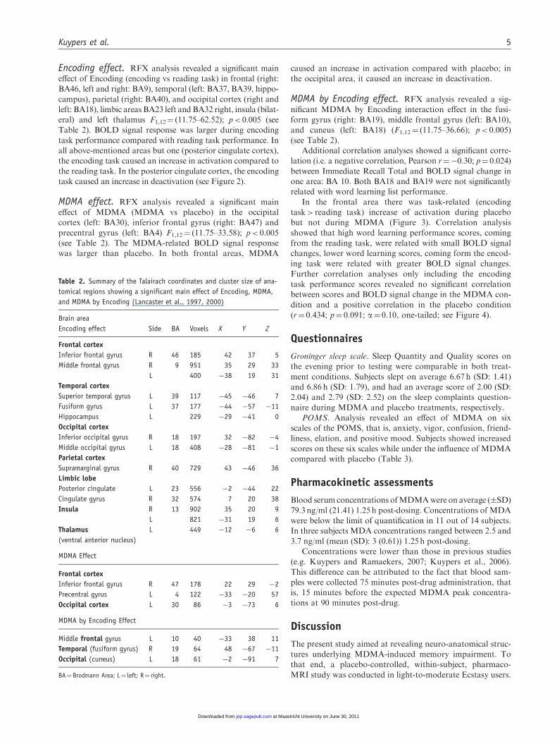
Encoding effect. RFX analysis revealed a significant maineffect of Encoding (encoding vs reading task) in frontal (right:BA46, left and right: BA9), temporal (left: BA37, BA39, hippo-
campus), parietal (right: BA40), and occipital cortex (right andleft: BA18), limbic areas BA23 left and BA32 right, insula (bilat-eral) and left thalamus F1,12¼ (11.75–62.52); p< 0.005 (seeTable 2). BOLD signal response was larger during encoding
task performance compared with reading task performance. Inall above-mentioned areas but one (posterior cingulate cortex),the encoding task caused an increase in activation compared to
the reading task. In the posterior cingulate cortex, the encodingtask caused an increase in deactivation (see Figure 2).
MDMA effect. RFX analysis revealed a significant maineffect of MDMA (MDMA vs placebo) in the occipitalcortex (left: BA30), inferior frontal gyrus (right: BA47) and
precentral gyrus (left: BA4) F1,12¼ (11.75–33.58); p< 0.005(see Table 2). The MDMA-related BOLD signal responsewas larger than placebo. In both frontal areas, MDMA
caused an increase in activation compared with placebo; inthe occipital area, it caused an increase in deactivation.
MDMA by Encoding effect. RFX analysis revealed a sig-nificant MDMA by Encoding interaction effect in the fusi-form gyrus (right: BA19), middle frontal gyrus (left: BA10),and cuneus (left: BA18) (F1,12¼ (11.75–36.66); p< 0.005)
(see Table 2).Additional correlation analyses showed a significant corre-
lation (i.e. a negative correlation, Pearson r¼�0.30; p¼ 0.024)
between Immediate Recall Total and BOLD signal change inone area: BA 10. Both BA18 and BA19 were not significantlyrelated with word learning list performance.
In the frontal area there was task-related (encodingtask> reading task) increase of activation during placebobut not during MDMA (Figure 3). Correlation analysis
showed that high word learning performance scores, comingfrom the reading task, were related with small BOLD signalchanges, lower word learning scores, coming form the encod-ing task were related with greater BOLD signal changes.
Further correlation analyses only including the encodingtask performance scores revealed no significant correlationbetween scores and BOLD signal change in the MDMA con-
dition and a positive correlation in the placebo condition(r¼ 0.434; p¼ 0.091; a¼ 0.10, one-tailed; see Figure 4).
Questionnaires
Groninger sleep scale. Sleep Quantity and Quality scores onthe evening prior to testing were comparable in both treat-
ment conditions. Subjects slept on average 6.67 h (SD: 1.41)and 6.86 h (SD: 1.79), and had an average score of 2.00 (SD:2.04) and 2.79 (SD: 2.52) on the sleep complaints question-
naire during MDMA and placebo treatments, respectively.POMS. Analysis revealed an effect of MDMA on six
scales of the POMS, that is, anxiety, vigor, confusion, friend-
liness, elation, and positive mood. Subjects showed increasedscores on these six scales while under the influence of MDMAcompared with placebo (Table 3).
Pharmacokinetic assessments
Blood serum concentrations ofMDMAwere on average (�SD)
79.3 ng/ml (21.41) 1.25 h post-dosing. Concentrations of MDAwere below the limit of quantification in 11 out of 14 subjects.In three subjects MDA concentrations ranged between 2.5 and
3.7 ng/ml (mean (SD): 3 (0.61)) 1.25 h post-dosing.Concentrations were lower than those in previous studies
(e.g. Kuypers and Ramaekers, 2007; Kuypers et al., 2006).
This difference can be attributed to the fact that blood sam-ples were collected 75 minutes post-drug administration, thatis, 15 minutes before the expected MDMA peak concentra-tions at 90 minutes post-drug.
Discussion
The present study aimed at revealing neuro-anatomical struc-tures underlying MDMA-induced memory impairment. Tothat end, a placebo-controlled, within-subject, pharmaco-
MRI study was conducted in light-to-moderate Ecstasy users.
Table 2. Summary of the Talairach coordinates and cluster size of ana-
tomical regions showing a significant main effect of Encoding, MDMA,
and MDMA by Encoding (Lancaster et al., 1997, 2000)
Brain area
Encoding effect Side BA Voxels X Y Z
Frontal cortex
Inferior frontal gyrus R 46 185 42 37 5
Middle frontal gyrus R 9 951 35 29 33
L 400 �38 19 31
Temporal cortex
Superior temporal gyrus L 39 117 �45 �46 7
Fusiform gyrus L 37 177 �44 �57 �11
Hippocampus L 229 �29 �41 0
Occipital cortex
Inferior occipital gyrus R 18 197 32 �82 �4
Middle occipital gyrus L 18 408 �28 �81 �1
Parietal cortex
Supramarginal gyrus R 40 729 43 �46 36
Limbic lobe
Posterior cingulate L 23 556 �2 �44 22
Cingulate gyrus R 32 574 7 20 38
Insula R 13 902 35 20 9
L 821 �31 19 6
Thalamus
(ventral anterior nucleus)
L 449 �12 �6 6
MDMA Effect
Frontal cortex
Inferior frontal gyrus R 47 178 22 29 �2
Precentral gyrus L 4 122 �33 �20 57
Occipital cortex L 30 86 �3 �73 6
MDMA by Encoding Effect
Middle frontal gyrus L 10 40 �33 38 11
Temporal (fusiform gyrus) R 19 64 48 �67 �11
Occipital (cuneus) L 18 61 �2 �91 7
BA¼Brodmann Area; L¼ left; R¼ right.
Kuypers et al. 5
at Maastricht University on June 30, 2011jop.sagepub.comDownloaded from

Behavioral effects of MDMA
A single dose of MDMA caused an impairment of immedi-ate recall, an indirect measure of word learning (encoding),but not delayed recall in the experimental word list
(encoding task). Under the influence of MDMA, subjectsrecalled on average 3.5 words less in total compared withthe number of words recalled during the placebo condition.
The finding that there was no effect of MDMA on delayedrecall suggests that there was no problem with storageof words. Behavioral data also showed that previouslylearnt words (control word list/reading task) were perfectly
recalled when under the influence of MDMA. In contrastto Quednow et al. (2006), who suggested that defectiverecall was the prime cause of impaired memory perfor-
mance, this suggests that the inferior memory performancein MDMA users is, rather, due to defective or slowerencoding.
Functional effects of Encoding
To determine the Encoding effect on brain activation patterns,
encoding task activation was compared with reading task acti-vation in both treatment conditions. Results showed significantinvolvement of a number of frontal (BA9, 46), temporal
(BA37, 39, hippocampus), parietal (BA40) and occipital(BA18) regions, the insular and limbic cortex, and thalamusduring Encoding in the present paradigm. According to previ-
ous studies in humans and non-human primates a number of
these above-mentioned regions and structures are included in anetwork supporting working memory (Buckner et al., 1999;
Dupont et al., 2002; Gabrieli et al., 1998; Mottaghy et al.,2002; Ojemann et al., 1997; Schacter and Wagner, 1999;Smith et al., 1998). Of interest is the right-sided activation
during Encoding in the dorsolateral prefrontal (BA9, 46)cortex. Previously it has been suggested that higher task com-plexity in a verbal working memory task is associated with
right-sided frontal activation (Mottaghy et al., 2002). In thepresent study, the encoding task was more complex than thecontrol word list as the former required reading and encoding,whereas the latter only required reading processes.
An odd result, perhaps, was the deactivation in the poste-rior cingulate gyrus (BA23) during Encoding. This area ispart of the so-called default brain network, displaying high
resting-state activation as a result of continuously gatheringinformation about the inner and outer world. When the taskat hand demands focused attention, activity in the network,
and thus BA23, is diminished (Gusnard and Raichle, 2001).In addition it has previously been shown that deactivation inBA23 increases with increasing task complexity (McKiernanet al., 2003). Together, this adequately explains the greater
increase in deactivation during the higher demanding encod-ing task condition.
The paradigm in the present study was previously used by
other investigators, though in a slightly different form(Andreasen et al., 1995; Dupont et al., 2002). Dupont et al.(2002) investigated recall and encoding processes and showed
activation in five regions during the encoding process,
Figure 2. Statistical F-map showing activation (encircled with green line and indicated by green arrow) in (from left to right) middle frontal gyrus,
superior temporal gyrus, supramarginal gyrus, and posterior cingulate. In event related plots, corresponding task-related BOLD signal change during the
experimental word list (EWL) or encoding task (blue) and control word list (CWL) or reading task (orange) is shown (for coordinates and number of
voxels, see Table 2).
6 Journal of Psychopharmacology 0(0)
at Maastricht University on June 30, 2011jop.sagepub.comDownloaded from

namely, occipital, left parietal, left superior temporal cortex,
bilaterally ventrolateral frontal cortex, and fusiform gyrus.This activation was observed after subtraction of the experi-mental task-activation (encoding of 17 words) and the control
task-activation (fixation of a single letter). The contrastanalysis of Dupont et al. (2002) and the analysis used inthe present study disclose consistencies concerning theregions active during the encoding of words. These findings
together support the notion that our new paradigm addressedareas that are generally active during encoding of verbalmaterial.
Functional effects of MDMA and limitations. MDMA
caused a significant increase in activation and deactivationin frontal and occipital areas, respectively. A general limita-tion of pharmaco-MRI studies is that drugs may directly
affect the BOLD signal response by vasodilation or vasocon-striction. Because MDMA acts generally on the serotonin(5-HT) system, and 5-HT has vasoconstrictor properties, itis possible that MDMA has a general effect on the cerebral
blood flow (CBF) in the whole brain (Cohen et al., 1996;Frackowiak, 2004; Meyer et al., 2006). The expected physio-logical effect of MDMA on CBF is a general decrease in
BOLD signal response, independent of task type (Brevardet al., 2006; Meyer et al., 2006). The fact that MDMAcaused an increase in activation and deactivation in selective
areas in the brain counteracts the notion that brain activa-tions in the present study were secondary to a general phys-iological effect of MDMA. In support, previous acuteMDMA studies in humans have also shown a specific effect
of MDMA on task-related activation contrary to a generaleffect (Bedi et al., 2009; Ramaekers et al., 2009).
Functional effects of MDMA on Encoding: Interactioneffect. Functional imaging data showed that a single dose of
MDMA caused a selective effect on Encoding-related BOLD
signal response (encoding task minus reading task) in threeregions involved, respectively, in memory processes, visual
recognition, and basic visual processing: that is, the middlefrontal gyrus (BA10), the fusiform gyrus (BA19), and thecuneus (BA18). Functional and behavioral data only corre-
lated in one of these three areas, being the left middle frontalgyrus. BOLD signal response in the left middle frontal gyrus,in the placebo condition, was increased during encoding task
performance but not during reading task performance. Thiswas according to expectation, as the reading task contained‘pre-encoded’ words opposite to the encoding task which con-tained new information that had to be encoded.
Subjects under the influence of MDMA showed a smallincrease in BOLD signal response during encoding and read-ing tasks. The resemblance of a BOLD signal response during
the encoding task and the reading task after taking MDMA isvery noteworthy. It suggests that subjects under influence ofMDMA invested equal effort during both lists. Behavioral
data showed that performance on the reading task was max-imal but the presence of BOLD signal response during this listsuggested that subjects had to invest effort in these ‘pre-encoded’ words, while this was not the case during placebo
treatment. The small increase in BOLD signal responseduring the encoding task in the MDMA condition relativeto the placebo condition could be an indication for the
absence of efficient encoding processes during experimentalword learning under the influence of MDMA. Previousmemory studies have shown that left frontal areas are a cru-
cial link in the memory formation process, as they initiate the
MDMA
40
45
50
55
60
65
70
75
BOLD Signal Change (%)
Co
rrec
t R
ecal
led
Wo
rds
(Nu
mb
er)
Placebo
40
45
50
55
60
65
70
75
BOLD Signal Change (%)C
orr
ect
Rec
alle
d W
ord
s (N
um
ber
)
r= .434; p<.10
–1 –0,8 –0,6 –0,4 –0,2 0 0,2 0,4 0,6 0,8 1
–0,2 0 0,2 0,4 0,6 0,8 1
Figure 4. Correlation between Encoding (experimental word list
performance) and BOLD signal change in BA10 during MDMA and placebo
treatments.
Figure 3. MDMA by Encoding Interaction. Bar chart indicating percent-
age BOLD signal change in BA10. EWL¼ Experimental Word List or
Encoding task; CWL¼ Control Word List or Reading task (for coordinates
and number of voxels, see Table 2).
Kuypers et al. 7
at Maastricht University on June 30, 2011jop.sagepub.comDownloaded from

formation of the memory trace. It is to be expected that dys-functions of these regions can result in defective memory for-
mation (Cabeza and Nyberg, 2000; Kelley et al., 1998; Owenet al., 2005).
Conclusion
A single dose of MDMA impaired memory performance and
affected encoding-related areas (BA10, BA19, BA18) as indi-cated by a change in BOLD signal response during the encod-ing task compared to the reading task, relative to placebo.Behavioral and functional data correlated only in one of
these three mentioned areas, namely BA10. Good perfor-mance, as indicated by a high Immediate Recall Total scorewas related to small BOLD signal change and lower scores to
higher BOLD signal change. The highest scores were obtainedduring the reading task, where performance was maximal, andthe lower scores were obtained during the encoding task.When
looking at the encoding task scores, it was shown that theseonly correlated to BOLD signal change in the placebo but notthe MDMA condition. These results suggest an absence of
effective encoding processing during MDMA influence.The MDMA effect was not due to a general physiological
effect on CBF, as indicated by a selective increase in activa-tion and deactivation in three brain areas. The Encoding
effect showed an absence of a BOLD signal response duringthe reading task, which proved there was no encoding duringthe reading task. This was expected, as the reading task was
pre-memorized or pre-encoded. The behavioral data alsoshowed that the words of the reading task could perfectlybe recalled during MDMA treatment. Together these data
supported the hypothesis that MDMA-induced memoryimpairment was due to defective or slower encoding andnot due to defective retrieval or storage problems.
Funding
This research received no specific grant from any funding agency in
the public, commercial, or not-for-profit sectors.
Conflict of interest
The authors declare no conflicts of interest.
Acknowledgments
We would like to thank Maria del Mar Ramirez Fernandez, Nele
Samyn and Gert De Boeck from the National Institute for
Criminalistics and Criminology (NICC) in Brussels (Belgium), for
analyzing MDMA blood plasma samples. Trial registration
(Nederlands Trialregister): http://www.trialregister.nl/trialreg/
admin/rctview.asp?TC¼1416, NTR number: NTR1416; trial name:
MDMA and memory.
References
Andreasen NC, O’Leary DS, Cizadlo T, et al. (1995) II. PET studies
of memory: novel versus practiced free recall of word lists.
NeuroImage 2: 296–305.
Bedi G, Phan KL, Angstadt M, et al. (2009) Effects of MDMA on
sociability and neural response to social threat and social reward.
Psychopharmacology 207: 73–83.
Brevard ME, Meyer JS, Harder JA, et al. (2006) Imaging brain activ-
ity in conscious monkeys following oral MDMA (‘ecstasy’).Magn
Reson Imaging 24: 707–714.
Buckner RL, Kelley WM and Petersen SE (1999) Frontal cortex con-
tributes to human memory formation. Nat Neurosci 2: 311–314.
Cabeza R and Nyberg L (2000) Imaging cognition II: An empirical
review of 275 PET and fMRI studies. J Cogn Neurosci 12: 1–47.
Cohen Z, Bonvento G, Lacombe P, et al. (1996) Serotonin in the
regulation of brain microcirculation. Prog Neurobiol 50: 335–362.
Craik FIM and Lockhart RS (1972) Levels of processing: A frame-
work for memory research. J Verbal Learn Verbal Behav 11:
671–684.
Cusack R, Cumming N, Bor D, et al. (2005) Automated post-hoc
noise cancellation tool for audio recordings acquired in an MRI
scanner. Hum Brain Mapping 24: 299–304.
De Wit H, Enggasser JL and Richards JB (2002) Acute administra-
tion of d-amphetamine decreases impulsivity in healthy volun-
teers. Neuropsychopharmacology 27: 813–825.
Dupont S, Samson Y, Le Bihan D, et al. (2002) Anatomy of verbal
memory: a functional MRI study. Surg Radiol Anat 24: 57–63.
Frackowiak RS (2004) Functional imaging of cognitive psychophar-
macology. In: Frackowiak RS (ed.) Human Brain Function,
2nd ed. Amsterdam: Elsevier Academic Press, 303–327.
Gabrieli JD, Poldrack RA and Desmond JE (1998) The role of left
prefrontal cortex in language and memory. Proc Natl Acad Sci
USA 95: 906–913.
Gusnard DA and Raichle ME (2001) Searching for a baseline:
Functional imaging and the resting human brain. Nat Rev
Neurosci 2: 685–694.
Table 3. Means (�SEM) of scores on scales of the POMS and t- and p-values
Mean (�SE) t-test placebo-MDMA
Scale MDMA placebo t p-values
Anxiety 6.36 (�0.676) 3.57 (�0.626) 2.724 0.017
Depression 0.21 (�0.114) 0.50 (�0.251) �1.170 0.263
Anger 1.00 (�0.296) 1.14 (�0.345) �0.342 0.738
Vigor 16.07 (�1.510) 13.07 (�1.659) 2.352 0.035
Fatigue 1.86 (�0.512) 1.86 (�0.783) 0.000 1.000
Confusion 6.36 (�0.843) 3.21 (�0.482) 3.217 0.007
Friendliness 20.00 (�1.726) 16.50 (�1.912) 2.511 0.026
Elation 12.64 (�0.809) 10.14 (�0.994) 3.736 0.002
Arousal 14.21 (�2.349) 11.57 (�2.163) 1.281 0.223
Positive mood 12.43 (�0.789) 9.64 (�1.020) 4.044 0.001
8 Journal of Psychopharmacology 0(0)
at Maastricht University on June 30, 2011jop.sagepub.comDownloaded from

Jonides J, Schumacher EH, Smith EE, et al. (1998) The role of pari-
etal cortex in verbal working memory. J Neurosci 18: 5026–5034.
Kelley WM, Miezin FM, McDermott KB, et al. (1998) Hemispheric
specialization in human dorsal frontal cortex and medial temporal
lobe for verbal and nonverbal memory encoding. Neuron 20:
927–936.
Kuypers KP and Ramaekers JG (2007) Acute dose of MDMA (75 mg)
impairs spatial memory for location but leaves contextual process-
ing of visuospatial information unaffected. Psychopharmacology
189: 557–563.
Kuypers KPC andRamaekers JG (2005) Transient memory impairment
after acute dose of 75 mg 3.4-Methylenedioxymethamphetamine.
J Psychopharmacol 19: 633–639.
Kuypers KPC, Samyn N and Ramaekers JG (2006) MDMA and
alcohol effects, combined and alone, on objective and subjective
measures of actual driving performance and psychomotor func-
tion. Psychopharmacology 187: 467–475.
Lancaster JL, Rainey LH, Summerlin JL, et al. (1997) Automated
labeling of the human brain: A preliminary report on the devel-
opment and evaluation of a forward-transform method. Hum
Brain Mapping 5: 238–242.
Lancaster JL, Woldorff MG, Parsons LM, et al. (2000) Automated
Talairach atlas labels for functional brain mapping. Hum Brain
Mapping 10: 120–131.
McAvoy MP and Buckner RL (2001) Cluster size thresholds for
assessments of significant activation in fMRI. NeuroImage 6: 198.
McKiernan KA, Kaufman JN, Kucera-Thompson J, et al. (2003) A
parametric manipulation of factors affecting task-induced deacti-
vation in functional neuroimaging. J Cogn Neurosci 15: 394–408.
Meyer JS, Brevard ME, Piper BJ, et al. (2006) Neural effects of
MDMA as determined by functional magnetic resonance and
magnetic resonance spectroscopy in awake marmoset monkeys.
Ann NY Acad Sci 1074: 365–376.
Mottaghy FM, Doring T, Muller Gartner HW, et al. (2002) Bilateral
parieto-frontal network for verbal working memory: an
interference approach using repetitive transcranial magnetic stim-
ulation (rTMS). Eur J Neurosci 16: 1627–1632.
Mulder-Hajonides van der Meulen WREH, Wijnberg JR, Hollanders
JJ, et al. (1980) Measurement of subjective sleep quality. Fifth
European Congress on Sleep Research. Amsterdam.
Ojemann JG, Buckner RL, Corbetta M, et al. (1997) Imaging studies
of memory and attention. Neurosurg Clin N Am 8: 307–319.
Owen AM, McMillan KM, Laird AR, et al. (2005) N-back working
memory paradigm: a meta-analysis of normative functional neu-
roimaging studies. Hum Brain Mapping 25: 46–59.
Poldrack RA, Fletcher P, Henson RN, et al. (2008) Guidelines for
reporting an fMRI study. NeuroImage 40: 409–414.
Quednow BB, Jessen F, Kuhn KU, et al. (2006) Memory deficits in
abstinent MDMA (ecstasy) users: neuropsychological evidence of
frontal dysfunction. J Psychopharmacol 20: 373–384.
Ramaekers JG, Kuypers KPC, Wingen M, et al. (2009) Involvement
of inferior parietal lobules in prospective memory impairment
during acute MDMA (Ecstasy) intoxication: an event-related
fMRI study. Neuropsychopharmacology 34: 1641–1648.
Rey A (1958) The Clinical Examination in Psychology/L’Examen
Clinique en Psychologie. Oxford, UK: Presses Universitaries De
France.
Schacter DL and Wagner AD (1999) Medial temporal lobe activa-
tions in fMRI and PET studies of episodic encoding and retrieval.
Hippocampus 9: 7–24.
Smith EE, Jonides J, Marshuetz C, et al. (1998) Components of
verbal working memory: evidence from neuroimaging. Proc
Natl Acad Sci USA 95: 876–882.
Squire LR and Zola Morgan S (1991) The medial temporal lobe
memory system. Science 253: 1380–1386.
Ward J, Hall K and Haslam C (2006) Patterns of memory dysfunc-
tion in current and 2-year abstinent MDMA users. J Clin Exp
Neuropsychol 28: 306–324.
Kuypers et al. 9
at Maastricht University on June 30, 2011jop.sagepub.comDownloaded from