Changes in interleukin-1 signal modulators induced by 3,4-methylenedioxymethamphetamine (MDMA):...
14
RESEARCH Open Access Changes in interleukin-1 signal modulators induced by 3,4-methylenedioxymethamphetamine (MDMA): regulation by CB2 receptors and implications for neurotoxicity Elisa Torres, Maria D Gutierrez-Lopez, Andrea Mayado, Ana Rubio, Esther O’Shea and Maria I Colado * Abstract Background: 3,4-Methylenedioxymethamphetamine (MDMA) produces a neuroinflammatory reaction in rat brain characterized by an increase in interleukin-1 beta (IL-1b) and microglial activation. The CB2 receptor agonist JWH- 015 reduces both these changes and partially protects against MDMA-induced neurotoxicity. We have examined MDMA-induced changes in IL-1 receptor antagonist (IL-1ra) levels and IL-1 receptor type I (IL-1RI) expression and the effects of JWH-015. The cellular location of IL-1b and IL-1RI was also examined. MDMA-treated animals were given the soluble form of IL-1RI (sIL-1RI) and neurotoxic effects examined. Methods: Dark Agouti rats received MDMA (12.5 mg/kg, i.p.) and levels of IL-1ra and expression of IL-1RI measured 1 h, 3 h or 6 h later. JWH-015 (2.4 mg/kg, i.p.) was injected 48 h, 24 h and 0.5 h before MDMA and IL-1ra and IL- 1RI measured. For localization studies, animals were sacrificed 1 h or 3 h following MDMA and stained for IL-1b or IL-1RI in combination with neuronal and microglial markers. sIL-1RI (3 μg/animal; i.c.v.) was administered 5 min before MDMA and 3 h later. 5-HT transporter density was determined 7 days after MDMA injection. Results: MDMA produced an increase in IL-ra levels and a decrease in IL-1RI expression in hypothalamus which was prevented by CB2 receptor activation. IL-1RI expression was localized on neuronal cell bodies while IL-1b expression was observed in microglial cells following MDMA. sIL-1RI potentiated MDMA-induced neurotoxicity. MDMA also increased IgG immunostaining indicating that blood brain-barrier permeability was compromised. Conclusions: In summary, MDMA produces changes in IL-1 signal modulators which are modified by CB2 receptor activation. These results indicate that IL-1b may play a partial role in MDMA-induced neurotoxicity. Background Administration of the recreationally used drug 3,4- methylenedioxymethamphetamine (MDMA) induces a long-term toxicity of 5-HT nerve terminals in several areas of rat brain. The damage is reflected by a substan- tial decrease in the concentration of 5-HT and its meta- bolite, (5-HIAA), a reduction in the density of 5-HT uptake sites labelled with [ 3 H]-paroxetine [1-4], a decrease in tryptophan hydroxylase activity [5] and a decrease in the immunoreactivity of fine 5-HT axons in cortex, striatum and hippocampus [6]. MDMA produces signs of neuroinflammation reflected as microglial activation and an increase in the release of interleukin-1b (IL-1b). Within 1-3 h following MDMA administration to rats there is an acute increase in IL-1b concentration in the hypothalamus and frontal cortex. The up-regulation of IL-1b levels appears at an early time-point after MDMA and is of short duration, levels returning to basal values 12 h after drug injection [7]. The IL-1b response is partially a consequence of MDMA hyperthermia and seems to be involved in the long-term 5-HT neurotoxicity since the i.c.v. injection of IL-1b in the rat brain enhances the long-lasting reduction in 5-HT transporters and 5-HT concentration induced by MDMA [8]. Nevertheless, the contribution * Correspondence: [email protected] Departamento de Farmacologia, Facultad de Medicina, Universidad Complutense, 28040 Madrid, Spain Torres et al. Journal of Neuroinflammation 2011, 8:53 http://www.jneuroinflammation.com/content/8/1/53 JOURNAL OF NEUROINFLAMMATION © 2011 Torres et al; licensee BioMed Central Ltd. This is an Open Access article distributed under the terms of the Creative Commons Attribution License (http://creativecommons.org/licenses/by/2.0), which permits unrestricted use, distribution, and reproduction in any medium, provided the original work is properly cited.
-
Upload
independent -
Category
Documents
-
view
0 -
download
0
Transcript of Changes in interleukin-1 signal modulators induced by 3,4-methylenedioxymethamphetamine (MDMA):...
RESEARCH Open Access
Changes in interleukin-1 signal modulators inducedby 3,4-methylenedioxymethamphetamine (MDMA):regulation by CB2 receptors and implications forneurotoxicityElisa Torres, Maria D Gutierrez-Lopez, Andrea Mayado, Ana Rubio, Esther O’Shea and Maria I Colado*
Abstract
Background: 3,4-Methylenedioxymethamphetamine (MDMA) produces a neuroinflammatory reaction in rat braincharacterized by an increase in interleukin-1 beta (IL-1b) and microglial activation. The CB2 receptor agonist JWH-015 reduces both these changes and partially protects against MDMA-induced neurotoxicity. We have examinedMDMA-induced changes in IL-1 receptor antagonist (IL-1ra) levels and IL-1 receptor type I (IL-1RI) expression andthe effects of JWH-015. The cellular location of IL-1b and IL-1RI was also examined. MDMA-treated animals weregiven the soluble form of IL-1RI (sIL-1RI) and neurotoxic effects examined.
Methods: Dark Agouti rats received MDMA (12.5 mg/kg, i.p.) and levels of IL-1ra and expression of IL-1RI measured1 h, 3 h or 6 h later. JWH-015 (2.4 mg/kg, i.p.) was injected 48 h, 24 h and 0.5 h before MDMA and IL-1ra and IL-1RI measured. For localization studies, animals were sacrificed 1 h or 3 h following MDMA and stained for IL-1b orIL-1RI in combination with neuronal and microglial markers. sIL-1RI (3 μg/animal; i.c.v.) was administered 5 minbefore MDMA and 3 h later. 5-HT transporter density was determined 7 days after MDMA injection.
Results: MDMA produced an increase in IL-ra levels and a decrease in IL-1RI expression in hypothalamus whichwas prevented by CB2 receptor activation. IL-1RI expression was localized on neuronal cell bodies while IL-1bexpression was observed in microglial cells following MDMA. sIL-1RI potentiated MDMA-induced neurotoxicity.MDMA also increased IgG immunostaining indicating that blood brain-barrier permeability was compromised.
Conclusions: In summary, MDMA produces changes in IL-1 signal modulators which are modified by CB2 receptoractivation. These results indicate that IL-1b may play a partial role in MDMA-induced neurotoxicity.
BackgroundAdministration of the recreationally used drug 3,4-methylenedioxymethamphetamine (MDMA) induces along-term toxicity of 5-HT nerve terminals in severalareas of rat brain. The damage is reflected by a substan-tial decrease in the concentration of 5-HT and its meta-bolite, (5-HIAA), a reduction in the density of 5-HTuptake sites labelled with [3H]-paroxetine [1-4], adecrease in tryptophan hydroxylase activity [5] and adecrease in the immunoreactivity of fine 5-HT axons incortex, striatum and hippocampus [6].
MDMA produces signs of neuroinflammationreflected as microglial activation and an increase in therelease of interleukin-1b (IL-1b). Within 1-3 h followingMDMA administration to rats there is an acute increasein IL-1b concentration in the hypothalamus and frontalcortex. The up-regulation of IL-1b levels appears at anearly time-point after MDMA and is of short duration,levels returning to basal values 12 h after drug injection[7]. The IL-1b response is partially a consequence ofMDMA hyperthermia and seems to be involved in thelong-term 5-HT neurotoxicity since the i.c.v. injectionof IL-1b in the rat brain enhances the long-lastingreduction in 5-HT transporters and 5-HT concentrationinduced by MDMA [8]. Nevertheless, the contribution
* Correspondence: [email protected] de Farmacologia, Facultad de Medicina, UniversidadComplutense, 28040 Madrid, Spain
Torres et al. Journal of Neuroinflammation 2011, 8:53http://www.jneuroinflammation.com/content/8/1/53
JOURNAL OF NEUROINFLAMMATION
© 2011 Torres et al; licensee BioMed Central Ltd. This is an Open Access article distributed under the terms of the Creative CommonsAttribution License (http://creativecommons.org/licenses/by/2.0), which permits unrestricted use, distribution, and reproduction inany medium, provided the original work is properly cited.
of IL-1b to MDMA-induced neurotoxicity is not wellestablished yet since IL-1b potentiates MDMA-induceddamage at doses that exacerbates the immediatehyperthermic response induced by the drug. Therefore,actions of IL-1b on body temperature may contribute tothe injury. In addition to increasing IL-1b, MDMA alsoincreased the density of peripheral benzodiazepinereceptor binding sites, labelled with [3H]-PK11195 ([(1-(2-chlorophenyl)-N-methyl-N-(1-methylpropyl)3-isoqui-nolinecarboxamide)], in hypothalamus and frontal cortexof the rat. This parameter could be reflecting an activa-tion of microglia as revealed by subsequent immunohis-tochemical studies which show an increased in OX-42immunoreactivity in both brain areas which is evident 3h after drug injection and remains 24 h later [7].Recent evidence indicates that MDMA increases the
expression of cannabinoid CB2 receptors in frontal cor-tex and hypothalamus shortly after injection and thatCB2 immunoreactivity co-localizes with the staining forthe microglial cell marker OX-42, indicating the presenceof CB2 receptors in microglial cells. Repeated administra-tion of the CB2 agonist JWH-015 prevents the MDMA-induced microglial activation, reduces IL-1b release andprovides partial protection against the serotonergic neu-rotoxicity induced by the drug [9]. Overexpression of thecannabinoid CB2 receptor in microglia during non-immune and immune pathological conditions is thoughtto be aimed at controlling the production of neurotoxicfactors such as pro-inflammatory cytokines.We have now examined the effect of JWH-015 on the
changes induced by MDMA on endogenous IL-1 signalmodulators in the frontal cortex and hypothalamus andexplored the relevance of these actions on the protectionexerted by this compound against MDMA neurotoxicity.The specific objectives of this study were as follows: 1) todetermine the effects of MDMA on the levels of the IL-1receptor antagonist (IL-1ra) and on the expression of theIL-1 receptor type I (IL-1RI) and evaluate the effect ofJWH-015 on these changes; 2) to define the cellular loca-tion of IL-1b and IL-1RI by immunohistochemical stain-ing; 3) to study the effect of i.c.v. administration of thesoluble IL-1 receptor type I (sIL-1RI) on MDMA-inducedhyperthermia and neurotoxicity; and 4) to evaluate theeffect of MDMA on blood-brain barrier (BBB) permeabil-ity. There is evidence showing that BBB integrity isaltered by a number of factors including increased levelsof inflammatory cytokines [10] and free radicals [11-13],factors/mediators which are increased following MDMAinjection [3,7].
MethodsAnimals and drug administrationMale Dark Agouti rats (175-200 g, Harlan LaboratoriesModels, Barcelona) were used. MDMA induces a
reproducible acute hyperthermic response in this strain[14,15] and also a reproducible long-term neurotoxicloss of 5-HT after a single dose [14]. Rats were housedin groups of 5 in conditions of constant temperature(21°C ± 2°C) and a 12 h light/dark cycle (lights on: 08 h00 min) and given free access to food and water.MDMA was given at the dose of 12.5 mg/kg (i.p.), ani-mals being sacrificed 1 h, 3 h, 6 h or 7 days after treat-ment. Room temperature at the time of MDMAadministration was 21-22°C. The cannabinoid CB2 ago-nist, JWH-015 (2.4 mg/kg, i.p.), was given 48 h, 24 hand 0.5 h before MDMA. Doses and protocols of thecannabinoid agent used have been shown previously tobe effective at modulating microglial functions [9,16,17].MDMA (LIPOMED, Arlesheim, Suiza) was dissolved
in saline (0.9% NaCl) and given in a volume of 1 mL/kg.Dose is reported in terms of the base.JWH-015 was initially dissolved in DMSO (10 mg/
mL), then diluted with 5% BSA in PBS and administeredin a volume of 2 mL/kg.Studies were performed in two brain areas, frontal
cortex and hypothalamus since previous studies [7,8]showed that MDMA administration to Dark Agouti ratsinduces an increase in IL-1b levels in the hypothalamusand frontal cortex and that MDMA alters, in a region-specific manner, the mechanisms which regulate IL-1bproduction in the brain of Dark Agouti rats.All experimental procedures were performed in accor-
dance with the guidelines of the Animal Welfare Com-mittee of the Universidad Complutense de Madrid(following European Council Directives 86/609/CEE and2003/65/CE).
Measurement of rectal temperatureImmediately before and up to 6 h after MDMA injec-tion, temperature was measured by use of a digital read-out thermocouple (BAT12 thermometer, Physitemp, NJ,USA) with a resolution of 0.1°C and accuracy of ± 0.1°Cattached to a RET-2 Rodent Sensor which was inserted2.5 cm into the rectum of the rat, the animal beinglightly restrained by holding it in the hand. A steadyreadout was obtained within 10 s of probe insertion.
IL-1ra immunoassaysBrain levels of IL-1ra were determined using a commer-cially available sandwich enzyme-linked immunosorbentassay (ELISA) system (human IL-1ra/IL-1F3, Quanti-kine® Immunoassay, R&D Systems, Minneapolis, MN,USA). Samples were prepared by homogenization offrontal cortex and hypothalamus in 6 volumes of ice-cold buffer containing 50 mM Tris, 320 mM sucrose, 1mM dithiothreitol, 10 μg/mL leupeptin, 10 μg/mL soy-bean trypsin, 2 μg/mL aprotinin and 0.2% phenantroline,pH 7.0. Samples were centrifuged at 14 000 × g for 10
Torres et al. Journal of Neuroinflammation 2011, 8:53http://www.jneuroinflammation.com/content/8/1/53
Page 2 of 14
min at 4°C. Protein was determined in the supernatant[18]. Samples were assayed in triplicate following themanufacturer’s guidelines. The quantification of IL-1rawas performed using a standard curve of increasing con-centrations of IL-1ra (31.2-2000 pg/mL). The opticaldensity of each well was determined using a microplatereader (ELX808 IU, Ultra Microplate Reader, Bio-TekInstruments, Inc) set to 450 nm (correction wavelengthset at 540 nm). Intra-assay and inter-assay variationswere less than 5% and 15%, respectively.
ImmunohistochemistryRats were anesthetized with sodium pentobarbital andperfused transcardially through the left ventricle with200 mL of phosphate buffered saline (0.1 M PBS, pH7.4) followed by 200 mL of 4% paraformaldehyde-PBS.Brains were removed, postfixed in the same solution for4 h at room temperature and cryoprotected by immer-sion in 30% sucrose-PBS at 4°C. Brains were then frozenin powdered dry ice and stored at -80°C. The brainswere sliced at 40 μm in the coronal plane through thewhole frontal cortex and hypothalamus, and stored incryoprotectant solution. Immunohistochemical studieswere performed in hypothalamus and the sections werelocalized using a rat brain stereotaxic atlas [19].For double labelling studies, cerebral free-floating sec-
tions were blocked by incubation with 0.5% BSA, 10%normal goat serum and 0.1% Triton X-100 for 30 minand incubated at 4°C with the appropriate primary anti-bodies (OX-42, Serotec, 1:400; anti-NeuN, Millipore,1:100; anti rat-IL-1b, R&D Systems, 1:200; anti IL-1RI,Santa Cruz Biotechnology, 1:200) followed by the AlexaFluor™ 488 donkey anti-mouse IgG (1:200), AlexaFluor™ 594 donkey anti-rabbit IgG or Alexa Fluor™555 donkey anti-goat IgG (Invitrogen, Spain) secondaryantibodies (1:500) and mounted in ProLong®Gold (Invi-trogen). Images were acquired sequentially on a LeicaTCS-SP2AOBS confocal microscope (Leica Microsys-tems, Heidelberg, Germany) for each fluorophore toavoid any cross-signal between them. Control experi-ments were performed in which sections were stainedwith each of the secondary antibodies or with the com-bination of them to rule out the possibility of reactionbetween them and images were taken using the samesettings for each antibody staining.IgG leakage from serum into the brain was assessed as
a marker of vasculature damage. Rats were anesthetizedwith sodium pentobarbital and sacrificed. Brains werethen frozen in powdered dry ice and stored at -80°C.The brains were sliced at 20 μm in the coronal planethrough the hypothalamus. After three washes with 0.1M PBS, sections were blocked by incubation with 0.5%BSA, 10% horse serum and 0.1% Triton X-100 for 60min, incubated at 4°C overnight with the antibody Alexa
FluorTM 488 donkey anti-rat IgG (1:500) and coveredwith ProLong®Gold (Invitrogen). Images were acquiredas described above and ImageJ (NIH) software was usedfor image analysis. All images were converted to grayscale and blood vessels were outlined to provide an inte-grated gray scale value for statistical analysis.
Western blot for IL-1RI immunoreactivityRats were killed by cervical dislocation and decapitation1 h, 3 h or 6 h after MDMA administration, the brainsrapidly removed and frontal cortex and hypothalamusdissected out on ice. Tissue was homogenized by sonica-tion in ice-cold buffer containing 50 mM Tris, 320 mMsucrose and a number of protease inhibitors (CompleteMini, Roche, Madrid, Spain). Samples were centrifugedat 14 000 × g for 15 min at 4°C and protein determinedin the supernatant [18].The samples were boiled in laemmli buffer and pro-
teins were resolved by 10% SDS-polyacrylamide gel elec-trophoresis (SDS-PAGE) and transferred to PVDFmembranes. Nonspecific binding was blocked by incuba-tion for 2 h in TBS buffer containing 5% skimmed milk.Membranes were incubated overnight at 4°C with thepolyclonal anti IL-1RI as primary antibody (1:200) fol-lowed by incubation with goat anti-rabbit IgG-horserad-ish peroxidase (Amersham GE; 1:2000) for 2 h. Equalprotein sample loading was confirmed by quantificationof the b-actin signal.
Quantification of IL-1RI immunoreactivityImmunoreactivity was detected with an enhanced chemi-luminescence (ECL) western blot detection system(Amersham Bioscience), followed by exposure to Amer-sham Hyperfilm ECL for 1-5 min. Different film exposuretimes were used to ensure that bands were not saturated.Quantification of specific bands on the film was per-formed using the Quantity One (USA) program. EachIL1-RI band density was normalised for protein contentby referring it to its b-actin band density and then theIL1-RI expression in the different experimental condi-tions was expressed as a percentage of the control group.
I.c.v. sIL-1RI administrationRats were anesthetized with a mixture of isoflurane(3.5% for induction, 1-2% for maintenance; flow rate 1.5L/min) and nitrous oxygen/oxygen mixture (30/70%) inair and placed in a stereotaxic frame secured in a Kopfstereotaxic frame with the tooth bar 3.3 mm belowinteraural zero. A 22 G guide cannula was implanted inthe right lateral ventricle (according to the followingcoordinates: 7.9 mm rostral to the interaural line, 0.8mm lateral to the midline and 3.1 mm from the skullsurface; [19]). The cannula was secured to the skull aspreviously described [20]. I.c.v. injections took place 5
Torres et al. Journal of Neuroinflammation 2011, 8:53http://www.jneuroinflammation.com/content/8/1/53
Page 3 of 14
days after surgery through a 28 G injector (Plastics One,Roanoke, VA, USA) fitted in the guide cannula whichprotruded 1 mm beyond the guide. sIL-1RI (Sigma-Aldrich, Madrid, Spain) (3 μg/animal in 5 μl over 5min) or PBS containing 0.1% BSA were administered i.c.v. 5 min before and 3 h after MDMA (12.5 mg/kg, i.p.)or saline (1 mL/kg, i.p.).
Quantification of 5-HT transporter density by [3H]-paroxetine binding[3H]-Paroxetine binding was measured in fresh hypotha-lamical and cortical tissue [2]. Briefly, tissue was homo-genized in ice-cold Tris-HCl (50 mM; pH 7.4)containing NaCl (120 mM) and KCl (5 mM) using anUltra-Turrax. The homogenate was centrifuged at 30000 × g for 10 min at 4°C. The supernatant was dis-carded and the wash procedure repeated twice more.The pellet was finally resuspended in the Tris buffer ata concentration of 10 mg tissue/mL. Aliquots of tissue(800 μL) were incubated with [3H]-paroxetine (1 nM)for 90 min at room temperature in the absence and pre-sence of 5-HT (100 μM) for determination of total andnon-specific binding, respectively. Assays were termi-nated by rapid filtration through glass fibre filters andradioactivity determined by scintillation spectrometry.Protein was determined by the method of Lowry [21].
Measurement of 5-HT concentrationSeven days after MDMA administration, rats were killedby cervical dislocation and decapitation, the brainsrapidly removed and frontal cortex dissected out on ice.Tissue was homogenized and 5-HT measured by HPLCwith electrochemical detection [3]. Briefly, the mobilephase consisted of KH2PO4 (0.05 M), octanesulfonicacid (0.16 mM), EDTA (0.1 mM) and methanol (16%),and was adjusted to pH 3 with phosphoric acid, filteredand degassed. The flow rate was 1 mL/min and theworking electrode potential was set at +0.4 V.The HPLC system consisted of a pump (Waters510)
linked to an automatic sample injector (Loop 200 μL,Waters 717 plus Autosampler), a stainless steelreversed-phase column (Spherisorb ODS2, 5 μm, 150 ×4.6 mm) fitted with a precolumn, and a coulometricdetector (Coulochem II, Esa, USA). The current pro-duced was monitored by using an integration softwarepackage (Unipoint, Gilson).
StatisticsData from the ELISA, quantification of immunoreactiv-ity and serotonergic parameters were analyzed usingone-way ANOVA followed by the Newman-Keuls multi-ple-comparisons test when a significant F value wasobtained. Statistical analyses of the temperature mea-surements were performed by two-way ANOVA with
repeated measures using treatment as the between sub-jects factor and time as the repeated measure followedby Bonferroni as post test (GraphPad Prism 5).
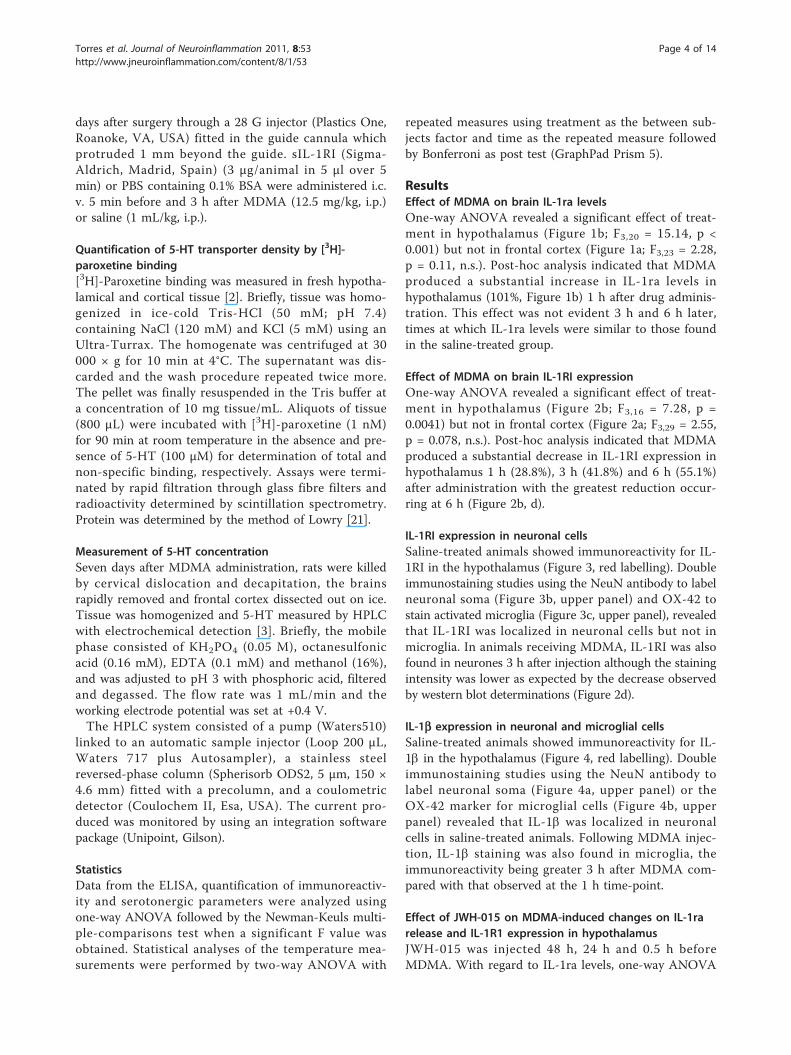
ResultsEffect of MDMA on brain IL-1ra levelsOne-way ANOVA revealed a significant effect of treat-ment in hypothalamus (Figure 1b; F3,20 = 15.14, p <0.001) but not in frontal cortex (Figure 1a; F3,23 = 2.28,p = 0.11, n.s.). Post-hoc analysis indicated that MDMAproduced a substantial increase in IL-1ra levels inhypothalamus (101%, Figure 1b) 1 h after drug adminis-tration. This effect was not evident 3 h and 6 h later,times at which IL-1ra levels were similar to those foundin the saline-treated group.
Effect of MDMA on brain IL-1RI expressionOne-way ANOVA revealed a significant effect of treat-ment in hypothalamus (Figure 2b; F3,16 = 7.28, p =0.0041) but not in frontal cortex (Figure 2a; F3,29 = 2.55,p = 0.078, n.s.). Post-hoc analysis indicated that MDMAproduced a substantial decrease in IL-1RI expression inhypothalamus 1 h (28.8%), 3 h (41.8%) and 6 h (55.1%)after administration with the greatest reduction occur-ring at 6 h (Figure 2b, d).
IL-1RI expression in neuronal cellsSaline-treated animals showed immunoreactivity for IL-1RI in the hypothalamus (Figure 3, red labelling). Doubleimmunostaining studies using the NeuN antibody to labelneuronal soma (Figure 3b, upper panel) and OX-42 tostain activated microglia (Figure 3c, upper panel), revealedthat IL-1RI was localized in neuronal cells but not inmicroglia. In animals receiving MDMA, IL-1RI was alsofound in neurones 3 h after injection although the stainingintensity was lower as expected by the decrease observedby western blot determinations (Figure 2d).
IL-1b expression in neuronal and microglial cellsSaline-treated animals showed immunoreactivity for IL-1b in the hypothalamus (Figure 4, red labelling). Doubleimmunostaining studies using the NeuN antibody tolabel neuronal soma (Figure 4a, upper panel) or theOX-42 marker for microglial cells (Figure 4b, upperpanel) revealed that IL-1b was localized in neuronalcells in saline-treated animals. Following MDMA injec-tion, IL-1b staining was also found in microglia, theimmunoreactivity being greater 3 h after MDMA com-pared with that observed at the 1 h time-point.
Effect of JWH-015 on MDMA-induced changes on IL-1rarelease and IL-1R1 expression in hypothalamusJWH-015 was injected 48 h, 24 h and 0.5 h beforeMDMA. With regard to IL-1ra levels, one-way ANOVA
Torres et al. Journal of Neuroinflammation 2011, 8:53http://www.jneuroinflammation.com/content/8/1/53
Page 4 of 14
revealed a significant effect of treatment (Figure 5a; F3,27= 13.95, p < 0.0001). Post-hoc analysis indicated thatJWH-015 significantly reduced the increase in IL-1ralevels induced by MDMA (126.6%, Figure 5a) such thatthe increase in IL-1ra levels was 32.7%. JWH-015 didnot alter IL-1ra levels in the brain of saline-treated rats.
IL-1RI expression was studied by western blot (Figure5c). Statistical analysis by one-way ANOVA revealed asignificant effect of treatment in hypothalamus (Figure5b; F3,25 = 4.56, p = 0.013). Post-hoc analysis indicatedthat JWH-015 prevented the reduction in IL-1RI expres-sion induced by MDMA (Figure 5b) such that theimmunoreactivity of IL-1RI was similar to that observedin the vehicle-treated group. JWH-015 did not alter IL-1RI expression in the brain of saline-treated rats.JWH-015 affected neither the hyperthermia of MDMA
nor the rectal temperature of saline-treated animals (Fig-ure 5d, e). Two-way ANOVA indicated that there was asignificant effect of treatment (Figure 5e: F3,120 = 45,7, p< 0.0001; Figure 5d: F3,154 = 152,9, p < 0.0001), time (Fig-ure 5e: F4,120 = 27,3, p < 0.0001; Figure 5d: F6,154 = 28,43,p < 0.0001) and interaction (Figure 5e: F12,120 = 10.0, p <0.0001; Figure 5d: F18,154 = 18,9, p < 0.0001). Bonferronipost test revealed that MDMA produced a hyperthermicthat was not modified by JWH-015.
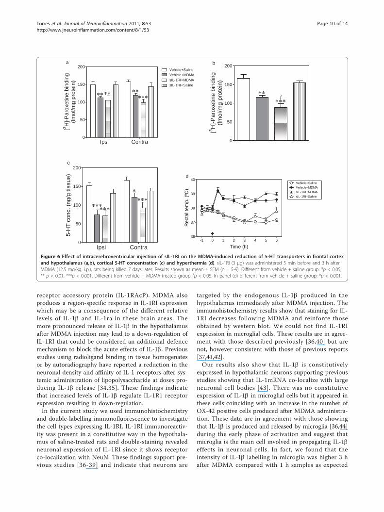
Effect of sIL-1RI on the MDMA-induced neurotoxicitySeven days after MDMA administration, the density of5-HT transporters and the concentration of 5-HT wassignificantly reduced in the ipsi- and contralateral fron-tal cortex (Figure 6a,c). The density of 5-HT transpor-ters was also reduced in hypothalamus (Figure 6b).Administration of sIL-1RI (3 μg, i.c.v.) significantlypotentiated the reduction of 5-HT transporters inducedby MDMA in the hypothalamus (Figure 6b) but it didnot alter the 5-HT parameters in frontal cortex (Figure6a,c). sIL-1RI did not modify the 5-HT levels (Figure 6a,c) nor density of 5-HT transporters (Figure 6a,b) in thebrain of saline-treated rats.
Effect of sIL-1RI on the MDMA-induced hyperthermiaTwo-way ANOVA indicated that there was a significanteffect of treatment (F3,268 = 128.2, p < 0.0001), time(F9,268 = 5.69, p < 0.0001) and interaction (F27,268 = 5.14,p < 0.0001). Bonferroni post test revealed that MDMAproduced a hyperthermic response which peaked 30min after treatment and was sustained for up to 6 hafter treatment. sIL-1RI (3 μg, i.c.v.) affected neither thehyperthermia of MDMA nor the rectal temperature ofsaline-treated animals (Figure 6d).
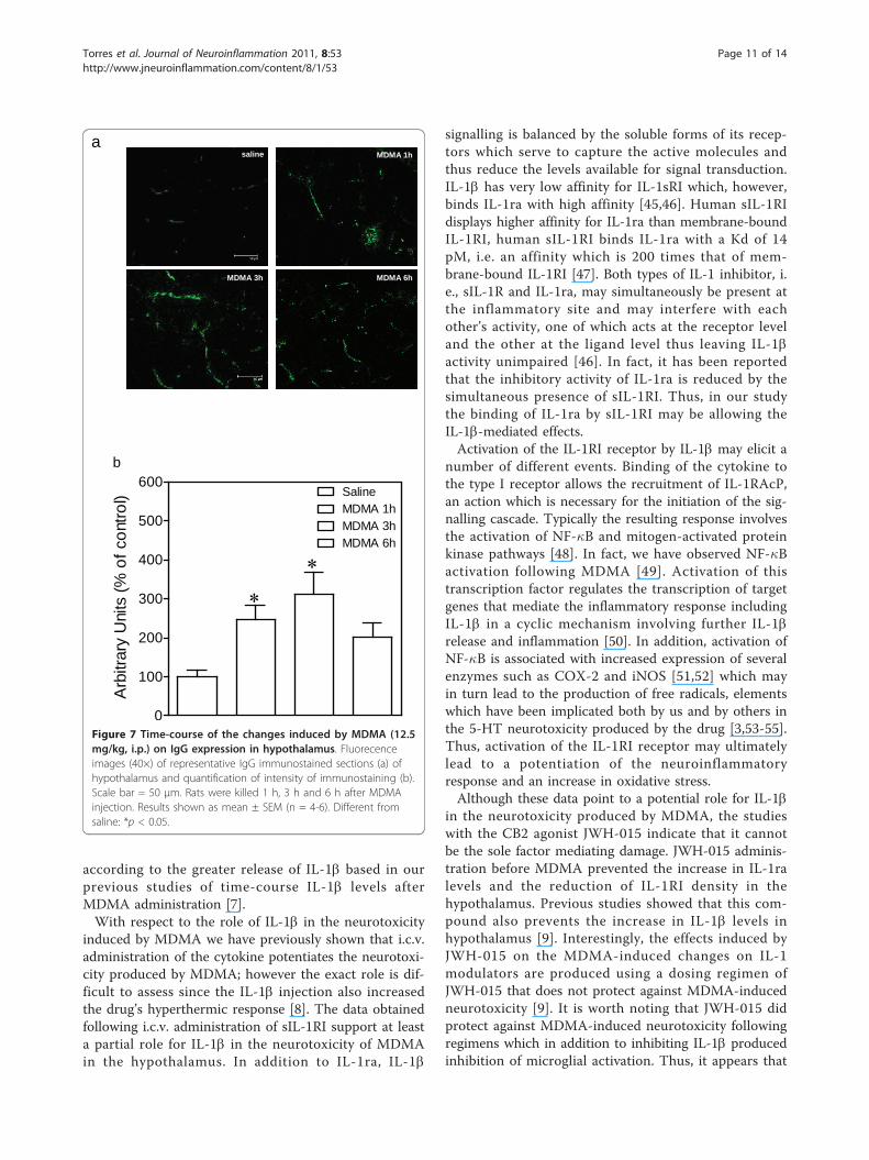
Effect of MDMA on BBB permeabilityBBB permeability was visualized and quantified bymeans of IgG immunostaining (Figure 7a). One-wayANOVA revealed a significant effect of treatment inhypothalamus (Figure 7b; F3,17 = 5.122, p = 0.0113).Post-hoc analysis indicated that MDMA produced asubstantial increase in hypothalamic IgG leakage (147%,Figure 7b) 1 h and (212%, Figure 7b) 3 h post-injection.This effect was not evident at the 6 h time point.
0.0
0.5
1.0
1.5
2.0 SalineMDMA 1hMDMA 3hMDMA 6h
a
IL-1
ra le
vels
(pg/
mg
prot
ein)
0.0
1.5
3.0
4.5
6.0 SalineMDMA 1hMDMA 3hMDMA 6h
b
IL-1
ra le
vels
(pg/
mg
prot
ein)
Figure 1 Time-course of the changes induced by MDMA (12.5mg/kg, i.p.) on IL-1ra (endogenous antagonist of IL-1 receptor)levels in a) frontal cortex and b) hypothalamus. IL-1ra levelswere measured 1 h, 3 h and 6 h after drug injection. Results shownas mean ± SEM (n = 6-8). Different from saline: ***p < 0.001.Different from MDMA-treated group at 1 h: fffp < 0.001.
Torres et al. Journal of Neuroinflammation 2011, 8:53http://www.jneuroinflammation.com/content/8/1/53
Page 5 of 14

DiscussionThe current study demonstrates that MDMA producesdifferential regulation of several modulators of IL-1 sig-nalling in hypothalamus and frontal cortex. Thus, in thehypothalamus, the drug induces a pronounced and tran-sient increase in IL-1ra levels and a sustained markedreduction of IL-1RI density that lasted at least 6 h.Neither of these two changes is observed in frontal cor-tex. The differential effects induced by MDMA on IL-1b signal modulators could be a consequence of theintensity of action of MDMA on IL-1b release in thesebrain areas. Previous studies from our laboratory haveshown that following MDMA, IL-1b levels are muchhigher in the hypothalamus than in frontal cortex [7];the basal levels of IL-1b also being lower in frontalcortex.The IL-1b pro-inflammatory effects are balanced by
IL-1ra, a naturally occurring IL-1 receptor antagonistwhich is structurally related to IL-1b and binds to thetype I IL-1 receptor (IL-1RI) competitively displacingIL-1b from the receptor complex without inducing anybiological response [22]. In fact, there is an up-regula-tion of IL-1ra in various models of neurodegeneration
[23-25] or after infectious stimuli [26]. In line with this,IL-1ra administration to rodents attenuates the neuroin-flammatory response and neuronal damage mediated byIL-1b following several insults [27-32]. The greater IL-1b content in the hypothalamus compared with frontalcortex as early as 1 h after MDMA injection could trig-ger the release of IL-1ra in an attempt to limit theeffects of the released IL-1b. In fact, much evidenceindicates that the effectiveness of IL-1b signal transduc-tion at the IL-1RI depends on the ratio between IL-1raand IL-1b. In vivo studies reveal that relatively high IL-1ra to IL-1b molecular ratios are necessary for an effec-tive blockade of IL-1b effects (at least 25:1, [33]). In ourhands, in the hypothalamus, IL-1b levels increased 400%after MDMA dosing [7], while the increase in IL-1ralevels was 100% (this paper). This means that althoughan increase in IL-1ra levels may contribute to limitingthe effects of IL-1b mediated through IL-1RI, the extentof this increase might not be sufficient to circumventIL-1b signalling.IL-1b signal transduction occurs via IL-1RI that is
expressed at the cell surface; it binds IL-1b and leads tointracellular signalling by association with the IL-1
0
50
100
150
200
250a
SalineMDMA 1hMDMA 3hMDMA 6h
Rel
ativ
e IL
-1R
I lev
els
(arb
itrar
y un
its)
0
50
100
150b
SalineMDMA 1hMDMA 3hMDMA 6h
Rel
ativ
e IL
-1R
I lev
els
(arb
itrar
y un
its)
Figure 2 Time-course of the changes induced by MDMA (12.5 mg/kg, i.p.) on IL-1RI expression in the frontal cortex (a) andhypothalamus (b). IL-1RI expression was measured 1 h, 3 h and 6 h after drug injection. Results shown as mean ± SEM (n = 5-13). Differentfrom saline: *p < 0.05, **p < 0.01. A representative western blot shows immunoreactivity in frontal cortex (c) and hypothalamus (d).
Torres et al. Journal of Neuroinflammation 2011, 8:53http://www.jneuroinflammation.com/content/8/1/53
Page 6 of 14
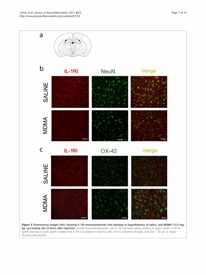
Figure 3 Fluorescence images (40×) showing IL-1RI immunoreactivity (red staining) in hypothalamus of saline- and MDMA (12.5 mg/kg, i.p.)-treated rats (3 hours after injection). Double immunofluorescence with IL-1RI and NeuN (green staining, b: upper panel) or OX-42(green staining, b: lower panel) revealed that IL-1RI is localized on neuronal cells, not on activated microglia. Scale bar = 50 μm. a: imageshowing area studied.
Torres et al. Journal of Neuroinflammation 2011, 8:53http://www.jneuroinflammation.com/content/8/1/53
Page 7 of 14
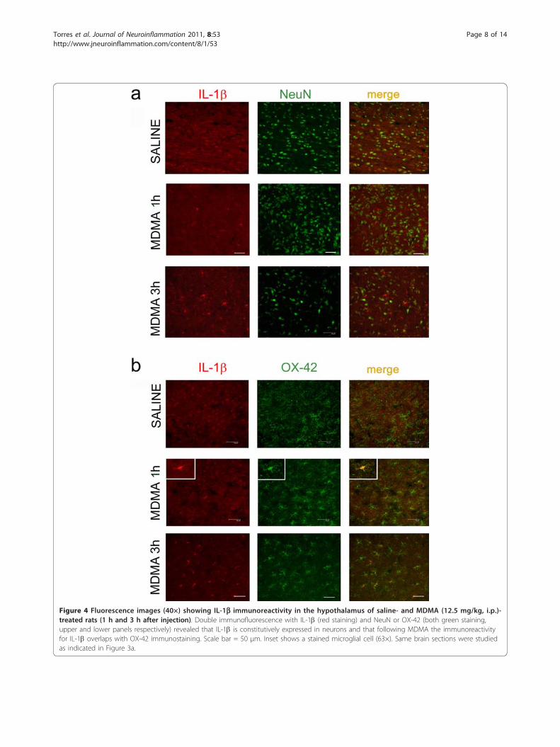
Figure 4 Fluorescence images (40×) showing IL-1b immunoreactivity in the hypothalamus of saline- and MDMA (12.5 mg/kg, i.p.)-treated rats (1 h and 3 h after injection). Double immunofluorescence with IL-1b (red staining) and NeuN or OX-42 (both green staining,upper and lower panels respectively) revealed that IL-1b is constitutively expressed in neurons and that following MDMA the immunoreactivityfor IL-1b overlaps with OX-42 immunostaining. Scale bar = 50 μm. Inset shows a stained microglial cell (63×). Same brain sections were studiedas indicated in Figure 3a.
Torres et al. Journal of Neuroinflammation 2011, 8:53http://www.jneuroinflammation.com/content/8/1/53
Page 8 of 14

0.0
0.5
1.0
1.5
2.0
2.5a
Vehicle+SalineVehicle+MDMAJWH-015+MDMAJWH-015+Saline
IL-1
ra le
vels
(pg/
mg
prot
ein)
0
50
100
150
bVehicle+SalineVehicle+MDMAJWH-015+MDMAJWH-015+Saline
Rel
ativ
e IL
-1R
I lev
els
(arb
itrar
y un
its)
c
-1 0 1 2 3
36
37
38
39
40e
Time (h)
Rec
tal t
emp.
(ºC
)
-1 0 1 2 3
36
37
38
39
40
Vehicle+Saline Vehicle+MDMAJWH-015+MDMA JWH-015+Saline
d
Time (h)
Rec
tal t
emp.
(ºC
)
Figure 5 Effect of JWH-015 on the MDMA-induced changes in IL-1ra levels (a) and IL-1RI expression (b) in hypothalamus. JWH-015(2.4 mg/kg, i.p.) was injected 48 h, 24 h, and 0.5 h before MDMA (12.5 mg/kg, i.p.). For IL-1ra levels determination rats were killed 1 hafter MDMA administration (a). IL-1RI expression was measured 3 h after MDMA (b). The effect of JWH-015 on MDMA-induced hyperthermia isalso shown (d,e). Results shown as mean ± SEM (n = 4-8). Different from vehicle + saline group: *p < 0.05, ***p < 0.001. Different from vehicle +MDMA-treated group: fp < 0.05, ffp < 0.01. In panels (d, e) different from vehicle + saline group: *p < 0.001. A representative western blot showsimmunoreactivity in hypothalamus (c).
Torres et al. Journal of Neuroinflammation 2011, 8:53http://www.jneuroinflammation.com/content/8/1/53
Page 9 of 14
receptor accessory protein (IL-1RAcP). MDMA alsoproduces a region-specific response in IL-1RI expressionwhich may be a consequence of the different relativelevels of IL-1b and IL-1ra in these brain areas. Themore pronounced release of IL-1b in the hypothalamusafter MDMA injection may lead to a down-regulation ofIL-1RI that could be considered an additional defencemechanism to block the acute effects of IL-1b. Previousstudies using radioligand binding in tissue homogenatesor by autoradiography have reported a reduction in theneuronal density and affinity of IL-1 receptors after sys-temic administration of lipopolysaccharide at doses pro-ducing IL-1b release [34,35]. These findings indicatethat increased levels of IL-1b regulate IL-1R1 receptorexpression resulting in down-regulation.In the current study we used immunohistochemistry
and double-labelling immunofluorescence to investigatethe cell types expressing IL-1RI. IL-1RI immunoreactiv-ity was present in a constitutive way in the hypothala-mus of saline-treated rats and double-staining revealedneuronal expression of IL-1RI since it shows receptorco-localization with NeuN. These findings support pre-vious studies [36-39] and indicate that neurons are
targeted by the endogenous IL-1b produced in thehypothalamus immediately after MDMA injection. Theimmunohistochemistry results show that staining for IL-1RI decreases following MDMA and reinforce thoseobtained by western blot. We could not find IL-1RIexpression in microglial cells. These results are in agree-ment with those described previously [36,40] but arenot, however consistent with those of previous reports[37,41,42].Our results also show that IL-1b is constitutively
expressed in hypothalamic neurons supporting previousstudies showing that IL-1mRNA co-localize with largeneuronal cell bodies [43]. There was no constitutiveexpression of IL-1b in microglial cells but it appeared inthese cells coinciding with an increase in the number ofOX-42 positive cells produced after MDMA administra-tion. These data are in agreement with those showingthat IL-1b is produced and released by microglia [36,44]during the early phase of activation and suggest thatmicroglia is the main cell involved in propagating IL-1beffects in neuronal cells. In fact, we found that theintensity of IL-1b labelling in microglia was higher 3 hafter MDMA compared with 1 h samples as expected
Ipsi Contra0
50
100
150
200a
Vehicle+SalineVehicle+MDMAsIL-1RI+MDMAsIL-1RI+Saline
[3 H]-P
arox
etin
e bi
ndin
g(fm
ol/m
g pr
otei
n)
0
50
100
150
200b
[3 H]-P
arox
etin
e bi
ndin
g(fm
ol/m
g pr
otei
n)
Ipsi Contra0
50
100
150
200c
5-H
T co
nc. (
ng/g
tiss
ue)
-1 0 1 2 3 4 5 636
37
38
39
40Vehicle+SalineVehicle+MDMAsIL-1RI+MDMA
d
sIL-1RI+Saline
Time (h)
Rec
tal t
emp.
(ºC
)
Figure 6 Effect of intracerebroventricular injection of sIL-1RI on the MDMA-induced reduction of 5-HT transporters in frontal cortexand hypothalamus (a,b), cortical 5-HT concentration (c) and hyperthermia (d). sIL-1RI (3 μg) was administered 5 min before and 3 h afterMDMA (12.5 mg/kg, i.p.), rats being killed 7 days later. Results shown as mean ± SEM (n = 5-9). Different from vehicle + saline group: *p < 0.05,** p < 0.01, ***p < 0.001. Different from vehicle + MDMA-treated group: fp < 0.05. In panel (d) different from vehicle + saline group: *p < 0.001.
Torres et al. Journal of Neuroinflammation 2011, 8:53http://www.jneuroinflammation.com/content/8/1/53
Page 10 of 14
according to the greater release of IL-1b based in ourprevious studies of time-course IL-1b levels afterMDMA administration [7].With respect to the role of IL-1b in the neurotoxicity
induced by MDMA we have previously shown that i.c.v.administration of the cytokine potentiates the neurotoxi-city produced by MDMA; however the exact role is dif-ficult to assess since the IL-1b injection also increasedthe drug’s hyperthermic response [8]. The data obtainedfollowing i.c.v. administration of sIL-1RI support at leasta partial role for IL-1b in the neurotoxicity of MDMAin the hypothalamus. In addition to IL-1ra, IL-1b
signalling is balanced by the soluble forms of its recep-tors which serve to capture the active molecules andthus reduce the levels available for signal transduction.IL-1b has very low affinity for IL-1sRI which, however,binds IL-1ra with high affinity [45,46]. Human sIL-1RIdisplays higher affinity for IL-1ra than membrane-boundIL-1RI, human sIL-1RI binds IL-1ra with a Kd of 14pM, i.e. an affinity which is 200 times that of mem-brane-bound IL-1RI [47]. Both types of IL-1 inhibitor, i.e., sIL-1R and IL-1ra, may simultaneously be present atthe inflammatory site and may interfere with eachother’s activity, one of which acts at the receptor leveland the other at the ligand level thus leaving IL-1bactivity unimpaired [46]. In fact, it has been reportedthat the inhibitory activity of IL-1ra is reduced by thesimultaneous presence of sIL-1RI. Thus, in our studythe binding of IL-1ra by sIL-1RI may be allowing theIL-1b-mediated effects.Activation of the IL-1RI receptor by IL-1b may elicit a
number of different events. Binding of the cytokine tothe type I receptor allows the recruitment of IL-1RAcP,an action which is necessary for the initiation of the sig-nalling cascade. Typically the resulting response involvesthe activation of NF-�B and mitogen-activated proteinkinase pathways [48]. In fact, we have observed NF-�Bactivation following MDMA [49]. Activation of thistranscription factor regulates the transcription of targetgenes that mediate the inflammatory response includingIL-1b in a cyclic mechanism involving further IL-1brelease and inflammation [50]. In addition, activation ofNF-�B is associated with increased expression of severalenzymes such as COX-2 and iNOS [51,52] which mayin turn lead to the production of free radicals, elementswhich have been implicated both by us and by others inthe 5-HT neurotoxicity produced by the drug [3,53-55].Thus, activation of the IL-1RI receptor may ultimatelylead to a potentiation of the neuroinflammatoryresponse and an increase in oxidative stress.Although these data point to a potential role for IL-1b
in the neurotoxicity produced by MDMA, the studieswith the CB2 agonist JWH-015 indicate that it cannotbe the sole factor mediating damage. JWH-015 adminis-tration before MDMA prevented the increase in IL-1ralevels and the reduction of IL-1RI density in thehypothalamus. Previous studies showed that this com-pound also prevents the increase in IL-1b levels inhypothalamus [9]. Interestingly, the effects induced byJWH-015 on the MDMA-induced changes on IL-1modulators are produced using a dosing regimen ofJWH-015 that does not protect against MDMA-inducedneurotoxicity [9]. It is worth noting that JWH-015 didprotect against MDMA-induced neurotoxicity followingregimens which in addition to inhibiting IL-1b producedinhibition of microglial activation. Thus, it appears that
asaline
MDMA 3h MDMA 6h
MDMA 1hsalinesaline
MDMA 3hMDMA 3h MDMA 6hMDMA 6h
MDMA 1hMDMA 1h
0
100
200
300
400
500
600SalineMDMA 1hMDMA 3hMDMA 6h
b
Arb
itrar
y U
nits
(% o
f con
trol)
Figure 7 Time-course of the changes induced by MDMA (12.5mg/kg, i.p.) on IgG expression in hypothalamus. Fluorecenceimages (40×) of representative IgG immunostained sections (a) ofhypothalamus and quantification of intensity of immunostaining (b).Scale bar = 50 μm. Rats were killed 1 h, 3 h and 6 h after MDMAinjection. Results shown as mean ± SEM (n = 4-6). Different fromsaline: *p < 0.05.
Torres et al. Journal of Neuroinflammation 2011, 8:53http://www.jneuroinflammation.com/content/8/1/53
Page 11 of 14
inhibition of microglial activation in addition to IL-1bproduction may play essential roles in MDMAneurotoxicity.What can be stated unequivocally is that the ability of
JWH-015 to prevent the MDMA-induced changes onIL-1 modulators is not related to an effect on body tem-perature. The hyperthermic response immediately fol-lowing MDMA was not modified in rats also given theCB2 agonist (this paper; [9]).What seems to be clear is that the changes induced by
MDMA on IL-1b signal modulators are regulated byCB2 receptors. Following MDMA there is an increase inCB2 expression in activated microglia which is pre-vented in rats receiving concomitantly the CB2 agonistJWH-015 [9]. Activation of CB2 receptors might inhibitthe conversion of microglia to a phenotype able toexpress and release IL-1b [9] and thus is probablyresponsible for the inhibition of IL-1b release from thiscell type following MDMA. As a consequence of this,IL-1ra levels do not rise and IL-1RI are not down-regu-lated in MDMA-treated animals also given JWH-015.There is evidence that expression of IL-1b in the CNS
induces reversible breakdown of the BBB [56,57] andthis effect has been linked to increased expression ofmatrix metalloproteinases (MMPs), a gene family ofextracellular matrix enzymes which degrade junctionproteins and change the permeability of the BBB [58].The current study shows that MDMA increases IgGexpression in the hypothalamus at 1 h and was maximal3 h postinjection. BBB breakdown is often associatedwith increased extravasation of plasma proteins andhigh levels of immunoglobulin G (IgG) in brain, there-fore, our results indicate that MDMA induces BBB dis-ruption. The effect of MDMA on BBB integrity has notbeen studied in detail. To the best of our knowledgethere is only one study examining the effects of acuteMDMA on BBB dysfunction, brain edema, and cellinjury in rats and mice [59]. This investigation showsthe leakage of Evans blue dye, particularly in the cere-bellum, hippocampus, cortex, thalamus, and hypothala-mus shortly after MDMA and therefore, support theresults reported in the current paper. Further studies arerequired in order to investigate possible mechanisticexplanation.
ConclusionsThese findings indicate for the first time that MDMAinduces changes in the upstream regulation of IL-1 sig-nalling, producing an increase in IL-1ra and a decreasein IL-1RI. These changes are observed selectively inhypothalamus, but not in frontal cortex where the IL-1brelease following drug injection is less pronounced. Thechanges are prevented by CB2 receptor activation. IL-1bis expressed in microglial cells shortly after MDMA
while IL-1RI is expressed in the neuronal cell body ofneurons suggesting that the release of IL-1b frommicroglia plays an important role in the neuroinflamma-tory response and that hypothalamic neurons are themain target cells for IL-1b. The fact that sIL-1RI admin-istration potentiates the MDMA-induced loss of 5-HTtransporters suggests a partial role for IL-1b signallingin MDMA-induced neurotoxicity.
List of Abbreviations5-HIAA: 5-hydroxyindoleacetic acid; 5-HT: 5-hydroxytryptamine; BBB: blood-brain barrier; BSA: bovine serum albumin; DMSO: dimethyl sulfoxide; IL-1β:interleukin-1beta; IL-1ra: IL-1 receptor antagonist; IL-1RI: IL-1 receptor type I;MDMA: 3,4-methylenedioxymethamphetamine; PBS: phosphate bufferedsaline; sIL-1RI: soluble IL-1 receptor type I.
AcknowledgementsThis work was supported by MICINN (Grant no. SAF2007-65175 and PI07/0892), Plan Nacional sobre Drogas (Grant no. PR75/06-15077 and PR61/08-16410), Red de Trastornos Adictivos (Grant no. RD06/0001/006), UCM-CAM(Grant no. CCG07-UCM/SAL-2588 and CCG08-UCM/SAL-3935) and FundacionMutua Madrileña. The authors received predoctoral funding from Ministeriode Ciencia e Innovación (ET, AM).
Authors’ contributionsET carried out ELISA and western blot assays and participated inimmunohistochemical studies and design of the study. MDGL carried outimmunohistochemistry, confocal microscopy and participated in theinterpretation of the data. AM participated in western blot analysis andneurotoxicity study. AR carried out the study on IgG extravasation. EOSparticipated in the interpretation of data and helped to draft the manuscript.MIC participated in the design of the study, data analysis and drafting andrevising the manuscript. All authors read and approved the final manuscript.
Competing interestsThe authors declare that they have no competing interests.
Received: 28 December 2010 Accepted: 19 May 2011Published: 19 May 2011
References1. Sharkey J, McBean DE, Kelly PA: Alterations in hippocampal function
following repeated exposure to the amphetamine derivativemethylenedioxymethamphetamine ("Ecstasy”). Psychopharmacology 1991,105:113-118.
2. Hewitt KE, Green AR: Chlormethiazole, dizocilpine and haloperidolprevent the degeneration of serotonergic nerve terminals induced byadministration of MDMA (’Ecstasy’) to rats. Neuropharmacology 1994,33:1589-1595.
3. Colado MI, O’Shea E, Granados R, Murray TK, Green AR: In vivo evidencefor free radical involvement in the degeneration of rat brain 5-HTfollowing administration of MDMA (’ecstasy’) and p-chloroamphetaminebut not the degeneration following fenfluramine. Br J Pharmacol 1997,121:889-900.
4. Sanchez V, Camarero J, Esteban B, Peter MJ, Green AR, Colado MI: Themechanisms involved in the long-lasting neuroprotective effect offluoxetine against MDMA (’ecstasy’)-induced degeneration of 5-HT nerveendings in rat brain. Br J Pharmacol 2001, 134:46-57.
5. O’Shea E, Orio L, Escobedo I, Sanchez V, Camarero J, Green AR, Colado MI:MDMA-induced neurotoxicity: long-term effects on 5-HT biosynthesisand the influence of ambient temperature. Br J Pharmacol 2006,148:778-785.
6. O’Hearn E, Battaglia G, De Souza EB, Kuhar MJ, Molliver ME:Methylenedioxyamphetamine (MDA) andmethylenedioxymethamphetamine (MDMA) cause selective ablation ofserotonergic axon terminals in forebrain: immunocytochemical evidencefor neurotoxicity. J Neurosci 1988, 8:2788-2803.
Torres et al. Journal of Neuroinflammation 2011, 8:53http://www.jneuroinflammation.com/content/8/1/53
Page 12 of 14
7. Orio L, O’Shea E, Sanchez V, Pradillo JM, Escobedo I, Camarero J, Moro MA,Green AR, Colado MI: 3,4-Methylenedioxymethamphetamine increasesinterleukin-1beta levels and activates microglia in rat brain: studies onthe relationship with acute hyperthermia and 5-HT depletion. JNeurochem 2004, 89:1445-1453.
8. O’Shea E, Sanchez V, Orio L, Escobedo I, Green AR, Colado MI: 3,4-Methylenedioxymethamphetamine increases pro-interleukin-1betaproduction and caspase-1 protease activity in frontal cortex, but not inhypothalamus, of Dark Agouti rats: role of interleukin-1beta inneurotoxicity. Neuroscience 2005, 135:1095-1105.
9. Torres E, Gutierrez-Lopez MD, Borcel E, Peraile I, Mayado A, O’Shea E,Colado MI: Evidence that MDMA (’ecstasy’) increases cannabinoid CB2receptor expression in microglial cells: role in the neuroinflammatoryresponse in rat brain. J Neurochem 2010, 113:67-78.
10. Blamire AM, Anthony DC, Rajagopalan B, Sibson NR, Perry VH, Styles P:Interleukin-1b-induced changes in blood-brain barrier permeability,apparent diffusion coefficient, and cerebral blood volume in the ratbrain: A magnetic resonance study. J Neurosci 2000, 20:8153-8159.
11. Greenwood J: Mechanisms of blood-brain barrier breakdown.Neuroradiology 1991, 33:95-100.
12. Heo JH, Han SW, Lee SK: Free radicals as triggers of brain edemaformation after stroke. Free Radic Biol Med 2005, 39:51-70.
13. Zlokovic BV: The blood-brain barrier in health and chronicneurodegenerative disorders. Neuron 2008, 57:178-201.
14. O’Shea E, Granados R, Esteban B, Colado MI, Green AR: The relationshipbetween the degree of neurodegeneration of rat brain 5-HT nerveterminals and the dose and frequency of administration of MDMA(’ecstasy’). Neuropharmacology 1998, 37:919-926.
15. Colado MI, Esteban B, O’Shea E, Granados R, Green AR: Studies on theneuroprotective effect of pentobarbitone on MDMA-inducedneurodegeneration. Psychopharmacology 1999, 142:421-425.
16. Arevalo-Martín A, Vela JM, Molina-Holgado E, Borrell J, Guaza C:Therapeutic action of cannabinoids in a murine model of multiplesclerosis. J Neurosci 2003, 23:2511-2516.
17. Chin CL, Tovcimak AE, Hradil VP, Seifert TR, Hollingsworth PR, Chandran P,Zhu CZ, Gauvin D, Pai M, Wetter J, Hsieh GC, Honore P, Frost JM, Dart MJ,Meyer MD, Yao BB, Cox BF, Fox GB: Differential effects of cannabinoidreceptor agonists on regional brain activity using pharmacological MRI.Br J Pharmacol 2008, 153:367-379.
18. Bradford MM: A rapid and sensitive method for the quantitation ofmicrogram quantities of protein utilizing the principle of protein-dyebinding. Anal Biochem 1976, 72:248-254.
19. König JFR, Klippel RA: The Rat Brain. A Stereotaxic Atlas of the Forebrainand Lower Parts of the Brain Stem. New York, Robert E Krieger PublishingCo Inc; 1963.
20. Baldwin HA, Colado MI, Murray TK, De Souza RJ, Green AR: Striataldopamine release in vivo following neurotoxic doses ofmethamphetamine and effect of the neuroprotective drugs,chlormethiazole and dizocilpine. Br J Pharmacol 1993, 108:590-596.
21. Lowry OH, Rosebrough NJ, Farr AL, Randal RJ: Protein measurement withthe folin phenol reagent. J Biol Chemistry 1951, 193:265-275.
22. Arend WP: The balance between IL-1 and IL-1ra in disease. CytokineGrowth Factor Rev 2002, 13:323-340.
23. Eriksson C, Zou LP, Ahlenius S, Winblad B, Schultzberga M: Inhibition ofkainic acid induced expression of interleukin-1b and interleukin-1receptor antagonist mRNA in the rat brain by NMDA receptorantagonists. Molecular Brain Res 2000, 85:103-113.
24. Hailer NP, Vogt C, Korf HW, Dehghani F: Interleukin-1beta exacerbates andinterleukin-1 receptor antagonist attenuates neuronal injury andmicroglial activation after excitotoxic damage in organotypichippocampal slice cultures. Eur J Neurosci 2005, 21:2347-2360.
25. Vroon A, Drukarch B, Bol JG, Cras P, Brevé JJ, Allan SM, Relton JK,Hoogland PV, Van Dam AM: Neuroinflammation in Parkinson’s patientsand MPTP-treated mice is not restricted to the nigrostriatal system:microgliosis and differential expression of interleukin-1 receptors in theolfactory bulb. Exp Gerontol 2007, 42:762-771.
26. van Deuren M, van der Ven-Jongekrijg J, Vannier E, van Dalen R, Pesman G,Bartelink AK, Dinarello CA, van der Meer JW: The pattern of interleukin-1beta (IL-1beta) and its modulating agents IL-1 receptor antagonist andIL-1 soluble receptor type II in acute meningococcal infections. Blood1997, 90:1101-1108.
27. Relton JK, Rothwell NJ: Interleukin-1 receptor antagonist inhibitsischaemic and excitotoxic neuronal damage in the rat. Brain Res Bull1992, 29:243-246.
28. Loddick SA, Rothwell NJ: Neuroprotective effects of human recombinantinterleukin-1 receptor antagonist in focal cerebral ischaemia in the rat. JCereb Blood Flow Metab 1996, 16:932-940.
29. Lawrence CB, Allan SM, Rothwell NJ: Interleukin-1beta and the interleukin-1 receptor antagonist act in the striatum to modify excitotoxic braindamage in the rat. Eur J Neurosci 1998, 10:1188-1195.
30. Jones NC, Prior MJ, Burden-Teh E, Marsden CA, Morris PG, Murphy S:Antagonism of the interleukin-1 receptor following traumatic braininjury in the mouse reduces the number of nitric oxide synthase-2-positive cells and improves anatomical and functional outcomes. Eur JNeurosci 2005, 22:72-78.
31. Koprich JB, Reske-Nielsen C, Mithal P, Isacson O: Neuroinflammationmediated by IL-1beta increases susceptibility of dopamine neurons todegeneration in an animal model of Parkinson’s disease. JNeuroinflammation 2008, 5:8.
32. Schmid AW, Lynch MA, Herron CE: The effects of IL-1 receptor antagoniston beta amyloid mediated depression of LTP in the rat CA1 in vivo.Hippocampus 2009, 19:670-676.
33. Plata-Salamán CR, Ffrench-Mullen JM: Intracerebroventricularadministration of a specific IL-1 receptor antagonist blocks food andwater intake suppression induced by interleukin-1 beta. Physiol Behav1992, 51:1277-1279.
34. Takao T, Culp SG, De Souza EB: Reciprocal modulation of interleukin-1beta (IL-1 beta) and IL-1 receptors by lipopolysaccharide (endotoxin)treatment in the mouse brain-endocrine-immune axis. Endocrinology1993, 132:1497-1504.
35. Takao T, Nakata H, Tojo C, Kurokawa H, Nishioka T, Hashimoto K, DeSouza EB: Regulation of interleukin-1 receptors and hypothalamic-pituitary-adrenal axis by lipopolysaccharide treatment in the mouse.Brain Res 1994, 649:265-270.
36. Sairanen TR, Lindsberg PJ, Brenner M, Sirén AL: Global forebrain ischemiaresults in differential cellular expression of interleukin-1beta (IL-1beta)and its receptor at mRNA and protein level. J Cereb Blood Flow Metab1997, 17:1107-1120.
37. Friedman WJ: Cytokines regulate expression of the type 1 interleukin-1receptor in rat hippocampal neurons and glia. Exp Neurol 2001, 168:23-31.
38. Parnet P, Kelley KW, Bluthé RM, Dantzer R: Expression and regulation ofinterleukin-1 receptors in the brain. Role in cytokines-induced sicknessbehavior. J Neuroimmunol 2002, 125:5-14.
39. Ravizza T, Vezzani A: Status epilepticus induces time-dependent neuronaland astrocytic expression of interleukin-1 receptor type I in the ratlimbic system. Neuroscience 2006, 137:301-308.
40. French RA, VanHoy RW, Chizzonite R, Zachary JF, Dantzer R, Parnet P,Bluthé RM, Kelley KW: Expression and localization of p80 and p68interleukin-1 receptor proteins in the brain of adult mice. JNeuroimmunol 1999, 93:194-202.
41. Pinteaux E, Parker LC, Rothwell NJ, Luheshi GN: Expression of interleukin-1receptors and their role in interleukin-1 actions in murine microglialcells. J Neurochem 2002, 83:754-763.
42. Wang XF, Huang LD, Yu PP, Hu JG, Yin L, Wang L, Xu XM, Lu PH:Upregulation of type I interleukin-1 receptor after traumatic spinal cordinjury in adult rats. Acta Neuropathol 2006, 111:220-228.
43. Kwon MS, Seo YJ, Lee JK, Lee HK, Jung JS, Jang JE, Park SH, Suh HW: Therepeated immobilization stress increases IL-1beta immunoreactivities inonly neuron, but not astrocyte or microglia in hippocampal CA1 region,striatum and paraventricular nucleus. Neurosci Lett 2008, 430:258-263.
44. Eriksson C, Van Dam AM, Lucassen PJ, Bol JG, Winblad B, Schultzberg M:Immunohistochemical localization of interleukin-1beta, interleukin-1receptor antagonist and interleukin-1beta converting enzyme/caspase-1in the rat brain after peripheral administration of kainic acid.Neuroscience 1999, 93:915-930.
45. Svenson M, Hansen MB, Heegaard P, Abell K, Bendtzen K: Specific bindingof interleukin 1 (IL-1) beta and IL-1 receptor antagonist (IL-1ra) tohuman serum. High-affinity binding of IL-1ra to soluble IL-1 receptortype I. Cytokine 1993, 5:427-435.
46. Burger D, Chicheportiche R, Giri JG, Dayer JM: The inhibitory activity ofhuman interleukin-1 receptor antagonist is enhanced by type II
Torres et al. Journal of Neuroinflammation 2011, 8:53http://www.jneuroinflammation.com/content/8/1/53
Page 13 of 14
interleukin-1 soluble receptor and hindered by type I interleukin-1soluble receptor. J Clin Invest 1995, 96:38-41.
47. McMahan CJ, Slack JL, Mosley B, Cosman D, Lupton SD, Brunton LL,Grubin CE, Wignall JM, Jenkins NA, Brannan CI, Copeland NG, Huebner K,Croce CM, Cannizzarro LA, Benjamin D, Dower SK, Spriggs MK, Sims JE: Anovel IL-1 receptor, cloned from B cells by mammalian expression, isexpressed in many cell types. EMBO J 1991, 10:2821-2832.
48. Sims JE, Smith DE: The IL-1 family: regulators of immunity. Nat RevImmunol 2010, 10:89-102.
49. Orio L, Llopis N, Torres E, Izco M, O’Shea E, Colado MI: A study on themechanisms by which minocycline protects against MDMA (’ecstasy’)-induced neurotoxicity of 5-HT cortical neurons. Neurotox Res 2010,18:187-99.
50. Ahn KS, Aggarwal BB: Transcription factor NF-kappaB: a sensor for smokeand stress signals. Ann N Y Acad Sci 2005, 1056:218-233.
51. Xie QW, Kashiwabara Y, Nathan C: Role of transcription factor NF-kappa B/Rel in induction of nitric oxide synthase. J Biol Chem 1994, 269:4705-4708.
52. Nadjar A, Bluthé RM, May MJ, Dantzer R, Parnet P: Inactivation of thecerebral NFkappaB pathway inhibits interleukin-1beta-induced sicknessbehavior and c-Fos expression in various brain nuclei.Neuropsychopharmacology 2005, 30:1492-1499.
53. Aguirre N, Barrionuevo M, Ramírez MJ, Del Río J, Lasheras B: Alpha-lipoicacid prevents 3,4-methylenedioxy-methamphetamine (MDMA)-inducedneurotoxicity. Neuroreport 1999, 10:3675-3680.
54. Sanchez V, Camarero J, O’Shea E, Green AR, Colado MI: Differential effectof dietary selenium on the long-term neurotoxicity induced by MDMAin mice and rats. Neuropharmacology 2003, 44:449-461.
55. Darvesh AS, Yamamoto BK, Gudelsky GA: Evidence for the involvement ofnitric oxide in 3,4-methylenedioxymethamphetamine-induced serotonindepletion in the rat brain. J Pharmacol Exp Ther 2005, 312:694-701.
56. Martiney JA, Litwak M, Berman JW, Arezzo JC, Brosnan CF:Pathophysiologic effect of interleukin-1in the rabbit retina. Am J Pathol1990, 137:1411-1423.
57. Jedrzejowska-Szypułka H, Straszak G, Larysz-Brysz M, Karpe J, Marcol W,Olakowska E, Woszczycka-Korczyńska I, Lewin-Kowalik J: Interleukin-1betaplays a role in the activation of peripheral leukocytes after blood-brainbarrier rupture in the course of subarachnoid hemorrhage. CurrNeurovasc Res 2010, 7:39-48.
58. Rosenberg GA, Estrada EY, Dencoff JE: Matrix metalloproteinases andTIMPs are associated with blood-brain barrier opening after reperfusionin rat brain. Stroke 1998, 29:2189-2195.
59. Sharma HS, Ali SF: Acute administration of 3,4-methylenedioxymethamphetamine induces profound hyperthermia,blood-brain barrier disruption, brain edema formation, and cell injury.Ann N Y Acad Sci 2008, 1139:242-258.
doi:10.1186/1742-2094-8-53Cite this article as: Torres et al.: Changes in interleukin-1 signalmodulators induced by 3,4-methylenedioxymethamphetamine (MDMA):regulation by CB2 receptors and implications for neurotoxicity. Journal ofNeuroinflammation 2011 8:53.
Submit your next manuscript to BioMed Centraland take full advantage of:
• Convenient online submission
• Thorough peer review
• No space constraints or color figure charges
• Immediate publication on acceptance
• Inclusion in PubMed, CAS, Scopus and Google Scholar
• Research which is freely available for redistribution
Submit your manuscript at www.biomedcentral.com/submit
Torres et al. Journal of Neuroinflammation 2011, 8:53http://www.jneuroinflammation.com/content/8/1/53
Page 14 of 14


![The Importance of Hydrogen Bonding and Aromatic Stacking to the Affinity and Efficacy of Cannabinoid Receptor CB2 Antagonist, 5-(4-chloro-3-methylphenyl)-1-[(4-methylphenyl)methyl]-N-[(1S,2S,4R)-1,3,3-trimethylbicyclo[2.2.1]hept-2-yl]-1H-pyrazole-3-carboxamide](https://static.fdokumen.com/doc/165x107/63122683c3611ef94d0cf31a/the-importance-of-hydrogen-bonding-and-aromatic-stacking-to-the-affinity-and-efficacy.jpg)


