neurophysiological correlates of ecstasy/mdma use on executive
Upload
independentCategory
view
1download
0
Elevation of Ambient Room Temperature has DifferentialEffects on MDMA-Induced 5-HT and Dopamine Releasein Striatum and Nucleus Accumbens of Rats
Esther O’Shea1, Isabel Escobedo1, Laura Orio1, Veronica Sanchez1, Miguel Navarro2, A Richard Green3 andM Isabel Colado*,1
1Departamento de Farmacologia, Facultad de Medicina, Universidad Complutense, Madrid, Spain; 2Departamento de Psicobiologia, Facultad de
Psicologia, Universidad Complutense, Madrid, Spain; 3Pharmacology Research Group, School of Pharmacy, De Montfort University, Leicester, UK
3,4-Methylenedioxymethamphetamine (MDMA) produces acute dopamine and 5-HT release in rat brain and a hyperthermic response,
which is dependent on the ambient room temperature in which the animal is housed. We examined the effect of ambient room
temperature (20 and 301C) on MDMA-induced dopamine and 5-HT efflux in the striatum and shell of nucleus accumbens (NAc) of
freely moving rats by using microdialysis. Locomotor activity and rectal temperature were also evaluated. In the NAc, MDMA (2.5 or
5 mg/kg, i.p.) produced a substantial increase in extracellular dopamine, which was more marked at 301C. 5-HT release was also
increased by MDMA given at 301C. In contrast, MDMA-induced extracellular dopamine and 5-HT increases in the striatum were
unaffected by ambient temperature. At 201C room temperature, MDMA did not modify the rectal temperature but at 301C it produced
a rapid and sustained hyperthermia. MDMA at 201C room temperature produced a two-fold increase in activity compared with saline-
treated controls. The MDMA-induced increase in locomotor activity was more marked at 301C due to a decrease in the activity of the
saline-treated controls at this high ambient temperature. These results show that high ambient temperature enhances MDMA-induced
locomotor activity and monoamine release in the shell of NAc, a region involved in the incentive motivational properties of drugs of
abuse, and suggest that the rewarding effects of MDMA may be more pronounced at high ambient temperature.
Neuropsychopharmacology (2005) 30, 1312–1323, advance online publication, 9 February 2005; doi:10.1038/sj.npp.1300673
Keywords: MDMA; dopamine; 5-HT; nucleus accumbens; locomotor activity; room and rectal temperature
������������������������������������������������
3,4-Methylenedioxymethamphetamine (MDMA or ‘ecstasy’)is a commonly used recreational drug, often ingested atcrowded and warm dance clubs and raves. The mainadverse effect related to acute MDMA toxicity is hyperther-mia, with body temperatures as high as 431C havingbeen reported (Henry, 1992). The hyperthermic responseis responsible for most of the deaths caused by thedrug since many of the other toxicological problems thatare seen, particularly rhabdomyolysis, disseminated intra-venous coagulation, and acute renal failure (Brownand Osterloh, 1987; Henry et al, 1992; Screaton et al,1992) result from hyperthermia. Hyperthermia is alsoobserved in experimental animals immediately after drug
injection and its magnitude is very dependent on ambientroom temperature during drug exposure (eg Green et al,2004).
Systemic MDMA administration has also been shown toincrease extracellular dopamine and 5-HT levels in meso-limbic brain areas such as the nucleus accumbens (NAc)(Yamamoto and Spanos, 1988; Marona-Lewicka et al, 1996;Kankaanpaa et al, 1998). The NAc is a brain area that isresponsible for the incentive motivational properties ofmost drugs of abuse and the rewarding effects of MDMAhave been shown using the appropriate paradigms. Thus,rats treated with MDMA developed a positive and dose-dependent response in the conditioned place preference(CPP) test (Marona-Lewicka et al, 1996; Bilsky et al, 1991;Bilsky and Reid, 1991; Schechter, 1991). Since CPP isbelieved to be a measure of appetitive behavior where theanimal associates contextual cues with either a positive ornegative feeling produced by the drug, these results providedirect evidence of the rewarding properties of MDMA inrats. A rewarding effect of MDMA has also been shown inthe self-stimulation paradigm in rats (Hubner et al, 1988),
Online publication: 17 December 2004 at http://www.acnp.org/citations/Npp121704040123/default.pdf
Received 17 March 2004; revised 3 November 2004; accepted 1December 2004
*Correspondence: Professor MI Colado, Departamento de Farmaco-logia, Facultad de Medicina, Universidad Complutense, Madrid 28040,Spain, Tel: þ 34 91 394 1213, Fax: þ 34 91 394 1463,E-mail: [email protected]
Neuropsychopharmacology (2005) 30, 1312–1323& 2005 Nature Publishing Group All rights reserved 0893-133X/05 $30.00
www.neuropsychopharmacology.org
where MDMA lowers the reward threshold of electricalstimulation, and in the drug self-administration test in rats(Schenk et al, 2003; Daniela et al, 2004) and baboons (Lamband Griffiths, 1987). The locomotor hyperactivity observedafter MDMA injection is also consistent with this drugexerting a positive rewarding effect (Gold and Koob, 1988;Gold et al, 1989a). 6-Hydroxydopamine lesions of the NAcattenuated the locomotor response produced by MDMA(Gold et al, 1989b), and dopaminergic activity thus appearsto be selectively responsible for MDMA- and amphetamine-induced locomotor activity, since blockade of dopaminereceptors or 6-OHDA lesions of mesolimbic dopaminefibers did not block caffeine, scopolamine, heroin, orcorticotrophin-releasing factor-induced locomotor activa-tion (Swerdlow and Koob, 1985; Vaccarino et al, 1986). Onthe other hand, the fact that 6-OHDA lesions of the NAcattenuated, but did not abolish, the MDMA-inducedhyperactivity (Gold et al, 1989b) indicates that theMDMA-induced dopamine release in the NAc is not thesole cause of the rewarding effects of MDMA and there issome evidence that serotoninergic activity may also beinvolved.
Recently, it was shown that an elevation of ambient roomtemperature enhanced the prosocial effects of MDMA andthe number of MDMA infusions self-administered by rats(Cornish et al, 2003). This suggests that the rewardingeffects of MDMA are more pronounced at high ambienttemperature and that the enthusiasm of recreational usersfor consuming the drug in hot environments might not becoincidental. Nevertheless, it is not known whether theneurochemical changes related to the rewarding effects ofMDMA are in any way dependent on the ambient roomtemperature in which the drug is ingested.
Using in vivo microdialysis, we have now examined theeffect of ambient temperature on MDMA-induced dopa-mine and 5-HT output and metabolism in the striatum andin the shell of NAc of freely moving rats. Locomotor activityand body temperature during drug exposure was alsoevaluated at standard (201C) and high (301C) room ambienttemperature.
MATERIALS AND METHODS
Animals and Drug Administration
Male Dark Agouti rats (175–200 g, Interfauna, Barcelona)were used. They were housed in groups of five, inconditions of constant temperature (21721C) and a 12 hlight/dark cycle (lights on: 0700), and given free access tofood and water. In order to habituate the animals to thedifferent ambient temperatures studied, on the day of theexperiment, rats were maintained at an ambient roomtemperature between either 19 and 211C (referred to in thetext as 201C) or 30 and 321C (referred to as 301C) for 2.5 hbefore MDMA (2.5 or 5 mg/kg, i.p.) administration andthese conditions were maintained for the entire experi-mental procedure. Rectal temperature data and microdia-lysate data were obtained from the same animals andseparate animals were used for the locomotor activityassessment. Animals were only treated at one dose level andused to study one brain area.
MDMA (NIDA, Research Triangle Park, NC) wasdissolved in saline (0.9% NaCl) and given in a volume of1 ml/kg. Dose is reported in terms of the base.
All experimental procedures were performed in accor-dance with the guidelines of the Animal Welfare Committeeof the Complutense University (following DC86/609/EU).
Measurement of Rectal Temperature
Immediately before and up to 5 h after MDMA injection,temperature was measured every 30 min by use of a digitalreadout thermocouple (Type K thermometer, Portec, UK)with a resolution of70.11C and accuracy of70.21C attachedto a CAC-005 Rodent Sensor, which was inserted 2.5 cm intothe rectum of the rat, the animal being lightly restrained byholding in the hand. A steady readout was obtained within10 s of probe insertion.
Measurement of Locomotor Activity
Animals were placed at either ambient room temperature(20 and 301C) 2.5 h before treatment and this temperaturewas maintained for the entire experimental procedure. Ratswere treated with MDMA (5 mg/kg, i.p.) or saline andimmediately placed in a locomotor activity chamber. Nohabituation to the chamber was performed. The opaqueplastic chamber measured 35� 37� 44 cm (w� l� h) witheight infrared beams and photocells distributed in two rowsalong the length of the chamber. The upper row was raised10.5 cm from the base of the cage and the lower row 5.5 cm.Spacing between adjacent beams was 7.5 cm. The locomotoractivity measurement consisted of the total number ofphotocell beam breaks (upper and lower beams) recordedin 30 min analyzed by a personal computer. Counting began10 min after injection and placement of the animals in thechamber area.
Implantation of Microdialysis Probe in the NAc andStriatum
The day before the experiment rats were anaesthetised withpentobarbitone (Euta-Lender, 40 mg/kg) and secured in aKopf stereotaxic frame with the tooth bar at �3.3 mm belowthe interaural zero. A guide cannula was implanted in theright side of the brain according to the followingcoordinates: þ 9.4 mm from the interaural line, �1.0 mmmediolateral, and �5.4 mm below the skull for the NAc, andþ 7.9 mm from the interaural line, �2.5 mm mediolateral,and �4.0 mm below the skull for the striatum (Konigand Klippel, 1963). Cannulae were secured to the skullas described by Baldwin et al (1994). On the day of theexperiment, the dialysis probes (membrane length:2.0 mm� 500 mm for the NAc and 4.0 mm� 500 mm forthe striatum; CMA/12, Sweden) were inserted in the guidecannulae such that the membrane protruded its full lengthfrom the end of the probe.
Measurement of Dopamine, 5-HT and Their Metabolitesin the Dialysate
Catechol and indole efflux in the brain in vivo was measuredby the method described in detail by Colado et al (1999).
Ambient temperature and monoamine release following MDMAE O’Shea et al
1313
Neuropsychopharmacology
At 24 h after implantation, probes were perfused withartificial cerebrospinal fluid (aCSF; KCl: 2.5 mM; NaCl:125 mM; MgCl2 � 6H2O: 1.18 mM; CaCl2 � 2H2O: 1.26 mM) ata rate of 1 ml/min and samples collected from the freelymoving animals at 30 min intervals in tubes containing 5mlof a solution composed of HClO4 (0.01 M), cysteine (0.2%),and sodium metabisulfite (0.2%). The first 60 min samplewas discarded and the next three 30 min baseline samplescollected. After injection, samples were collected every30 min for 5 h.
Dopamine, 5-HT and the metabolites, 3,4-dihydroxyphe-nylacetic acid (DOPAC), homovanillic acid (HVA), and5-hydroxyindole acetic acid (5-HIAA) were measured inthe dialysate by HPLC and electrochemical detection. Themobile phase consisted of KH2PO4 (0.05 M), octanesulfonicacid (0.4 mM), EDTA (0.1 mM), and methanol (16%) andwas adjusted to pH 3 with phosphoric acid, filtered anddegassed. The flow rate was 1 ml/min. The HPLC systemconsisted of a pump (Waters 510) linked to an automaticsample injector (Loop 200 ml, Waters 717 plus Autosam-pler), a stainless-steel reversed-phase column (SpherisorbODS2, 5 mm, 150� 4.6 mm) with a precolumn and acoulometric detector (Coulochem II, Esa, USA). The work-ing electrode potential was set at 400 mV with a gain of 1 mA(for dopamine) and 500 nA (for the remaining compounds).The current produced was monitored by using integrationsoftware (Unipoint, Gilson).
Measurement of MDMA Concentration in StriatalTissue
Brain concentrations of MDMA were determined followinga previously described method with minor modifications(Sanchez et al, 2001). The striatal tissue was homogenizedin ice-cold sodium carbonate–sodium bicarbonate buffer(pH 11.5) using an ultrasonicator. The homogenate wascentrifuged at 27 000g for 20 min at 41C. The supernatantwas applied to a 145 mg C8 end-capped SPE light column(International Sorbent Technology, Waters). The columnwas washed with methanol (2 ml) followed by distilled water(2 ml) before applying the sample (400 ml of supernatantþ350 ml of distilled water). The column was washed withwater (2 ml) before selective elution of MDMA withmethanol (1 ml).
An aliquot (20 ml) of the resulting eluate was injectedinto a Waters HPLC system, which consisted of a pump(Waters 510) linked to a manual sample injector (Loop20 ml, Rheodyne), a stainless-steel column (RP 18, 5 mm,150� 4.6 mm, XTerra) fitted with a precolumn (RP 18,5mm, 20� 3.9 mm, XTerra), and a UV/visible detector(Waters 2487). The current produced was monitored usingan integrator (Waters M745). The mobile phase consisted of20 mM potassium dihydrogen phosphate (75%) and aceto-nitrile (25%), pH 2.5; the flow rate was set to 0.8 ml/min andUV absorption was measured at 235 nm.
Statistics
Data from the locomotor activity experiments wereanalyzed using one-way ANOVA followed by Tukey’smultiple comparison test where significant differencesoccurred. Data from brain MDMA levels were analyzed by
Student’s t-test. Statistical analyses of the temperaturemeasurements and dialysis were performed using thestatistical computer package BMDP/386 Dynamic (BMDPStatistical Solutions, Cork, Eire). Data were analyzed byanalysis of variance (ANOVA) with repeated measures(program 2V) or, where missing values occurred, anunbalanced repeated measure model (program 5V) wasused. Both used treatment as the between subjects factorand time as the repeated measure. To evaluate the effect ofambient temperature, the tests used treatment and ambienttemperature as between subjects factors and time as therepeated measure. ANOVA was performed on bothpretreatment and post-treatment data. Differences wereconsidered significant at Po0.05. The results of thestatistical comparisons are included in the figure legends.
RESULTS
Effect of Ambient Temperature on MDMA-InducedChanges in Rectal Temperature
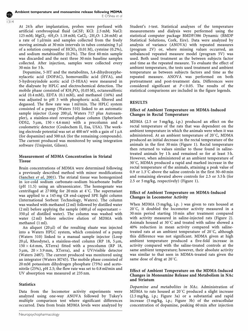
MDMA (2.5 or 5 mg/kg, i.p.) produced an effect on therectal temperature of the rats that was dependent on theambient temperature in which the animals were when it wasadministered. At an ambient temperature of 201C, MDMAproduced an initial decrease in the rectal temperature of theanimals in the first 30 min (Figure 1). Rectal temperaturethen returned to values similar to those found in saline-treated animals by 1 h and remained so for at least 5 h.However, when administered at an ambient temperature of301C, MDMA produced a rapid and marked increase in therectal temperature of the animals, attaining a peak value of0.9 or 1.31C above the saline controls in the first 30–60 minand remaining elevated above controls for 2.5 or 3.5 h (for2.5 or 5 mg/kg, respectively) (Figure 1).
Effect of Ambient Temperature on MDMA-InducedChanges in Locomotor Activity
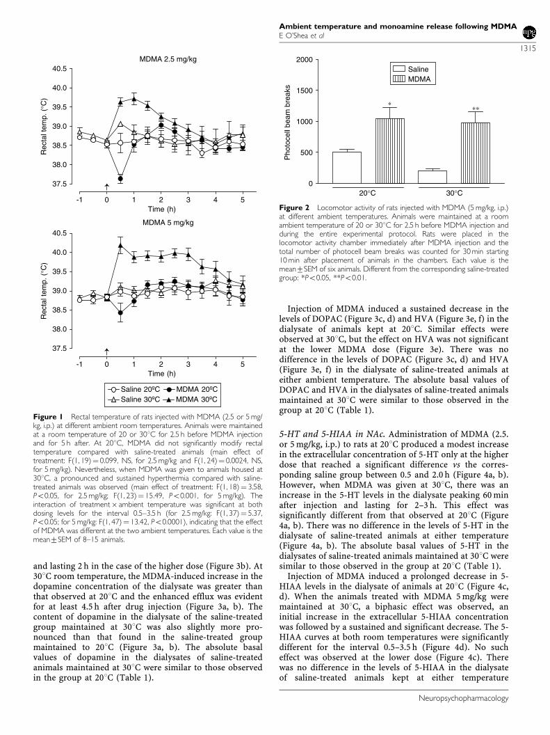
When MDMA (5 mg/kg, i.p. ) was given to rats housed at201C, it doubled the locomotor activity measured in a30 min period starting 10 min after treatment comparedwith activity measured in saline-injected rats (Figure 2).Animals housed at 301C and treated with saline showed a40% reduction in mean activity compared with saline-treated rats at an ambient temperature of 201C, althoughthis difference was not significant. MDMA given at highambient temperature produced a five-fold increase inactivity compared with the saline-treated controls at thesame ambient temperature; however, their absolute activitywas similar to that seen in MDMA-treated rats given thesame dose of drug at 201C.
Effect of Ambient Temperature on the MDMA-InducedChanges in Monoamine Release and Metabolism in NAcand Striatum
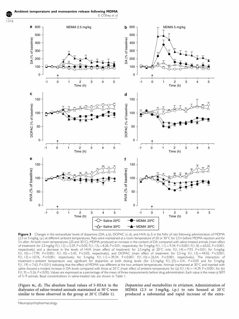
Dopamine and metabolites in NAc. Administration ofMDMA to rats housed at 201C produced a slight increase(2.5 mg/kg, i.p.; Figure 3a) or a substantial and rapidincrease (5 mg/kg, i.p.; Figure 3b) of the extracellularconcentration of dopamine, peaking 60 min after injection
Ambient temperature and monoamine release following MDMAE O’Shea et al
1314
Neuropsychopharmacology
and lasting 2 h in the case of the higher dose (Figure 3b). At301C room temperature, the MDMA-induced increase in thedopamine concentration of the dialysate was greater thanthat observed at 201C and the enhanced efflux was evidentfor at least 4.5 h after drug injection (Figure 3a, b). Thecontent of dopamine in the dialysate of the saline-treatedgroup maintained at 301C was also slightly more pro-nounced than that found in the saline-treated groupmaintained to 201C (Figure 3a, b). The absolute basalvalues of dopamine in the dialysates of saline-treatedanimals maintained at 301C were similar to those observedin the group at 201C (Table 1).
Injection of MDMA induced a sustained decrease in thelevels of DOPAC (Figure 3c, d) and HVA (Figure 3e, f) in thedialysate of animals kept at 201C. Similar effects wereobserved at 301C, but the effect on HVA was not significantat the lower MDMA dose (Figure 3e). There was nodifference in the levels of DOPAC (Figure 3c, d) and HVA(Figure 3e, f) in the dialysate of saline-treated animals ateither ambient temperature. The absolute basal values ofDOPAC and HVA in the dialysates of saline-treated animalsmaintained at 301C were similar to those observed in thegroup at 201C (Table 1).
5-HT and 5-HIAA in NAc. Administration of MDMA (2.5.or 5 mg/kg, i.p.) to rats at 201C produced a modest increasein the extracellular concentration of 5-HT only at the higherdose that reached a significant difference vs the corres-ponding saline group between 0.5 and 2.0 h (Figure 4a, b).However, when MDMA was given at 301C, there was anincrease in the 5-HT levels in the dialysate peaking 60 minafter injection and lasting for 2–3 h. This effect wassignificantly different from that observed at 201C (Figure4a, b). There was no difference in the levels of 5-HT in thedialysate of saline-treated animals at either temperature(Figure 4a, b). The absolute basal values of 5-HT in thedialysates of saline-treated animals maintained at 301C weresimilar to those observed in the group at 201C (Table 1).
Injection of MDMA induced a prolonged decrease in 5-HIAA levels in the dialysate of animals at 201C (Figure 4c,d). When the animals treated with MDMA 5 mg/kg weremaintained at 301C, a biphasic effect was observed, aninitial increase in the extracellular 5-HIAA concentrationwas followed by a sustained and significant decrease. The 5-HIAA curves at both room temperatures were significantlydifferent for the interval 0.5–3.5 h (Figure 4d). No sucheffect was observed at the lower dose (Figure 4c). Therewas no difference in the levels of 5-HIAA in the dialysateof saline-treated animals kept at either temperature
MDMA 2.5 mg/kg
-1 0 1 2 3 4 5
37.5
38.0
38.5
39.0
39.5
40.0
40.5
Time (h)
Rec
tal t
emp.
(°C
)
MDMA 5 mg/kg
Saline 20ºC MDMA 20ºCSaline 30ºC MDMA 30ºC
-1 0 1 2 3 4 5
37.5
38.0
38.5
39.0
39.5
40.0
40.5
Time (h)
Rec
tal t
emp.
(°C
)
Figure 1 Rectal temperature of rats injected with MDMA (2.5 or 5 mg/kg, i.p.) at different ambient room temperatures. Animals were maintainedat a room temperature of 20 or 301C for 2.5 h before MDMA injectionand for 5 h after. At 201C, MDMA did not significantly modify rectaltemperature compared with saline-treated animals (main effect oftreatment: F(1, 19)¼ 0.099, NS, for 2.5 mg/kg and F(1, 24)¼ 0.0024, NS,for 5 mg/kg). Nevertheless, when MDMA was given to animals housed at301C, a pronounced and sustained hyperthermia compared with saline-treated animals was observed (main effect of treatment: F(1, 18)¼ 3.58,Po0.05, for 2.5 mg/kg; F(1, 23)¼ 15.49, Po0.001, for 5 mg/kg). Theinteraction of treatment� ambient temperature was significant at bothdosing levels for the interval 0.5–3.5 h (for 2.5 mg/kg: F(1, 37)¼ 5.37,Po0.05; for 5 mg/kg: F(1, 47)¼ 13.42, Po0.0001), indicating that the effectof MDMA was different at the two ambient temperatures. Each value is themean7SEM of 8–15 animals.
0
500
1000
1500
2000SalineMDMA
20°C 30°C
∗∗∗
Pho
toce
ll be
am b
reak
s
Figure 2 Locomotor activity of rats injected with MDMA (5 mg/kg, i.p.)at different ambient temperatures. Animals were maintained at a roomambient temperature of 20 or 301C for 2.5 h before MDMA injection andduring the entire experimental protocol. Rats were placed in thelocomotor activity chamber immediately after MDMA injection and thetotal number of photocell beam breaks was counted for 30 min starting10 min after placement of animals in the chambers. Each value is themean7SEM of six animals. Different from the corresponding saline-treatedgroup: *Po0.05, **Po0.01.
Ambient temperature and monoamine release following MDMAE O’Shea et al
1315
Neuropsychopharmacology
(Figure 4c, d). The absolute basal values of 5-HIAA in thedialysates of saline-treated animals maintained at 301C weresimilar to those observed in the group at 201C (Table 1).
Dopamine and metabolites in striatum. Administration ofMDMA (2.5 or 5 mg/kg, i.p.) to rats housed at 201Cproduced a substantial and rapid increase of the extra-
MDMA 2.5 mg/kg
-1 0 1 2 3 4 5
0
100
200
300
400
500
600
Time (h)
DA
(%
of b
asel
ine)
MDMA 5 mg/kg
-1 0 1 2 3 4 5
0
50
100
150
0
50
100
150
Time (h)
-1 0 1 2 3 4 5Time (h)
DO
PA
C (
% o
f bas
elin
e)H
VA
(%
of
ba
se
line
)
Saline 20ºC
Saline 30ºC
MDMA 20ºC
MDMA 30ºC
-1 0 1 2 3 4 5
0
100
200
300
400
500
600
Time (h)
DA
(%
of b
asel
ine)
0
50
100
150
-1 0 1 2 3 4 5Time (h)
HV
A (
% o
f b
ase
line
)
-1 0 1 2 3 4 5
0
50
100
150
Time (h)
DO
PA
C (
% o
f bas
elin
e)
a b
c d
e f
Figure 3 Changes in the extracellular levels of dopamine (DA; a, b), DOPAC (c, d), and HVA (e, f) in the NAc of rats following administration of MDMA(2.5 or 5 mg/kg, i.p.) at different ambient temperatures. Rats were maintained at a room temperature of 20 or 301C for 2.5 h before MDMA injection and for5 h after. At both room temperatures (20 and 301C), MDMA produced an increase in the content of DA compared with saline-treated animals (main effectof treatment: for 2.5 mg/kg: F(1, 12)¼ 5.29, Po0.05; F(1, 13)¼ 8.28, Po0.01, respectively; for 5 mg/kg: F(1, 11)¼ 9.34, Po0.001; F(1, 8)¼ 65.02, Po0.001,respectively) and a decrease in the levels of HVA (main effect of treatment: for 2.5 mg/kg at 201C only: F(1, 14)¼ 7.93, Po0.01; for 5 mg/kg:F(1, 10)¼ 17.99, Po0.001; F(1, 10)¼ 5.45, Po0.05, respectively), and DOPAC (main effect of treatment: for 2.5 mg: F(1, 13)¼ 44.02, Po0.001;F(1, 13)¼ 10.76, Po0.001, respectively; for 5 mg/kg: F(1, 11)¼ 39.34, Po0.001; F(1, 10)¼ 26.54, Po0.001, respectively). The interaction oftreatment� ambient temperature was significant for dopamine at both dosing levels (for 2.5 mg/kg: F(1, 25)¼ 3.41, Po0.05 and for 5 mg/kg:F(1, 19)¼ 7.63, Po0.01) indicating that the effect of MDMA was different at the two ambient temperatures. Animals maintained at 301C and injected withsaline showed a modest increase in DA levels compared with those at 201C (main effect of ambient temperature: for (a) F(1,14)¼ 14.39, Po0.001; for (b)F(1, 9)¼ 5.26, Po0.05). Values are expressed as a percentage of the mean of three measurements before drug administration. Each value is the mean7SEMof 5–9 animals. Basal concentrations in saline-treated rats are shown in Table 1.
Ambient temperature and monoamine release following MDMAE O’Shea et al
1316
Neuropsychopharmacology
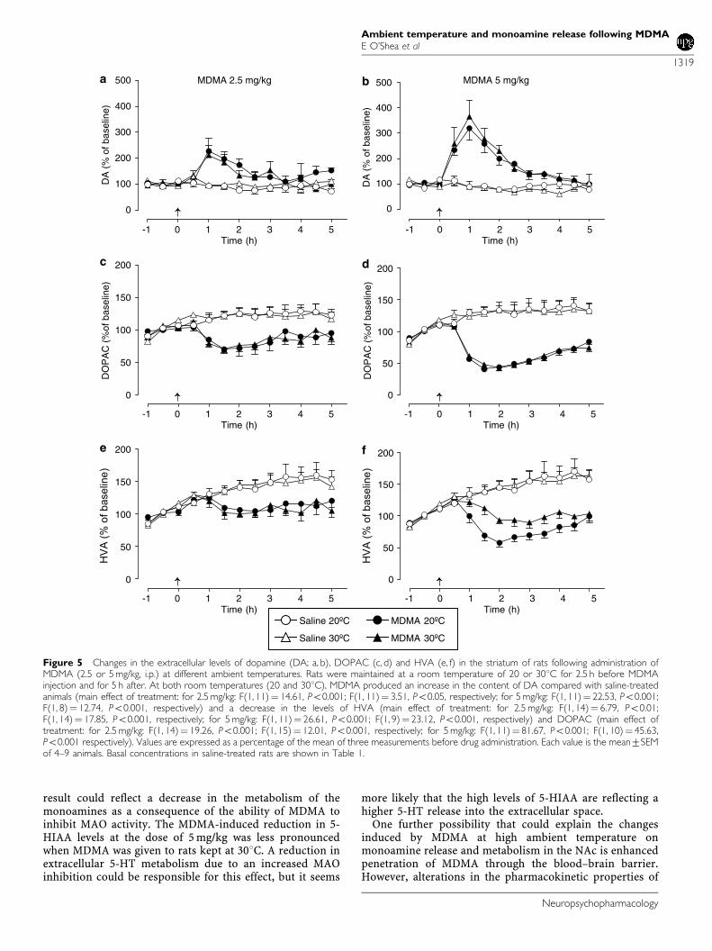
cellular concentration of striatal dopamine, peaking at60 min and lasting 3–4 h. At 301C, the changes induced byMDMA on the dialysate dopamine concentration weresimilar in magnitude and duration to those observed at201C (Figure 5a, b). There was no difference in theextracellular concentration of dopamine of saline-treatedanimals kept at either temperature (Figure 5a, b). Theabsolute basal values of dopamine in the dialysates ofsaline-treated animals maintained at 301C were similar tothose observed in the group at 201C (Table 1).
Injection of MDMA induced a sustained decrease in thelevels of DOPAC (Figure 5c, d) and HVA (Figure 5e, f) in thestriatal dialysate of animals kept at either ambienttemperature. There was no difference in the levels ofDOPAC (Figure 5c, d) or HVA (Figure 5e, f) in the dialysateof saline-treated animals at either temperature. The absolutebasal values of DOPAC and HVA in the dialysates of saline-treated animals maintained at 301C were similar to thoseobserved in the group at 201C (Table 1).
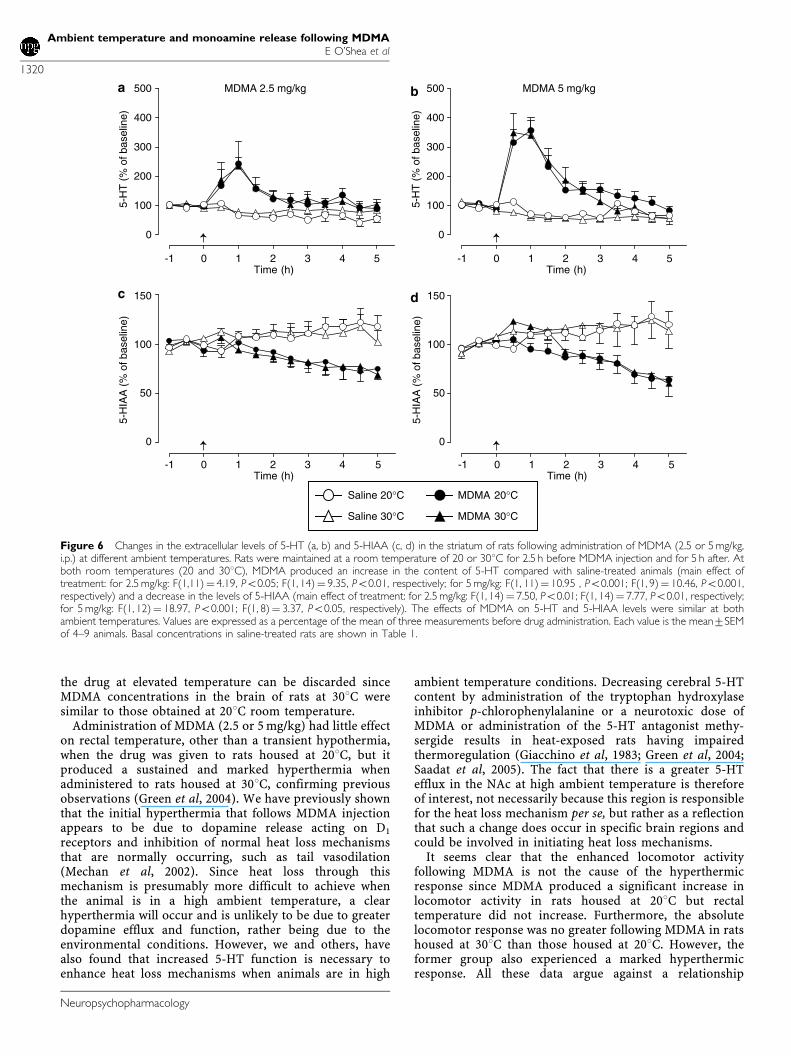
5-HT and 5-HIAA in striatum. Administration of MDMA(2.5 or 5 mg/kg, i.p.) to animals housed at 201C produced asubstantial and rapid increase in the extracellular concen-tration of 5-HT in the striatum peaking 60 min afterinjection and lasting 2–3 h (Figure 6a, b). Similar changeswere seen in animals housed at 301C (Figure 6a, b). Therewas no difference in the levels of 5-HT in the dialysate ofsaline-treated animals at either temperature (Figure 6a, b).The absolute basal values of 5-HT in the dialysates of saline-treated animals maintained at 301C were similar to thoseobserved in the group at 201C (Table 1).
Low-dose MDMA (2.5 mg/kg, i.p.) produced a sustaineddecrease in the extracellular levels of 5-HIAA, which wasnot modified by an increase in ambient temperature(Figure 6c). While injection of MDMA at the higher doseto animals at 201C also induced a sustained decrease indialysate 5-HIAA levels, in animals kept at 301C, it induced
a biphasic response consisting of an initial rise followed bya decrease (Figure 6d). Nevertheless, 5-HIAA curves at bothroom temperatures were not significantly different(Figure 6d). There was no difference in the levels of 5-HIAA in the dialysate of saline-treated animals kept atdifferent temperatures (Figure 6c, d). The absolute basalvalues of 5-HIAA in the dialysates of saline-treated animalsmaintained at 301C were similar to those observed in thegroup at 201C (Table 1).
Effect of Ambient Temperature on Striatal Levels ofMDMA
In order to investigate the possible effect of ambienttemperature on the concentration of MDMA in the brain,rats were given MDMA (5 mg/kg, i.p.) and killed 1 h later.This time point was chosen because the cerebral concentra-tion of MDMA normally peaks 60 min after MDMAinjection (Esteban et al, 2001) and it was the time point atwhich neurotransmitter release peaked. There were nodifferences between the MDMA levels found in the striatumof rats treated with the drug at 201C (22.973.4 nmol/gtissue, n¼ 4) and those treated at 301C (22.670.2 nmol/gtissue, n¼ 4).
DISCUSSION
Using intracerebral microdialysis in freely moving rats, thisstudy has shown that elevation of ambient temperatureenhances the effect induced by low and medium doses ofMDMA on dopamine release in the shell of the NAc but notin the striatum. The output of 5-HT is also enhanced in theNAc, but not striatum, by high ambient temperatureconditions.
Although MDMA increased both dopamine and 5-HTrelease in the NAc of animals at 301C, the magnitude of theMDMA effect on extracellular dopamine levels (260 and
Table 1 Basal Values for Extracellular Dopamine (DA), 5-HT, and Corresponding Metabolites in Saline-Treated Rats and StatisticalComparison of the Effect of Temperature
201C 301C
Brain area Compound �60 min �30 min 0 min �60 min �30 min 0 min F-value
NAc DA 0.4670.09 0.4870.09 0.3670.06 0.4770.09 0.4170.08 0.4570.17 F(1, 21)¼ 0.001, P¼ 0.97
DOPAC 204723 219723 195722 165720 182721 188720 F(1, 23)¼ 0.90, P¼ 0.35
HVA 112712 125715 117714 102711 112712 115712 F(1, 22)¼ 0.07, P¼ 0.79
5-HT 0.7070.16 0.5670.08 0.5370.07 0.5770.21 0.5670.20 0.4170.08 F(1, 20)¼ 0.17, P¼ 0.66
5-HIAA 4975 5275 4875 4974 5274 5375 F(1, 22)¼ 0.11, P¼ 0.74
Striatum DA 0.6370.07 0.6370.08 0.7070.09 0.5470.06 0.4670.05 0.4770.05 F(1, 21)¼ 3.21, P¼ 0.09
DOPAC 338744 399755 435763 279738 361749 440758 F(1, 23)¼ 0.07, P¼ 0.79
HVA 296738 339741 375748 254733 313739 390749 F(1, 22)¼ 0.01, P¼ 0.92
5-HT 0.6570.08 0.6470.08 0.6170.07 1.0070.15 0.9070.11 0.7670.12 F(1, 22)¼ 3.54, P¼ 0.07
5-HIAA 9576 10077 10077 111713 123714 131717 F(1, 22)¼ 2.57, P¼ 0.12
Three 30 min baseline samples were collected immediately before injecting MDMA. Values are mean (pg/ml)7SEM of 22–25 animals.
Ambient temperature and monoamine release following MDMAE O’Shea et al
1317
Neuropsychopharmacology

490% increase for 2.5 and 5 mg/kg at 201C) was larger thanthat elicited on 5-HT concentration (197 and 265%). Thismore pronounced effect of MDMA on dopamine release isnot a consequence of the high ambient temperature sinceWhite et al (1994) observed a similar difference at standardroom temperature. Nor can the difference in increases inthe levels of DA and 5-HT in the NAc following MDMA beattributed to a different affinity of MDMA for monoaminetransporters or to its different potency for inducing [3H]-DA or [3H]-5-HT release in vitro. Using hippocampal andstriatal synaptosomal preparations, it has been shown thatMDMA is only slightly more potent at inhibiting [3H]-5-HTthan [3H]-dopamine uptake (1.7 and 3.2 times, in each area,respectively) and that it is 10 times more potent atincreasing [3H]-5-HT than [3H]-dopamine release (Crespi
et al, 1997). Nevertheless, in NAc, there is a greater densityof dopamine than 5-HT terminals and this morphologicdifference may account for the relatively larger MDMA-induced increase in extracellular dopamine levels comparedwith 5-HT (Dewar et al, 1991; Mennicken et al, 1992). Inaddition, although 5-HT axon density is higher in the shellof NAc than in the dorsal striatum (Deutch and Cameron,1992) few axons in the area express 5-HT transporters. It isworth mentioning that MDMA penetrates into 5-HT nerveterminals by means of 5-HT uptake system (Sanchez et al,2004) and increases 5-HT release through the 5-HTtransporter operating in reverse (Rudnick and Wall, 1992).
In addition to increasing dopamine release, MDMA alsodecreased the extracellular concentrations of DOPAC, HVA,and 5-HIAA in the NAc at both ambient temperatures. This
MDMA 2.5 mg/kg
-1 0 1 2 3 4 5
0
100
200
300
Time (h)
-1 0 1 2 3 4 5Time (h)
5-H
T(%
of b
asel
ine)
MDMA 5 mg/kg
0
50
100
150
5-H
IAA
(%
ofb
asel
ine)
Saline 20°C
Saline 30°C
MDMA 20°C
MDMA 30°C
-1 0 1 2 3 4 5
0
100
200
300
Time (h)
5-H
T(%
of b
asel
ine)
-1 0 1 2 3 4 5Time (h)
0
50
100
150
5-H
IAA
(%
ofb
asel
ine)
a b
c d
Figure 4 Changes in the extracellular levels of 5-HT (a, b) and 5-HIAA (c, d) in the NAc of rats following administration of MDMA (2.5 or 5 mg/kg, i.p.) atdifferent ambient temperatures. Rats were maintained at a room temperature of 20 or 301C for 2.5 h before MDMA injection and for 5 h after. At 201CMDMA (5 mg/kg, i.p.) reduced 5-HIAA levels compared with saline-treated animals (main effect of treatment: F(1, 12)¼ 21.21, Po0.001) and produced asignificant increase in 5-HT concentration vs the corresponding saline from 0.5-2.0 h (main effect of treatment: F(1, 9)¼ 3.97, Po0.05). At 2.5 mg/kg, MDMAonly reduced 5-HIAA levels (main effect of treatment: F(1, 11)¼ 30.58, Po0.001). When MDMA was given at 301C, it produced a pronounced andprolonged increase in 5-HT concentration compared with saline-treated controls (main effect of treatment: for 2.5 mg/kg between 0.5 and 2.0 h:F(1, 13)¼ 5.86, Po0.01; for 5 mg/kg between 0.5 and 5 h: F(1, 9)¼ 18.49, Po0.001). The interaction of treatment� ambient temperature was significant for5-HT at both dosing levels (for 2.5 mg/kg between 0.5 and 2.0 h: F(1, 26)¼ 4.08, Po0.05; for 5 mg/kg between 0.5 and 5 h: F(1, 18)¼ 4.24, Po0.05),indicating that the effect of MDMA was different at the two ambient temperatures. MDMA also produced a reduction in 5-HIAA levels (main effect oftreatment: for 2.5 mg/kg: F(1, 8)¼ 8.51, Po0.01; for 5 mg/kg: F(1, 8)¼ 6.06, Po0.01). The interaction of treatment� ambient temperature was notsignificant for 5-HIAA at either dosing level (for 2.5 mg/kg: F(1, 19)¼ 0.00, NS; for 5 mg/kg: F(1, 20)¼ 2.27, NS), indicating that the effect of MDMA was thesame at the two ambient temperatures, excepting for the interval 0.5–3.5 h at the higher dose (F(1, 20)¼ 3.80, Po0.05). Values are expressed as apercentage of the mean of three measurements before drug administration. Each value is the mean7SEM of 5–8 animals. Basal concentrations in saline-treated rats are shown in Table 1.
Ambient temperature and monoamine release following MDMAE O’Shea et al
1318
Neuropsychopharmacology
result could reflect a decrease in the metabolism of themonoamines as a consequence of the ability of MDMA toinhibit MAO activity. The MDMA-induced reduction in 5-HIAA levels at the dose of 5 mg/kg was less pronouncedwhen MDMA was given to rats kept at 301C. A reduction inextracellular 5-HT metabolism due to an increased MAOinhibition could be responsible for this effect, but it seems
more likely that the high levels of 5-HIAA are reflecting ahigher 5-HT release into the extracellular space.
One further possibility that could explain the changesinduced by MDMA at high ambient temperature onmonoamine release and metabolism in the NAc is enhancedpenetration of MDMA through the blood–brain barrier.However, alterations in the pharmacokinetic properties of
MDMA 2.5 mg/kg
-1 0 1 2 3 4 5
0
100
200
300
400
500
Time (h)
-1 0 1 2 3 4 5Time (h)
-1 0 1 2 3 4 5Time (h)
DA
(%
of b
asel
ine)
MDMA 5 mg/kg
0
50
100
150
200
0
50
100
150
200
DO
PA
C (
%of
bas
elin
e)H
VA
(%
of
ba
se
line
)
Saline 20ºC
Saline 30ºC
MDMA 20ºC
MDMA 30ºC
-1 0 1 2 3 4 5Time (h)
0
50
100
150
200
HV
A (
% o
f b
ase
line
)
-1 0 1 2 3 4 5Time (h)
0
50
100
150
200
DO
PA
C (
%of
bas
elin
e)
-1 0 1 2 3 4 5
0
100
200
300
400
500
Time (h)
DA
(%
of b
asel
ine)
a b
c d
e f
Figure 5 Changes in the extracellular levels of dopamine (DA; a, b), DOPAC (c, d) and HVA (e, f) in the striatum of rats following administration ofMDMA (2.5 or 5 mg/kg, i.p.) at different ambient temperatures. Rats were maintained at a room temperature of 20 or 301C for 2.5 h before MDMAinjection and for 5 h after. At both room temperatures (20 and 301C), MDMA produced an increase in the content of DA compared with saline-treatedanimals (main effect of treatment: for 2.5 mg/kg: F(1, 11)¼ 14.61, Po0.001; F(1, 11)¼ 3.51, Po0.05, respectively; for 5 mg/kg: F(1, 11)¼ 22.53, Po0.001;F(1, 8)¼ 12.74, Po0.001, respectively) and a decrease in the levels of HVA (main effect of treatment: for 2.5 mg/kg: F(1, 14)¼ 6.79, Po0.01;F(1, 14)¼ 17.85, Po0.001, respectively; for 5 mg/kg: F(1, 11)¼ 26.61, Po0.001; F(1, 9)¼ 23.12, Po0.001, respectively) and DOPAC (main effect oftreatment: for 2.5 mg/kg: F(1, 14)¼ 19.26, Po0.001; F(1, 15)¼ 12.01, Po0.001, respectively; for 5 mg/kg: F(1, 11)¼ 81.67, Po0.001; F(1, 10)¼ 45.63,Po0.001 respectively). Values are expressed as a percentage of the mean of three measurements before drug administration. Each value is the mean7SEMof 4–9 animals. Basal concentrations in saline-treated rats are shown in Table 1.
Ambient temperature and monoamine release following MDMAE O’Shea et al
1319
Neuropsychopharmacology
the drug at elevated temperature can be discarded sinceMDMA concentrations in the brain of rats at 301C weresimilar to those obtained at 201C room temperature.
Administration of MDMA (2.5 or 5 mg/kg) had little effecton rectal temperature, other than a transient hypothermia,when the drug was given to rats housed at 201C, but itproduced a sustained and marked hyperthermia whenadministered to rats housed at 301C, confirming previousobservations (Green et al, 2004). We have previously shownthat the initial hyperthermia that follows MDMA injectionappears to be due to dopamine release acting on D1
receptors and inhibition of normal heat loss mechanismsthat are normally occurring, such as tail vasodilation(Mechan et al, 2002). Since heat loss through thismechanism is presumably more difficult to achieve whenthe animal is in a high ambient temperature, a clearhyperthermia will occur and is unlikely to be due to greaterdopamine efflux and function, rather being due to theenvironmental conditions. However, we and others, havealso found that increased 5-HT function is necessary toenhance heat loss mechanisms when animals are in high
ambient temperature conditions. Decreasing cerebral 5-HTcontent by administration of the tryptophan hydroxylaseinhibitor p-chlorophenylalanine or a neurotoxic dose ofMDMA or administration of the 5-HT antagonist methy-sergide results in heat-exposed rats having impairedthermoregulation (Giacchino et al, 1983; Green et al, 2004;Saadat et al, 2005). The fact that there is a greater 5-HTefflux in the NAc at high ambient temperature is thereforeof interest, not necessarily because this region is responsiblefor the heat loss mechanism per se, but rather as a reflectionthat such a change does occur in specific brain regions andcould be involved in initiating heat loss mechanisms.
It seems clear that the enhanced locomotor activityfollowing MDMA is not the cause of the hyperthermicresponse since MDMA produced a significant increase inlocomotor activity in rats housed at 201C but rectaltemperature did not increase. Furthermore, the absolutelocomotor response was no greater following MDMA in ratshoused at 301C than those housed at 201C. However, theformer group also experienced a marked hyperthermicresponse. All these data argue against a relationship
MDMA 2.5 mg/kg
-1 0 1 2 3 4 5
0
100
200
300
400
500
Time (h)
-1 0 1 2 3 4 5Time (h)
5-H
T (
% o
f bas
elin
e)
MDMA 5 mg/kg
0
50
100
150
5-H
IAA
(%
of b
asel
ine)
Saline 20°C
Saline 30°C
MDMA 20°C
MDMA 30°C
-1 0 1 2 3 4 5Time (h)
0
50
100
150
5-H
IAA
(%
of b
asel
ine)
-1 0 1 2 3 4 5
0
100
200
300
400
500
Time (h)
5-H
T (
% o
f bas
elin
e)
a b
c d
Figure 6 Changes in the extracellular levels of 5-HT (a, b) and 5-HIAA (c, d) in the striatum of rats following administration of MDMA (2.5 or 5 mg/kg,i.p.) at different ambient temperatures. Rats were maintained at a room temperature of 20 or 301C for 2.5 h before MDMA injection and for 5 h after. Atboth room temperatures (20 and 301C), MDMA produced an increase in the content of 5-HT compared with saline-treated animals (main effect oftreatment: for 2.5 mg/kg: F(1,11)¼ 4.19, Po0.05; F(1, 14)¼ 9.35, Po0.01, respectively; for 5 mg/kg: F(1, 11)¼ 10.95 , Po0.001; F(1, 9)¼ 10.46, Po0.001,respectively) and a decrease in the levels of 5-HIAA (main effect of treatment: for 2.5 mg/kg: F(1, 14)¼ 7.50, Po0.01; F(1, 14)¼ 7.77, Po0.01, respectively;for 5 mg/kg: F(1, 12)¼ 18.97, Po0.001; F(1, 8)¼ 3.37, Po0.05, respectively). The effects of MDMA on 5-HT and 5-HIAA levels were similar at bothambient temperatures. Values are expressed as a percentage of the mean of three measurements before drug administration. Each value is the mean7SEMof 4–9 animals. Basal concentrations in saline-treated rats are shown in Table 1.
Ambient temperature and monoamine release following MDMAE O’Shea et al
1320
Neuropsychopharmacology
between locomotor activity and MDMA-induced hyper-thermia, a conclusion also made by Dafters (1995) followinghis studies on hyperkinesis and hyperthermia followingMDMA administration.
Locomotor activity following administration of ampheta-mines is generally considered to result from increaseddopaminergic activity in the mesolimbic region, whilestereotypy is associated with dopamine release in thestriatum (Kelly et al, 1975; Fibiger and Phillips, 1974), andtherefore the greater increase in extracellular dopamine inthe NAc may account for the greater increase in locomotoractivity, above control values, in animals housed at highambient room temperature.
What is particularly interesting with regard to theenhanced dopamine release in the NAc in rats housed athigh room temperature is its possible relationship to therewarding action of MDMA. The mesolimbic dopaminesystem has long been suggested to be involved in therewarding properties of recreational drugs (Fibiger andPhillips, 1974; Wise and Bozarth, 1982), including MDMA(Beardsley et al, 1986). Low-dose MDMA has been shown tohave rewarding properties when given at standard roomtemperature as it induces a positive effect in the CPP test(Marona-Lewicka et al, 1996). Recently, Cornish et al (2003)reported greater MDMA-induced social interaction beha-vior in rats housed at 301C compared with 201C and alsogreater self-administration of MDMA at high temperature.It is tempting to suggest that the enhanced mesolimbicdopamine release seen at high temperature is responsiblefor the behavioral changes seen by Cornish et al (2003).
In addition, evidence suggests that the 5-HT system mayalso be involved in mediating the rewarding and stimulantproperties of MDMA either by direct or indirect mechan-isms. Dopamine release in the ventral tegmental area maybe facilitated by 5-HT, possibly acting indirectly through aGABAergic mechanism (Kalivas, 1993; Prisco et al, 1994;Trifunovic and Brodie, 1996). Furthermore, various 5-HTreceptor subtypes have been shown to modulate dopaminerelease in the NAc and the striatum (Benloucif andGalloway, 1991; Chen et al, 1991; Benloucif et al, 1993;Parsons and Justice, 1993; Lucas et al, 1997). Moreover, itappears that 5-HT release and subsequent 5-HT1B receptor(and possibly 5-HT2A receptor) stimulation is required forMDMA-induced hyperlocomotion to develop (Callawayet al, 1990; Kehne et al, 1996; McCreary et al, 1999). Inparticular, since dopamine release in the NAc is positivelycontrolled by 5-HT1B (Boulenguez et al, 1996) and 5-HT2A
receptors (De Deurwaerdere and Spampinato, 1999), it maybe that the increase in locomotor activity induced byMDMA is a consequence of an increase in dopaminergictransmission mediated by 5-HT in the NAc (McCreary et al,1999).
Therefore, the facilitatory effect of MDMA on dopamineand 5-HT efflux in the NAc suggests that the rewardingproperties of MDMA could be more pronounced at highambient temperature.
Human recreational users of MDMA may experiencegreater psychoactive effects of MDMA when taking it inwarm crowded conditions. The corollary of this, however, isthat high ambient temperature produces a greater hy-perthermic response to the same MDMA dose, which isassociated with greater long-term neurotoxicity in rat
models (Broening et al, 1995; Malberg and Seiden, 1998;O’Shea et al, 1998; Sanchez et al, 2004). This may suggestthat human recreational users put themselves at greater riskby ingesting the drug in hot room conditions.
ACKNOWLEDGEMENTS
MIC thanks Ministerio de Ciencia y Tecnologia (GrantSAF2001-1437), Ministerio de Sanidad (Grant FIS02/1885,Grant G03/005), Plan Nacional sobre Drogas (Ministerio delInterior), and Fundacion Mapfre Medicina for financialsupport. VS thanks FIS for a studentship.
REFERENCES
Baldwin HA, Williams JL, Snares M, Ferreira T, Cross AJ, GreenAR (1994). Attenuation by chlormethiazole administration of therise in extracellular amino acids following focal ischaemia in thecerebral cortex of the rat. Br J Pharmacol 112: 188–194.
Beardsley PM, Balster RL, Harris LS (1986). Self-administration ofmethylenedioxymethamphetamine (MDMA) by rhesus mon-keys. Drug Alcohol Depend 18: 149–157.
Benloucif S, Galloway MP (1991). Facilitation of dopamine releasein vivo by serotonin agonists: studies with microdialysis. Eur JPharmacol 200: 1–8.
Benloucif S, Keegan MJ, Galloway MP (1993). Serotonin-facilitateddopamine release in vivo: pharmacological characterization.J Pharmacol Exp Ther 265: 373–377.
Bilsky EJ, Hubbell CL, Delconte JD, Reid LD (1991). MDMAproduces a conditioned place preference and elicits ejaculationin male rats: a modulatory role for the endogenous opioids.Pharmacol Biochem Behav 40: 443–447.
Bilsky EJ, Reid LD (1991). MDL72222, a serotonin 5-HT3 receptorantagonist, blocks MDMA’s ability to establish a conditionedplace preference. Pharmacol Biochem Behav 39: 509–512.
Boulenguez P, Rawlins JN, Chauveau J, Joseph MH, Mitchell SN,Gray JA (1996). Modulation of dopamine release in the nucleusaccumbens by 5-HT1B agonists: involvement of the hippocam-po-accumbens pathway. Neuropharmacology 35: 1521–1529.
Broening HW, Bowyer JF, Slikker Jr W (1995). Age-dependentsensitivity of rats to the long term effects of the seroton-ergic neurotoxicant (7)-3,4-methylenedioxymethampheramine(MDMA) correlates with the magnitude of the MDMA-inducedthermal response. J Pharmacol Exp Ther 275: 325–333.
Brown C, Osterloh J (1987). Multiple severe complications fromrecreational ingestion of MDMA (‘Ecstasy’). J Am Med Assoc 258:780–781.
Callaway CW, Wing LL, Geyer MA (1990). Serotonin releasecontributes to the locomotor stimulant effects of 3,4-methyle-nedioxymethamphetamine in rats. J Pharmacol Exp Ther 254:456–464.
Chen JP, van Praag HM, Gardner EL (1991). Activation of 5-HT3receptor by 1-phenylbiguanide increases dopamine release in therat nucleus accumbens. Brain Res 543: 354–357.
Colado MI, O’Shea E, Granados R, Esteban B, Martın AB, Green AR(1999). Studies on the role of dopamine in the degenerationof 5-HT nerve endings in the brain of Dark Agouti rats following3,4-methylenedioxymethamphetamine (MDMA or ‘ecstasy’)administration. Br J Pharmacol 126: 911–924.
Cornish JL, Shahnawaz Z, Thompson MR, Wong S, Morley KC,Hunt GE et al (2003). Heat increases 3,4-methylenedioxy-methamphetamine self-administration and social effects in rats.Eur J Pharmacol 482: 339–341.
Crespi D, Mennini T, Gobbi M (1997). Carrier-dependentand Ca(2+)-dependent 5-HT and dopamine release inducedby (+)-amphetamine, 3,4-methylendioxymethamphetamine,
Ambient temperature and monoamine release following MDMAE O’Shea et al
1321
Neuropsychopharmacology
p-chloroamphetamine and (+)-fenfluramine. Br J Pharmacol121: 1735–1743.
Dafters RI (1995). Effect of ambient temperature on hyperthermiaand hyperkinesis induced by 3,4-methylenedioxymethampheta-mine (MDMA or ‘ecstasy’) in rats. Psychopharmacology 114:505–518.
Daniela E, Brennan K, Gittings D, Hely L, Schenk S (2004). Effect ofSCH 23390 on (7)-3,4-methylenedioxymethamphetamine hy-peractivity and self-administration in rats. Pharmacol BiochemBehav 77: 745–750.
De Deurwaerdere P, Spampinato U (1999). Role of serotonin(2A)and serotonin(2B/2C) receptor subtypes in the control ofaccumbal and striatal dopamine release elicited in vivo bydorsal raphe nucleus electrical stimulation. J Neurochem 73:1033–1042.
Deutch AY, Cameron DS (1992). Pharmacological characterizationof dopamine systems in the nucleus accumbens core and shell.Neuroscience 46: 49–56.
Dewar KM, Reader TA, Grondin L, Descarries L (1991).[3H]paroxetine binding and serotonin content of rat andrabbit cortical areas, hippocampus, neostriatum, ventral me-sencephalic tegmentum, and midbrain raphe nuclei region.Synapse 9: 14–26.
Esteban B, O’Shea E, Camarero J, Sanchez V, Green AR, Colado MI(2001). 3,4-Methylenedioxymethamphetamine induces monoa-mine release, but not toxicity, when administered centrally at aconcentration occurring following a peripherally injectedneurotoxic dose. Psychopharmacology 154: 251–260.
Fibiger HC, Phillips AG (1974). Role of dopamine and norepi-nephrine in the chemistry of reward. J Psychiatr Res 11: 135–143.
Giacchino JL, Schertel ER, Horowitz JM, Horwitz BA (1983). Effectof p-chlorophenylalanine on thermoregulation in unrestrainedrats. Am J Physiol 244: R299–R302.
Gold LH, Geyer MA, Koob GF (1989a). Neurochemical mechan-isms involved in behavioral effects of amphetamines and relateddesigner drugs. NIDA Res Monogr 94: 101–126.
Gold LH, Hubner CB, Koob GF (1989b). A role for the mesolimbicdopamine system in the psychostimulant actions of MDMA.Psychopharmacology 99: 40–47.
Gold LH, Koob GF (1988). Methysergide potentiates the hyper-activity produced by MDMA in rats. Pharmacol Biochem Behav29: 645–648.
Green AR, Sanchez V, O’Shea E, Saadat KS, Elliott JM, Colado MI(2004). Effect of ambient temperature and a prior neurotoxicdose of 3,4-methylenedioxymethamphetamine (MDMA) onthe hyperthermic response of rats to a single or repeated(‘binge’ ingestion) low dose of MDMA. Psychopharmacology 173:264–269.
Henry JA (1992). Ecstasy and the dance of death. BMJ 305: 5–6.Henry JA, Jeffreys KJ, Dawling S (1992). Toxicity and deaths from
3,4-methylenedioxymethamphetamine (‘ecstasy’). Lancet 340:384–387.
Hubner CB, Bird M, Rassnick S, Kornetsky C (1988). The thresholdlowering effects of MDMA (ecstasy) on brain-stimulationreward. Psychopharmacology 95: 49–51.
Kalivas PW (1993). Neurotransmitter regulation of dopamineneurons in the ventral tegmental area. Brain Res Brain Res Rev18: 75–113.
Kankaanpaa A, Meririnne E, Lillsunde P, Seppala T (1998). Theacute effects of amphetamine derivatives on extracellularserotonin and dopamine levels in rat nucleus accumbens.Pharmacol Biochem Behav 59: 1003–1009.
Kehne JH, Ketteler HJ, McCloskey TC, Sullivan CK, Dudley MW,Schmidt CJ (1996). Effects of the selective 5-HT2A receptorantagonist MDL 100,907 on MDMA-induced locomotor stimula-tion in rats. Neuropsychopharmacology 15: 116–124.
Kelly PH, Seviour PW, Iversen SD (1975). Amphetamine andapomorphine responses in the rat following 6-OHDA lesions of
the nucleus accumbens septi and corpus striatum. Brain Res 94:507–522.
Konig JFR, Klippel RA (1963). The Rat Brain. A Stereotaxic Atlas ofthe Forebrain and Lower Parts of the Brain Stem. Robert EKrieger Publishing Co Inc.: New York.
Lamb RJ, Griffiths RR (1987). Self-injection of d,1-3,4-methylene-dioxymethamphetamine (MDMA) in the baboon. Psychophar-macology 91: 268–272.
Lucas JJ, Segu L, Hen R (1997). 5-Hydroxytryptamine1B receptorsmodulate the effect of cocaine on c-fos expression: convergingevidence using 5-hydroxytryptamine1B knockout mice and the5-hydroxytryptamine1B/1D antagonist GR127935. Mol Pharma-col 51: 755–763.
Malberg JE, Seiden LS (1998). Small changes in ambienttemperature cause large changes in 3,4-methylenedioxymetham-phetamine (MDMA)-induced serotonin neurotoxicity and corebody temperature in the rat. J Neurosci 18: 5086–5094.
Marona-Lewicka D, Rhee GS, Sprague JE, Nichols DE (1996).Reinforcing effects of certain serotonin-releasing amphetaminederivatives. Pharmacol Biochem Behav 53: 99–105.
McCreary AC, Bankson MG, Cunningham KA (1999). Pharmaco-logical studies of the acute and chronic effects of (+)-3, 4-methylenedioxymethamphetamine on locomotor activity: roleof 5-hydroxytryptamine(1A) and 5-hydroxytryptamine(1B/1D)receptors. J Pharmacol Exp Ther 290: 965–973.
Mechan AO, Esteban B, O’Shea E, Elliott JM, Colado MI, Green AR(2002). The pharmacology of the acute hyperthermic responsethat follows administration of 3,4-methylenedioxymethamphe-tamine (MDMA, ‘ecstasy’) to rats. Br J Pharmacol 135: 170–180.
Mennicken F, Savasta M, Peretti-Renucci R, Feuerstein C (1992).Autoradiographic localization of dopamine uptake sites in therat brain with 3H-GBR 12935. J Neural Transm Gen Sect 87: 1–14.
O’Shea E, Granados R, Esteban B, Colado MI, Green AR (1998).The relationship between the degree of neurodegeneration of ratbrain 5-HT nerve terminals and the dose and frequency ofadministration of MDMA (‘ecstasy’). Neuropharmacology 37:919–926.
Parsons LH, Justice Jr JB (1993). Perfusate serotonin increasesextracellular dopamine in the nucleus accumbens as measuredby in vivo microdialysis. Brain Res 606: 195–199.
Prisco S, Pagannone S, Esposito E. (1994). Serotonin-dopamineinteraction in the rat ventral tegmental area: an electrophysio-logical study in vivo. J Pharmacol Exp Ther 271: 83–90.
Rudnick G, Wall SC (1992). The molecular mechanism of ‘ecstasy’[3,4-methylenedioxymethamphetamine (MDMA)]: serotonintransporters are targets for MDMA-induced serotonin release.Proc Natl Acad Sci 89: 1817–1821.
Saadat KS, O’Shea E, Colado MI, Elliott JM, Green AR (2005). Therole of 5-HT in the impairment of thermoregulation observed inrats administered MDMA (‘ecstasy’) when housed at hightemperature. Psychopharmacology (in press).
Sanchez V, Camarero J, Esteban B, Peter MJ, Green AR, Colado MI(2001). The mechanisms involved in the long-lasting neuropro-tective effect of fluoxetine against MDMA (‘ecstasy’)-induceddegeneration of 5-HT nerve endings in rat brain. Br J Pharmacol134: 46–57.
Sanchez V, O’Shea E, Saadat KS, Elliott JM, Colado MI, Green AR(2004). Effect of repeated (‘binge’) dosing of MDMA to ratshoused at normal and high temperature on neurotoxic damageto cerebral 5-HT and dopamine neurones. J Psychopharmacol 18:412–416.
Schechter MD (1991). Effect of MDMA neurotoxicity upon itsconditioned place preference and discrimination. PharmacolBiochem Behav 38: 539–544.
Schenk S, Gittings D, Johnstone M, Daniela E (2003). Development,maintenance and temporal pattern of self-administrationmaintained by ecstasy (MDMA) in rats. Psychopharmacology169: 21–27.
Ambient temperature and monoamine release following MDMAE O’Shea et al
1322
Neuropsychopharmacology

Screaton GR, Singer M, Cairns HS, Thrasher A, Sarner M, CohenSL (1992). Hyperpyrexia and rhabdomyolysis after MDMA(‘ecstasy’) abuse. Lancet 339: 677–678.
Swerdlow NR, Koob GF (1985). Separate neural substrates of thelocomotor activating-properties of amphetamine, heroin, caf-feine and corticotrophin releasing factor (CRF) in the rat.Pharmacol Biochem Behav 23: 303–307.
Trifunovic RD, Brodie MS (1996). The effects of clomipramine onthe excitatory action of ethanol on dopaminergic neurons of theventral tegmental area in vitro. J Pharmacol Exp Ther 276: 34–40.
Vaccarino FJ, Amalric M, Swerdlow NR, Koob GF (1986). Blockadeof amphetamine but not opiate-induced locomotion following
antagonism of dopamine function in the rat. Pharmacol BiochemBehav 24: 61–65.
White SR, Duffy P, Kalivas PW (1994). Methylenedioxymetham-phetamine depresses glutamate-evoked neuronal firing andincreases extracellular levels of dopamine and serotonin in thenucleus accumbens in vivo. Neuroscience 62: 41–50.
Wise RA, Bozarth MA (1982). Action of drugs of abuse on brainreward systems: an update with specific attention to opiates.Pharmacol Biochem Behav 17: 239–243.
Yamamoto BK, Spanos LJ (1988). The acute effects of methylene-dioxymethamphetamine on dopamine release in the awake-behaving rat. Eur J Pharmacol 148: 195–203.
Ambient temperature and monoamine release following MDMAE O’Shea et al
1323
Neuropsychopharmacology
Copyright © 2022 FDOKUMEN


![Dopamine release in nucleus accumbens during rewarded task switching measured by [¹¹C]raclopride](https://static.fdokumen.com/doc/165x107/633ae2b3ea31206fd207b694/dopamine-release-in-nucleus-accumbens-during-rewarded-task-switching-measured-by.jpg)