Chronic exposure to ethanol exacerbates MDMA-induced hyperthermia and exposes liver to severe...
8
Toxicology 252 (2008) 64–71 Contents lists available at ScienceDirect Toxicology journal homepage: www.elsevier.com/locate/toxicol Chronic exposure to ethanol exacerbates MDMA-induced hyperthermia and exposes liver to severe MDMA-induced toxicity in CD1 mice Helena Pontes a,∗ , José Alberto Duarte b , Paula Guedes de Pinho a , Maria Elisa Soares a , Eduarda Fernandes c , Ricardo Jorge Dinis-Oliveira a,d,e , Carla Sousa a , Renata Silva a , Helena Carmo a , Susana Casal f , Fernando Remião a , Félix Carvalho a , Maria Lourdes Bastos a,∗ a REQUIMTE, Toxicology Department, Faculty of Pharmacy, University of Porto, Rua Aníbal Cunha 164, 4099-030 Porto, Portugal b CIAFEL, Faculty of Sport, University of Porto, Rua Dr. Plácido Costa 91, 4200-450 Porto, Portugal c REQUIMTE, Physical-Chemistry Department, Faculty of Pharmacy, University of Porto, Rua Aníbal Cunha 164, 4099-030 Porto, Portugal d Clinical Analysis and Public Health Department, Cooperativa de Ensino Superior, Politécnico e Universitário, CRL Rua José António Vidal, 81, 4760-409 Vila Nova de Famalicão, Portugal e Institute of Legal Medicine, Faculty of Medicine, University of Porto, Jardim Carrilho Videira, 4050-167 Porto, Portugal f REQUIMTE, Bromatology Department, Faculty of Pharmacy, University of Porto, Rua Aníbal Cunha 164, 4099-030 Porto, Portugal article info Article history: Received 17 June 2008 Received in revised form 24 July 2008 Accepted 26 July 2008 Available online 7 August 2008 Keywords: Ethanol 3,4-Methylenedioxymethamphetamine (MDMA) Hyperthermia Oxidative stress Hepatotoxicity abstract 3,4-Methylenedioxymethamphetamine (MDMA; ecstasy) is an amphetamine derivative drug with entac- togenic, empathogenic and hallucinogenic properties, commonly consumed at rave parties in a polydrug abuse pattern, especially with cannabis, tobacco and ethanol. Since both MDMA and ethanol may cause deleterious effects to the liver, the evaluation of their putative hepatotoxic interaction is of great interest, especially considering that most of the MDMA users are regular ethanol consumers. Thus, the aim of the present study was to evaluate, in vivo, the acute hepatotoxic effects of MDMA (10mg/kg i.p.) in CD-1 mice previously exposed to 12% ethanol as drinking fluid (for 8 weeks). Body temperature was continuously measured for 12 h after MDMA administration and, after 24 h, hepatic damage was evaluated. The administration of MDMA to non pre-treated mice resulted in sustained hyperthermia, which was significantly increased in ethanol pre-exposed mice. A correspondent higher increase of hepatic heat shock transcription factor (HSF-1) activation was also observed in the latter group. Furthermore, MDMA administration resulted in liver damage as confirmed by histological analysis, slight decrease in liver weight and increased plasma transaminases levels. These hepatotoxic effects were also exacerbated when mice were pre-treated with ethanol. The activities of some antioxidant enzymes (such as SOD, GPx and Catalase) were modified by ethanol, MDMA and their joint action. The hepatotoxicity resulting from the simultaneous exposure to MDMA and ethanol was associated with a higher activation of NF-B, indicating a pro-inflammatory effect in this organ. In conclusion, the obtained results strongly suggest that the consumption of ethanol increases the hyperthermic and hepatotoxic effects associated with MDMA abuse. © 2008 Elsevier Ireland Ltd. All rights reserved. Abbreviations: MDMA, 3,4-methylenedioxymethamphetamine, ecstasy; MDA, methylenedioxyamphetamine; ROW, relative organ weight; %DLWC, percent dry lipid weight content; HSF-1, heat shock factor 1; NF-B, nuclear factor kappa-B; GOT, glutamic oxalic transaminase; GPT, glutamic pyruvic transaminase; SOD, superoxide dismutase; GPx, glutathione peroxidase; GST, glutathione-S-transferase; GSH, glu- tathione (reduced form); GSSG, glutathione (oxidised form); TBARS, thiobarbituric acid reactive substances; IHC, immunohistochemistry. ∗ Corresponding authors. Tel.: +351 222078979; fax: +351 222003977. E-mail addresses: [email protected] (H. Pontes), [email protected] (M.L. Bastos). 1. Introduction 3,4-Methylenedioxymethamphetamine (MDMA; ecstasy) is an amphetamine derivative drug with entactogenic, empathogenic and hallucinogenic properties, commonly consumed at rave par- ties in a polydrug abuse pattern, especially with cannabis, amphetamine, cocaine, tobacco and ethanol (Green et al., 2003; Schifano, 2004; Breen et al., 2006; Barrett et al., 2006). The com- mon concomitant use of ethanol and MDMA has a great interaction potential since both compounds are metabolized in the liver to hep- atotoxic metabolites (Gemma et al., 2006; Green et al., 2003; Carmo et al., 2006) with higher toxicity than their parent compounds 0300-483X/$ – see front matter © 2008 Elsevier Ireland Ltd. All rights reserved. doi:10.1016/j.tox.2008.07.064
-
Upload
independent -
Category
Documents
-
view
1 -
download
0
Transcript of Chronic exposure to ethanol exacerbates MDMA-induced hyperthermia and exposes liver to severe...
Ce
HEHa
b
c
d
8e
f
a
ARRAA
KE3(HOH
mlgdta
0d
Toxicology 252 (2008) 64–71
Contents lists available at ScienceDirect
Toxicology
journa l homepage: www.e lsev ier .com/ locate / tox ico l
hronic exposure to ethanol exacerbates MDMA-induced hyperthermia andxposes liver to severe MDMA-induced toxicity in CD1 mice
elena Pontesa,∗, José Alberto Duarteb, Paula Guedes de Pinhoa, Maria Elisa Soaresa,duarda Fernandesc, Ricardo Jorge Dinis-Oliveiraa,d,e, Carla Sousaa, Renata Silvaa,elena Carmoa, Susana Casal f, Fernando Remiãoa, Félix Carvalhoa, Maria Lourdes Bastosa,∗
REQUIMTE, Toxicology Department, Faculty of Pharmacy, University of Porto, Rua Aníbal Cunha 164, 4099-030 Porto, PortugalCIAFEL, Faculty of Sport, University of Porto, Rua Dr. Plácido Costa 91, 4200-450 Porto, PortugalREQUIMTE, Physical-Chemistry Department, Faculty of Pharmacy, University of Porto, Rua Aníbal Cunha 164, 4099-030 Porto, PortugalClinical Analysis and Public Health Department, Cooperativa de Ensino Superior, Politécnico e Universitário, CRL Rua José António Vidal,1, 4760-409 Vila Nova de Famalicão, PortugalInstitute of Legal Medicine, Faculty of Medicine, University of Porto, Jardim Carrilho Videira, 4050-167 Porto, PortugalREQUIMTE, Bromatology Department, Faculty of Pharmacy, University of Porto, Rua Aníbal Cunha 164, 4099-030 Porto, Portugal
r t i c l e i n f o
rticle history:eceived 17 June 2008eceived in revised form 24 July 2008ccepted 26 July 2008vailable online 7 August 2008
eywords:thanol,4-MethylenedioxymethamphetamineMDMA)yperthermiaxidative stressepatotoxicity
a b s t r a c t
3,4-Methylenedioxymethamphetamine (MDMA; ecstasy) is an amphetamine derivative drug with entac-togenic, empathogenic and hallucinogenic properties, commonly consumed at rave parties in a polydrugabuse pattern, especially with cannabis, tobacco and ethanol. Since both MDMA and ethanol may causedeleterious effects to the liver, the evaluation of their putative hepatotoxic interaction is of great interest,especially considering that most of the MDMA users are regular ethanol consumers.
Thus, the aim of the present study was to evaluate, in vivo, the acute hepatotoxic effects of MDMA(10 mg/kg i.p.) in CD-1 mice previously exposed to 12% ethanol as drinking fluid (for 8 weeks). Bodytemperature was continuously measured for 12 h after MDMA administration and, after 24 h, hepaticdamage was evaluated.
The administration of MDMA to non pre-treated mice resulted in sustained hyperthermia, which wassignificantly increased in ethanol pre-exposed mice. A correspondent higher increase of hepatic heatshock transcription factor (HSF-1) activation was also observed in the latter group. Furthermore, MDMAadministration resulted in liver damage as confirmed by histological analysis, slight decrease in liver
weight and increased plasma transaminases levels. These hepatotoxic effects were also exacerbated whenmice were pre-treated with ethanol. The activities of some antioxidant enzymes (such as SOD, GPx andCatalase) were modified by ethanol, MDMA and their joint action. The hepatotoxicity resulting from thesimultaneous exposure to MDMA and ethanol was associated with a higher activation of NF-�B, indicatinga pro-inflammatory effect in this organ.In conclusion, the obtained results strongly suggest that the consumption of ethanol increases thetoxic
hyperthermic and hepatoAbbreviations: MDMA, 3,4-methylenedioxymethamphetamine, ecstasy; MDA,ethylenedioxyamphetamine; ROW, relative organ weight; %DLWC, percent dry
ipid weight content; HSF-1, heat shock factor 1; NF-�B, nuclear factor kappa-B; GOT,lutamic oxalic transaminase; GPT, glutamic pyruvic transaminase; SOD, superoxideismutase; GPx, glutathione peroxidase; GST, glutathione-S-transferase; GSH, glu-athione (reduced form); GSSG, glutathione (oxidised form); TBARS, thiobarbituriccid reactive substances; IHC, immunohistochemistry.∗ Corresponding authors. Tel.: +351 222078979; fax: +351 222003977.
E-mail addresses: [email protected] (H. Pontes), [email protected] (M.L. Bastos).
1
aataSmpae
300-483X/$ – see front matter © 2008 Elsevier Ireland Ltd. All rights reserved.oi:10.1016/j.tox.2008.07.064
effects associated with MDMA abuse.© 2008 Elsevier Ireland Ltd. All rights reserved.
. Introduction
3,4-Methylenedioxymethamphetamine (MDMA; ecstasy) is anmphetamine derivative drug with entactogenic, empathogenicnd hallucinogenic properties, commonly consumed at rave par-ies in a polydrug abuse pattern, especially with cannabis,mphetamine, cocaine, tobacco and ethanol (Green et al., 2003;
chifano, 2004; Breen et al., 2006; Barrett et al., 2006). The com-on concomitant use of ethanol and MDMA has a great interactionotential since both compounds are metabolized in the liver to hep-totoxic metabolites (Gemma et al., 2006; Green et al., 2003; Carmot al., 2006) with higher toxicity than their parent compounds
icology
(vapaiaeMcaidstrjtcpoewMtwM
2
2
afg21tSw2c(pp(
2
3aw6cigafatge(ddcGoAAl
2
eobawttacctb
2
gAsRs(3
2
2
vdIR
22peAgao
nhc
2wDo
2sae1
2D
2(a�
2w
H. Pontes et al. / Tox
Carvalho et al., 2004; Meier and Seitz, 2008). This metabolic acti-ation observed with MDMA seems to be a common feature ofmphetamine derivatives, since it was already described for com-ounds such as 4-MTA (Carmo et al., 2004b, 2007), 2-CB (Carmo etl., 2004a, 2005) which showed a variable toxicity profile accord-ngly to the individual metabolic capacity. In addition, ethanol canlter the expression and/or activity of some drug-metabolizingnzymes (Jang and Harris, 2007), including those involved inDMA metabolism. Furthermore, MDMA users are regular ethanol
onsumers before their first contact with MDMA (Ben Hamida etl., 2006), which may represent a putative risk factor for the toxicnteraction between these two drugs. In fact, our group has recentlyemonstrated that the repeated exposure of CD1 mice to ethanoleems to increase the vulnerability of freshly isolated hepatocytesowards pro-oxidant conditions (Pontes et al., 2008). Additionally,esults reported by Pacifici and Hernández-Lopes in human sub-ects (Hernández-López et al., 2002; Pacifici et al., 2001) showedhat also the physiologic and psychopathologic effects of MDMAould be increased in presence of ethanol. Thus, the objective of theresent study was to evaluate, in vivo, the acute hepatotoxic effectsf MDMA (10 mg/kg i.p.) in CD-1 mice previously exposed to 12%thanol as drinking fluid (for 8 weeks). This experimental approachas designed to simulate the most common pattern of ethanol andDMA abuse, in which the individuals are exposed to ethanol prior
o the first contact with MDMA. To our knowledge, experimentalork addressing the in vivo effects of chronic ethanol exposure onDMA-induced hepatotoxicity has not been previously reported.
. Materials and methods
.1. Chemicals
All the reagents used in this study were of analytical grade. Bovine serumlbumin (fraction V), �-nicotinamide adenine dinucleotide phosphate reducedorm (�-NADPH), pyruvic acid, 2-vinylpyridine, glutathione reduced form (GSH),lutathione oxidised form (GSSG), glutathione reductase (EC 1.6.4.2), 5,5-dithiobis--nitrobenzoic acid (DTNB), guanidine hydrochloride, 2,4-dinitrophenylhydrazine,-chloro-2,4-dinitrobenzene (CDNB), xanthine, nitrotetrazolium blue chloride, xan-hine oxidase from bovine milk and hydrogen peroxide solution were obtained fromigma–Aldrich Co. (St. Louis, MO, USA). The injectable solution of sodic heparineas obtained from B. Braun (Lisbon, Portugal). Perchloric acid, trichloroacetic acid,-thiobarbituric acid, Folin Ciocalteau reagent and all other chemicals were pur-hased from Merck (Darmstadt, Germany). 3,4-MethylenedioxymethamphetamineHCl salt) was extracted and purified from high purity MDMA tablets that wererovided by the Portuguese Criminal Police Department. The obtained salt wasure and fully characterized by NMR and mass spectrometry methodologiespurity > 99%).
.2. Animals
Adult male CD1 mice (Charles-River Laboratories, Barcelona, Spain), weighing0–40 g, were used in all experiments. For at least 1 week prior to the treatment,nimals were acclimatized in polyethylene cages with wire-mesh tops, lined withood shavings, at an ambient temperature of 20 ± 2 ◦C, humidity between 40 and0% and 12 h/12 h light/dark cycle (light on from 8.00 to 20.00 h), having standardhow and tap drinking water ad libitum. After this period, animals were dividednto two main groups: ethanol (n = 12) and non-ethanol (n = 12). In the ethanol mainroup the tap drinking water was replaced by a 5% ethanol solution for 1 weeknd, afterwards, by a 12% ethanol solution for 8 weeks. The replacement of wateror 5% ethanol before 12% ethanol corresponds to a required adaptation period, tovoid aversion of mice to a high ethanol concentration in the drinking fluid, sincehese animals do not have natural preference for 12% ethanol. The non-ethanol mainroup had drinking tap water ad libitum for the same period. There were no differ-nces in body weight between the main groups after the 8 weeks of the experimentdata not shown). During the 24-h period after MDMA administration, animals wereeprived of food but the drinking solution was allowed ad libitum. Surgical proce-ures for blood collection and liver excision were performed under anaesthesia and
arried out between 9 and 11 a.m. Animal experiments were licensed by Portugueseeneral Directorate of Veterinary Medicine. Housing and experimental treatmentf animals were in accordance with the Guide for the Care and Use of Laboratorynimals from the Institute for Laboratory Animal Research (Institute for Laboratorynimal Research, 1996). The experiments complied with the current Portugueseaws.
cTa
2g
252 (2008) 64–71 65
.3. MDMA challenge
Mice were housed in four groups of six animals (control, ethanol, MDMA andthanol + MDMA groups) and were injected intraperitoneally with saline (0.9% NaCl)r 10 mg/kg MDMA (dissolved in saline) in a volume of 0.1 mL/30 g body weightetween 9:00 and 10:00 a.m. The experiment was performed under controlledmbient temperature fixed at 20 ◦C (±2 ◦C). Subcutaneous temperature of animalsas measured repeatedly after MDMA administration (see measurement of body
emperature). After 24 h, animals were anaesthetized and blood was removed fromhe inferior vena cava and collected into heparinized tubes. The liver was excisednd weighed, after body perfusion with 0.9% NaCl solution, and used for biochemi-al analysis. Samples were kept frozen (−80 ◦C) until assay. Liver sections were alsoollected for histological examination. The relative organ weight (ROW), an indica-or of tissue harm, was also calculated for each animal as a percentage of the totalody weight at the sacrifice day.
.4. Measurement of body temperature
Subcutaneous temperatures of mice were measured using BioMedic pro-rammable temperature transponders (microchips BMDS IPTT-100, Plexx BV, 6660E ELST, Netherlands). The microchips were implanted subcutaneously between thehoulder blades, using the BioMedic implant device, 2 days before the experiment.eadings of subcutaneous temperature were taken immediately prior to MDMA oraline injection and for 12 h thereafter using a hand held Biomedic Pocket ScannerDAS-5007). Temperature was recorded every 15 min during the first hour, every0 min during the second and third hours and every hour thereafter.
.5. Plasma biochemical analysis
.5.1. Plasma transaminasesPlasma activities of glutamic oxalic transaminase (GOT) and glutamic pyru-
ic transaminase (GPT) were evaluated as biomarkers hepatic damage. Theseeterminations were performed in a Reflotron Analyser (Boehringer Mannheim,
ndianapolis, IN) using 32 �L of plasma for each analysis (GOT, Cat no. 10745120,oche Diagnostics; GPT, Cat no. 10745138, Roche Diagnostics).
.5.2. Oxidative stress parameters
.5.2.1. Sample preparation. For the quantification of protein carbonyl groups, lipideroxidation extent and GSH and GSSG concentrations, tissue samples were homog-nized (1:4 m/v) in ice-cold 5% perchloric acid with an Ultra-Turrax® homogenizer.fter the homogenization, aliquots were taken to determine protein carbonylroups content and protein levels. The remaining homogenates were centrifugedt 3200 × g for 10 min at 4 ◦C and the supernatants were used for the quantificationf lipid peroxidation and GSH/GSSG.
For measurement of antioxidant enzymes activities, liver samples were homoge-ized (1:4 m/v) in ice cold 0.1% Triton X-100 in 50 mM phosphate buffer (pH 7.4). Theomogenates were centrifuged at 3200 × g for 10 min at 4 ◦C, and the supernatantsollected for the enzymatic and protein assays.
.5.2.2. Protein carbonyl groups. Protein carbonyl groups (ketones and aldehydes)ere quantified according to Levine et al. (1994). Results were expressed as nmol ofNPH incorporated per mg of protein, calculated by using an extinction coefficientf 2.2 × 10⁴ M−1 cm−1.
.5.2.3. Non-specific lipid peroxidation. The extent of lipid peroxidation was mea-ured by the thiobarbituric acid reactive substances (TBARS) assay at 535 nm (Buegend Aust, 1978; Carvalho et al., 1997). TBARS were expressed as malondialdehydequivalents per milligram of protein, calculated by using an extinction coefficient of.56 × 105 M−1 cm−1.
.5.2.4. GSH and GSSG. Tissue GSH and GSSG contents were determined by theTNB-GSSG reductase recycling assay as described before (Pontes et al., 2008).
.5.2.5. Catalase. Catalase activity was measured according to the method of Aebi1984), by monitoring the decomposition of H2O2 at 240 nm, calculated by usingn extinction coefficient of 39.4 M−1 cm−1). The enzyme activity was expressed asmol of H2O2 consumed per min and per mg of protein.
.5.2.6. Glutathione-S-transferase (GST). Glutathione-S-transferase (GST) activityas assayed according to the method of Habig et al. (1974). The formation of GSH
onjugate with 1-chloro-2,4-dinitrobenzene was monitored for 5 min at 340 nm.
he GST activity was calculated by using an extinction coefficient of 9.6 mM−1 cm−1nd expressed as �mol per min per mg of protein.
.5.2.7. Selenium-dependent glutathione peroxidase (GPx). Selenium-dependentlutathione peroxidase (GPx) activity was assayed by NADPH oxidation at 340 nm
6 icology
(tm
2ao(
2
s
2
e
2
eieBE2
cw3B0ipt−e
ebwDbNlbPeCaT
2
mgbwdE
h7
pwawwUTtrwuM
0c
0lctieWw5uamw
2
nibbt
3
3
Maem
figsSa
3
i((
3
sp3tert
3
mtcat
6 H. Pontes et al. / Tox
Flohe and Gunzler, 1984). The enzyme activity was calculated by using an extinc-ion coefficient of 6.22 mM−1 cm−1 and expressed as nmol of NADPH oxidized per
in per mg of protein.
.5.2.8. Superoxide dismutase (SOD). Copper/zinc superoxide dismutase (CuZnSOD)nd manganese superoxide dismutase (MnSOD) were assayed using the methodf Flohé and Ötting (1984), with some modifications previously reported beforeDinis-Oliveira et al., 2006).
.5.3. Protein quantificationProtein was determined by the Lowry method (Lowry et al., 1951) using bovine
erum albumin as the standard.
.5.4. Dry lipid weight contentThe total liver lipid content was determined according to Folch et al. (1957) and
xpressed on a dry weight percentage (%DLWC).
.6. Nuclear extracts and fEMSA
Transcriptional activations of NF-�B and HSF-1 were determined by fluorescentlectrophoretic mobility shift assay (fEMSA) using nuclear protein extracts accord-ng to a previously reported method (Dinis-Oliveira et al., 2007). The nuclear proteinxtracts were obtained from tissue homogenates (1:2.5 m/v) in cell lysis buffer (ACuffer) containing 10 mM HEPES, 1.5 mM MgCl2, 10 mM KCl, 0.5 mM EDTA, 0.1 mMGTA, 1 mM ditiothreitol (DTT), 0.25 mM phenyl methylsulfonyl fluoride (PMSF) and0 �L/mL igepal (pH 7.9).
To obtain the nuclear extracts, the homogenates were exposed to a freeze/thawycle and centrifuged for 10 min at 850 × g, 4 ◦C. The pellets were washed againith AC Buffer, incubated for 15 min on ice, and centrifuged at 16,000 × g, 4 ◦C, for
0 s. The obtained nuclear pellets were resuspended in complete lysis Buffer (BCuffer, containing 20 mM HEPES, 20% (m/v) Glycerol, 420 mM NaCl, 1.5 mM MgCl2,.5 mM EDTA, 1 mM DTT, 0.25 mM PMSF, 20 �L/mL igepal and 5 �g/mL of a protease
nhibitors cocktail containing 1 mg/mL aprotinin, 1 mg/mL leupeptin and 1 mg/mLepsatin), incubated for 30 min on ice on a rocking platform at 150 rpm and cen-rifuged for 10 min at 16,000 × g at 4 ◦C. Aliquots of the supernatant were stored at80 ◦C until assay. Protein quantification was determined by the Lowry assay andach extract aliquot was diluted to 2 mg/mL of protein.
Thereafter, to perform the fEMSA electrophoresis, 20 �g of protein from nuclearxtracts were incubated with Cy5-labeled NF-�B or HSF-1 consensus probe in assayuffer (200 mM HEPES, 500 mM KCl, 10 mM EDTA and 10 mM DTT) supplementedith glycerol, poly(dI-dC), DTT and igepal, at 4 ◦C over-night. Specificity of theNA–protein complex was confirmed by the addition of a 50-fold excess of an unla-eled specific competitor (SC) (equal to the specific probe but without the Cy5 label).ine microliters of each mixture were loaded on a 5% non-denaturating polyacry-
amide gel at 10 ◦C, 800 V, 50 mA and 30 W, in TBE buffer 1× (90 mM Tris base, 90 mMoric acid, 2 mM EDTA, pH 8.3) using an ALFexpress II DNA Analyser (Amershamharmacia Biotech, Sweden). The temperature was regulated by an external ALF-xpress II Cooler system (Amersham Pharmacia Biotech, Sweden). The followingy5-labeled specific double stranded probes were used for NF-�B and HSF-1 gelssays: NF-�B, 5′-GCC TGG GAA AGT CCC CTC AAC T-3′; HSF-1, 5′-GAT CCT CGA ATGTC GCG AAA AG-3′ .
.7. Tissue preparation for light and transmission electron microscopy
For light microscopy, after excision, liver was sliced in 2–4 mm3 pieces, approxi-ately, and fixed in 4% formaldehyde. The fixed pieces were, then, dehydrated with
raded ethanol and included in paraffin blocks. The compound used in the transitionetween dehydration and impregnation was benzene. Semi-thin sections (4 �m)ere cut in a microtome, applied on silane-coated slides and deparaffinated. Theeparaffinated sections were treated for different staining protocols (Haematoxylin-osin, Van Gieson) and for immunohistochemistry assays for the detection of NF-�B.
The haematoxylin–eosin staining was performed by immersion in Mayer’saematoxylin solution for 3–4 min followed by immersion in 1% eosin solution formin, dehydration with graded alcohols through xylene, and mounting with DPX.
To perform the NF-�B immunohistochemical detection, NF-�B p50 (NLS) rabbitolyclonal antibody (SC-114, 1:20, Santa Cruz Biotechnology Inc., California, USA)as applied on deparaffinated liver sections and these samples were incubated
t 37 ◦C for 2 h in a humidified chamber. The same samples were then incubatedith a secondary anti-immunoglobulin goat anti-rabbit IgG, F(ab′)2 conjugatedith alkaline phosphatase (SC-3838, 1:50, Santa Cruz Biotechnology Inc., California,SA), under the same conditions, for 1 h. SIGMAFAST® Fast Red TR/Naphthol AS-MXablets were used as substrate according to manufacturer’s instructions. The sec-ions were counterstained with Mayer’s haematoxylin. The primary antibody waseplaced by PBS for negative control sections. Wistar rat 14th day placenta slides
ere used as positive control. All stained sections were mounted on glass slidessing Aquatex® . For the light microscopy study, an optic photomicroscope Nikon,odel Eclipse E400 was used.For electron microscopy, 1 mm3 tissue pieces were fixed in 2% gluteraldehyde (in.2 M sodium cacodylate) for 2 h at 4 ◦C. After three 15 min washes with 0.2 M sodiumacodylate buffer at 4 ◦C, the pieces were post-fixed with 2% osmium tetroxide (in
oadea
252 (2008) 64–71
.2 M sodium cacodylate), for 2 h at 4 ◦C. After a new wash with sodium cacody-ate (5 min), the samples were submitted to a dehydration process using increasingoncentrations of ethanol (from 50% to 100%) for 90 min. In the impregnation stage,he pieces were placed in mixtures containing absolute ethanol and LR White, withncreasing concentrations of resin (50% and 75% (v/v) of absolute ethanol) for 30 minach. In the next stage, samples were kept for two periods of 30 min each, in 100% LRhite, and afterwards were mounted on covered gelatine capsules. The inclusionas processed in a drying oven at 60 ◦C for 22–26 h. Ultra-thin (thickness equal to00–600 Å) sections were cut and contrasted with a saturated aqueous solution ofranyl acetate, for 30 min, and with a solution of lead citrate, for 15 min, with washest the beginning and at the end of each one of these procedures. For the electronicroscopy study a transmission electron microscope Zeiss EM 10 A, at 60 Kvoltsas used.
.8. Statistical analysis
Results are presented as mean ± S.E.M. (from six animals) and were tested forormality with the Kolmogorov–Smirnov test. For temperature analysis, compar-
sons were made by analysis of variance (ANOVA) with repeated measures, followedy the Bonferroni’s post-hoc test. The other statistical comparisons were performedy one-way analysis of variance (ANOVA) followed by Tukey’s multiple comparisonest. Significance was accepted at P < 0.05.
. Results
.1. Body temperature
After administration of 10 mg/kg MDMA, animals from theDMA group showed a typical amphetaminic reaction char-
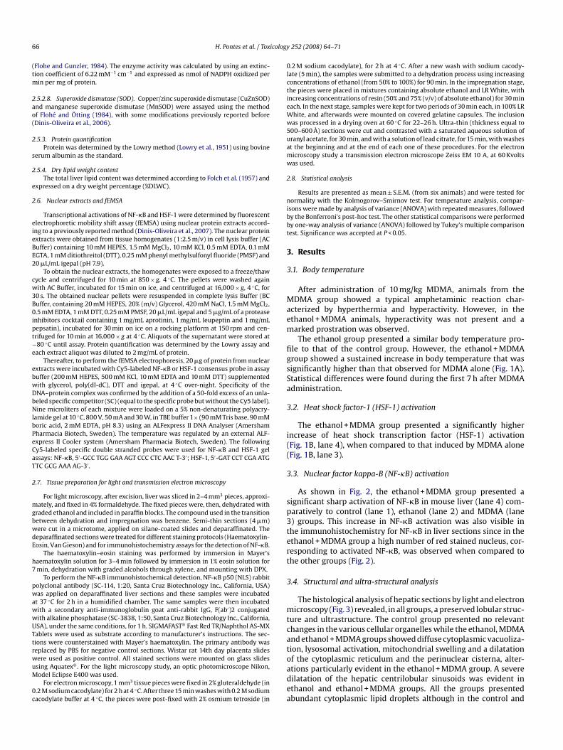
cterized by hyperthermia and hyperactivity. However, in thethanol + MDMA animals, hyperactivity was not present and aarked prostration was observed.The ethanol group presented a similar body temperature pro-
le to that of the control group. However, the ethanol + MDMAroup showed a sustained increase in body temperature that wasignificantly higher than that observed for MDMA alone (Fig. 1A).tatistical differences were found during the first 7 h after MDMAdministration.
.2. Heat shock factor-1 (HSF-1) activation
The ethanol + MDMA group presented a significantly higherncrease of heat shock transcription factor (HSF-1) activationFig. 1B, lane 4), when compared to that induced by MDMA aloneFig. 1B, lane 3).
.3. Nuclear factor kappa-B (NF-�B) activation
As shown in Fig. 2, the ethanol + MDMA group presented aignificant sharp activation of NF-�B in mouse liver (lane 4) com-aratively to control (lane 1), ethanol (lane 2) and MDMA (lane) groups. This increase in NF-�B activation was also visible inhe immunohistochemistry for NF-�B in liver sections since in thethanol + MDMA group a high number of red stained nucleus, cor-esponding to activated NF-�B, was observed when compared tohe other groups (Fig. 2).
.4. Structural and ultra-structural analysis
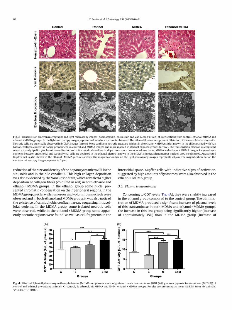
The histological analysis of hepatic sections by light and electronicroscopy (Fig. 3) revealed, in all groups, a preserved lobular struc-
ure and ultrastructure. The control group presented no relevanthanges in the various cellular organelles while the ethanol, MDMAnd ethanol + MDMA groups showed diffuse cytoplasmic vacuoliza-ion, lysosomal activation, mitochondrial swelling and a dilatation
f the cytoplasmic reticulum and the perinuclear cisterna, alter-tions particularly evident in the ethanol + MDMA group. A severeilatation of the hepatic centrilobular sinusoids was evident inthanol and ethanol + MDMA groups. All the groups presentedbundant cytoplasmic lipid droplets although in the control and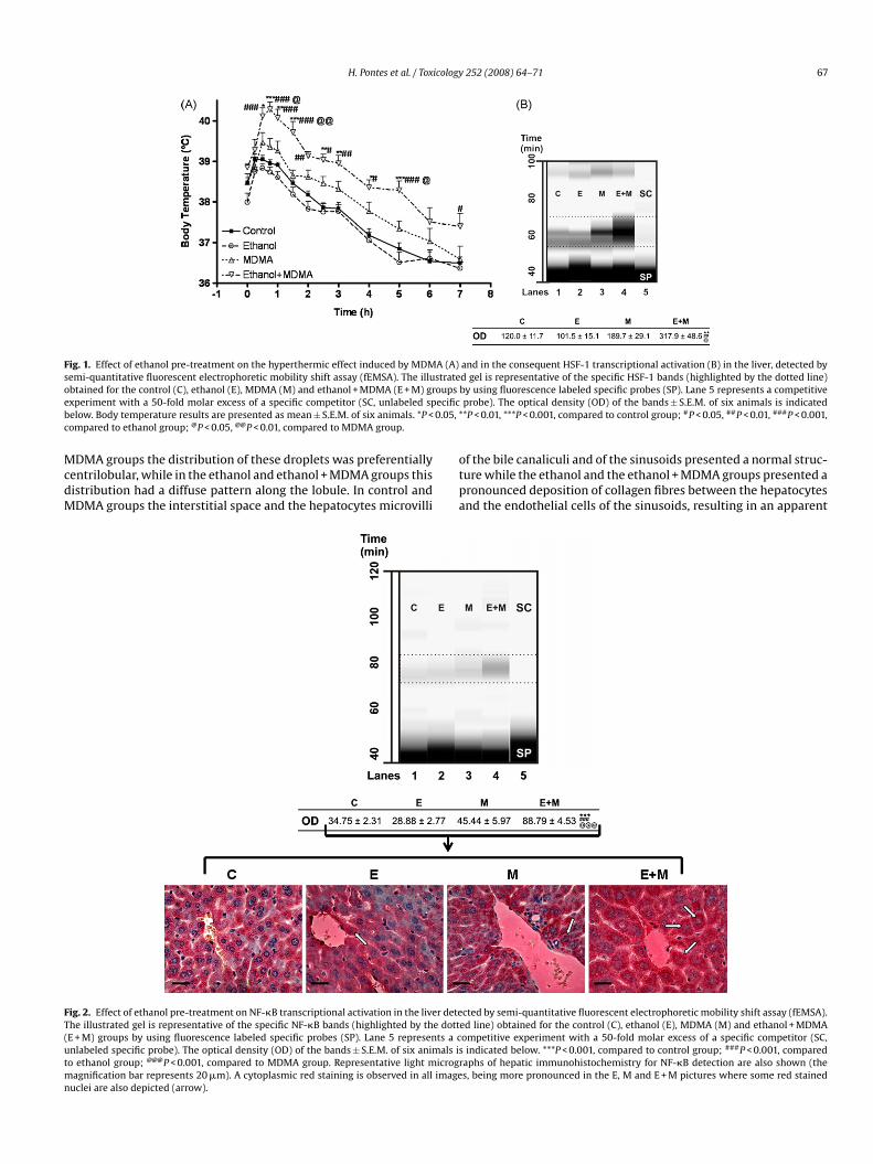
H. Pontes et al. / Toxicology 252 (2008) 64–71 67
Fig. 1. Effect of ethanol pre-treatment on the hyperthermic effect induced by MDMA (A) and in the consequent HSF-1 transcriptional activation (B) in the liver, detected bysemi-quantitative fluorescent electrophoretic mobility shift assay (fEMSA). The illustrated gel is representative of the specific HSF-1 bands (highlighted by the dotted line)o oupse ecificb 0.05, *c
McdM
FT(utmn
btained for the control (C), ethanol (E), MDMA (M) and ethanol + MDMA (E + M) grxperiment with a 50-fold molar excess of a specific competitor (SC, unlabeled spelow. Body temperature results are presented as mean ± S.E.M. of six animals. *P <ompared to ethanol group; @P < 0.05, @@P < 0.01, compared to MDMA group.
DMA groups the distribution of these droplets was preferentiallyentrilobular, while in the ethanol and ethanol + MDMA groups thisistribution had a diffuse pattern along the lobule. In control andDMA groups the interstitial space and the hepatocytes microvilli
otpa
ig. 2. Effect of ethanol pre-treatment on NF-�B transcriptional activation in the liver detehe illustrated gel is representative of the specific NF-�B bands (highlighted by the dottE + M) groups by using fluorescence labeled specific probes (SP). Lane 5 represents a cnlabeled specific probe). The optical density (OD) of the bands ± S.E.M. of six animals iso ethanol group; @@@P < 0.001, compared to MDMA group. Representative light microgr
agnification bar represents 20 �m). A cytoplasmic red staining is observed in all imageuclei are also depicted (arrow).
by using fluorescence labeled specific probes (SP). Lane 5 represents a competitiveprobe). The optical density (OD) of the bands ± S.E.M. of six animals is indicated*P < 0.01, ***P < 0.001, compared to control group; #P < 0.05, ##P < 0.01, ###P < 0.001,
f the bile canaliculi and of the sinusoids presented a normal struc-ure while the ethanol and the ethanol + MDMA groups presented aronounced deposition of collagen fibres between the hepatocytesnd the endothelial cells of the sinusoids, resulting in an apparent
cted by semi-quantitative fluorescent electrophoretic mobility shift assay (fEMSA).ed line) obtained for the control (C), ethanol (E), MDMA (M) and ethanol + MDMAompetitive experiment with a 50-fold molar excess of a specific competitor (SC,indicated below. ***P < 0.001, compared to control group; ###P < 0.001, compared
aphs of hepatic immunohistochemistry for NF-�B detection are also shown (thes, being more pronounced in the E, M and E + M pictures where some red stained
68 H. Pontes et al. / Toxicology 252 (2008) 64–71
Fig. 3. Transmission electron micrographs and light microscopy images (haematoxylin–eosin stain and Van Gieson’s stain) of liver sections from control, ethanol, MDMA andethanol + MDMA groups. In the light microscopy images, a preserved lobular structure is observed. The ethanol illustrations present dilatation of the centrilobular sinusoids.Necrotic cells are punctually observed in MDMA images (arrow). More confluent necrotic areas are evident in the ethanol + MDMA slide (arrow). In the slides stained with VanGieson, collagen content is poorly pronounced in control and MDMA images and more marked in ethanol exposed groups (arrow). The transmission electron micrographsr icturec ure (aK on bare
rswdesMotlwe
ise
3
Fc*
eveal a mainly lipidic cytoplasmic vacuolization and mitochondrial swelling in all pontents between endothelial and parenchymal cells are depicted in the ethanol pictupffer cell is also shown in the ethanol + MDMA picture (arrow). The magnificatilectron microscopy images represents 2 �m.
eduction of the size and density of the hepatocytes microvilli in theinusoids and in the bile canaliculi. This high collagen depositionas also evidenced by the Van Gieson stain, which revealed a highereposition of collagen fibres (coloured in red) in both ethanol andthanol + MDMA groups. In the ethanol group some nuclei pre-ented chromatin condensation on their peripheral regions. In theDMA group, nuclei with numerous and voluminous nucleoli were
bserved and in both ethanol and MDMA groups it was also noticedhe existence of osmiophobic confluent areas, suggesting intracel-ular oedema. In the MDMA group, some isolated necrotic cells
ere observed, while in the ethanol + MDMA group some appar-ntly necrotic regions were found, as well as cell fragments in the
itoto
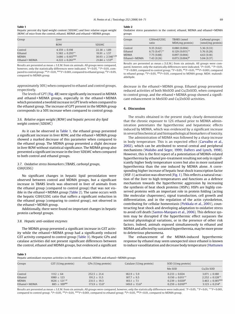
ig. 4. Effect of 3,4-methylenedioxymethamphetamine (MDMA) on plasma levels of gluontrol and ethanol pre-treated animals. C: control, E: ethanol, M: MDMA and E + M: eP < 0.05, ***P < 0.001.
s, more pronounced in ethanol, MDMA and ethanol + MDMA images. Large collagenrrow). In the MDMA micrograph numerous nucleoli are also observed. An activatedon the light microscopy images represents 20 �m. The magnification bar on the
nterstitial space. Kupffer cells with indicative signs of activation,uggested by high amounts of lysosomes, were also observed in thethanol + MDMA group.
.5. Plasma transaminases
Concerning to GOT levels (Fig. 4A), they were slightly increased
n the ethanol group compared to the control group. The adminis-ration of MDMA produced a significant increase of plasma levelsf this transaminase in both MDMA and ethanol + MDMA groups,he increase in this last group being significantly higher (increasef approximately 35%) than in the MDMA group (increase oftamic oxalic transaminase (GOT (A)), glutamic pyruvic transaminase (GPT (B)) ofthanol + MDMA groups. Results are presented as mean ± S.E.M. from six animals.
H. Pontes et al. / Toxicology 252 (2008) 64–71 69
Table 1Hepatic percent dry lipid weight content (%DLWC) and liver relative organ weight(ROW) of mice from the control, ethanol, MDMA and ethanol + MDMA groups
Group Liver
ROW %DLWC
Control 4.359 ± 0.198 22.38 ± 1.89Ethanol 5.582 ± 0.293** 19.91 ± 1.57MDMA 3.686 ± 0.165### 30.93 ± 2.546*,##
Ethanol + MDMA 4.222 ± 0.261## 21.84 ± 1.53@
Rhpc
ar
awtc
3w
astipt
3G
oitittt
p
3
iGct
Table 2Oxidative stress parameters in the control, ethanol, MDMA and ethanol + MDMAgroups
Groups GSHred/GSSGratio
TBARS (nmolMDA/mg protein)
Carbonyl groups(nmol/mg protein)
Control 9.35 (0.62) 0.080 (0.004) 5.36 (0.33)Ethanol 6.73 (0.47)** 0.129 (0.015)** 5.76 (0.28)MDMA 7.75 (0.88) 0.097 (0.004) 4.63 (0.18)Ethanol + MDMA 7.43 (0.26) 0.073 (0.004)@ 5.04 (0.37)
Results are presented as mean ± (S.E.M.) from six animals. All groups were com-p*ta
drtc
4
tiii
i2mHhihs(tmtsldctttnf
TH
G
CEME
Rc
esults are presented as mean ± S.E.M. from six animals. All groups were compared,owever, only the statistically differences were indicated. *P < 0.05, **P < 0.01, com-ared to control group; ##P < 0.01, ###P < 0.001, compared to ethanol group; @P < 0.05,ompared to MDMA group.
pproximately 30%) when compared to ethanol and control groups,espectively.
The levels of GPT (Fig. 4B) were significantly increased in MDMAnd ethanol + MDMA groups, especially in the ethanol + MDMAhich presented a twofold increase in GPT levels when compared to
he ethanol group. The increase of GPT present in the MDMA grouporresponds to a 50% increase when compared to control group.
.6. Relative organ weight (ROW) and hepatic percent dry lipideight content (%DLWC)
As it can be observed in Table 1, the ethanol group presentedsignificant increase in liver ROW, and the ethanol + MDMA group
howed a marked decrease of this parameter when compared tohe ethanol group. The MDMA group presented a slight decreasen liver ROW without statistical significance. The MDMA group alsoresented a significant increase in the liver %DLWC when comparedo both control and ethanol groups.
.7. Oxidative stress biomarkers (TBARS, carbonyl groups,SH/GSSG)
No significant changes in hepatic lipid peroxidation werebserved between control and MDMA groups, but a significantncrease in TBARS levels was observed in liver of animals fromhe ethanol group (compared to control group) that was not vis-ble in the ethanol + MDMA group (Table 2). The same occurs withhe hepatic GSH/GSSG ratio that suffers a significant reduction inhe ethanol group (comparing to control group), not observed inhe ethanol + MDMA group.
Additionally, there were found no important changes in hepaticrotein carbonyl groups.
.8. Hepatic anti-oxidant enzymes
The MDMA group presented a significant increase in GST activ-ty while the ethanol + MDMA group had a significantly reducedST activity compared to control group (Table 3). Hepatic GPx andatalase activities did not present significant differences betweenhe control, ethanol and MDMA groups, but evidenced a significant
Mt
rt
able 3epatic antioxidant enzymes activities in the control, ethanol, MDMA and ethanol + MDM
roups GST (U/mg protein) GPx (U/mg protein)
ontrol 1112 ± 64 252.5 ± 21.4thanol 1080 ± 123 191.2 ± 11.5DMA 1626 ± 121*,# 231.5 ± 19.3
thanol + MDMA 885 ± 109@@ 173.9 ± 13.0*
esults are presented as mean ± S.E.M. from six animals. All groups were compared, howevompared to control group; #P < 0.05, ##P < 0.01, ###P < 0.001, compared to ethanol group;
ared, however, only the statistically differences were indicated. *P < 0.05, **P < 0.01,**P < 0.001, compared to control group; #P < 0.05, ##P < 0.01, ###P < 0.001, comparedo ethanol group; @P < 0.05, @@P < 0.01, compared to MDMA group. MDA: malondi-ldehyde.
ecrease in the ethanol + MDMA group. Ethanol group presentededuced activities of both MnSOD and Cu/ZnSOD, when comparedo control group, and the ethanol + MDMA group showed a signifi-ant enhancement in MnSOD and Cu/ZnSOD activities.
. Discussion
The results obtained in the present study clearly demonstratehat the chronic exposure to 12% ethanol prior to MDMA admin-stration potentiates the hyperthermic and hepatotoxic effectsnduced by MDMA, which was evidenced by a significant increasen several biochemical and histopathological biomarkers of toxicity.
The administration of MDMA was followed by a sharp increasen body temperature. This is an expected effect (Carvalho et al.,002), which can be attributed to several central and peripheralechanisms (Walubo and Seger, 1999; Dafters and Lynch, 1998).owever, this is the first report of a potentiation of MDMA-relatedyperthermia by ethanol pre-treatment resulting not only in signif-
cantly higher body temperature scores but also in more sustainedyperthermia than the one induced by MDMA alone. A corre-ponding higher increase of hepatic heat shock transcription factorHSF-1) activation was observed (Fig. 1). This reflects a natural reac-ion of the liver to high temperatures and functions as a defence
echanism towards the hyperthermic aggression by increasinghe synthesis of heat shock proteins (HSPs). HSPs are highly con-erved proteins with an important role in protein folding (actingike molecular chaperones), signal transduction, cell growth andifferentiation, and in the regulation of the actin cytoskeleton,ontributing for cellular homeostasis (Pirkkala et al., 2001), coun-eracting heat shock and developing adaptation to oxidative stresso avoid cell death (Santos-Marques et al., 2006). This defence sys-em may be disrupted if the hyperthermic effect surpasses theormal physiological variations, or in the presence of other risk
actors. Indeed, animals exposed simultaneously to ethanol and
DMA and affected by sustained hyperthermia, may be more proneo deleterious phenomena.The enhancement of the MDMA-induced hyperthermic
esponse by ethanol may seem unexpected since ethanol is knowno induce vasodilatation and decrease body temperature (Huttunen
A groups
Catalase (U/mg protein) SOD (U/mg protein)
Mn SOD Cu/Zn SOD
182.9 ± 5.9 0.233 ± 0.024 3.071 ± 0.180187.7 ± 9.5 0.150 ± 0.011** 2.252 ± 0.128**183.1 ± 7.1 0.239 ± 0.020# 3.463 ± 0.185###
149.0 ± 13.6# 0.259 ± 0.010## 3.123 ± 0.214#
er, only the statistically differences were indicated. *P < 0.05, **P < 0.01, ***P < 0.001,@P < 0.05, @@P < 0.01, compared to MDMA group.
7 icology
eane1atbe
iseadaawbmeti
truaraHcbptiitacemgoil
totMpaoiMptoce
atr
1aHeee
setr
oer
C
A
2(o(
Sop
R
AB
B
B
B
B
C
C
C
C
C
0 H. Pontes et al. / Tox
t al., 1998). However, this hypothermic effect is only evident forcute ethanol administration and it is affected by tolerance phe-omena that can appear as early as between two consecutivexposures with a delay of 24 h after the first one (Khanna et al.,996). The result obtained in the present study is in agreement withprevious report of Cassel et al., who showed that ethanol inhibits
he MDMA-induced hyperthermia by the first day of treatment,ut not on subsequent treatment days, suggesting a toleranceffect on ethanol-induced hypothermia (Cassel et al., 2004).
Ethanol pre-treatment was also able to exacerbate MDMA-nduced hepatotoxicity, which could be ascertained by theignificant increase of plasma transaminases activities, biomark-rs of hepatic lesion, when animals were exposed to ethanolnd MDMA. The increase in GOT and GPT activities were alreadyescribed for both compounds in humans (Ellis et al., 1996; Yue etl., 2006) and rats (Beitia et al., 2000; Montet et al., 2002). This hep-totoxic effect was also confirmed by the decrease in liver weighthen MDMA was administered to ethanol pre-exposed mice and
y histological analysis of liver sections by light and electronicroscopy, which gives further evidences that the concomitant
xposure to MDMA and ethanol results in a marked aggravation ofhe hepatotoxic effects exerted by each one of the compounds atsolated exposures.
The marked increase in the activation of NF-�B, one of theranscription factors involved in the activation of immediate earlyesponse genes in response to injurious and inflammatory stim-li (Chen and Shi, 2002), indicates a pro-inflammatory effect asscertained by the observation of activated Kupffer cells and cor-esponds to another cellular defence mechanism by increasing thectivity of antioxidant enzymes such as SOD (Lenart et al., 2007).owever, the activation of both NF-�B and HSF-1 was not suffi-ient to protect cells from the toxicity of MDMA and ethanol despiteeing efficient in avoiding sharp modifications in oxidative stressarameters such as GSH/GSSG ratio, protein carbonyl groups con-ent and TBARS. Unchanged glutathione levels were also detectedn rats after a single dose of MDMA by Beitia et al. (2000). Thencrease in SOD activity due to MDMA administration verified inhis study was previously reported in the kidney by Ninkovic etl. (2008). The decrease of SOD activity following chronic ethanolonsumption is also in accordance with previous reports by Chent al. (2002). The increase in SOD activity is a specific adaptiveechanism to a stress condition due to an increased superoxide
eneration associated with MDMA exposure. Increased expressionf SOD leads to removal of reactive superoxide radical anions, min-mizing the generation of cytotoxic peroxynitrite and terminatingipid peroxidation-induced chain reactions.
Catalase and GPx are other antioxidant enzymes whose activi-ies protect cells from the toxicity of H2O2. The observed decreasef their activities in the ethanol + MDMA group evidenced thathe hepatic oxidative injury was significantly more intense when
DMA was administered to ethanol pre-exposed mice when com-ared to the other studied groups. The effect of MDMA itself on thectivities of antioxidant enzymes confirmed the results previouslybtained by our group showing no significant changes in the activ-ties of hepatic Mn SOD, Cu/Zn SOD, GPx and Catalase by 10 mg/kg
DMA (Carvalho et al., 2002). The only exception was GST, whichresented an increase in its activity never reported before. The pre-reatment with 12% ethanol seems to avoid the observed increasen GST activity provoked by the administration of MDMA, whichan be explained by the GST-lowering effect already described for
thanol (Pari and Suresh, 2008).Our results also confirm the results of previous reports thatddressed an increase in hepatic lipid content to MDMA consump-ion (Beitia et al., 2000) probably due to an increase of �-adrenergiceceptors-dependent lipolysis in the skeletal muscle tissue (Unger,
C
252 (2008) 64–71
995), and the subsequent increase of circulating levels of free fattycids (Sprague et al., 2007) that are thereafter taken up by the liver.owever, in the present study, ethanol was not able to modify thisffect despite being widely described that the chronic exposure tothanol leads to an increase in hepatic lipid content (de la Montet al., 2008; Pritchard and Nagy, 2005).
In conclusion, the obtained results strongly suggest that the con-umption of ethanol increases the toxic effects induced by MDMAxposure, with special relevance to its hyperthermic and hepato-oxic effects. If fact, signs of hepatic necrosis and inflammatoryesponse were evident and consistent with drug related hepatitis.
These results will certainly contribute for the understandingf the health risks undertaken by polydrug abusers who combinecstasy and ethanol and who were already associated with MDMA-elated fatalities (Schifano et al., 2003).
onflict of interest statement
The authors declare that there are no conflicts of interest.
cknowledgements
This work was supported by a project from FCT and POCI-010, Portugal, by FEDER European Community co-fundingPOCI/SAU-FCF/57187/2004). Helena Pontes was the recipientf a PhD grant from “Fundacão para a Ciência e Tecnologia”SFRH/BD/18527/2004).
The authors would like to thank Doctor Georgina Correia-da-ilva from the Biochemistry Department of the Faculty of Pharmacyf the University of Porto for kindly provide the Wistar rat 14th daylacenta slides.
eferences
ebi, H., 1984. Catalase in vitro. Methods Enzymol. 105, 121–126.arrett, S.P., Darredeau, C., Pihl, R.O., 2006. Patterns of simultaneaous polysubstance
use in drug using university students. Hum. Psychopharmacol. 21, 255–263.eitia, G., Cobreros, A., Sainz, L., Cenarruzabeitia, E., 2000. Ecstasy-induced toxicity
in rat liver. Liver 20, 8–15.en Hamida, S., Bach, S., Plute, E., Jones, B.C., Kelche, C., Cassel, J.C., 2006. Ethanol-
ecstasy (MDMA) interactions in rats: preserved attenuation of hyperthermiaand potentiation of hyperactivity by ethanol despite prior ethanol treatment.Pharmacol. Biochem. Behav. 84, 162–168.
reen, C., Degenhardt, L., Kinner, S., Bruno, R., Jenkinson, R., Matthews, A., Newman,J., 2006. Alcohol use and risk taking among regular ecstasy users. Subst. UseMisuse 41, 1095–1109.
uege, J.A., Aust, S.D., 1978. Microsomal lipid peroxidation. Methods Enzymol. 52,302–310.
armo, H., Brulport, M., Hermes, M., Oesch, F., de Boer, D., Remião, F., Carvalho, F.,Schön, M.R., Krebsfaenger, N., Doehmer, J., Bastos, M.L., Hengstler, J.G., 2007.CYP2D6 increases toxicity of the designer drug 4-methylthioamphetamine (4-MTA). Toxicology 229, 236–244.
armo, H., Brulport, M., Hermes, M., Oesch, F., Silva, R., Ferreira, L.M., Branco,P.S., Boer, D., Remião, F., Carvalho, F., Schön, M.R., Krebsfaenger, N., Doehmer,J., Bastos, M.L., Hengstler, J.G., 2006. Influence of CYP2D6 polymorphism on3,4-methylenedioxymethamphetamine (‘Ecstasy’) cytotoxicity. Pharmacogenet.Genomics 16, 789–799.
armo, H., de Boer, D., Remião, F., Carvalho, F., dos Reys, L.A., de Lour-des Bastos, M., 2004a. Metabolism of the designer drug 4-bromo-2,5-dimethoxyphenethylamine (2C-B) in mice, after acute administration. J.Chromatogr. B Anal. Technol. Biomed. Life Sci. 811, 143–152.
armo, H., Hengstler, J.G., de Boer, D., Ringel, M., Carvalho, F., Fernandes, E., Remião, F.,dos Reys, L.A., Oesch, F., de Lourdes Bastos, M., 2004b. Comparative metabolismof the designer drug 4-methylthioamphetamine by hepatocytes from man, mon-key, dog, rabbit, rat and mouse. Naunyn Schmiedebergs Arch Pharmacol. 369,198–205.
armo, H., Hengstler, J.G., de Boer, D., Ringel, M., Remião, F., Carvalho, F., Fernandes,E., dos Reys, L.A., Oesch, F., de Lourdes Bastos, M., 2005. Metabolic pathways of
4-bromo-2,5-dimethoxyphenethylamine (2C-B): analysis of phase I metabolismwith hepatocytes of six species including human. Toxicology 206, 75–89.arvalho, F., Remião, F., Soares, M.E., Catarino, R., Queiroz, G., Bastos, M.L., 1997. d-Amphetamine-induced hepatotoxicity: possible contribution of catecholaminesand hyperthermia to the effect studied in isolated rat hepatocytes. Arch. Toxicol.71, 429–436.
icology
C
C
C
C
C
D
d
D
D
E
F
F
F
G
G
H
H
H
I
J
K
L
L
L
M
M
N
P
P
P
P
P
S
S
S
S
U
H. Pontes et al. / Tox
arvalho, M., Carvalho, F., Remiao, F., Pereira, M.L., Pires-das-Neves, R., Bastos, M.L.,2002. Effect of 3,4-methylenedioxymethamphetamine (“ecstasy”) on body tem-perature and liver antioxidant status in mice: influence of ambient temperature.Arch. Toxicol. 76, 166–172.
arvalho, M., Remião, F., Milhazes, N., Borges, F., Fernandes, E., Carvalho, F., Bastos,M.L., 2004. The toxicity of N-methyl-alpha-methyldopamine to freshly isolatedrat hepatocytes is prevented by ascorbic acid and N-acetylcysteine. Toxicology200, 193–203.
assel, J.C., Jeltsch, H., Koenig, J., Jones, B.C., 2004. Locomotor and pyretic effects ofMDMA—ethanol associations in rats. Alcohol 34, 285–289.
hen, F., Shi, X., 2002. Signaling from toxic metals to NF-kappaB and beyond: notjust a matter of reactive oxygen species. Environ. Health Perspect. 110, 807–811.
hen, L.H., Thielen, V., Ciccia, R., Langlais, P.J., 2002. Effects of chronic ethanol feedingand thiamin deficiency on antioxidant defenses in kidney and lung of rats. Nutr.Res. 22, 835–845.
afters, R.I., Lynch, E., 1998. Persistent loss of thermoregulation in the rat inducedby 3,4-methylenedioxymethamphetamine (MDMA or “Ecstasy”) but not by fen-fluramine. Psychopharmacology (Berlin) 138, 207–212.
e la Monte, S.M., Yeon, J.E., Tong, M., Longato, L., Chaudhry, R., Pang, M.Y., Duan,K., Wands, J.R., 2008. Insulin resistance in experimental alcohol-induced liverdisease. J. Gastroenterol. Hepatol., doi:10.1111/j.1440-1746.2008.05339.x.
inis-Oliveira, R.J., Remião, F., Duarte, J.A., Ferreira, R., Sánchez Navarro, A., Bas-tos, M.L., Carvalho, F., 2006. P-glycoprotein induction: an antidotal pathway forparaquat-induced lung toxicity. Free Radic. Biol. Med. 41, 1213–1224.
inis-Oliveira, R.J., Sousa, C., Remião, F., Duarte, J.A., Navarro, A.S., Bastos, M.L.,Carvalho, F., 2007. Full survival of paraquat-exposed rats after treatment withsodium salicylate. Free Radic. Biol. Med. 42, 1017–1028.
llis, A.J., Wendon, J.A., Portmann, B., Williams, R., 1996. Acute liver damage andecstasy ingestion. Gut 38, 454–458.
lohe, L., Gunzler, W.A., 1984. Assays of glutathione peroxidase. Methods Enzymol.105, 114–121.
lohé, L., Ötting, F., 1984. Superoxide dismutase assays. Methods Enzymol. 105,93–104.
olch, J., Lees, M., Sloane-Stanley, G.H., 1957. A simple method for the isolation andpurification of total lipids from animal tissues. J. Biol. Chem. 226, 497–509.
emma, S., Vichi, S., Testai, E., 2006. Individual susceptibility and alcoholeffects:biochemical and genetic aspects. Ann. Ist. Super Sanita 42, 8–16.
reen, A.R., Mechan, A.O., Elliott, J.M., O’Shea, E., Colado, M.I., 2003. The phar-macology and clinical pharmacology of 3,4-methylenedioxymethamphetamine(MDMA, “ecstasy”). Pharmacol. Rev. 55, 508–563.
abig, W.H., Pabst, M.J., Jakoby, W.B., 1974. Glutathione S-transferases. The firstenzymatic step in mercapturic acid formation. J. Biol. Chem. 249, 7130–7139.
ernández-López, C., Farré, M., Roset, P.N., Menoyo, E., Pizerro, N., Ortuno, J., Tor-rens, M., Cami, J., de la Torre, R., 2002. 3,4-Methylenedioxymethamphetamine(ecstasy) and alcohol interactions in humans: psychomotor performance, sub-jective effects, and pharmacokinetics. J. Pharmacol. Exp. Ther. 300, 236–244.
uttunen, P., Sämpi, M., Myllylä, R., 1998. Ethanol-induced hypothermia and ther-mogenesis of brown adipose tissue in the rat. Alcohol 15, 315–318.
nstitute for Laboratory Animal Research, 1996. Guide for the Care and Use of Labo-ratory Animals. National Academy Press, Washington, D.C.
ang, G.R., Harris, R., 2007. Drug interactions involving ethanol and alcoholic bever-ages. Expert Opin. Drug Metab. Toxicol. 3, 719–731.
W
Y
252 (2008) 64–71 71
hanna, J.M., Chau, A., Shah, G., 1996. Characterization of the phenomenon of rapidtolerance to ethanol. Alcohol 13, 621–628.
enart, J., Dombrowski, F., Görlach, A., Kietzmann, T., 2007. Deficiency of man-ganese superoxide dismutase in hepatocytes disrupts zonated gene expressionin mouse liver. Arch. Biochem. Biophys. 462, 238–244.
evine, R.L., Williams, J.A., Stadtman, E.R., Shacter, E., 1994. Carbonyl assays for deter-mination of oxidatively modified proteins. Methods Enzymol. 233, 346–357.
owry, O.H., Rosebrough, N.J., Farr, A.L., Randall, R.J., 1951. Protein measurement withthe Folin phenol reagent. J. Biol. Chem. 193, 265–275.
eier, P., Seitz, H.K., 2008. Age, alcohol metabolism and liver disease. Curr. Opin.Clin. Nutr. Metab. Care 11, 21–26.
ontet, A.M., Oliva, L., Beaugé, F., Montet, J.C., 2002. Bile salts modulate chronicethanol-induced hepatotoxicity. Alcohol Alcoholism 37, 25–29.
inkovic, M., Selakovic, V., Ethukic, M., Milosavljevic, P., Vasiljevic, I., Jovanovic,M., Malicevic, Z., 2008. Oxidative stress in rat kidneys due to 3,4-methylenedioxymetamphetamine (ecstasy) toxicity. Nephrology (Carlton) 13,33–37.
acifici, R., Zuccaro, R., Hernandez-Lopez, C., Pichini, S., Di Carlo, S., Farre, M.,Roset, P.N., Ortuno, J., Segura, J., Torre, R.L., 2001. Acute effects of 3,4-methylenedioxymethamphetamine alone and in combination with ethanol onthe immune system in humans. J. Pharmacol. Exp. Ther. 296, 207–215.
ari, L., Suresh, A., 2008. Effect of grape (Vitis vinifera L.) leaf extract on alcoholinduced oxidative stress in rats. Food Chem. Toxicol. 46, 1627–1634.
irkkala, L., Nykänen, P., Sistonen, L., 2001. Roles of the heat shock transcrip-tion factors in regulation of the heat shock response and beyond. FASEB J. 15,1118–1131.
ontes, H., Santos-Marques, M.J., Fernandes, E., Duarte, J.A., Remião, F., Carvalho, F.,Bastos, M.L., 2008. Effect of chronic ethanol exposure on the hepatotoxicity ofecstasy in mice. An ex-vivo study. Toxicol. In Vitro 22, 910–920.
ritchard, M.T., Nagy, L.E., 2005. Ethanol-induced liver injury: potential roles foregr-1. Alcohol. Clin. Exp. Res. 29, 146S–150S.
antos-Marques, M.J., Carvalho, F., Sousa, C., Remião, F., Vitorino, R., Amado, F., Fer-reira, R., Duarte, J.A., Bastos, M.L., 2006. Cytotoxicity and cell signalling inducedby continuous mild hyperthermia in freshly isolated mouse hepatocytes. Toxi-cology 224, 210–218.
chifano, F., 2004. A bitter pill. Overview of ecstasy (MDMA, MDA) related fatalities.Psychopharmacology (Berlin) 173, 242–248.
chifano, F., Oyefeso, A., Corkery, J., Cobain, K., Jambert-Gray, R., Martinotti, G.,Ghodse, A.H., 2003. Death rates from ecstasy (MDMA, MDA) and polydrug usein England and Wales 1996–2002. Hum. Psychopharmacol. 18, 519–524.
prague, J.E., Yang, X., Sommers, J., Gilman, T.L., Mills, E.M., 2007. Roles of nore-pinephrine, free fatty acids, thyroid status, and skeletal muscle uncouplingprotein 3 expression in sympathomimetic-induced thermogenesis. J. Pharmacol.Exp. Ther. 320, 274–280.
nger, R.H., 1995. Lipotoxicity in the pathogenesis of obesity-dependent NIDDM.Genetic and clinical implications. Diabetes 44, 863–870.
alubo, A., Seger, D., 1999. Fatal multi-organ failure after suicidal overdose withMDMA, ‘ecstasy’: case report and review of the literature. Hum. Exp. Toxicol. 18,119–125.
ue, M., Ni, Q., Yu, C.H., Ren, K.M., Chen, W.X., Li, Y.M., 2006. Transient elevation ofhepatic enzymes in volunteers after intake of alcohol. Hepatobiliary Pancreat.Dis. Int. 5, 52–55.