Effect of fluoride intoxication on lipidperoxidation and antioxidant status in experimental rats
10
Toxicology 204 (2004) 219–228 Effect of fluoride intoxication on lipidperoxidation and antioxidant status in experimental rats Dhanarajan Shanthakumari a , Seshachalam Srinivasalu b , Sorimuthu Subramanian a,∗ a Department of Biochemistry and Molecular Biology, University of Madras, Guindy Campus, Chennai 600025, Tamil Nadu, India b Department of Geology, Anna University, Chennai 600025, India Received 19 March 2004; received in revised form 28 May 2004; accepted 27 June 2004 Available online 9 August 2004 Abstract Fluoride is a potent enzyme poison. Thirty ground water samples from Vellore District, Tamil Nadu, India were analysed for fluoride content and it was revealed that the fluoride content of 24 samples were over and above the permissible limits. In the present study, the experimental rats were orally treated with 25 ppm of fluoride/rat/day for 8 and 16 weeks, respectively, and the levels of lipid peroxidation and antioxidant enzymes were studied to evaluate fluoride intoxication. An increase in the level of lipid peroxides along with a concomitant decrease in the activities of superoxide dismutase (SOD), catalase (CAT), glutathione peroxidase (GPx) and reduced glutathione content were observed in fluoride administered groups of rats. The altered antioxidant status may be attributed to the increased generation of free radicals. © 2004 Elsevier Ireland Ltd. All rights reserved. Keywords: Fluoride; Lipidperoxidation; Antioxidant; Tissue damage 1. Introduction Fluoride is ubiquitous in the environment as it is the 13th most abundant element on earth’s crust (Whitford, 1983). Fluorine is never found free in nature. Fluorine in drinking water is totally in an ionic form and hence, it rapidly, totally and passively pass through the intestinal mucosa and interferes with major metabolic pathways ∗ Corresponding author. Tel.: +91 44 22351269; fax: +91 44 22300488. E-mail address: [email protected] (S. Subramanian). of the living system. Fluorine is often called a two-edge sword. Fluoride in small doses has remarkable prophy- lactic influence on the dental system by inhibiting the dental caries while in higher doses it causes dental and skeletal fluorosis (Pandit, 1944; Siddiqui, 1955; Singh, 1963). Other manifestations of the toxic effect of fluo- ride include the effect on kidney (Voznaya, 1981; Guan et al., 2000), parathyroid glands (Ream et al., 1982), skeletal fluorosis, excess bone development, calcifica- tion of ligament, stiffening of bone joints and outward bending of legs from knees (knock-knee syndrome). In advanced cases of fluorosis, the person becomes im- 0300-483X/$ – see front matter © 2004 Elsevier Ireland Ltd. All rights reserved. doi:10.1016/j.tox.2004.06.058
Transcript of Effect of fluoride intoxication on lipidperoxidation and antioxidant status in experimental rats

Toxicology 204 (2004) 219–228
Effect of fluoride intoxication on lipidperoxidationand antioxidant status in experimental rats
Dhanarajan Shanthakumaria, Seshachalam Srinivasalub, Sorimuthu Subramaniana,∗a Department of Biochemistry and Molecular Biology, University of Madras, Guindy Campus, Chennai 600025, Tamil Nadu, India
b Department of Geology, Anna University, Chennai 600025, India
Received 19 March 2004; received in revised form 28 May 2004; accepted 27 June 2004Available online 9 August 2004
Abstract
Fluoride is a potent enzyme poison. Thirty ground water samples from Vellore District, Tamil Nadu, India were analysed forfluoride content and it was revealed that the fluoride content of 24 samples were over and above the permissible limits. In thepresent study, the experimental rats were orally treated with 25 ppm of fluoride/rat/day for 8 and 16 weeks, respectively, and thelevels of lipid peroxidation and antioxidant enzymes were studied to evaluate fluoride intoxication. An increase in the level oflipid peroxides along with a concomitant decrease in the activities of superoxide dismutase (SOD), catalase (CAT), glutathioneperoxidase (GPx) and reduced glutathione content were observed in fluoride administered groups of rats. The altered antioxidantstatus may be attributed to the increased generation of free radicals.©
K
1
11irm
f
dgephy-thel andh,o-n
ifica-rd). Ins im-
0
2004 Elsevier Ireland Ltd. All rights reserved.
eywords:Fluoride; Lipidperoxidation; Antioxidant; Tissue damage
. Introduction
Fluoride is ubiquitous in the environment as it is the3th most abundant element on earth’s crust (Whitford,983). Fluorine is never found free in nature. Fluorine
n drinking water is totally in an ionic form and hence, itapidly, totally and passively pass through the intestinalucosa and interferes with major metabolic pathways
∗ Corresponding author. Tel.: +91 44 22351269;ax: +91 44 22300488.
E-mail address:[email protected] (S. Subramanian).
of the living system. Fluorine is often called a two-esword. Fluoride in small doses has remarkable prolactic influence on the dental system by inhibitingdental caries while in higher doses it causes dentaskeletal fluorosis (Pandit, 1944; Siddiqui, 1955; Sing1963). Other manifestations of the toxic effect of fluride include the effect on kidney (Voznaya, 1981; Guaet al., 2000), parathyroid glands (Ream et al., 1982),skeletal fluorosis, excess bone development, calction of ligament, stiffening of bone joints and outwabending of legs from knees (knock-knee syndromeadvanced cases of fluorosis, the person become
300-483X/$ – see front matter © 2004 Elsevier Ireland Ltd. All rights reserved.doi:10.1016/j.tox.2004.06.058
220 D. Shanthakumari et al. / Toxicology 204 (2004) 219–228
mobile and bed ridden. About 96% of the fluoride inthe body is found in bones and teeth (Voznaya, 1981).
High concentration of fluorine is a noxious envi-ronment affecting the health of human and animals.There are two patterns of fluoride toxicity in the world,endemic fluorosis and industrial fluorosis. Endemicfluorosis is related to the high concentration of fluo-ride present in the drinking water (Saralakumari andRamakrishna Rao, 1991), while industrial fluorosis ismainly due to air pollution of fluorine. However, thetoxicity kinetics and pathogenesis of fluoride on wholebody is still unclear. According to a recent report, anestimated 62 million people in India in 17 states areafflicted with fluorosis (Susheela, 1999).
According to a recent report in Tamil Nadu, 3555habitations have been identified as fluoride affectedsettlements spreading over the Vellore, Dharmapuri,Trichy, Karur, Salem, Namakkal, Erode, Coimbatoreand Virdunagar Districts (Mariappan et al., 1999).
In the present study, a total number of thirty wa-ter samples primarily meant for human consumptionwere collected in Tirupattur and Alangayam Blocksof Vellore District in Tamil Nadu, India and analysedfor fluoride content. Analytical results of these sam-ples revealed the presence of fluoride over and abovethe permissible limits in twenty four samples. Twosamples found to contain alarming fluoride content(4.56 ppm).
Increased generation of reactive oxygen species(ROS) and lipid peroxidation have been found to be in-v owna anyc to tra-t eks,r iox-i sesst ngl
2
2
td.,I ticalg
Sodium fluoride (AR, BDH) was used as the sourceof fluoride. Distilled water was always used to preparethe fluoride solutions. A stock solution of fluoride wasprepared by dissolving 2.21 g of sodium fluoride andthe solution was made up to 1 litre in a standard flask(Bellack and Schouboe, 1968). 25 ppm fluoride solu-tion was prepared freshly from the stock and used fororal administration. 25 ppm of fluoride solution indi-cate 25 mg of fluoride in 1000 ml of distilled water; 0.5ml of 25 ppm of fluoride solution contains 0.0125 mgof fluoride.
2.2. Collection of water samples
The ground water sampling was carried out sys-tematically from dug wells, shallow hand pumps andover head tanks. Thirty ground water samples coveringthe entire area of Poongulam and Sowedakuppam pan-chayats of Tirupattur block and Marimanikuppam andNarasimha Puram panchayats of Alangayam blocks ofVellore district, Tamil Nadu were collected. The watersamples were collected in pre cleaned 500 ml polythenebottles with air tight lids.
2.3. Fluoride assay
To determine the fluoride concentration in the watersamples, Orion ion analyser (Orion, EA, USA) withfluoride in selective electrode was used. By construct-ing the cell using the fluoride ion-selective electrodea flu-o gtha bed
oft 0 go ridei hep singg ask.I oredi
tan-d 10,1 raphw susl dec
olved in the pathogenesis of many diseases of knnd unknown etiology and in the toxic actions of mompounds (Shivarajashankara et al., 2001). The effecf oral administration of sodium fluoride at a concen
ion of 25 ppm/rat/day/single dose for 8 and 16 weespectively, on lipid peroxides and enzymatic antdants in experimental rats was carried out to ashe toxic nature of fluoride in living systems duriong-term exposure.
. Materials and methods
.1. Chemicals
Sodium fluoride was purchased from Ranbaxy Lndia and all other chemicals used were of analyrade.
nd calomel reference electrode in a solution ofride at pH 5.35, adjusted with total ionic strendjusting buffer (TISAB), the cell potential canetermined.
TISAB buffer was prepared by dissolving 4 grans-1,2-diaminocyclohexane-tetra-acetic acid, 16f ammonium acetate and 56 g of ammonium chlo
n 800 ml of double distilled water in 1 l beaker. TH of the resulting solution was adjusted to 5.35 ulacial acetic acid and made up to 1 l in a standard fl
t was then transferred to a plastic container and stn a refrigerator.
By measuring the cell potential for a series of sard fluoride solutions of concentration 0.1, 1.0,00 and 1000 ppm and the standard calibration gas constructed by plotting the cell potential ver
og(F), it is possible to find out the unknown fluorioncentration from measured cell potential.
D. Shanthakumari et al. / Toxicology 204 (2004) 219–228 221
2.4. Experimental animals
Male albino rats of wistar strain weighing around120–160 g obtained from Tamil Nadu University ofVeterinary and Animal Sciences (TANUVAS), Mad-havaram, Chennai were used for the study. The animalswere housed in solid bottomed polypropylene cages,acclimatized to animal house conditions. The rats werefed with commercial rat diet (Hindustan Lever Lim-ited, Mumbai, India) and water ad libitum. Autoclavedpaddy husk was used as the bedding material. The ex-periments were designed and conducted in accordancewith the ethical norms approved by Ministry of So-cial Justices and Empowerment, Government of Indiaand Institutional Animal Ethics Committee Guidelines(approval No. 360/01/A/CPCSEA).
2.5. Experimental setup
The animals were divided into three groups with aminimum of six rats in each group.
Group I: Control rats receiving unfluoridated water.Group II: Experimental rats orally treated with 0.5 mlwater containing 25 ppm fluoride/rat/day for 8 weeks.Group III: Experimental rats orally treated with0.5 ml water containing 25 ppm fluoride/rat/day for16 weeks.
During the course of treatment, daily water con-sumption, body weight gain and feed consumptionw er-i alsw cap-i
2
lantw d fort ing(B
sedi Clb sedf e-a eree )
andJiang et al. (1992). Reduced glutathione (GSH) wasestimated by the methodSedlak and Lindsay (1968).The activity of superoxide dismutase (SOD) was as-sayed by the method ofMisra and Fridovich (1972).The activity of glutathione peroxidase (GPx) was as-sayed according to the method ofRotruck et al. (1973).Catalase (CAT) activity was assayed by the method ofTakahara et al. (1960).
2.7. Histopathological studies
A portion of the liver and kidney tissues were fixedin 10% buffered neutral formal saline solution for his-tological studies. After fixation, tissues were embeddedin paraffin, solid sections were cut at 5�m and stainedwith haematoxylin and eosin. The sections were ex-amined under light microscope and photomicrographswere taken.
2.8. Statistical analysis
All the grouped data were statistically evaluatedwith SPSS/7.5 software. Hypothesis testing methodsincluded one-way analysis of variance (ANOVA) fol-lowed by least significant difference (LSD) test.P-values of less than 0.05 were considered to indicatestatistical significance. All the results were expressedas mean± S.D. for six animals in each group.
3
nde inedi theb timed s IIa terc s ofr
o-p lka-l per-i kere eri-m toc
ere recorded periodically. At the end of the expmental period, the control and experimental animere fasted overnight and sacrificed by cervical de
tation.
.6. Biochemical assays
Blood samples collected without the anticoaguere used for serum preparation. Serum was use
he estimation of GPT, GOT by the method of K1972) and ALP by the method ofMac Comb andowers (1972).The liver and kidney tissues were excised, rin
n ice-cold saline and then homogenized in Tris–Huffer (pH 7.4). The tissue homogenates were uor the following estimations: thiobarbituric acid rctive substances (TBARS) and hydroperoxides wstimated according to methods ofOkhawa et al. (1979
. Results
Fig. 1 shows the body weight gain in control axperimental groups of rats. From the results obta
t is evident that fluoride administration decreaseody weight gain in a dose-dependent manner anduration in fluoride treated groups of rats (groupnd III). No significant change in the feed and waonsumption was observed in experimental groupats.
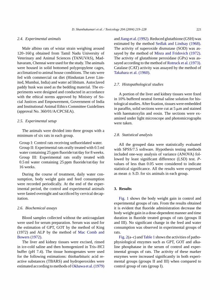
Fig. 2(a–c) andTable 1shows the activities of pathhysiological enzymes such as GPT, GOT and a
ine phosphatase in the serum of control and exmental groups of rats. The activity of these marnzymes were increased significantly in both expental groups (groups II and III) when compared
ontrol group of rats (group I).

222 D. Shanthakumari et al. / Toxicology 204 (2004) 219–228
Fig. 1. Change in body weight of control and experimental group of rats after 4th, 8th, 12th and 16th weeks of fluoride (25 ppm) treatment,respectively.
Table 1Activities of GPT, GOT and ALP in serum of control and experi-mental groups of rats
Parameters Group I Group II Group III
GPT IU/l 38.43± 5.94 49.02± 6.41∗ 50.7± 6.77∗GOT IU/l 26.41± 1.80 28.26± 2.31∗ 29.63± 2.85∗ALP IU/l 86.78± 9.21 88.86± 9.63∗ 106.62± 11.01∗
Values are expressed as mean± S.D. for six animals in each group.Groups II, III compared with group I.
∗ Values are statistically significant at (P < 0.05).
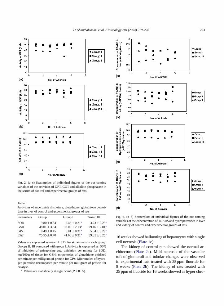
Fig. 3(a–d) andTable 2shows the concentrationof TBARS and hydroperoxides in liver and kidney ofcontrol and experimental groups of rats. There was asignificant elevation in tissue TBARS and hydroper-
Table 2Levels of TBARS and hydroperoxides in liver and kidney of control and experimental groups of rats
Parameters Group I Group II Group III
TBARS (mM/100 g tissue) Liver 0.93± 0.02 1.81± 0.29∗ 1.88± 0.30∗Kidney 1.34± 0.22 2.27± 0.28∗ 2.39± 0.32∗
Hydroperoxides (mM/100 g tissue) Liver 72.13± 1.8 95.23± 0.5∗ 96.24± 0.53∗Kidney 58.03± 1.9 74.46± 0.5∗ 75.41± 0.51∗
Values are expressed as mean± S.D. for six animals in each group. Groups II, III compared with group I.∗ Values are statistically significant at (P < 0.05).
oxides in both experimental groups (groups II andIII).
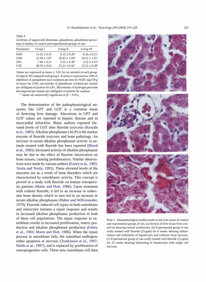
Figs. 4(a–d) and 5(a–d), Tables 3 and 4show the ac-tivities of SOD, GSH, GPx and CAT in liver and kidneyof control and experimental groups of rats. A signifi-cant decrease was observed in the activities of SOD,GSH, GPx and CAT in liver and kidney of experimen-tal animals (groups II and III).
The histopathological examination revealed exten-sive alterations in liver and kidney of fluoride treatedrats. The liver of control rats (Plate 1a) shows nor-mal architecture. In liver, inflammation and infiltrationand cohesive focal necrosis were found in experimen-tal rats treated with 25 ppm of fluoride for 8 weeks(Plate 1b) and rats treated with 25 ppm of fluoride for
D. Shanthakumari et al. / Toxicology 204 (2004) 219–228 223
Fig. 2. (a–c) Scatterplots of individual figures of the out comingvariables of the activities of GPT, GOT and alkaline phosphatase inthe serum of control and experimental groups of rats.
Table 3Activities of superoxide dismutase, glutathione, glutathione peroxi-dase in liver of control and experimental groups of rats
Parameters Group I Group II Group III
SOD 9.80± 0.34 5.45± 0.21∗ 3.23± 0.23∗GSH 48.01± 3.34 33.09± 2.13∗ 29.16± 2.01∗GPx 9.49± 0.45 6.01± 0.31∗ 5.04± 0.29∗CAT 75.55± 0.40 41.60± 0.31∗ 39.31± 0.25∗
Values are expressed as mean± S.D. for six animals in each group.Groups II, III compared with group I. Activity is expressed as: 50%of inhibition of epinephrine auto oxidation per minute for SOD;mg/100 g of tissue for GSH; micromoles of glutathione oxidizedper minute per milligram of protein for GPx. Micromoles of hydro-gen peroxide decomposed per minute per milligram of protein forcatalyse.
∗ Values are statistically at significant (P < 0.05).
Fig. 3. (a–d) Scatterplots of individual figures of the out comingvariables of the concentration of TBARS and hydroperoxides in liverand kidney of control and experimental groups of rats.
16 weeks showed ballooning of hepatocytes with singlecell necrosis (Plate 1c).
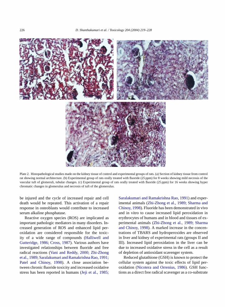
The kidney of control rats showed the normal ar-chitecture (Plate 2a). Mild necrosis of the vasculartuft of glomeruli and tubular changes were observedin experimental rats treated with 25 ppm fluoride for8 weeks (Plate 2b). The kidney of rats treated with25 ppm of fluoride for 16 weeks showed as hyper chro-

224 D. Shanthakumari et al. / Toxicology 204 (2004) 219–228
Fig. 4. (a–d) Scatterplots of individual figures of the out comingvariables of the activities of SOD, GSH, GPx and CAT in liver ofcontrol and experimental groups of rats.
matic changes in glomerulus and necrosis of tuft of theglomerulus.
4. Discussion
Every trace element is potentially toxic when thesafe and adequate exposure is exceeded. Fluoride ion
Fig. 5. (a–d) Scatterplots of individual figures of the out comingvariables of the activities of SOD, GSH, GPx and CAT in kidney ofcontrol and experimental groups of rats.
is protoplasmic poison and only a small amount canbe tolerated by any living cell and known to causeseveral biochemical alterations. The decreased bodyweight observed in experimental groups (groups II andIII) in comparison to normal rats (group I) indicatesloss of weight due to excessive break down of tissueproteins (Chatterjea and Shinde, 2002).
D. Shanthakumari et al. / Toxicology 204 (2004) 219–228 225
Table 4Activities of superoxide dismutase, glutathione, glutathione peroxi-dase in kidney of control and experimental groups of rats
Parameters Group I Group II Group III
SOD 15.62± 0.25 9.12± 0.29∗ 8.34± 0.21∗GSH 32.58± 1.87 26.42± 1.04∗ 24.31± 1.01∗GPx 7.46± 0.21 4.31± 0.39∗ 3.22± 0.31∗CAT 40.35± 0.62 25.21± 0.41∗ 23.32± 0.28∗
Values are expressed as mean± S.D. for six animals in each group.Groups II, III compared with group I. Activity is expressed as: 50% ofinhibition of epinephrine auto oxidation per min for SOD; mg/100 gof tissue for GSH; micromoles of glutathione oxidized per minuteper milligram of protein for GPx. Micromoles of hydrogen peroxidedecomposed per minute per milligram of protein for catalase.
∗ Values are statistically significant at (P < 0.05).
The determination of the pathophysiological en-zymes like GPT and GOT is a common meanof detecting liver damage. Alterations in GPT andGOT values are reported in hepatic disease and inmyocardial infraction. Many authors reported ele-vated levels of GOT after fluoride toxicosis (Kessabiet al., 1983). Alkaline phosphatase (ALP) is the markerenzyme of fluoride toxicosis and bone pathology. Anincrease in serum alkaline phosphatase activity in an-imals treated with fluoride has been reported (Bloodet al., 1983). Increased activity of alkaline phosphatasemay be due to the effect of fluorine intoxication onbone tissues, causing protuberances. Similar observa-tions were made by various authors (Farley et al., 1983;Teotia and Teotia, 1991). These elevated levels of theenzymes are as a result of bone disorders which arecharacterised by osteoblastic activity. This concept isproved in a study with fluoride on human osteoporo-sis patients (Marie and Hott, 1986). Upon treatmentwith sodium fluoride, it led to an increase in trabec-ular bone density which in turn led to an increase inserum alkaline phosphatase (Haber and Willvonseder,1979). Fluoride induced cell injury in both osteoblastsand osteocytes initiates a repair response and resultsin increased alkaline phosphatase production in bothof these cell populations. The repair response in os-teoblasts results in increased proliferation, matrix pro-duction and alkaline phosphatase production (Farleyet al., 1983; Marie and Hott, 1986). When the repairprocess in osteoblasts fails, the osteoblast undergoese ;N ofo then
Plate 1. Histopathological studies made on the liver tissue of controland experimental groups of rats. (a) Section of liver tissue from con-trol rat showing normal architecture. (b) Experimental group of ratsorally treated with fluoride (25 ppm) for 8 weeks showing inflam-mation and infiltration of hepatocytes and cohesive focal necrosis.(c) Experimental group of rats orally treated with fluoride (25 ppm)for 16 weeks showing ballooning of hepatocytes with single cellnecrosis.
ither apoptosis or necrosis (Tomkinson et al., 1997oble et al., 1997), and is replaced by proliferationsteoprogenitor cells. These new osteoblasts will
226 D. Shanthakumari et al. / Toxicology 204 (2004) 219–228
Plate 2. Histopathological studies made on the kidney tissue of control and experimental groups of rats. (a) Section of kidney tissue from controlrat showing normal architecture. (b) Experimental group of rats orally treated with fluoride (25 ppm) for 8 weeks showing mild necrosis of thevascular tuft of glomeruli, tubular changes. (c) Experimental group of rats orally treated with fluoride (25 ppm) for 16 weeks showing hyperchromatic changes in glomerulus and necrosis of tuft of the glomerulus.
be injured and the cycle of increased repair and celldeath would be repeated. This activation of a repairresponse in osteoblasts would contribute to increasedserum alkaline phosphatase.
Reactive oxygen species (ROS) are implicated asimportant pathologic mediators in many disorders. In-creased generation of ROS and enhanced lipid per-oxidation are considered responsible for the toxic-ity of a wide range of compounds (Halliwell andGutteridge, 1986; Cross, 1987). Various authors haveinvestigated relationships between fluoride and freeradical reactions (Vani and Reddy, 2000; Zhi-Zhonget al., 1989; Saralakumari and Ramakrishna Rao, 1991;Patel and Chinoy, 1998). A close association be-tween chronic fluoride toxicity and increased oxidativestress has been reported in humans (Jeji et al., 1985;
Saralakumari and Ramakrishna Rao, 1991) and exper-imental animals (Zhi-Zhong et al., 1989; Sharma andChinoy, 1998). Fluoride has been demonstrated in vivoand in vitro to cause increased lipid peroxidation inerythrocytes of humans and in blood and tissues of ex-perimental animals (Zhi-Zhong et al., 1989; Sharmaand Chinoy, 1998). A marked increase in the concen-trations of TBARS and hydroperoxides are observedin liver and kidney of experimental rats (groups II andIII). Increased lipid peroxidation in the liver can bedue to increased oxidative stress in the cell as a resultof depletion of antioxidant scavenger system.
Reduced glutathione (GSH) is known to protect thecellular system against the toxic effects of lipid per-oxidation (Nicotera and Orrenius, 1986). GSH func-tions as a direct free radical scavenger as a co-substrate

D. Shanthakumari et al. / Toxicology 204 (2004) 219–228 227
for GPx activity and as a co-factor for many enzymesand forms conjugates in endo and xenobiotic reactions(Gregus et al., 1996). Enhanced lipid peroxidation anddecreased activities of antioxidant enzymes have beenrecorded in soft tissues of fluoride treated mice. Theobserved decrease in GSH level in fluoride treated ratsrepresents increased utilization due to oxidative stress.GPx metabolizes hydrogen peroxide to water by usingGSH as a hydrogen donor (Sies, 1993). The depletionin the activity of GPx may result in the involvement ofdeleterious oxidative changes due to the accumulationof toxic products.
Decreased SOD levels have been reported in the tis-sues of mice exposed to high fluoride intake. Reducedactivities of SOD and CAT in liver and kidney of exper-imental rats have been observed in our study. SOD is animportant defense enzyme, which converts superoxideradicals to hydrogen peroxide (Mc-Cord et al., 1984).CAT is a heme protein, which decomposes hydrogenperoxide and protects the tissues from highly reactivehydroxyl radicals (Chance et al., 1982). The reductionin the activity of these enzymes may be due to oxidativestress exerted by fluoride intoxication.
Hence, it may be concluded that ingestion of drink-ing water containing high fluoride may lead to in-creased free radical generation which may result intissue damage and other secondary complications. Fur-ther studies are in progress, in alleviating the fluoridetoxicity by antioxidant supplementation isolated fromplant origin.
R
B ation32.
B Gay,dal
C ech-hem.
C em-ew
C Cross,Ann.
F ctlyone-
Gregus, Z., Fekete, T., Halaszi, E., Klaassen, C.D., 1996. Lipoic acidimpairs glycine conjugation of banzoic acid and renal excretionof benzoyl glycine. Drug. Metab. Disp. 24, 682–688.
Guan, Z.Z., Xiao, K.O., Zeng, X.Y., Long, Y.G., Cheng, Y.H., Jiang,S.F., Wang, Y.N., 2000. Changed cellular membrane lipid com-position and lipid peroxidation of kidney in rats with chronicfluorosis. Arch. Toxicol. 74 (10), 602.
Haber, P., Willvonseder, R., 1979. Therapy of senile osteoporosiswing sodium fluoride continuous and intermittent long term ther-apy. Schweiz.-Med.-Wochenschr. 109 (18), 688–692.
Halliwell, B., Gutteridge, J.M.C., 1986. Oxygen free radicals andiron in relation to biology and medicine: some problems andconcepts. Arch. Biochem. Biophys. 246, 501–514.
Jeji, J., Sharma, R., Jolly, S.S., Pamnani, S., 1985. Implication ofglutathione in endemic fluorosis. Fluoride 18, 117–119.
Jiang, Z.Y., Hunt, J.V., Wolff, S.D., 1992. Ferrous ion oxidation in thepresence of xylenol orange for detection of lipid hydroperoxidein low density lipoprotein. Anal. Biochem. 202, 384–389.
Kessabi, M., Khouzaimi, M., Braun, J.P., Hamliri, 1983. A serumbiochemical effects of fluoride on cattle in the darmous area.Vet. Hum. Toxicol. 25 (6), 403–406.
Mac Comb, R.B., Bowers, G.N., 1972. Optimum buffer conditionsfor measuring alkaline phosphatase activity in human serum.Clin. Chem. 18, 97.
Mariappan, P., Vasudevan, T., Yegnanaraman, V., 1999. Behaviour offluoride with respect to static water level in Salem Distrct, TamilNadu. In: Proceedings of the International Seminar on AppliedGeochemistry, Annamalai Nagar, pp. 31–46.
Marie, P.J., Hott, M., 1986. Short-term effects of fluoride and stron-tium on bone formation and resorption in the mouse. Metabolism35, 547–551.
Mc-Cord, J.M., Keele, B.B., Fridovich, I., 1984. An enzyme basedtheory of obligate anaerobiosis, the physiological functionsof superoxide dismutase. Proc. Natl. Acad. Sci. U.S.A. 68,1024–1027.
M theoxide
N ainst41–
N tifica-logic
O desem.
P f therens. 28,
P icalride
R n theluo-
eferences
ellack, E., Schouboe, P.J., 1968. Rapid photometric determinof fluoride with SPADNS-zirconium lake. Anal. Chem. 30, 20
lood, D.C., Radostits, O.M., Henderson, J.A., Arundel, J.H.,C.C., 1983. In: Veterinary Medicine, sixth ed. Bailliere Tinand Cassel Ltd., London.
hance, B., Green Stein, D.S., Roughton, R.J.W., 1982. The manism of catalyse action steady state analysis. Arch. BiocBiophys. 37, 301–339.
hatterjea, M.N., Shinde, R., 2002. Text Book of Medical Biochistry, fifth ed. Jaypee Brothers, Medical Publishers Ltd., NDelhi, p. 317.
ross, C.E., 1987. The spectrum of diseases (pp. 531–533). In:C.E. (moderator), Oxygen Radicals and Human Disease.Intern. Med. 107, 526–545.
arley, J.R., Wergedal, J.E., Baylink, D.J., 1983. Fluoride direstimulates proliferatin and alkaline phosphatase activity of bforming cells. Science 222, 330–332.
isra, H.P., Fridovich, I., 1972. The role of superoxide anion inautooxidation of epinephrine and a simple assay for superdismutase. J. Biol. Chem. 247, 3170–3175.
icotera, P., Orrenius, S., 1986. Role of thiols in protection agbiological reactive intermediates. Adv. Exp. Med. Biol. 197,49.
oble, B.S., Stevens, H., Loveridge, N., Reeve, J., 1997. Idention of apoptotic changes in osteocytes in normal and pathohuman bone. Bone 20, 273–282.
khawa, H., Ohishi, N., Yagi, K., 1979. Assay for lipid peroxiin animal tissues by thiobarbituric acid reaction. Anal. Bioch95, 351–358.
andit, C.G., 1944. Endemic fluorosis in South India: a study ofactors involved in the production of mottled enamel in childand severe bone manifestations in adults. Indian J. Med. Re533.
atel, P.D., Chinoy, N.J., 1998. Influence of fluoride on biologfree radical reactions in ovary of mice and its reversal. Fluo31, 27.
eam, L.J., Dayton, Ohio, 1982. Ultrastructural observations oeffect of fluoride ingestion on the parathyroid gland of rat. Fride 15 (4), 208.
228 D. Shanthakumari et al. / Toxicology 204 (2004) 219–228
Rotruck, J.T., Pope, A.L., Gasther, H.E., Hafeman, D.G., Hoekstra,W.G., 1973. Biochemical role as a component of glutathioneperoxidase. Science 179, 588–590.
Saralakumari, D., Ramakrishna Rao, P., 1991. Red cell membranealterations in human chronic fluoride toxicity. Biochem. Int. 23,639–648.
Sedlak, J., Lindsay, R.H., 1968. Estimation of total protein boundand non-protein sulfhydryl groups in tissue with Ellmans reagent.Anal. Biochem. 25, 293–305.
Sharma, A., Chinoy, N.J., 1998. Role of free radicals in fluoride-induced toxicity in liver and kidney of mice and its reversal.Fluoride 31, 26.
Shivarajashankara, Y.M., Shivashankara, A.R., Hanumanth Rao, S.,Gopalakrishna Bhat, S., 2001. Oxidative stress in children withendemic skeletal florosis. Fluoride 34, 103.
Siddiqui, A.H., 1955. Fluorosis in Nalagende district, Hyderabad-Deccan. J. Br. Med. 2, 1408.
Sies, H., 1993. Damage to plasmid DNA by singlet oxygen and itsprotection. Mut. Res. 299, 183–191.
Singh, A., 1963. Medicine. Baltimore 42, 229.
Susheela, A.K., 1999. Fluorosis management programme in India.Curr. Sci. 77, 1250–1255.
Takahara, S., Hamilton, B.H., Nell, J.V., Ogura, Y., Nishimura, E.T.,1960. Hypocatalasemia a new genetic carrier state. J. Clin. Invest.29, 610–619.
Teotia, S.P.S., Teotia, M., 1991. Endemic fluoride: bone and teeth-update. Ind. J. Environ. Toxicol. 1, 1.
Tomkinson, A., Reeve, J., Shaw, R.W., Noble, B.S., 1997. The deathof osteocytes via apoptosis accompanies estrogen withdrawal inhuman bone. J. Clin. Endocrinol. Metab. 83, 3128–3135.
Vani, M.L., Reddy, K.P., 2000. Efects of fluoride accumulation onsome enzymes of brain and gastrocnemius muscle of mice. Flu-oride 33, 17–27.
Voznaya, N.F., 1981. Chemistry of Water and Microbiology. MirPublishers, Moscow.
Whitford, G.M., 1983. Fluorides: metabolism, mechanisms actionand safety. Dental Hygiene 16.
Zhi-Zhong, G., Pei-Si, Y., Nai-den, Y., Zong-Jie, Z., 1989. An experi-mental study of blood biochemical diagnostic indices for chronicfluorosis. Fluoride 22, 112–118.