Behavioral and neurotoxic effects seen during and after subchronic exposure of rats to organic...
12
Environmental Toxicology and Pharmacology 19 (2005) 785–796 Behavioral and neurotoxic effects seen during and after subchronic exposure of rats to organic mercury T¨ unde Vez´ er a , Andr´ as Papp a,∗ , Anita Kurunczi b , ´ Arp´ ad P´ arducz b , Mikl´ os N´ aray c , L´ aszl´ o Nagymajt´ enyi a a Department of Public Health, University of Szeged, H-6720 Szeged, D´ om t´ er 10, Hungary b Biological Research Center of the Hungarian Academy of Sciences, Szeged, Hungary c National Center for Public Health, Budapest, Hungary Available online 30 January 2005 Abstract Young adult male Wistar rats (24/group) were treated for 5 weeks with methyl mercury(II)chloride (corresponding to 0.5 and 2.0 mgHg ◦ /kg b.w., control: distilled water) by gavage, followed by a 19 weeks post-treatment period. Spontaneous motility, psychomotor performance and sensorimotor gating was repeatedly tested, electrophysiological recordings done, in the rats throughout the whole experiment. Decreased horizontal open field activity, reduced number of “noise positive” startle responses, as well as increase of startle response onset latency and peak time, and decrease of peak amplitude, was seen in the treated animals. Most changes disappeared in the post-treatment period. In the spontaneous cortical and hippocampal activity, altered distribution of the frequency bands was seen after 5 weeks of treatment but not at the end of the post-treatment period. Hippocampal population spikes in the treated animals were depressed and showed less potentiation, which effect was still present 19 weeks after finishing the treatment. The duration of the sensory cortical evoked potentials was shorter than in the controls. In the treated rats, tyrosine hydroxylase-immunoreactive boutons in the substantia nigra pars reticulata were shrunk; blood and brain Hg levels were significantly higher and decreased only slowly. Considering the continuous presence of low levels of mercurials in the human environment, effects of this kind may be supposed as the background of some human neurobehavioral abnormalities. © 2005 Elsevier B.V. All rights reserved. Keywords: Methyl mercury; Spontaneous exploratory activity; Psychomotor performance; Electrocorticogram; Evoked activity; Rat 1. Introduction In the exposure of the population to organic mercury compounds, phenyl mercury in dispersion wall paints and methyl mercury (MEM) as contaminant in food of sea (and freshwater) origin are the most important sources. Within the organism, dealkylation can occur (Gallagher and Lee, 1980) so that both organic and inorganic forms of Hg must be taken into account when investigating toxic effects. MEM was found to affect several of the known transmit- ter systems. Hippocampal population spikes (generated by glutamatergic transmission) in in vitro slices were reduced ∗ Corresponding author. Tel.: +36 62 545 119; fax: +36 62 545 120. E-mail address: [email protected] (A. Papp). by MEM most probably by a postsynaptic mechanism (Yuan and Atchison, 1994). In the extracellular space, glutamate (Glu) level is likely to increase, as suggested by the MEM- induced inhibition of Glu uptake (Aschner et al., 2000) and conversion to glutamine (Kwon and Park, 2003) in astrocytes. Muscarinic ACh receptors in the rat brain, and especially in the hippocampus, increased in number in animals with oral MEM exposure (Coccini et al., 2000), possibly leading to altered memory and psychomotor functions. Extracellular level of dopamine (DA), a transmitter with a crucial role in extrapyramidal motor control and in several elements of behavior, was increased by systemic MEM treatment (Faro et al., 1997), probably due to the action of MEM on the DA transporter (Faro et al., 2002a) and/or, indirectly, to a glutamatergic mechanism (Faro et al., 2002b). GABAergic 1382-6689/$ – see front matter © 2005 Elsevier B.V. All rights reserved. doi:10.1016/j.etap.2004.12.045
Transcript of Behavioral and neurotoxic effects seen during and after subchronic exposure of rats to organic...
Environmental Toxicology and Pharmacology 19 (2005) 785–796
Behavioral and neurotoxic effects seen during and after subchronicexposure of rats to organic mercury
Tunde Vezera, Andras Pappa,∗, Anita Kurunczib, Arpad Parduczb,Mikl os Narayc, Laszlo Nagymajtenyia
a Department of Public Health, University of Szeged, H-6720 Szeged, D´om ter 10, Hungaryb Biological Research Center of the Hungarian Academy of Sciences, Szeged, Hungary
c National Center for Public Health, Budapest, Hungary
Available online 30 January 2005
Abstract
Young adult male Wistar rats (24/group) were treated for 5 weeks with methyl mercury(II)chloride (corresponding to 0.5 and 2.0 mgHg◦/kgb.w., control: distilled water) by gavage, followed by a 19 weeks post-treatment period. Spontaneous motility, psychomotor performance andsensorimotor gating was repeatedly tested, electrophysiological recordings done, in the rats throughout the whole experiment.
ponse onsetl ost-treatmentp
ent but nota potentiation,w horter thani runk; blooda
sed as theb©
K
1
cmft1b
tg
ateM-
.ly inoral
olularolets of
hea
1d
Decreased horizontal open field activity, reduced number of “noise positive” startle responses, as well as increase of startle resatency and peak time, and decrease of peak amplitude, was seen in the treated animals. Most changes disappeared in the period.In the spontaneous cortical and hippocampal activity, altered distribution of the frequency bands was seen after 5 weeks of treatm
t the end of the post-treatment period. Hippocampal population spikes in the treated animals were depressed and showed lesshich effect was still present 19 weeks after finishing the treatment. The duration of the sensory cortical evoked potentials was s
n the controls. In the treated rats, tyrosine hydroxylase-immunoreactive boutons in the substantia nigra pars reticulata were shnd brain Hg levels were significantly higher and decreased only slowly.Considering the continuous presence of low levels of mercurials in the human environment, effects of this kind may be suppo
ackground of some human neurobehavioral abnormalities.2005 Elsevier B.V. All rights reserved.
eywords:Methyl mercury; Spontaneous exploratory activity; Psychomotor performance; Electrocorticogram; Evoked activity; Rat
. Introduction
In the exposure of the population to organic mercuryompounds, phenyl mercury in dispersion wall paints andethyl mercury (MEM) as contaminant in food of sea (and
reshwater) origin are the most important sources. Withinhe organism, dealkylation can occur (Gallagher and Lee,980) so that both organic and inorganic forms of Hg muste taken into account when investigating toxic effects.
MEM was found to affect several of the known transmit-er systems. Hippocampal population spikes (generated bylutamatergic transmission) in in vitro slices were reduced
∗ Corresponding author. Tel.: +36 62 545 119; fax: +36 62 545 120.E-mail address:[email protected] (A. Papp).
by MEM most probably by a postsynaptic mechanism (Yuanand Atchison, 1994). In the extracellular space, glutam(Glu) level is likely to increase, as suggested by the MEinduced inhibition of Glu uptake (Aschner et al., 2000) andconversion to glutamine (Kwon and Park, 2003) in astrocytesMuscarinic ACh receptors in the rat brain, and especialthe hippocampus, increased in number in animals withMEM exposure (Coccini et al., 2000), possibly leading taltered memory and psychomotor functions. Extracellevel of dopamine (DA), a transmitter with a crucial rin extrapyramidal motor control and in several elemenbehavior, was increased by systemic MEM treatment (Faroet al., 1997), probably due to the action of MEM on tDA transporter (Faro et al., 2002a) and/or, indirectly, toglutamatergic mechanism (Faro et al., 2002b). GABAergic
382-6689/$ – see front matter © 2005 Elsevier B.V. All rights reserved.oi:10.1016/j.etap.2004.12.045
786 T. Vezer et al. / Environmental Toxicology and Pharmacology 19 (2005) 785–796
transmission, involved in sensorimotor regulation and loco-motor activity (Koch et al., 2000) is also known to be affectedby MEM (Yuan and Atchison, 1994). The effects of MEMon Ca2+ homeostasis (Denny and Atchison, 1996) may influ-ence neural functions via the mentioned transmitter systemsor by direct action. MEM-dependent block of mitochondrialCa-ATPase (Freitas et al., 1996) causes increase of cytosolicCa2+ levels, affecting the gating function of ion channels.
The behavioral methods used in the present experiment(see below) are known to be sensitive in detecting changes inthe above-mentioned transmitter systems. There have been,in fact, several publications on the effect of mercury exposureon behavioral outcomes in rats (open field:Rossi et al., 1997;T-maze:Zenick, 1974; open field and several maze types:Fredriksson et al., 1996). These, however, provided no cor-relation with morphological and physiological parameters,and included no follow-up in the after-exposure period. Inthe present study, a comprehensive analysis of MEM effectson functions of the nervous system was attempted; by oralexposure of rats for 5 weeks with two doses, which provedto be toxicologically relevant in behavioral endpoints in dif-ferent settings from prenatal (Fredriksson et al., 1996; Rossiet al., 1997) to young adult (Coccini et al., 2000). Observa-tions were continued for a long post-treatment period, andtwo doses of d-amphetamine (d-AM), a dopaminerigc ago-nist, were applied to detect possible alterations in the neu-r ves-t hip-p ylasei ) toc andb , pre-p
2
2
ingC in
a week, with methylmercury chloride (CH3HgCl; analyticalgrade, Aldrich). The animals were housed under conventionalconditions (22–24◦C, 12 h light/dark cycle with light startingat 6:00 a.m., up to four rats in one cage). Standard rodent chowand drinking water was given ad libitum.
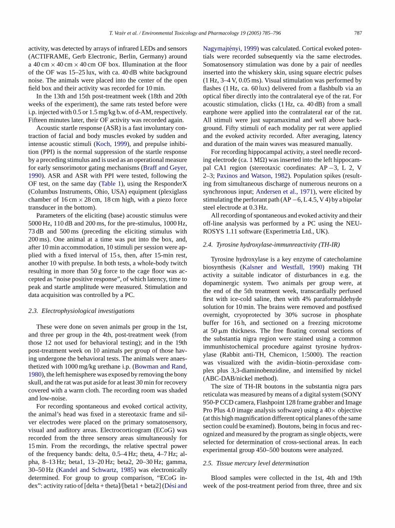
Three groups of rats were used, consisting of 24 animalseach, one receiving 2.0 mg/kg b.w. MEM (high dose group),another receiving 0.5 mg/kg, and a third, control group re-ceiving distilled water. MEM was dissolved in distilled wa-ter b.w. to 1 ml/kg administration volume, pH neutralized asnecessary, and administered by gavage. All 24 animals in thegroups received the same treatment for 5 weeks (1st to 5thtreatment week,Table 1). From each group, 12 animals werekept, without treatment, for further 19 weeks (1st to 19thpost-treatment week) to see the elimination of MEM; andwere used for the behavioral tests before, during and afterthe treatment period; and for electrophysiological recordingat the end of the post-treatment period. From the other 12 ratsper group, seven were used for electrophysiological record-ing in the 1st, and three in the 4th, post-treatment week, andtwo went for tyrosine hydroxylase immunochemistry at theend of the 5th treatment week. Blood and brain (cortex andhippocampus) samples were taken in the 1st, 4th and 19thpost-treatment week.
During the whole study, the principles of the Ethical Com-mittee for the Protection of Animals in Research of the Uni-v
2g
sted;i n),t mentw perg tot ting.O ionsb runl ing)
TT treatme
whole
ent per
4th 19th
–––3 0
3
T rrespo l) in,t nt test hibI try; ele ,b l, corte het.
otransmitter systems. A spectrum of endpoints was inigated, from mercury levels (in the blood, cortex andocampus) and histological changes (tyrosine hydrox
mmunoreactivity in the substantia nigra pars reticulataortical electrophysiological (ECoG, evoked potential)ehavioral (open field activity, acoustic startle responseulse inhibition) alterations.
. Methods
.1. Animals and treatment
Male Wistar rats (obtained at the University’s Breedenter, 10 weeks old, 160± 10 g b.w.) were treated, 5 days
able 1ime scheme of the experiment in the pre-treatment week and in the
Investigations Number of investigated animals per group over the
Pre-treatment Treatment period Post-treatm
0th 1st–4th 5th 1st 2nd–3rd
Open field12
–12
– –ASR/PPI – – –TH-IR – – – 2 –Electrophys. – – – 7 –Blood Hg level – – –
3–
Tissue Hg level – – – –
ests and investigations performed are indicated in the column of the cohe number in the cells show the number of animals used in the differeR, brain tissue sampling for tyrosine hydroxylase immunhistochemislood sampling from tail vein for Hg level determination; tissue Hg leve
ersity were strictly followed.
.2. Open field, startle response and psychomotorating tests
The animals’ spontaneous motor behavior was first ten the 0th week (before commencing MEM administratiohen in the 5th treatment week, and the 12th post-treateek (17th week of the experiment); on the 12 animalsroup defined above (Table 1). The rats were brought
he test room for accommodation 30–40 min before tespen field (OF) activity was investigated in 10-min sessetween 9.00 a.m. and 2.00 p.m. Horizontal motiliy (
ength), and vertical (rearing) as well as local (groom
nt and post-treatment period
course (0–24th weeks) of the experiment
iod
5th–11th 12th 13th 14th 15th 16–18th
–12
0.5 d-AM 12 – 1.5 d-AM 12 – –– – – – – –– – – – – – –– – – – – – 1
– – – – – –6– – – – – –
nding week. There were three groups (high dose, low dose and controvestigateds. Abbreviations: ASR/PPI, acoustic startle response and pre-pulse inition; TH-ctrophys., electrocorticogram (ECoG) and evoked potential (EP); blood Hg levelx and hippocampus sampled for Hg level determination; d-AM, d-ampamine
T. Vezer et al. / Environmental Toxicology and Pharmacology 19 (2005) 785–796 787
activity, was detected by arrays of infrared LEDs and sensors(ACTIFRAME, Gerb Electronic, Berlin, Germany) arounda 40 cm× 40 cm× 40 cm OF box. Illumination at the floorof the OF was 15–25 lux, with ca. 40 dB white backgroundnoise. The animals were placed into the center of the openfield box and their activity was recorded for 10 min.
In the 13th and 15th post-treatment week (18th and 20thweeks of the experiment), the same rats tested before werei.p. injected with 0.5 or 1.5 mg/kg b.w. of d-AM, respectively.Fifteen minutes later, their OF activity was recorded again.
Acoustic startle response (ASR) is a fast involuntary con-traction of facial and body muscles evoked by sudden andintense acoustic stimuli (Koch, 1999), and prepulse inhibi-tion (PPI) is the normal suppression of the startle responseby a preceding stimulus and is used as an operational measurefor early sensorimotor gating mechanisms (Braff and Geyer,1990). ASR and ASR with PPI were tested, following theOF test, on the same day (Table 1), using the ResponderX(Columbus Instruments, Ohio, USA) equipment (plexiglasschamber of 16 cm× 28 cm, 18 cm high, with a piezo forcetransducer in the bottom).
Parameters of the eliciting (base) acoustic stimulus were5000 Hz, 110 dB and 200 ms, for the pre-stimulus, 1000 Hz,73 dB and 500 ms (preceding the eliciting stimulus with200 ms). One animal at a time was put into the box, and,after 10 min accommodation, 10 stimuli per session were ap-p st,a itchr ac-c e top andd
2
1st,a fromt 19thp hav-i naest ,1 bonys veryc deda
ivity,t sil-v sory,v wasr ly for1 wero ; al-p ma,3 yd in-d
Nagymajtenyi, 1999) was calculated. Cortical evoked poten-tials were recorded subsequently via the same electrodes.Somatosensory stimulation was done by a pair of needlesinserted into the whiskery skin, using square electric pulses(1 Hz, 3–4 V, 0.05 ms). Visual stimulation was performed byflashes (1 Hz, ca. 60 lux) delivered from a flashbulb via anoptical fiber directly into the contralateral eye of the rat. Foracoustic stimulation, clicks (1 Hz, ca. 40 dB) from a smallearphone were applied into the contralateral ear of the rat.All stimuli were just supramaximal and well above back-ground. Fifty stimuli of each modality per rat were appliedand the evoked activity recorded. After averaging, latencyand duration of the main waves was measured manually.
For recording hippocampal activity, a steel needle record-ing electrode (ca. 1 M�) was inserted into the left hippocam-pal CA1 region (stereotaxic coordinates: AP−3, L 2, V2–3;Paxinos and Watson, 1982). Population spikes (result-ing from simultaneous discharge of numerous neurons on asynchronous input;Andersen et al., 1971), were elicited bystimulating the perforant path (AP−6, L 4.5, V 4) by a bipolarsteel electrode at 0.3 Hz.
All recording of spontaneous and evoked activity and theiroff-line analysis was performed by a PC using the NEU-ROSYS 1.11 software (Experimetria Ltd., UK).
2.4. Tyrosine hydroxylase-immunreactivity (TH-IR)
ineba thed , att usedfi ydes xedo hateb mea oft moni rox-y tionw m-p ckel(
arsr ONY9 mageP( ames rec-o , weres eache
2
19thw d six
lied with a fixed interval of 15 s, then, after 15-min renother 10 with prepulse. In both tests, a whole-body twesulting in more than 50 g force to the cage floor wasepted as “noise positive response”, of which latency, timeak and startle amplitude were measured. Stimulationata acquisition was controlled by a PC.
.3. Electrophysiological investigations
These were done on seven animals per group in thend three per group in the 4th, post-treatment week (
hose 12 not used for behavioral testing); and in theost-treatment week on 10 animals per group of those
ng undergone the behavioral tests. The animals were ahetized with 1000 mg/kg urethane i.p. (Bowman and Rand980), the left hemisphere was exposed by removing thekull, and the rat was put aside for at least 30 min for recoovered with a warm cloth. The recording room was shand low-noise.
For recording spontaneous and evoked cortical acthe animal’s head was fixed in a stereotaxic frame ander electrodes were placed on the primary somatosenisual and auditory areas. Electrocorticogram (ECoG)ecorded from the three sensory areas simultaneous5 min. From the recordings, the relative spectral pof the frequency bands: delta, 0.5–4 Hz; theta, 4–7 Hzha, 8–13 Hz; beta1, 13–20 Hz; beta2, 20–30 Hz; gam0–50 Hz (Kandel and Schwartz, 1985) was electronicalletermined. For group to group comparison, “ECoGex”: activity ratio of [delta + theta]/[beta1 + beta2] (Desi and
-
Tyrosine hydroxylase is a key enzyme of catecholamiosynthesis (Kalsner and Westfall, 1990) making THctivity a suitable indicator of disturbances in e.g.opaminergic system. Two animals per group were
he end of the 5th treatment week, transcardially perfrst with ice-cold saline, then with 4% paraformaldeholution for 10 min. The brains were removed and postfivernight, cryoprotected by 30% sucrose in phospuffer for 16 h, and sectioned on a freezing microtot 50�m thickness. The free floating coronal sections
he substantia nigra region were stained using a commmunhistochemical procedure against tyrosine hydlase (Rabbit anti-TH, Chemicon, 1:5000). The reacas visualized with the avidin–biotin–peroxidase colex plus 3,3-diaminobenzidine, and intensified by niABC-DAB/nickel method).
The size of TH-IR boutons in the substantia nigra peticulata was measured by means of a digital system (S50-P CCD camera, Flashpoint 128 frame grabber and Iro Plus 4.0 image analysis software) using a 40× objective
at this high magnification different optical planes of the section could be examined). Boutons, being in focus andgnized and measured by the program as single objectselected for determination of cross-sectional areas. Inxperimental group 450–500 boutons were analyzed.
.5. Tissue mercury level determination
Blood samples were collected in the 1st, 4th andeek of the post-treatment period from three, three an
788 T. Vezer et al. / Environmental Toxicology and Pharmacology 19 (2005) 785–796
animals of each group, respectively (Table 1). The tail veinwas punctured and blood was taken to heparinized vacutain-ers of 7.0 ml capacity (Becton-Dickinson, BD367735). Allblood samples were stored at 4◦C until analyzed.
When the electrophysiological recording was finished(1st, 4th and 19th post-treatment week), the animals weresacrificed with an overdose of Nembutal. The thorax wasopened and the animals were transcardially perfused with500 ml saline to remove blood from the organs. The brainwas removed whole, without the meninges, and was, un-der a stereomicroscope, halved and dissected to isolatethe hippocampus. The whole hippocampus and 0.3 g ofthe cortex was taken as sample, kept at−18◦C untilanalyzed.
Total mercury was determined at the accredited labora-tory of the National Center for Public Health, Budapest. Thesamples were digested in 2 ml concentrated nitric acid, with0.5 ml hydrogen peroxide added for cortex and hippocampus.Total mercury content was measured by cold vapor atomicabsorption photometry.
2.6. Statistics
The number of animals giving “noise positive response”in the ASR and PPI tests was evaluated with the Chi2
t nor-m ont outbf riod( ent
variables). One-way ANOVA was used to evaluate the resultsof ASR/PPI, Hg level determination, electrophysiologicalrecordings, TH histochemistry, and OF (in the pre-treatmentand treatment periods). The confidence level was alwaysset to p< 0.05. Post hoc analysis of group differenceswas performed by Scheffe’s test; group comparisonsfollowing the Kruskal-Wallis test were performed by theMann–WithneyU-test (also here,p< 0.05). For the statisticalanalysis, Statistica for Windows 4.0 software package wasused.
3. Results
3.1. Blood and tissue mercury levels
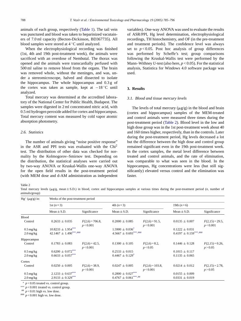
The levels of total mercury (�g/g) in the blood and brain(cortex and hippocampus) samples of the MEM-treatedand control animals were measured three times during thepost-treatment period (Table 2). Blood level in the low andhigh dose group was in the 1st post-treatment week about 40and 160 times higher, respectively, than in the controls. Laterduring the post-treatment period, Hg levels decreased a lotbut the difference between the high dose and control groupremained significant even in the 19th post-treatment week.I eent tion,w theh sig-n wasf
TT ampus fa
(n= 3)
an± S.D
00± 0
00± 067± 0
00± 0
33± 067± 0
47± 0
00± 067± 0
∗
#
est. The distribution of other data was checked forality by the Kolmogorov–Smirnov test. Depending
he distribution, the statistical analyses were carriedy two-way ANOVA or Kruskal-Wallis one-way ANOVA
or the open field results in the post-treatment pewith MEM dose and d-AM administration as independ
able 2otal mercury levels (�g/g, mean± S.D.) in blood, cortex and hippocnimals/group)
Hg◦ (�g/g) in: Weeks of the post-treatment period
1st (n= 3) 4th
Mean± S.D. Significance Me
BloodControl 0.2633± 0.035 F(2,6) = 706.8,
p< 0.0010.20
0.5 mg/kg 10.8233± 1.954*** 1.592.0 mg/kg 42.1467± 1.490*** ,### 4.56
HippocampusControl 0.1783± 0.083 F(2,6) = 42.5,
p< 0.0010.13
0.5 mg/kg 0.6200± 0.072*** 0.252.0 mg/kg 0.6633± 0.057*** 0.44
CortexControl 0.0250± 0.005 F(2,6) = 38.9,
p< 0.0010.02
0.5 mg/kg 2.1233± 0.637*** 0.282.0 mg/kg 2.9133± 0.329*** 0.47
∗ p< 0.05 treated vs. control group.∗∗ p< 0.001 treated vs. control group.## p< 0.01 high vs. low dose.## p< 0.001 high vs. low dose.
n the cortex samples, the metal level difference betwreated and control animals, and the rate of eliminaas comparable to what was seen in the blood. Inippocampus, Hg concentrations were less (but stillificantly) elevated versus control and the elimination
aster.
samples at various times during the post-treatment period (n, number o
19th (n= 6)
. Significance Mean± S.D. Significance
.085 F(2,6) = 91.5,p< 0.001
0.0135± 0.007 F(2,15) = 29.5,p< 0.001
.036* 0.1222± 0.031
.693*** ,### 0.4197± 0.159*** ,###
.105 F(2,6) = 8.2,p< 0.05
0.1446± 0.128 F(2,15) = 0.26,p> 0.05
.015 0.1015± 0.117
.129* 0.1135± 0.065
.005 F(2,6) = 103.8,p< 0.001
0.0214± 0.012 F(2,15) = 2.78,p> 0.05
.027*** 0.0155± 0.009
.061*** ,## 0.0331± 0.019
T. Vezer et al. / Environmental Toxicology and Pharmacology 19 (2005) 785–796 789
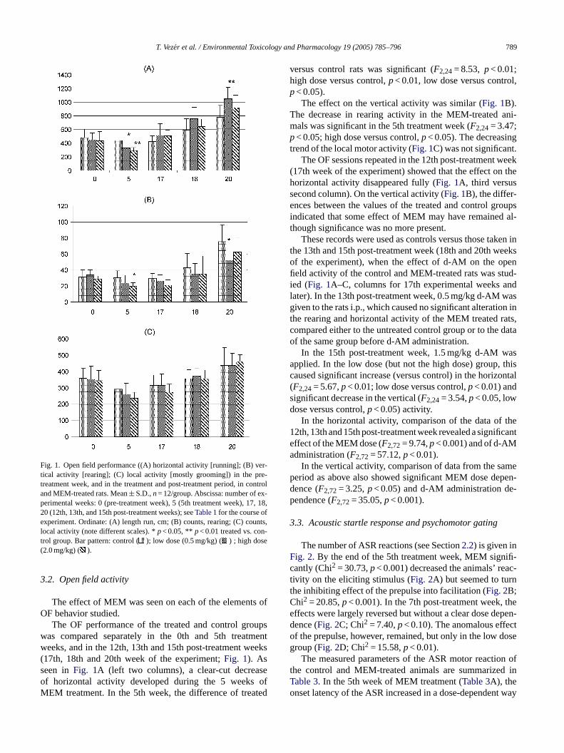
Fig. 1. Open field performance ((A) horizontal activity [running]; (B) ver-tical activity [rearing]; (C) local activity [mostly grooming]) in the pre-treatment week, and in the treatment and post-treatment period, in controland MEM-treated rats. Mean± S.D.,n= 12/group. Abscissa: number of ex-perimental weeks: 0 (pre-treatment week), 5 (5th treatment week), 17, 18,20 (12th, 13th, and 15th post-treatment weeks); seeTable 1for the course ofexperiment. Ordinate: (A) length run, cm; (B) counts, rearing; (C) counts,local activity (note different scales). *p< 0.05, ** p< 0.01 treated vs. con-trol group. Bar pattern: control ( ); low dose (0.5 mg/kg) ( ) ; high dose(2.0 mg/kg) ( ).
3.2. Open field activity
The effect of MEM was seen on each of the elements ofOF behavior studied.
The OF performance of the treated and control groupswas compared separately in the 0th and 5th treatmentweeks, and in the 12th, 13th and 15th post-treatment weeks(17th, 18th and 20th week of the experiment;Fig. 1). Asseen inFig. 1A (left two columns), a clear-cut decreaseof horizontal activity developed during the 5 weeks ofMEM treatment. In the 5th week, the difference of treated
versus control rats was significant (F2,24= 8.53, p< 0.01;high dose versus control,p< 0.01, low dose versus control,p< 0.05).
The effect on the vertical activity was similar (Fig. 1B).The decrease in rearing activity in the MEM-treated ani-mals was significant in the 5th treatment week (F2,24= 3.47;p< 0.05; high dose versus control,p< 0.05). The decreasingtrend of the local motor activity (Fig. 1C) was not significant.
The OF sessions repeated in the 12th post-treatment week(17th week of the experiment) showed that the effect on thehorizontal activity disappeared fully (Fig. 1A, third versussecond column). On the vertical activity (Fig. 1B), the differ-ences between the values of the treated and control groupsindicated that some effect of MEM may have remained al-though significance was no more present.
These records were used as controls versus those taken inthe 13th and 15th post-treatment week (18th and 20th weeksof the experiment), when the effect of d-AM on the openfield activity of the control and MEM-treated rats was stud-ied (Fig. 1A–C, columns for 17th experimental weeks andlater). In the 13th post-treatment week, 0.5 mg/kg d-AM wasgiven to the rats i.p., which caused no significant alteration inthe rearing and horizontal activity of the MEM treated rats,compared either to the untreated control group or to the dataof the same group before d-AM administration.
In the 15th post-treatment week, 1.5 mg/kg d-AM wasa thisc ontal(sd
the1 ficantea
mep pen-d e-p
3
F ifi-c ac-t ntC thee pen-d cto oseg
n oft d inTo t way
pplied. In the low dose (but not the high dose) group,aused significant increase (versus control) in the horizF2,24= 5.67,p< 0.01; low dose versus control,p< 0.01) andignificant decrease in the vertical (F2,24= 3.54,p< 0.05, lowose versus control,p< 0.05) activity.
In the horizontal activity, comparison of the data of2th, 13th and 15th post-treatment week revealed a signiffect of the MEM dose (F2,72= 9.74,p< 0.001) and of d-AMdministration (F2,72= 57.12,p< 0.01).
In the vertical activity, comparison of data from the saeriod as above also showed significant MEM dose deence (F2,72= 3.25,p< 0.05) and d-AM administration dendence (F2,72= 35.05,p< 0.001).
.3. Acoustic startle response and psychomotor gating
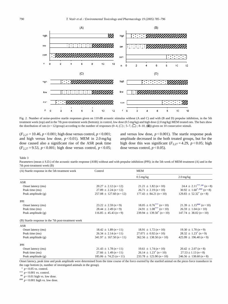
The number of ASR reactions (see Section2.2) is given inig. 2. By the end of the 5th treatment week, MEM signantly (Chi2 = 30.73,p< 0.001) decreased the animals’ reivity on the eliciting stimulus (Fig. 2A) but seemed to turhe inhibiting effect of the prepulse into facilitation (Fig. 2B;hi2 = 20.85,p< 0.001). In the 7th post-treatment week,ffects were largely reversed but without a clear dose deence (Fig. 2C; Chi2 = 7.40,p< 0.10). The anomalous effef the prepulse, however, remained, but only in the low droup (Fig. 2D; Chi2 = 15.58,p< 0.01).
The measured parameters of the ASR motor reactiohe control and MEM-treated animals are summarizeable 3. In the 5th week of MEM treatment (Table 3A), thenset latency of the ASR increased in a dose-dependen
790 T. Vezer et al. / Environmental Toxicology and Pharmacology 19 (2005) 785–796
Fig. 2. Number of noise-positive startle responses given on 110 dB acoustic stimulus without (A and C) and with (B and D) prepulse inhibition, in the 5thtreatment week (top) and in the 7th post-treatment week (bottom); in control, low dose (0.5 mg/kg) and high dose (2.0 mg/kg) MEM treated rats. The bars showthe distribution of rats (n= 12/group) according to the number of responses (0–4, (�) ; 5–7, ( ) ; 8–10, ( )) given on 10 consecutive stimuli.
(F2,27= 10.46,p< 0.001; high dose versus control,p< 0.001;and high versus low dose,p< 0.01). MEM in 2.0 mg/kgdose caused also a significant rise of the ASR peak time(F2,27= 9.53,p< 0.001; high dose versus control,p< 0.05;
and versus low dose,p< 0.001). The startle response peakamplitude decreased in the both treated groups, but for thehigh dose this was significant (F2,27= 4.29,p< 0.05; highdose versus control,p< 0.05).
Table 3Parameters (mean± S.D.) of the acoustic startle response (ASR) without and with prepulse inhibition (PPI); in the 5th week of MEM treatment (A) and in the7th post-treatment week (B)
(A) Startle response in the 5th treatment week Control MEM
0.5 mg/kg 2.0 mg/kg
ASROnset latency (ms) 20.27± 2.12 (n= 12) 21.21± 1.82 (n= 10) 24.4± 2.11*** ,## (n= 8)Peak time (ms) 27.89± 2.24 (n= 12) 26.71± 2.19 (n= 10) 30.92± 1.60* ,### (n= 8)Peak amplitude (g) 257.08± 127.60 (n= 12) 177.43± 84.21 (n= 10) 129.83± 52.32* (n= 8)
PPIOnset latency (ms) 23.22± 2.59 (n= 9) 18.05± 0.76*** (n= 10) 21.39± 1.15### (n= 10)Peak time (ms) 28.44± 2.49 (n= 9) 24.91± 1.08*** (n= 10) 26.59± 1.04 (n= 10)Peak amplitude (g) 116.85± 45.43 (n= 9) 239.94± 139.50* (n= 10) 147.74± 38.02 (n= 10)
(B) Startle response in the 7th post-treatment week
ASROnset latency (ms) 18.42± 1.89 (n= 11) 18.91± 1.72 (n= 10) 19.30± 1.70 (n= 9)Peak time (ms) 26.34± 2.14 (n= 11) 27.071± 0.92 (n= 10) 28.32± 1.23* (n= 9)Peak amplitude (g) 341.97± 167.50 (n= 11) 362.56± 138.50 (n= 10) 425.99± 196.40 (n= 9)
PPIOnset latency (ms) 21.43± 1.78 (n= 11) 19.61± 1.74 (n= 10) 20.42± 2.07 (n= 8)
9 (n= 125 (n=
O he time transduct
∗
#
Peak time (ms) 27.66± 1.4Peak amplitude (g) 185.06± 74.
nset latency, peak time and peak amplitude were determined from the cage bottom (n, number of investigated animals in the group).
∗ p< 0.05 vs. control.∗∗ p< 0.001 vs. control.## p< 0.01 high vs. low dose.## p< 0.001 high vs. low dose.
1) 26.14± 1.23* (n= 10) 27.53± 1.13 (n= 8)11) 233.78± 125.90 (n= 10) 246.56± 138.60 (n= 8)
course of the force exerted by the startled animal on the piezo forceer in
T. Vezer et al. / Environmental Toxicology and Pharmacology 19 (2005) 785–796 791
With prepulse, the response peak amplitude in the lowdose group was significantly higher (F2,26= 5.04,p< 0.05;low dose versus control,p< 0.05). It was also greater thanwithout prepulse in the same group. There was also a signifi-cant decrease in peak time (F2,26= 11.03,p< 0.001) and onsetlatency (F2,26= 24.14,p< 0.001) of the responses obtainedwith prepulse in the low dose group (in the high dose group,the changes showed the same trend without being significant).
In the 7th post-treatment week (Table 3B), ASR ampli-tude and onset latency was no more significantly differentbetween the treated and control animals. Peak time, how-ever, was still significantly longer in the high dose group(F2,27= 5.05,p< 0.05; high dose versus control,p< 0.05). Bythis time, no significant differences remained in the latencyand amplitude of the ASR with PPI in the treated groups ver-sus control, only the peak time of the low dose group was
significantly reduced (F2,26= 4.19,p< 0.05; low dose versuscontrol,p< 0.05).
3.4. Effects on electrophysiological parameters
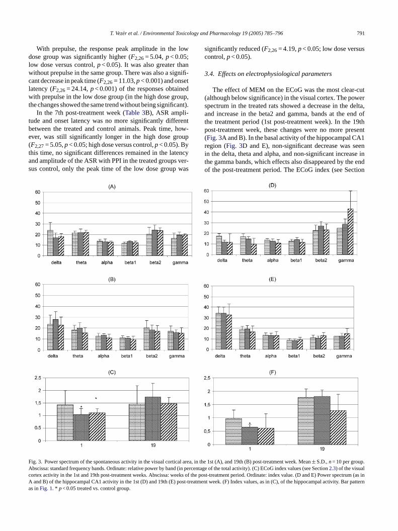
The effect of MEM on the ECoG was the most clear-cut(although below significance) in the visual cortex. The powerspectrum in the treated rats showed a decrease in the delta,and increase in the beta2 and gamma, bands at the end ofthe treatment period (1st post-treatment week). In the 19thpost-treatment week, these changes were no more present(Fig. 3A and B). In the basal activity of the hippocampal CA1region (Fig. 3D and E), non-significant decrease was seenin the delta, theta and alpha, and non-significant increase inthe gamma bands, which effects also disappeared by the endof the post-treatment period. The ECoG index (see Section
FAcAa
ig. 3. Power spectrum of the spontaneous activity in the visual cortical areabscissa: standard frequency bands. Ordinate: relative power by band (in peortex activity in the 1st and 19th post-treatment weeks. Abscissa: weeks ofand B) of the hippocampal CA1 activity in the 1st (D) and 19th (E) post-trea
s inFig. 1. * p< 0.05 treated vs. control group.
, in the 1st (A), and 19th (B) post-treatment week. Mean± S.D.,n= 10 per group.rcentage of the total activity). (C) ECoG index values (see Section2.3) of the visualthe post-treatment period. Ordinate: index value. (D and E) Power spectrum (as intment week. (F) Index values, as in (C), of the hippocampal activity. Bar pattern
792 T. Vezer et al. / Environmental Toxicology and Pharmacology 19 (2005) 785–796
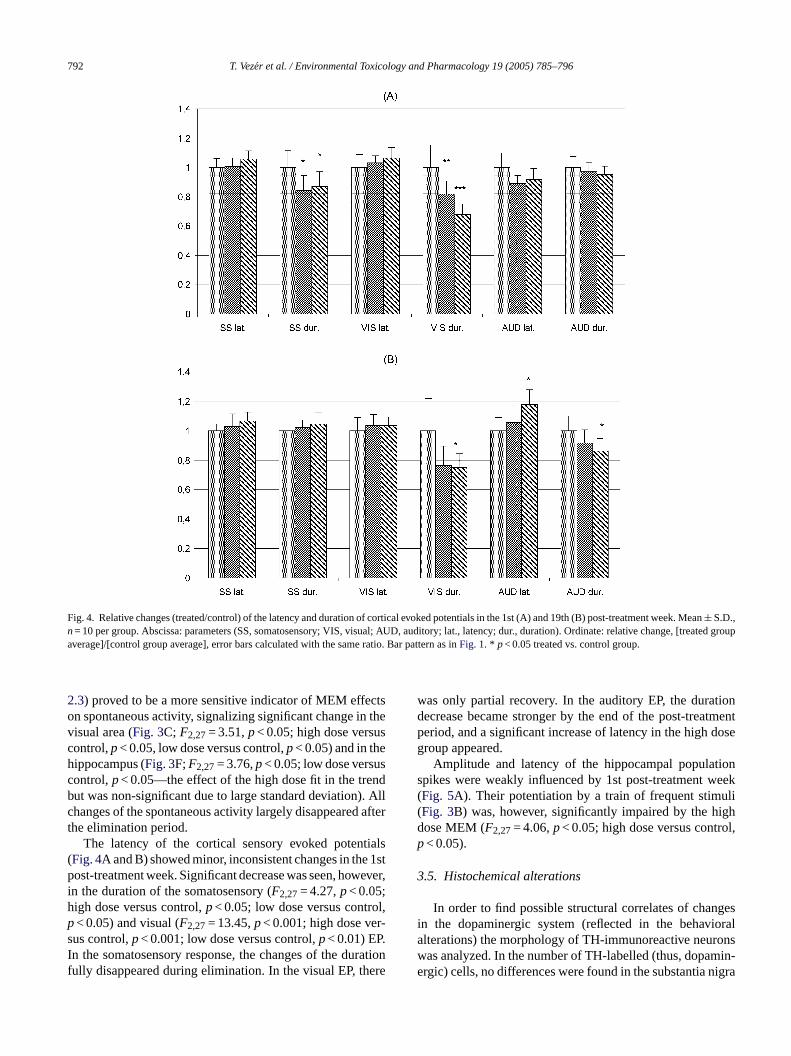
Fig. 4. Relative changes (treated/control) of the latency and duration of cortical evoked potentials in the 1st (A) and 19th (B) post-treatment week.Mean± S.D.,n= 10 per group. Abscissa: parameters (SS, somatosensory; VIS, visual; AUD, auditory; lat., latency; dur., duration). Ordinate: relative change, [treated groupaverage]/[control group average], error bars calculated with the same ratio. Bar pattern as inFig. 1. * p< 0.05 treated vs. control group.
2.3) proved to be a more sensitive indicator of MEM effectson spontaneous activity, signalizing significant change in thevisual area (Fig. 3C; F2,27= 3.51,p< 0.05; high dose versuscontrol,p< 0.05, low dose versus control,p< 0.05) and in thehippocampus (Fig. 3F;F2,27= 3.76,p< 0.05; low dose versuscontrol,p< 0.05—the effect of the high dose fit in the trendbut was non-significant due to large standard deviation). Allchanges of the spontaneous activity largely disappeared afterthe elimination period.
The latency of the cortical sensory evoked potentials(Fig. 4A and B) showed minor, inconsistent changes in the 1stpost-treatment week. Significant decrease was seen, however,in the duration of the somatosensory (F2,27= 4.27,p< 0.05;high dose versus control,p< 0.05; low dose versus control,p< 0.05) and visual (F2,27= 13.45,p< 0.001; high dose ver-sus control,p< 0.001; low dose versus control,p< 0.01) EP.In the somatosensory response, the changes of the durationfully disappeared during elimination. In the visual EP, there
was only partial recovery. In the auditory EP, the durationdecrease became stronger by the end of the post-treatmentperiod, and a significant increase of latency in the high dosegroup appeared.
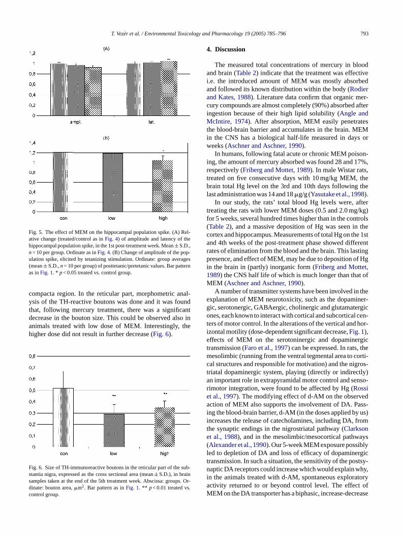
Amplitude and latency of the hippocampal populationspikes were weakly influenced by 1st post-treatment week(Fig. 5A). Their potentiation by a train of frequent stimuli(Fig. 3B) was, however, significantly impaired by the highdose MEM (F2,27= 4.06,p< 0.05; high dose versus control,p< 0.05).
3.5. Histochemical alterations
In order to find possible structural correlates of changesin the dopaminergic system (reflected in the behavioralalterations) the morphology of TH-immunoreactive neuronswas analyzed. In the number of TH-labelled (thus, dopamin-ergic) cells, no differences were found in the substantia nigra
T. Vezer et al. / Environmental Toxicology and Pharmacology 19 (2005) 785–796 793
Fig. 5. The effect of MEM on the hippocampal population spike. (A) Rel-ative change (treated/control as inFig. 4) of amplitude and latency of thehippocampal population spike, in the 1st post-treatment week. Mean± S.D.,n= 10 per group. Ordinate as inFig. 4. (B) Change of amplitude of the pop-ulation spike, elicited by tetanizing stimulation. Ordinate: group averages(mean± S.D.,n= 10 per group) of posttetanic/pretetanic values. Bar patternas inFig. 1. * p< 0.05 treated vs. control group.
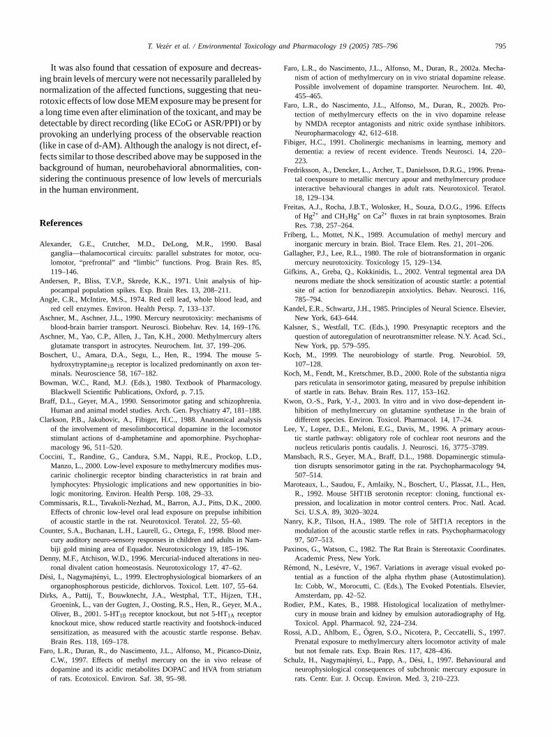
compacta region. In the reticular part, morphometric anal-ysis of the TH-reactive boutons was done and it was foundthat, following mercury treatment, there was a significantdecrease in the bouton size. This could be observed also inanimals treated with low dose of MEM. Interestingly, thehigher dose did not result in further decrease (Fig. 6).
Fig. 6. Size of TH-immunoreactive boutons in the reticular part of the sub-stantia nigra, expressed as the cross sectional area (mean± S.D.), in brainsamples taken at the end of the 5th treatment week. Abscissa: groups. Or-dinate: bouton area,�m2. Bar pattern as inFig. 1. ** p< 0.01 treated vs.control group.
4. Discussion
The measured total concentrations of mercury in bloodand brain (Table 2) indicate that the treatment was effectivei.e. the introduced amount of MEM was mostly absorbedand followed its known distribution within the body (Rodierand Kates, 1988). Literature data confirm that organic mer-cury compounds are almost completely (90%) absorbed afteringestion because of their high lipid solubility (Angle andMcIntire, 1974). After absorption, MEM easily penetratesthe blood-brain barrier and accumulates in the brain. MEMin the CNS has a biological half-life measured in days orweeks (Aschner and Aschner, 1990).
In humans, following fatal acute or chronic MEM poison-ing, the amount of mercury absorbed was found 28 and 17%,respectively (Friberg and Mottet, 1989). In male Wistar rats,treated on five consecutive days with 10 mg/kg MEM, thebrain total Hg level on the 3rd and 10th days following thelast administration was 14 and 18�g/g (Yasutake et al., 1998).
In our study, the rats’ total blood Hg levels were, aftertreating the rats with lower MEM doses (0.5 and 2.0 mg/kg)for 5 weeks, several hundred times higher than in the controls(Table 2), and a massive deposition of Hg was seen in thecortex and hippocampus. Measurements of total Hg on the 1stand 4th weeks of the post-treatment phase showed differentrates of elimination from the blood and the brain. This lastingp f Hgi t,1 ofM
thee ner-g rgico cen-t hor-ie rgict them orti-c ros-t tly)a nso-r ie eda ss-i us)i fromte ays( lyl rgict stsy-n hy,i torya t ofM rease
resence, and effect of MEM, may be due to deposition on the brain in (partly) inorganic form (Friberg and Motte989) the CNS half life of which is much longer than thatEM (Aschner and Aschner, 1990).A number of transmitter systems have been involved in
xplanation of MEM neurotoxicity, such as the dopamiic, serotonergic, GABAergic, cholinergic and glutamatenes, each known to interact with cortical and subcortical
ers of motor control. In the alterations of the vertical andzontal motility (dose-dependent significant decrease,Fig. 1),ffects of MEM on the serotoninergic and dopamine
ransmission (Faro et al., 1997) can be expressed. In rats,esolimbic (running from the ventral tegmental area to c
al structures and responsible for motivation) and the nigriatal dopaminergic system, playing (directly or indirecn important role in extrapyramidal motor control and seimotor integration, were found to be affected by Hg (Rosst al., 1997). The modifying effect of d-AM on the observction of MEM also supports the involvement of DA. Pa
ng the blood-brain barrier, d-AM (in the doses applied byncreases the release of catecholamines, including DA,he synaptic endings in the nigrostriatal pathway (Clarksont al., 1988), and in the mesolimbic/mesocortical pathwAlexander et al., 1990). Our 5-week MEM exposure possibed to depletion of DA and loss of efficacy of dopamineransmission. In such a situation, the sensitivity of the poaptic DA receptors could increase which would explain w
n the animals treated with d-AM, spontaneous exploractivity returned to or beyond control level. The effecEM on the DA transporter has a biphasic, increase-dec

794 T. Vezer et al. / Environmental Toxicology and Pharmacology 19 (2005) 785–796
dose dependence (Schweri, 1994) which is a possible ex-planation of the missing or anomalous dose-dependence ofcertain effects (e.g. in the open field) in our experiments.
Shrinkage of dopaminergic boutons (indicating DA de-pletion;Faro et al., 1997) in the substantia nigra pars reticu-lata (SNR) in the rats after 5 weeks MEM treatment furthersupports this hypothesis. SNR, connecting the dorsal andventral striatum with the thalamus, superior colliculus andpontomedullary brainstem, has a strategic role in locomo-tor activity (stereotypes), and in the sensorimotor regulationof behavior by a tonic nigroreticular GABAergic inhibitoryprojection to the dorsal and ventral striatal neurons (Kochet al., 2000). The number of TH-immunoreactive cells them-selves was, on the contrary, not different in the control andMEM-treated animals, similarly to the findings ofRossiet al. (1997).
In the regulation of locomotor activity and ASR/PPI, thefunctional connections of the dorsal and ventral hippocam-pus (the Hg content of which was significantly elevated after5 weeks MEM administration,Table 2) with the amygdala,nucleus accumbens and prefrontal cortex have also been im-plicated (Zhang et al., 2002). In the rat, ASR is primarilymediated by a glutamatergic excitatory pathway comprisingthe cochlear root nucleus and the caudal pontine reticular for-mation, projecting to facial, cranial and spinal motoneurons(Lee et al., 1996). The dorsal and ventral cochlear nuclei, thev olivefv ughtt ni de( n-d sioni lo thel 0a n int thev PPI(
ise-p on-s EMa ner-g odu-ls ns ofv asalg greya(s ndt
laint SRt n a
previous experiment (Vezer and Papp, 2002). ACh is one ofthe transmitters of the projection from the pedunculopontinetegmental nucleus to the caudal pontine reticular nucleus, apart of an inhibitory pathway which mediates the reduction inmagnitude of the ASR by PPI. In the MEM-treated animals,higher number of “noise positive” responses under prepulseapplication (that is, reduced inhibitory modulation of the star-tle response) and shortened onset latency and peak time wasseen (although with no clear dose-dependence). The Hg2+
sensitivity of hippocampal muscarinic receptors is extraor-dinarily high (Coccini et al., 2000) which is in line with thedecrease of locomotion and with the stronger alteration ofhippocampal versus cortical spontaneous activity (suggestedby the changes of the frequency spectra) seen in MEM-treatedrats in the present study.
As to the cortical activity, the primary event was likelythe shift of the spontaneous activity to higher frequencies,due (at least partly) to the effect of MEM on the ascend-ing cholinergic modulation. Decreased extracellular DA level(see above) may also have contributed to this frequency al-teration (Vorobyov et al., 2003). The general depression ofcortical evoked responses (longer latency and shorter dura-tion in the treated rats) may be a consequence of increasedhigh-frequency spontaneous activity, a state when intracor-tical connections predominate over thalamocortical input(Remond and Lesevre, 1967). Reduced glial glutamate up-t ,l rs onc cor-t duc-t cts ofM dt s andd arliersh d po-t etala
5
rela-t ls inb , andn entt ina-t ngere duet in-v y thee theb ner-g bei
entrolateral tegmental nucleus and the lateral superiororm late components of the pathway (Koch, 1999). In theentral tegmental area, DA-containing neurons are thoo play (by connection of D2 and GABAA receptors) amportant role in motivation, emotion, and ASR amplituGifkins et al., 2002). The prepulse inhibition (PPI) is uer strong tonic influence of the dopaminergic transmis
n the nucleus accumbens (Mansbach et al., 1988). Reversaf the PPI, as was seen in our work, was described in
iterature (from lead-treated rats;Commissaris et al., 200)nd was explained by altered dopaminergic modulatio
he nucleus accumbens of the GABAergic pathway toentral pallidum responsible for decrease in the ASR bySwerdlow et al., 1990).
Reduced OF activity and decreased number of “noositive” ASR responses, together with increased ASRet latency and peak time in the rats after 5 weeks Mdministration, pointed to the involvement of the serotoic system. Serotonin receptors (1A and 1B) indeed m
ate startle reactivity in rats (Nanry and Tilson, 1989). Theerotonin-1B receptor is predominantly located on neuroarious brain regions including motor control centers (banglia) and structures involved in mood control (centralnd hippocampus;Maroteaux et al., 1992), and its inhibitionBoschert et al., 1994) or absence (Dirks et al., 2001) re-ults indirectly in altered modulation of startle reactivity ahreshold.
The effect of Hg on the cholinergic system may exphe diminished reactivity of the treated animals in the Aest (Fibiger, 1991), and the decrement of memory found i
ake induced by Hg (Aschner et al., 2000) could, in the endead to desensitization of the postsynaptic Glu receptoortical neurons, contributing to the reduction of evokedical responses in a more specific way. Slowed axonal conion and increased synaptic delay due to ion channel effeEM (Sirois and Atchison, 1996) may also have contribute
o the altered evoked potentials. Increased spontaneouecreased evoked cortical activity was observed in our etudies with inorganic mercury, too (Schulz et al., 1997). Inumans, latency changes of brainstem auditory evoke
ential were found to correlate with body burden of both mnd organic mercury (Counter et al., 1998).
. Conclusion
The results of the present study demonstrated theionship between subtoxic MEM exposure, mercury levelood and parts of the brain (cortex and hippocampus)eurobehavioral alterations. Interestingly, during treatm
he animals receiving the higher dose, while during elimion those having received the lower dose showed stroffects (OF, ASR/PPI). The behavioral alterations can be
o the effect of MEM on neurotransmitter systems. Theolvement of the dopaminergic system was indicated bffect of d-AM, and by the morphological alteration ofoutons in the SNR. However, further systems (serotoic, GABAergic, glutamatergic and cholinergic) may also
nvolved.
T. Vezer et al. / Environmental Toxicology and Pharmacology 19 (2005) 785–796 795
It was also found that cessation of exposure and decreas-ing brain levels of mercury were not necessarily paralleled bynormalization of the affected functions, suggesting that neu-rotoxic effects of low dose MEM exposure may be present fora long time even after elimination of the toxicant, and may bedetectable by direct recording (like ECoG or ASR/PPI) or byprovoking an underlying process of the observable reaction(like in case of d-AM). Although the analogy is not direct, ef-fects similar to those described above may be supposed in thebackground of human, neurobehavioral abnormalities, con-sidering the continuous presence of low levels of mercurialsin the human environment.
References
Alexander, G.E., Crutcher, M.D., DeLong, M.R., 1990. Basalganglia—thalamocortical circuits: parallel substrates for motor, ocu-lomotor, “prefrontal” and “limbic” functions. Prog. Brain Res. 85,119–146.
Andersen, P., Bliss, T.V.P., Skrede, K.K., 1971. Unit analysis of hip-pocampal population spikes. Exp. Brain Res. 13, 208–211.
Angle, C.R., McIntire, M.S., 1974. Red cell lead, whole blood lead, andred cell enzymes. Environ. Health Persp. 7, 133–137.
Aschner, M., Aschner, J.L., 1990. Mercury neurotoxicity: mechanisms ofblood-brain barrier transport. Neurosci. Biobehav. Rev. 14, 169–176.
Aschner, M., Yao, C.P., Allen, J., Tan, K.H., 2000. Methylmercury altersglutamate transport in astrocytes. Neurochem. Int. 37, 199–206.
B e 5-ter-
B logy.
B enia.–188.
C lysisotorphar-
C L.D.,us-andbio-
C 000.ition
C mer-Nam-
D neu-
D an64.
D H.,.A.,
rucedBehav.
F iniz,of
tum
Faro, L.R., do Nascimento, J.L., Alfonso, M., Duran, R., 2002a. Mecha-nism of action of methylmercury on in vivo striatal dopamine release.Possible involvement of dopamine transporter. Neurochem. Int. 40,455–465.
Faro, L.R., do Nascimento, J.L., Alfonso, M., Duran, R., 2002b. Pro-tection of methylmercury effects on the in vivo dopamine releaseby NMDA receptor antagonists and nitric oxide synthase inhibitors.Neuropharmacology 42, 612–618.
Fibiger, H.C., 1991. Cholinergic mechanisms in learning, memory anddementia: a review of recent evidence. Trends Neurosci. 14, 220–223.
Fredriksson, A., Dencker, L., Archer, T., Danielsson, D.R.G., 1996. Prena-tal coexposure to metallic mercury apour and methylmercury produceinteractive behavioural changes in adult rats. Neurotoxicol. Teratol.18, 129–134.
Freitas, A.J., Rocha, J.B.T., Wolosker, H., Souza, D.O.G., 1996. Effectsof Hg2+ and CH3Hg+ on Ca2+ fluxes in rat brain synptosomes. BrainRes. 738, 257–264.
Friberg, L., Mottet, N.K., 1989. Accumulation of methyl mercury andinorganic mercury in brain. Biol. Trace Elem. Res. 21, 201–206.
Gallagher, P.J., Lee, R.L., 1980. The role of biotransformation in organicmercury neurotoxicity. Toxicology 15, 129–134.
Gifkins, A., Greba, Q., Kokkinidis, L., 2002. Ventral tegmental area DAneurons mediate the shock sensitization of acoustic startle: a potentialsite of action for benzodiazepin anxiolytics. Behav. Neurosci. 116,785–794.
Kandel, E.R., Schwartz, J.H., 1985. Principles of Neural Science. Elsevier,New York, 643–644.
Kalsner, S., Westfall, T.C. (Eds.), 1990. Presynaptic receptors and thequestion of autoregulation of neurotransmitter release. N.Y. Acad. Sci.,New York, pp. 579–595.
K 59,
K nigrabition
K t in-n of
L us-the
M ula-y 94,
M Hen,l ex-cad.
N theology
P nates.
R po-ion).vier,
R er-Hg.
R 97.male
S dure in
oschert, U., Amara, D.A., Segu, L., Hen, R., 1994. The moushydroxytryptamine1B receptor is localized predominantly on axonminals. Neuroscience 58, 167–182.
owman, W.C., Rand, M.J. (Eds.), 1980. Textbook of PharmacoBlackwell Scientific Publications, Oxford, p. 7.15.
raff, D.L., Geyer, M.A., 1990. Sensorimotor gating and schizophrHuman and animal model studies. Arch. Gen. Psychiatry 47, 181
larkson, P.B., Jakubovic, A., Fibiger, H.C., 1988. Anatomical anaof the involvement of mesolimbocortical dopamine in the locomstimulant actions of d-amphetamine and apomorphine. Psychomacology 96, 511–520.
occini, T., Randine, G., Candura, S.M., Nappi, R.E., Prockop,Manzo, L., 2000. Low-level exposure to methylmercury modifies mcarinic cholinergic receptor binding characteristics in rat brainlymphocytes: Physiologic implications and new opportunities inlogic monitoring. Environ. Health Persp. 108, 29–33.
ommissaris, R.L., Tavakoli-Nezhad, M., Barron, A.J., Pitts, D.K., 2Effects of chronic low-level oral lead exposure on prepulse inhibof acoustic startle in the rat. Neurotoxicol. Teratol. 22, 55–60.
ounter, S.A., Buchanan, L.H., Laurell, G., Ortega, F., 1998. Bloodcury auditory neuro-sensory responses in children and adults inbiji gold mining area of Equador. Neurotoxicology 19, 185–196.
enny, M.F., Atchison, W.D., 1996. Mercurial-induced alterations inronal divalent cation homeostasis. Neurotoxicology 17, 47–62.
esi, I., Nagymajtenyi, L., 1999. Electrophysiological biomarkers oforganophosphorous pesticide, dichlorvos. Toxicol. Lett. 107, 55–
irks, A., Pattij, T., Bouwknecht, J.A., Westphal, T.T., Hijzen, T.Groenink, L., van der Gugten, J., Oosting, R.S., Hen, R., Geyer, MOliver, B., 2001. 5-HT1B receptor knockout, but not 5-HT1A receptoknockout mice, show reduced startle reactivity and footshock-indsensitization, as measured with the acoustic startle response.Brain Res. 118, 169–178.
aro, L.R., Duran, R., do Nascimento, J.L., Alfonso, M., Picanco-DC.W., 1997. Effects of methyl mercury on the in vivo releasedopamine and its acidic metabolites DOPAC and HVA from striaof rats. Ecotoxicol. Environ. Saf. 38, 95–98.
och, M., 1999. The neurobiology of startle. Prog. Neurobiol.107–128.
och, M., Fendt, M., Kretschmer, B.D., 2000. Role of the substantiapars reticulata in sensorimotor gating, measured by prepulse inhiof startle in rats. Behav. Brain Res. 117, 153–162.
won, O.-S., Park, Y.-J., 2003. In vitro and in vivo dose-dependenhibition of methylmercury on glutamine synthetase in the braidifferent species. Environ. Toxicol. Pharmacol. 14, 17–24.
ee, Y., Lopez, D.E., Meloni, E.G., Davis, M., 1996. A primary acotic startle pathway: obligatory role of cochlear root neurons andnucleus reticularis pontis caudalis. J. Neurosci. 16, 3775–3789.
ansbach, R.S., Geyer, M.A., Braff, D.L., 1988. Dopaminergic stimtion disrupts sensorimotor gating in the rat. Psychopharmacolog507–514.
aroteaux, L., Saudou, F., Amlaiky, N., Boschert, U., Plassat, J.L.,R., 1992. Mouse 5HT1B serotonin receptor: cloning, functionapression, and localization in motor control centers. Proc. Natl. ASci. U.S.A. 89, 3020–3024.
anry, K.P., Tilson, H.A., 1989. The role of 5HT1A receptors inmodulation of the acoustic startle reflex in rats. Psychopharmac97, 507–513.
axinos, G., Watson, C., 1982. The Rat Brain is Stereotaxic CoordiAcademic Press, New York.
emond, N., Lesevre, V., 1967. Variations in average visual evokedtential as a function of the alpha rhythm phase (AutostimulatIn: Cobb, W., Morocutti, C. (Eds.), The Evoked Potentials. ElseAmsterdam, pp. 42–52.
odier, P.M., Kates, B., 1988. Histological localization of methylmcury in mouse brain and kidney by emulsion autoradiography ofToxicol. Appl. Pharmacol. 92, 224–234.
ossi, A.D., Ahlbom, E.,Ogren, S.O., Nicotera, P., Ceccatelli, S., 19Prenatal exposure to methylmercury alters locomotor activity ofbut not female rats. Exp. Brain Res. 117, 428–436.
chulz, H., Nagymajtenyi, L., Papp, A., Desi, I., 1997. Behavioural anneurophysiological consequences of subchronic mercury exposrats. Centr. Eur. J. Occup. Environ. Med. 3, 210–223.

796 T. Vezer et al. / Environmental Toxicology and Pharmacology 19 (2005) 785–796
Schweri, M.M., 1994. Mercuric chloride andp-chloromercuriphenylsul-fonate exert a biphasic effect on the binding of the stimu-lant [3H]methylphenidate to the dopamine transporter. Synapse 16,188–194.
Sirois, Y.E., Atchison, W.D., 1996. Effects of mercurials on ligand- andvoltage-gated ion channels: a review. Neurotoxicology 17, 63–84.
Swerdlow, N.R., Braff, D.L., Geyer, M.A., 1990. GABAergic projec-tion from nucleus accumbens to ventral pallidum mediates dopamine-induced sensorimotor gating deficits of acoustic startle in rats. BrainRes. 532, 146–150.
Vezer, T., Papp, A., 2002. Subchronic exposure to methylmercury in maleWistar rats: effects on neurobehavioral performance. Centr. Eur. J.Occup. Environ. Med. 8, 131–141.
Vorobyov, V.V., Schibaev, N.V., Morelli, M., Catra, A.P., 2003. EEG mod-ifications in the cortex and striatum after dopaminergic priming in the
6-hydroxydopamine rat model of Parkinson’s disease. Brain Res. 972,177–185.
Yasutake, A., Nakano, A., Hirayama, K., 1998. Inducing by mercurycompounds of brain metallothionein in rats: Hg
◦exposure induces
long-lived brain metallothionein. Arch. Toxicol. 72, 187–191.Yuan, Y., Atchison, W.D., 1994. Comparative effects of inorganic divalent
mercury, methylmercury and phenylmercury on membrane excitabilityand synaptic transmission of CA1 neurons in hippocampal slices ofthe rat. Neurotoxicology 15, 403–412.
Zenick, H., 1974. Behavioral and biochemical consequences inmethylmercury chloride toxicity. Pharmacol. Biochem. Behav. 2,709–713.
Zhang, W.-N., Bast, T., Feldon, J., 2002. Effects of hippocampalN-methyl-d-aspartate infusion on locomotor activity and prepulse in-hibition. Behav. Neurosci. 116, 72–84.