Behavioral and neurotoxicological effects of subchronic manganese exposure in rats
14
Environmental Toxicology and Pharmacology 19 (2005) 797–810 Behavioral and neurotoxicological effects of subchronic manganese exposure in rats T¨ unde Vez´ er a,∗ , Andr´ as Papp a , Zs ´ ofia Hoyk b , Csaba Varga b , Mikl´ os N´ aray c , L´ aszl´ o Nagymajt´ enyi a a Department of Public Health, University of Szeged, D´ om t´ er 10, H-6720 Szeged, Hungary b Biological Research Center of the Hungarian Academy of Sciences, Szeged, Hungary c National Center for Public Health, Budapest, Hungary Available online 30 January 2005 Abstract In male Wistar rats, behavioral and electrophysiological investigations, and blood and brain manganese level determinations, were performed; during 10 weeks treatment with low-dose manganese chloride and a 12 weeks post-treatment period. Three groups of 16 animals each received daily doses of 14.84 and 59.36mg/kg b.w. MnCl 2 (control: distilled water) via gavage. During treatment period, Mn accumulation was seen first in the blood, then in the brain samples of the high-dose animals. Short- and long-term spatial memory performance of the treated animals decreased, spontaneous open field activity (OF) was reduced. The number of acoustic startle responses (ASR), and the pre-pulse inhibition (PPI) of these, diminished. In the cortical and hippocampal spontaneous activity, power spectrum was shifted to higher frequencies. The latency of the sensory evoked potentials increased, and their duration, decreased. By the end of the post-treatment period, Mn levels returned to the control in all samples. The impairment of long-term spatial memory remained, as did the number of acoustic startle responses. Pre-pulse inhibition, however, returned to the pre-treatment levels. The changes of the open field activity disappeared but a residual effect could be revealed by administration of d-amphetamine. The electrophysiological effects were partially reversed. By applying a complex set of methods, it was possible to obtain new data for a better-based relationship between the known effects of Mn at neuronal level and the behavioral and electrophysiological outcomes of Mn exposure. © 2005 Elsevier B.V. All rights reserved. Keywords: Manganese chloride; Maze learning; Spontaneous locomotor activity; Cortical activity; Acoustic startle response; GFAP 1. Introduction Manganese (Mn) is, in small amounts, an essential micronutrient (WHO, 1981), being cofactor in metallo- enzymes. In prolonged exposure to higher doses it is, how- ever, a potential environmental neurotoxicant. In the general population, excess dietary intake (beyond the estimated safe and adequate daily amount) is more typical, as in case of ba- bies fed by cow milk or soybean-based formulas (Marlowe and Bliss, 1993), or in the population having environmental exposure via the drinking water. In Greece, 50 mg/l Mn in the drinking water was associated with neurological effects (Kondakis et al., 1989). Mn as man-made environmental con- ∗ Corresponding author. Tel.: +36 62 545 119; fax: +36 62 545 120. E-mail address: [email protected] (T. Vez´ er). taminant originates from Mn-containing waste (e.g. batter- ies), methylcyclopentadienyl manganese tricarbonyl (MMT) used as anti-knock petrol additive, and organo-Mn agricul- tural fungicides (ATSDR, 2000). Following ingestion, inorganic Mn is absorbed in the in- testine mostly in trivalent form (Cotzias et al., 1971) in a homeostatic self-limiting process. The main sites of depo- sition are the mitochondria-rich tissues, e.g. liver, pancreas, pituitary gland and the muscles (Barceloux, 1999). Brain is among the primary target organs in chronic Mn exposure (Roels et al., 1987), and the turnover of Mn there is much slower than in other parts of the body (Feldman, 1992). In subchronic dosage, Mn appears in the cerebral and cere- bellar cortex (Dorman et al., 2000). In case of chronic intake, Mn is found in motor control centers such as the basal ganglia, particularly the globus pallidus, striatum and substantia ni- 1382-6689/$ – see front matter © 2005 Elsevier B.V. All rights reserved. doi:10.1016/j.etap.2004.12.046
Transcript of Behavioral and neurotoxicological effects of subchronic manganese exposure in rats

Environmental Toxicology and Pharmacology 19 (2005) 797–810
Behavioral and neurotoxicological effects of subchronicmanganese exposure in rats
Tunde Vezera,∗, Andras Pappa, Zsofia Hoykb, Csaba Vargab,Mikl os Narayc, Laszlo Nagymajtenyia
a Department of Public Health, University of Szeged, D´om ter 10, H-6720 Szeged, Hungaryb Biological Research Center of the Hungarian Academy of Sciences, Szeged, Hungary
c National Center for Public Health, Budapest, Hungary
Available online 30 January 2005
Abstract
In male Wistar rats, behavioral and electrophysiological investigations, and blood and brain manganese level determinations, were performed;during 10 weeks treatment with low-dose manganese chloride and a 12 weeks post-treatment period. Three groups of 16 animals each receiveddaily doses of 14.84 and 59.36 mg/kg b.w. MnCl2 (control: distilled water) via gavage. During treatment period, Mn accumulation was seenfi ted animalsd lse inhibition( ncies. Thel els returnedt s. Pre-pulsei ct could ber
fects of Mna©
K
1
meepabaet(
tter-MT)icul-
in-
epo-reas,
surech
cere-e,glia,ni-
1d
rst in the blood, then in the brain samples of the high-dose animals. Short- and long-term spatial memory performance of the treaecreased, spontaneous open field activity (OF) was reduced. The number of acoustic startle responses (ASR), and the pre-puPPI) of these, diminished. In the cortical and hippocampal spontaneous activity, power spectrum was shifted to higher frequeatency of the sensory evoked potentials increased, and their duration, decreased. By the end of the post-treatment period, Mn levo the control in all samples. The impairment of long-term spatial memory remained, as did the number of acoustic startle responsenhibition, however, returned to the pre-treatment levels. The changes of the open field activity disappeared but a residual effeevealed by administration ofd-amphetamine. The electrophysiological effects were partially reversed.
By applying a complex set of methods, it was possible to obtain new data for a better-based relationship between the known eft neuronal level and the behavioral and electrophysiological outcomes of Mn exposure.2005 Elsevier B.V. All rights reserved.
eywords:Manganese chloride; Maze learning; Spontaneous locomotor activity; Cortical activity; Acoustic startle response; GFAP
. Introduction
Manganese (Mn) is, in small amounts, an essentialicronutrient (WHO, 1981), being cofactor in metallo-nzymes. In prolonged exposure to higher doses it is, how-ver, a potential environmental neurotoxicant. In the generalopulation, excess dietary intake (beyond the estimated safend adequate daily amount) is more typical, as in case of ba-ies fed by cow milk or soybean-based formulas (Marlowend Bliss, 1993), or in the population having environmentalxposure via the drinking water. In Greece, 50 mg/l Mn inhe drinking water was associated with neurological effectsKondakis et al., 1989). Mn as man-made environmental con-
∗ Corresponding author. Tel.: +36 62 545 119; fax: +36 62 545 120.E-mail address:[email protected] (T. Vezer).
taminant originates from Mn-containing waste (e.g. baies), methylcyclopentadienyl manganese tricarbonyl (Mused as anti-knock petrol additive, and organo-Mn agrtural fungicides (ATSDR, 2000).
Following ingestion, inorganic Mn is absorbed in thetestine mostly in trivalent form (Cotzias et al., 1971) in ahomeostatic self-limiting process. The main sites of dsition are the mitochondria-rich tissues, e.g. liver, pancpituitary gland and the muscles (Barceloux, 1999). Brain isamong the primary target organs in chronic Mn expo(Roels et al., 1987), and the turnover of Mn there is muslower than in other parts of the body (Feldman, 1992).
In subchronic dosage, Mn appears in the cerebral andbellar cortex (Dorman et al., 2000). In case of chronic intakMn is found in motor control centers such as the basal ganparticularly the globus pallidus, striatum and substantia
382-6689/$ – see front matter © 2005 Elsevier B.V. All rights reserved.oi:10.1016/j.etap.2004.12.046

798 T. Vezer et al. / Environmental Toxicology and Pharmacology 19 (2005) 797–810
gra where it causes degeneration (Yamada et al., 1986). Mnaccumulation was also described in the pons and medulla(Chan et al., 1992), and in the hippocampus (Takeda et al.,1998).
The high sensitivity of different behavior types and theintegration of different (motor, sensory, attentional, motiva-tional) behavioral functions is especially important in assess-ment neurotoxic risk due, e.g. to Mn. Excess Mn was shownto cause several neurotoxic effects such as childhood hyper-activity disorder (Barlow, 1983), manganese psychosis, ex-trapyramidal dysfunction (Quaghebeur et al., 1996), motordeficit (above 7.5�g/l blood Mn;Mergler et al., 1999); as wellas altered childhood psychomotor development (Takser et al.,2003) and impairments of several memory process (list learn-ing, visual recognition, digit span).Lucchini et al. (1999)found irritability and motor functional damage in exposedworkers having ca. 10�g/l blood Mn level. A child devel-oped severe epilepsy following exposure to welding fumesresulting in 15–20�g/l blood Mn (Hernandez et al., 2003).
Similar deficits were also induced by Mn in animals.Onerand Senturk (1992)saw impaired T-maze learning in rats re-ceiving 375�g/kg b.w. Mn for 30 days.Ingersoll et al. (1995)elicited spontaneous hypoactivity by intrathecal administra-tion of MnCl2. Dorman et al. (2000)described altered acous-tic startle responses (ASR) in neonatal rats receiving 25 and5
ons,n cat-e (e x-p ath-wd theM ctly,i
de-s rt hese( t-t ,1 ateu ofii cito-t se-i osis,w n-i Mneh reaso rats( ef-f bilityt d byM tion-s
There has been ample literature on the negative effectsof MnCl2 on behavioral outcomes in rats. Several mecha-nisms have been purposed, such as influence on transmittersystems (Tran et al., 2002; Gwiazda et al., 2002), or directlyon structural elements like hippocampal astrocytes (Aschner,1996) and the midbrain and basal ganglia (Erikson andAschner, 2002). The dentate gyrus (primarily the granulecells) plays an important role in the acquisition of new infor-mation (Ogura et al., 2002) and is possibly neural substrateof spatial reference and working memory, and of synapticplasticity (Ikegaya et al., 1995). Agents acting on the dentategyrus possibly affect learning and memory performance, lo-comotor activity, radial maze performance and spontaneousmotor activity, and psychomotor performance.
In the works cited above, however, behavioral effects werenot supported by histochemical or electrophysiological find-ings, and no follow-up in the after-exposure period was in-cluded. The electrophysiological effect of Mn exposure is initself an open question: some authors found disturbances ofspontaneous or stimulus-evoked cortical activity in workersexposed to comparable airborne Mn levels (Sinczuk-Walczaket al., 2001) while others did not (Deschamps et al., 2001).
Hence, our experiment involved a complex behavioral testbattery (8-arm radial maze, open field activity—OF, acous-tic startle response—ASR, and pre-pulse inhibition—PPI).O orti-c kedp ousp tioni erep nis-t f thed nisti
2
2
ighta ereu itionso -hd s tod ent,t s of2 thatd ricteda 5%)b tw ervede
g of1 4.84( ose
0 mg/kg b.w. MnCl2.Significant relationship between neurotoxic alterati
eurotransmitter or modulator metabolism (first of allcholamines), and Mn exposure have been describedNefft al., 1969). Mn, in case of prolonged low level eosure, accumulates in the nigrostrial dopaminergic pay (Subhash and Padmashree, 1991). The resultingopaminergic–glutamatergic interactions are involved inn effects on extrapyramidal motor functions and, indire
n the sensorimotor integration (Calabresi et al., 1997).Direct toxicity of Mn to dopaminergic neurons was
cribed byParenti et al. (1987). The involvement of otheransmitters can result from dopaminergic control upon tTakeda et al., 2002) or by direct effect of Mn. The laer is know to exist in the GABAergic (Trepper et al.998) and glutamatergic system. Reduced glial glutamptake (Aschner et al., 1999) leads, as a direct result
mpaired astrocytic-neuronal interactions (Hazell, 2002), toncreased extracellular glutamate concentration, the exoxic effect of which could play a key role in manganenduced neuronal cell death. Marked reactive astrocytith significant hypertrophy of glial fibrillary acid protei
mmunoreactive—GFAP-IR—astrocytes, was seen onxposure in the globus pallidus (Baeck et al., 2003). In theippocampal dentate gyrus, Mn exposure caused a decf GFAP-IR area in juvenile, but an increase in adult,Pappas et al., 1997). This, together with the numerousects of Mn exposure on the behavior, raised the possihat these functional and morphological changes inducen develop in parallel and are in detectable causal rela
hip.
e
ur behavioral investigations were supplemented by cal electrophysiology (electrocorticogram—ECoG, evootentials—EP), immunhistochemistry (GFAP in variarts of the hippocampus), and by Mn level determina
n blood, cortex and hippocampus. All investigations werformed both during and after the period of Mn admi
ration. It was also attempted to prove the involvement oopamine (DA) system by applying a dopaminergic ago
n the elimination period.
. Methods
.1. Animals, housing, treatment
Young adult male Wistar rats (220–250 g body wet start), obtained at the University’s Breeding Center, wsed. The animals were housed under controlled condf temperature (22–24◦C) and photoperiod (12-h light:12ark cycle with light starting at 06:00 h), with free accesrinking water. Three weeks before starting the treatm
he animals (up to 4 rats per cage) were put in cage8 cm× 40 cm× 20 cm. The memory test used requireduring the 10 weeks of treatment the animals had a restccess to food (1 h/day) resulting in a mild (ca. 20–2ody weight loss (Beatty and Shavalia, 1982). Body weighas measured and the animals’ general state was obsvery day.
The rats were divided into three groups, consistin6 animals each at start. One group was treated with 1low dose group), and another one with 59.36 (high d

T. Vezer et al. / Environmental Toxicology and Pharmacology 19 (2005) 797–810 799
group), mg/kg b.w. inorganic Mn (MnCl2·4H2O, minimum99% purity, REANAL Hungary) dissolved in distilled waterto 1 ml/kg administration volume, given by gavage, 5 daysa week, for 10 weeks (Table 1). These doses were 1/100and 1/25 of the acute oral rat-LD50, taken from the safetydata for manganese(II) chloride tetrahydrate (Physical andTheoretical Chemistry Laboratory, 2001). (Our own LD50determination gave a similar value. Literature with compara-ble figures, referred to in Section1, indicated that this doserange is likely to cause behavioral alterations.) Animals inthe third (control) group received distilled water.
The whole study was done in adherence to the require-ments by the Ethical Committee for the Protection of Animalsof the University.
2.2. Time scheme of the tests performed
The time scheme of the whole experiment, and the numberof animals used, is summarized inTable 1. The experimentlasted 22 weeks, whereby the 0th week was pre-treatmentperiod, the treatment period involved the 1st to 10th weeks,and the 11th to 22nd weeks constituted the post-treatmentperiod.
The spatial maze learning and memory test were donethroughout the whole treatment period. In the 0th, 5th and1 loco-m stics nder-w forb loods eek,fi icalr anda em-i upw ory,l ed tos ects.F sues
2
08:00a vel,d als.A werep
28-
a the1 -m nderp pel-l
Table 1), with one training per day, the rats learned to visit thefarthest points of each arm. This way, the rats were forced toacquire a win-shift food-rewarded learning strategy (Beattyand Shavalia, 1982). The rate of correct responses was givenin percents. (Incorrect responses consisted of revisiting anarm previously visited in the same session.) All animals hada run performance of over 85%. Based on this memory con-tent, the following tests were performed:
Short term spatial workingmemory testswere done in the 3rdand 5th treatment week (Mem 2handMem 4h; Table 1). Therats were one by one put for 10 min maximum in the center ofthe maze, but were allowed to enter only four of the eight openand baited arms (at their own random selection); this was the“event-to-be-remembered”. After visiting the four arms, theanimal was returned to its cage and kept there for 2 or 4 h.The rat was then put again in the maze center and allowedto complete arm choices 5–8 to obtain rewards in the fourbaited arms not visited before. In the working memory tests,errors (WM error) meant reentry into any of the baited armsvisited in the first run. Working memory performance wasthus counted as [(correct responses− WM errors)× 100].
Reference memorywas tested in the 4th week with 16 ratsper group. Here, food reward was put only in the four armspreferred by the individual rats. Entering an unbaited armconstituted a reference memory error, from which perfor-m
R the6 mentw singr newi
L e8 eks.I the9a thepT
2d, us-
i 0th,5 rats( ost-t yso ses-s e int oxo c-t and1 with1 ationa iteb enter
0th week, a subset of 12 per group was tested forotor activity, and in the 5th and 10th week, for acou
tartle response. In the 5th week, three rats per group uent electrophysiological recording, and were sacrificedlood and tissue sampling to determine Mn content (bamples were also taken in the 0th week). In the 10th wve animals per group were used for electrophysiologecording and sacrificed for blood and tissue sampling;nother two per group were sacrificed for GFAP histoch
stry (see Section2.4). The remaining six animals per groere kept for further 12 weeks, during which spatial mem
ocomotor activity and acoustic startle tests were repeatee any elimination of Mn and disappearance of its effinally, electrophysiological recording, and blood and tisampling, was done on these animals.
.3. Behavioral tests
The maze and open field tests were done betweennd 14:00 h, in a room of low illumination and noise leifferent from that used for keeping and treating the animcoustic startle response and pre-pulse inhibition testerformed under identical conditions later in the day.
.3.1. Test procedure for spatial learning and memoryThe animals’ spatial learning ability was tested in an
rm radial maze (Columbus Instruments, Ohio, USA). Inst week of treatment period (Table 1), the animals had 10in training sessions twice a day whereby they were (uartial food deprivation, see above) adapted to find food
ets in the maze arm ends. During acquisition (Acquisitionin
ance was calculated as above.
est periods,lasting 2 weeks, were introduced twice: inth to 7th treatment weeks and in the 1st to 2nd post-treateeks. In these times, the animals were kept in the hou
oom, did not have any testing, and were not exposed tonformation.
ong-termworkingmemory (retention) testswere done in thth to 10th treatment, and 3rd to 5th post-treatment we
n the 8th week, memory return was observed, and inth and 10th week, 2- and 4-h working memory (Mem R 2hndMem R 4h; Table 1). This sequence was repeated inost-treatment period (Returm(e),Mem(e)R2h,Mem(e)R4h;able 1).
.3.2. Locomotor activityThe rats’ spontaneous motor behavior was assesse
ng a fixed subset of 12 rats from each group, in theth and 10th week of treatment phase, and, with sixof the 12 tested previously) per group, in the 7th preatment week (Table 1). The test was performed alwance in the given weeks, following the maze-learningion. The animals were first allowed to accommodathe testing room for 30–40 min. An open field (OF) bf 40 cm× 40 cm× 40 cm size (ACTIFRAME, Gerb Ele
ronic, Berlin, Germany), equipped with two arrays (at 35 cm above floor level) of infrared movement detectors.1 cm distance between the beams, was used. Illumint the floor of the OF was 12–25 lx, with ca. 40 dB whackground noise. The animals were placed into the c

800T.V
ezeretal./E
nvironmentalTo
xicolog
yandPharm
acolog
y19(2005)797–810
Table 1Time scheme of experiment (lasting from the 0th to the 22nd weeks)—the pre-treatment week and treatment (A) and post-treatment (B) period
Investigations Pre-treatmentweek
Weeks of theMnCl2-treatment period (1st to 10th experimental weeks)
0th 1st 2nd 3rd 4th 5th 6th 7th 8th 9th 10th
(A)Learning and memory tests – Adaptation
(3× 16)Acquisition(3× 16)
Mem 2h(3× 16)
Reference(3× 16)
Mem 4h(3× 16)
Rest(3× 13)
Rest(3× 13)
Return(3× 13)
Mem R 2h(3× 13)
Mem R 4h(3× 13)
Open field (3× 12) – – – – (3× 12) – – – – (3× 12)ASR/PPI – – – – – – – – – – (3× 12)Electrophysiology – – – – – ECoG, EP
(3× 3)– – – – ECoG, EP
(3× 5)Histology – – – – – – – – – – (3× 2)Mn content in: blood (3× 3) – – – – Blood, cortex,
hippoc. (3× 3)– – – – Blood, cortex,
hippoc. (3× 5)
Investigations Weeks of the post-treatment (elimination) period (11th to 22nd experimental weeks)
1st 2nd 3rd 4th 5th 6th 7th 8th 9th 10th 11th 12th
(B)Learning and memory tests Rest(e)
(3× 6)Rest(e)(3× 6)
Return(e)(3× 6)
Mem(e)R 2h(3× 6)
Mem(e)R 4h(3× 6)
– – – – – – –
Open field – – – – – (3× 6) 0.5d-A (3 × 6) – 1.5d-A (3 × 6) – –ASR/PPI 6) – – – – –Electrophysiology – – – – ECoG, EP
(3× 6)Mn content in: – – – – Blood, cortex,
hippoc. (3× 6)
Tests and investigation (high dose, low dose and control) investigated, the number in the cells show the number ofanimals used in the diff m R 2h, 2-h working memory after first return; Mem R 4h, 4-h working memory after firstreturn; ASR/PPI, acous hippoc., hippocampus. (B)Rest(e), resting weeks in the post-treatment (elimination) period;Return(e), second mem econd return; Mem(e)R 2h, 2-h working memory after second return;d-A, d-amphetamine.
– – – – – (3×– – – – – –
– – – – – –
s performed are indicated in the column of the corresponding week. There were three groupserent tests. Abbreviations: (A) Mem 2h, 2-h working memory; Mem 4h, 4-h working memory; Metic startle response and pre-pulse inhibition; ECoG, electrocorticogram; EP, evoked potential;ory return in the post-treatment (elimination) period; Mem(e)R 2h, 2-h working memory after s

T. Vezer et al. / Environmental Toxicology and Pharmacology 19 (2005) 797–810 801
of the box and their spontaneous horizontal (running), ver-tical (rearing) and local (predominantly grooming) activitywas scored during a 10 min session. Movement scores werecomputed on the basis of beam interruptions with a PC soft-ware having preset conditions to discriminate between formsof movement.
2.3.3. Amphetamine-induced locomotor activityd-Amphetamine (d-A) is known to pass the blood–brain
barrier and increase the release of catecholamines (includingDA) from presynaptic endings (Clarkson et al., 1988). On the8th and 10th post-treatment week, the same rats tested beforein the open field were i.p. injected with 0.5 or 1.5 mg/kg b.w.of d-A, respectively (Table 1). Fifteen minutes later, their OFactivity was recorded again.
2.3.4. Acoustic startle response and pre-pulse inhibitionThe ASR and PPI test was performed on the same an-
imals as the OF test, in the 10th treatment and 7th post-treatment week (Table 1). In this test of sensorimotor con-trol and gating (Feifel et al., 1999), whole body muscletwitch as a response to a sudden acoustic stimulus is mea-sured. A commercially available reflex monitor (Respon-der X System, Columbus Instruments, Ohio, USA) wasused. The animals were, one by one, put in the plexiglasst ap und-a a se-r s in-t PPI( erei ibit-i msdwt se, ofw ea-s d bya
2
10w ost-ta ,1 g theb cee visuala e-d gion( dW or-t lyzedf fre-q
Then, the surface electrodes were used to record corti-cal evoked potentials. For somatosensory stimulation, a pairof needles was inserted into the area of the whiskers andrectangular stimuli (1 Hz, 3–4 V, 0.2 ms) were applied. Vi-sual stimulation was performed by flashes (1 Hz, 60 lx) de-livered by a flashbulb via an optical fiber directly into thecontralateral eye of the rat. For acoustic stimulation, clicks(1 Hz, 40 dB), from a small earphone were applied into thecontralateral ear of the rat. Fifty stimuli of each modalityper rat were applied and the EPs recorded. After averag-ing, latency and duration of the main waves was measuredmanually.
All recording of spontaneous and evoked activity and off-line analysis was performed by a PC using the NEUROSYS1.11 software (Experimetria Ltd., UK).
2.5. Immunohistochemistry
After finishing behavioral tests in the 10th treatment week,two rats per group (Table 1), were brought to terminal Nem-butal anesthesia and were perfused transcardially first with50 ml physiological saline (pre-rinse) then with 400 ml 4%paraformaldehyde fixative solved in 0.1 M phosphate buffer(PB, pH 7.4). The brains were removed from the skull, post-fixed for 4 h and cryoprotected by overnight storage in 30%s icro-t reds
ree-fl per-ob erei on-t tis-s FAP( 00,5 weret nti-m S)f edc ands ac
ip-p d fort
ideoi soft-w neds pusm itha digi-t mbero entsw s fore
est cage (16 cm× 28 cm× 18 cm) the floor of which wasiezo force transducer, enclosed within a light- and sottenuated chamber. After a 10-min accommodation,ies of 10 consecutive tones (5 kHz, 110 dB, 200 ms, 15erval) as test (eliciting) stimuli were applied. To testrepresenting sensorimotor gating), the eliciting stimuli wn a second series (after 15 min rest) preceded by inhng (non-startling) pre-pulses (1 kHz, 73 dB, 500 ms, 200elay between pre-pulse and test pulse;Graham, 1975). Ahole body twitch resulting in more than 50×g force to
he cage floor was accepted as “noise-positive” responhich latency, time to peak and peak amplitude were mured. Stimulation and data acquisition was controllePC.
.4. Electrophysiology
After finishing all behavioral tests (i.e. after 5 andeeks of Mn administration and at the end of the p
reatment period), the animals to be recorded (Table 1) werenesthetized with 1000 mg/kg urethane (Bowman and Rand980), and the left hemisphere was exposed by removinony skull. Following a recovery of 30 min minimum, surfalectrodes were placed on the primary somatosensory,nd auditory cortical focus (Zilles, 1984), and a steel nele electrode was inserted into the hippocampal CA1 restereotaxic coordinates: AP−3, L 2, V 2–3;Paxinos anatson, 1982). Spontaneous electrical activity (electroc
icogram) was recorded for 5 min, and subsequently anaor the relative power distribution among the standarduency bands (delta to gamma;Kandel and Schwartz, 1985).
ucrose in PB. Coronal sections were cut on a cryostat mome at a 50�m thickness and collected in phosphate buffealine.
Immunohistochemical reactions were performed on foating sections. After abolishing endogenous tissuexidase activity by treatment with 0.6% H2O2 in Trisuffered saline (TBS; 0.1 M, pH 7.4), the sections w
ncubated in 5% normal goat serum (NGS) in TBS caining 0.1% Triton X-100 for 1 h. Subsequently,ues samples were incubated in monoclonal anti-GSigma, 1:10,000 in TBS containing 0.1% Triton X-1% NGS) for 16 h at room temperature. The sections
horoughly rinsed and exposed to biotinylated goat aouse IgG (Dianova, 1:300 in TBS containing 5% NG
or 1 h. Finally, sections were processed with preformomplexes of streptavidin and biotinylated peroxidasetained with nickel-enhanced 3,3′-diaminobenzidine ashromogen.
The density of GFAP-IR was determined for the hocampal CA1 stratum radiatum and stratum oriens, an
he hilus of the dentate gyrus.The area of GFAP-IR structures was determined by v
maging using an Image Pro Plus 4.0 image analysisare (Media Cybernetics, Silver Spring, MD, USA). Staiections were examined under bright field with an Olymicroscope and a 10× objective. Images were recorded wSONY 950-P CCD camera (Sony Corp., Japan) and
ized. The program expressed the IR positive area as nuf pixels having densities above the threshold. Measuremere taken in a blinded fashion from at least 16 sectionach animal group and averaged.

802 T. Vezer et al. / Environmental Toxicology and Pharmacology 19 (2005) 797–810
2.6. Tissue manganese level determination
2.6.1. Blood collection, cortex and hippocampussampling
Blood and tissue samples were collected in the 5th, 10thtreatment and 12th post-treatment week from 3, 5 and 6animals per group, respectively (from three rats per group,blood was also taken in the 0th week). Before the elec-trophysiological recording, the tail vein was punctured andblood was taken to heparinized vacutainers of 7.0 ml capac-ity (Becton–Dickinson, BD367735). All blood samples werestored at 4◦C until analyzed. When the electrophysiologicalrecording was finished, the animals were sacrificed with anoverdose of Nembutal, the thorax was opened, and the ani-mals were transcardially perfused with 500 ml saline to re-move blood from the organs. The brain was removed whole,without the meninges, and was, under a stereomicroscope,halved and dissected to isolate the hippocampus. The wholehippocampus and 0.3 g of the cortex was taken as sample,kept at−18◦C until analyzed.
2.6.2. Manganese level determinationTotal manganese content was determined at the accred-
ited laboratory of the National Center for Public Health, Bu-dapest. The samples were digested in 2 ml concentrated nitrica hip-p minedu spec-t
2
wasc De-p car-r ay
ANOVA for the maze results, two-way ANOVA for the openfield and for tissue manganese content results, and one-wayANOVA for the electrophysiological and morphological re-sults. Dose was chosen for independent variable, and timefor second independent variable in two-way ANOVA (in thepost-treatment period of the open field tests, time as vari-able represented the administration of two doses ofd-A).The confidence level was set top< 0.05 in every case. Posthoc analysis of group differences after ANOVA was per-formed by the LSD test; and group comparisons following theKruskal–Wallis test were performed by the Mann–WhitneyU-test (also here,p< 0.05). The number of animals giving“noise-positive response” in the ASR and PPI tests was eval-uated with theχ2-test. For the statistical analysis, Statisticafor Windows 4.0 software package was used.
3. Results
3.1. Manganese levels
Levels of Mn in various tissues, at the end of the 5th and10th week of MnCl2 application and the 12th post-treatmentweek, are given inTable 2. Compared to the 0th week (pre-administration) value of 0.0181± 0.003�g/g, blood Mn lev-els increased in a dose- and time-dependent way. In the highd treat-mh ntwv ek,t andt
lsos tion.A eds am-
TM taken a ent, and 12tw
S
B
C
H
M dose v
cid, with 0.5 ml hydrogen peroxide added for cortex andocampus. Total manganese in the samples was detersing a flameless graphite furnace atomic absorption
rometry.
.7. Data and statistical analysis
The distribution of the data (except the ASR scores)hecked for normality by the Kolmogorov–Smirnov test.ending on the distribution, the statistical analyses wereied out by one-way ANOVA or Kruskal–Wallis one-w
able 2anganese content of the blood, cortex and hippocampus sampleseek of the post-treatment period)
ample Mn (�g/g)
Treatment
5th week (n= 3)
loodControl 0.0314± 0.00914.84 mg/kg 0.0537± 0.01259.36 mg/kg 0.3167± 0.136**,##
ortexControl 0.2167± 0.02914.84 mg/kg 0.2140± 0.03259.36 mg/kg 0.2823± 0.031
ippocampusControl 0.1710± 0.01214.84 mg/kg 0.2607± 0.08559.36 mg/kg 0.2307± 0.063
ean± S.D.;n, animals sampled in each group. *, **:p< 0.05, 0.01 high
ose group, the increase was significant both in the 5thent week (F2,6= 12.16; high dose versus control,p< 0.01;igh versus low dose,p< 0.01) and in the 10th treatmeeek (F2,12= 9.51; high dose versus control,p< 0.01; highersus low dose,p< 0.01). By the 12th post-treatment wehe difference between the blood Mn levels in the controlreated groups was no more present.
In the brain samples, a build-up of Mn level was aeen in control animals, indicating preferential deposifter 5 weeks MnCl2 treatment, cortical Mn level increaslightly versus control in the high dose group, and hippoc
t different times during the experiment (5th and 10th week of treatmh
Post-treatment
10th week (n= 5) 12th week (n= 6)
0.0118± 0.002 0.0138± 0.0010.0164± 0.005 0.0167± 0.0030.0364± 0.016**,## 0.0165± 0.004
0.3134± 0.015 0.3130± 0.0320.3220± 0.056 0.3095± 0.0560.4420± 0.107*,# 0.2872± 0.029
0.4604± 0.035 0.5897± 0.1020.4680± 0.050 0.6022± 0.1660.6813± 0.138**,## 0.5662± 0.126
s. control; #, ##:p< 0.05, 0.01 high vs. low dose.

T. Vezer et al. / Environmental Toxicology and Pharmacology 19 (2005) 797–810 803
Fig. 1. The animals’ memory performance during the whole course of the experiment. Abscissa: weeks and phases of the experiment (1st to 10th experimentalweeks, treatment period; 11th to 15th experimental weeks, post-treatment period; seeTable 1). Mem 2h, Mem 4h: 2 and 4 h working memory; Mem R 2h, MemR 4h: working memory after memory return; (e) indicates the post-treatment period (phase of elimination). Ordinate: weekly averages (mean± S.D.) of thespatial learning and memory performance in percents in the control and treated groups., , : p< 0.05, 0.01, 0.001 treated vs. control.#, ##, ###: p< 0.05,0.01, 0.001 high vs. low dose. Bar pattern: control (�); low dose (14.84 mg/kg) ( ); high dose (59.36 mg/kg) (�).
pal Mn level, in both treated groups. By the 10th week, theincrease became dose-dependently significant both in the cor-tex (F2,12= 5.22; high dose versus control,p< 0.05; high ver-sus low dose,p< 0.05) and hippocampus (F2,15= 9.41; highdose versus control,p< 0.01; high versus low dose,p< 0.01).These differences also disappeared during the 12 weeks ofpost-treatment phase.
3.2. Maze learning and memory
During all phases of the spatial maze learning and memorytest, including the treatment and post-treatment period, bothMnCl2 treated groups showed, compared to control animals,a decrease in the average memory performance (Fig. 1).
Acquisition (2nd treatment week) was dose-dependentlyimpaired in the MnCl2 treated rats (F2,15= 7.51; high doseversus control,p< 0.01; low,p< 0.05). Reference memory(4th treatment week) showed in both treated groups a sig-nificant performance deficit (F2,12= 24.68; high dose versuscontrol,p< 0.001; low versus control,p< 0.01; high versuslow dose,p< 0.05). During the 4 h short-term spatial work-ing memory tests (5th treatment week) the performance ofthe treated rats was significantly and dose-dependently lowerthan in the controls (F2,9= 16.85; high dose versus control,p< 0.001; low versus control,p< 0.05; high versus low dose,p lowd en ont imalsw em-o ss trol( ce
was also significant (p< 0.001). On comparison of the long-term (9th and 10th week) and short-term (3rd and 5th week)memory retention it was found that the group receiving lowdose Mn had a better performance in the 9th than in the 3rdweek. All other differences were insignificant.
At the end of the 10th week, Mn administration was fin-ished. In the 3rd post-treatment week, memory return in thehigh dose group was, compared to the 8th treatment week,greatly improved. In the repeated long-term spatial workingmemory test, however, no noteworthy change in any of thetreated groups was seen.
3.3. Open field activity
In the open field activity, MnCl2 treated animals had de-creased spontaneous activities.
Horizontal activity (running) of the treated animals de-creased in the 5th and 10th treatment week in a dose- andtime-dependent manner versus untreated (or versus 0th week)controls (Fig. 2A; F4,72= 11.04; 5th week: both doses ver-sus control,p< 0.01; 10th week: both doses versus control,p< 0.05). The interaction of dose and treatment time was notsignificant (F4,72= 2.17; n.s.).
Decrease of the local activity (consisting predominantlyof grooming;Fig. 2B) became significant by the 10th week( no
r s con-t rol,p k,n
< 0.05). After a 2 weeks rest period, the control andose group both showed a memory return to the level se
he 2nd week but the level reached by the high dose anas markedly less. In the long-term spatial working mry test (9th treatment week), both MnCl2 treated grouphowed a further significant memory deficit versus conF2,12= 26.14;p< 0.001) and the high versus low differen
F4,72= 3.18; both doses versus control,p< 0.01). Interactiof doses and treatment times:F4,72= 1.98; n.s.
Vertical activity (the number of rearings;Fig. 2C) waseduced in a dose- and time-dependent manner versurols by the 5th week (F4,72= 7.19; both doses versus cont< 0.001; interaction:F4,72= 2.17; n.s.). In the 10th weeear-control levels of vertical activity was seen.

804 T. Vezer et al. / Environmental Toxicology and Pharmacology 19 (2005) 797–810
Fig. 2. Open field performance (A) horizontal activity [running]; (B) localactivity [predominantly grooming]; (C) vertical activity [rearing] in differentphases of the experiment in control and treated rats. Abscissa: weeks of theexperiment: 0 (pre-treatment week), 5, 10 (5th and 10th treatment week),17, 18, 20 (7th, 8th and 10th post-treatment week). Ordinate: (A) length run,cm; (B) count, local activity; (C) count, rearing. Numerical performancevalue (mean± S.D.), note different scales and units., , : p< 0.05,0.01, 0.001 vs. control. Bar pattern as inFig. 1.
After 7 weeks without treatment the difference betweenthe control and treated groups was minimal in all three formsof activity. These records were used as controls versus thosetaken in the 8th and 10th post-treatment week (18th and 20thweek of the experiment), when the effect of two doses ofd-Aon the open field activity of the control and high- and low-dose MnCl2 treated rats was studied (Fig. 2A–C, columns forweek 17 and later). In the 8th post-treatment week, 0.5 mg/kgd-A was given to the rats i.p., which caused no significant al-teration in the rearing, grooming and horizontal activity ofboth treated groups, compared either to the untreated controlgroup or to the data of the same group befored-A adminis-tration.
Allowing 2 weeks ford-A to disappear, this test wasrepeated on the 10th post-treatment week with a higherdose (1.5 mg/kg). Here, local activity (Fig. 2B) decreasedin both treated groups (F2,12= 5.42; low dose versus con-trol, p< 0.01; high dose, n.s.). Horizontal activity (Fig. 2A)
showed an increasing trend in both groups versus controlbut the effect was significant only in the low dose group(F2,12= 4.15; p< 0.05). The high dose group also showedclearly (albeit not significantly) increased vertical activityafter 1.5 mg/kgd-A (Fig. 2C).
In the horizontal activity, comparison of the data ofthe 7th, 8th and 10th post-treatment week revealed a sig-nificant effect of the MnCl2 dose (F4,36= 3.43, p< 0.05)and ofd-A administration (F4,36= 51.05,p< 0.001). In thelocal (grooming) activity, comparison of the data of thesame post-treatment weeks revealed a significant effect ofMnCl2 dose (F4,36= 5.69,p< 0.01) and ofd-A administra-tion (F4,36= 20.63,p< 0.001).
3.4. Acoustic startle response and pre-pulse inhibition
In the 10th treatment week, the effect of Mn on thenumber (Fig. 3) and measured parameters (onset latency,peak time, peak amplitude;Table 3) of the “noise positive”acoustic startle responses (defined in Section 2.3.4) was be-low significance. Mn dose-dependently reduced the num-ber of acoustic responses given on 10 consecutive stimuli(Fig. 3A; χ2 = 16.63,p< 0.01). The effect of the pre-pulseinhibition—indicating sensorimotor gating—was strong inthe control group, but in both treated groups it was changedt lse,χ t-t of theg the1 atswp tionb een-g
ASRo enta treat-m tlyif asedi t andw Pre-p o ont er andsho ct ofth e,p n-t
3
ivity,t wer
o facilitation (i.e. more responses than without pre-pu2 = 22.98, p< 0.01; Fig. 3B versus A). In the 7th pos
reatment week, the number of ASR responses in eachroups was not significantly different from the values in0th treatment week (Fig. 3C), the reaction of the treated ras again significantly different from the controls (χ2 = 9.75,< 0.05). Now, the pre-pulses exerted their inhibiting acoth in the treated and control groups making the betwroup differences insignificant (Fig. 3D; χ2 = 0.55,p> 0.05).
The alterations in the measurable parameters of thef the control and Mn-treated animals in the 10th treatmnd 7th post-treatment week were moderate. In the 10thent week (Table 3A), onset latency increased significan
n both treated groups versus control (F2,24= 4.06;p< 0.05or both groups versus control). Peak amplitude decren the treated groups but the change was not significanas stronger in the low than in the high dose group.ulse inhibition in the treated groups was paradox als
hese parameters: the startle responses became fasttronger (F2,23= 10.01; low dose versus control,p< 0.001;igh,p< 0.01). After 7 weeks of treatment (Table 3B) mostf the effects were still present but the paradoxical effe
he pre-pulse was no more seen. ASR latency:F2,14= 8.20;igh dose versus control,p< 0.01; high versus low dos< 0.05. ASR peak time:F2,14= 11.58; high dose versus co
rol, p< 0.001; high versus low dose,p< 0.01.
.5. Electrophysiological parameters
In the spontaneous cortical and hippocampal acthere were minor, non-significant changes in the po

T. Vezer et al. / Environmental Toxicology and Pharmacology 19 (2005) 797–810 805
Fig. 3. Number of positive startle responses given on 110 dB acoustic stimulus, without (A, C) and with (B, D) pre-pulse inhibition, at the end of the treatmentperiod (10th week, top) and in the 7th week of the post-treatment period (bottom), in control and treated (low dose, 14.84 mg/kg; high dose, 59.36 mg/kg) rats.The bars show the distribution of rats (n= 12 per group in the 10th treatment week, andn= 6 per group in the 7th post-treatment week) according to the numberof responses: (0–4 (�); 5–7 ( ); 8–10 (�)) given on 10 consecutive stimuli.
distribution over the frequency range on Mn application(Fig. 4A–D). The general trend was increase in the thetaand decrease in the beta2 and gamma bands but the dose-dependence was not clear. In the 12th post-treatment week(Fig. 4E–H), the changes of the spontaneous activity inthe cortex disappeared but the decrease in the beta2 and
gamma band of the hippocampal activity became moreexpressed.
In the sensory EPs, the general effect after 10 weeks Mnadministration (Fig. 5A–C) was a lengthening of the latency(significant in the somatosensory and visual EP,p< 0.05).The alteration of the duration was mostly negligible. After 12
Table 3Measured parameters of the acoustic startle response in control and treated animals after 10 weeks Mn administration (A) and after subsequent 7 weekselimination (B)
Control MnCl2
14.84 mg/kg 59.36 mg/kg
(A) Startle response in the 10th week of the treatment periodAcoustic startle response
Onset latency (ms) 19.384± 1.8209 (n= 9) 22.095± 1.5013* (n= 9) 21.694± 2.9442* (n= 9)Peak time (ms) 26.709± 1.5104 (n= 9) 27.491± 2.0387 (n= 9) 27.127± 1.8735 (n= 9)Peak amplitude (g) 285.794± 150.9 (n= 9) 153.076± 71.1477 (n= 9) 252.200± 154.15 (n= 9)
ASR with pre-pulse inhibitionOnset latency (ms) 23.221± 1.8558 (n= 8) 19.520± 1.7919*** (n= 9) 19.837± 1.9564** (n= 9)Peak time (ms) 26.711± 1.6376 (n= 8) 25.812± 1.0045 (n= 9) 25.911± 1.4715 (n= 9)Peak amplitude (g) 128.130± 125.96 (n= 8) 203.648± 72.635 (n= 9) 185.696± 100.45 (n= 9)
(B) Startle response in the 7th week of the post-treatment periodAcoustic startle response
Onset latency (ms) 19.182± 1.3883 (n= 6) 20.800± 1.3509 (n= 6) 23.826± 2.8222**,# (n= 6)Peak time (ms) 25.527± 1.1923 (n= 6) 27.060± 0.9581 (n= 6) 30.56± 2.724***,## (n= 6)
O the time Meann
Peak amplitude (g) 197.030± 64.9463 (n= 6)
ASR with pre-pulse inhibitionOnset latency (ms) 21.951± 3.708 (n= 5)Peak time (ms) 28.207± 3.6450 (n= 5)Peak amplitude (g) 214.761± 120.709 (n= 5)
nset latency, peak time and peak amplitude were determined from
, animals investigated in each group. *, **, ***:p< 0.05, 0.01, 0.001 vs. contro203.494± 57.2813 (n= 6) 188.680± 100.367 (n= 6)
22.536± 1.3707 (n= 5) 25.250± 4.3373 (n= 5)28.260± 0.7829 (n= 5) 29.800± 2.7055 (n= 5)
155.970± 113.5942 (n= 5) 90.567± 55.0914 (n= 5)
course of the force, exerted by the startled rat on the cage bottom.± S.D.;
l; #, ##:p< 0.05, 0.01 high vs. low dose.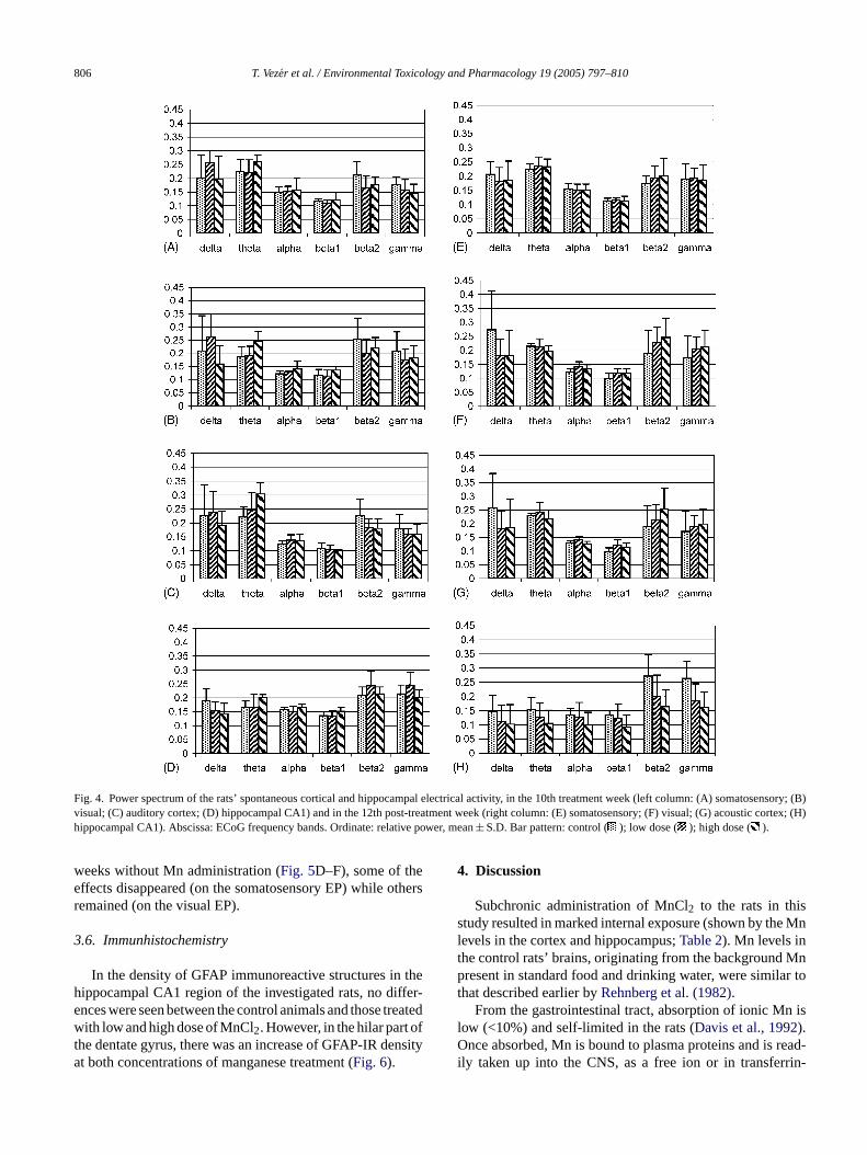
806 T. Vezer et al. / Environmental Toxicology and Pharmacology 19 (2005) 797–810
Fig. 4. Power spectrum of the rats’ spontaneous cortical and hippocampal electrical activity, in the 10th treatment week (left column: (A) somatosensory; (B)visual; (C) auditory cortex; (D) hippocampal CA1) and in the 12th post-treatment week (right column: (E) somatosensory; (F) visual; (G) acoustic cortex; (H)hippocampal CA1). Abscissa: ECoG frequency bands. Ordinate: relative power, mean± S.D. Bar pattern: control ( ); low dose ( ); high dose ( ).
weeks without Mn administration (Fig. 5D–F), some of theeffects disappeared (on the somatosensory EP) while othersremained (on the visual EP).
3.6. Immunhistochemistry
In the density of GFAP immunoreactive structures in thehippocampal CA1 region of the investigated rats, no differ-ences were seen between the control animals and those treatedwith low and high dose of MnCl2. However, in the hilar part ofthe dentate gyrus, there was an increase of GFAP-IR densityat both concentrations of manganese treatment (Fig. 6).
4. Discussion
Subchronic administration of MnCl2 to the rats in thisstudy resulted in marked internal exposure (shown by the Mnlevels in the cortex and hippocampus;Table 2). Mn levels inthe control rats’ brains, originating from the background Mnpresent in standard food and drinking water, were similar tothat described earlier byRehnberg et al. (1982).
From the gastrointestinal tract, absorption of ionic Mn islow (<10%) and self-limited in the rats (Davis et al., 1992).Once absorbed, Mn is bound to plasma proteins and is read-ily taken up into the CNS, as a free ion or in transferrin-

T. Vezer et al. / Environmental Toxicology and Pharmacology 19 (2005) 797–810 807
Fig. 5. Latency and duration of the sensory evoked potentials in the 10th treatment week (left column: (A) somatosensory; (B) visual; (C) auditory EP)and inthe 12th post-treatment week (right column: (D) somatosensory; (E) visual; (F) auditory EP). Ordinate: latency (lat.) and duration (dur.) in ms, mean± S.D. ,
: p< 0.05, 0.01 treated vs. control. Bar pattern as inFig. 4.
Fig. 6. Presence of GFAP-immunoreactive elements in different parts of thehippocampus (stratum radiatum and stratum oriens of CA1, and hilus ofthe dentate gyrus) in control and treated rats in the 10th week of treatment.Ordinate: GFAP positivity in percent of the total area (mean± S.D., on thebasis of numerous sections from two animals per group).: p< 0.05 treatedvs. control. Bar pattern as inFig. 4.
bound form (Aschner and Gannon, 1994). There is a slowshift of Mn from the parenchymal organs to the CNS whereMn will be deposited. Elimination of Mn from the CNS, firstof all from the cerebrum, is much slower than from the wholebody (Cotzias et al., 1968). Following a shorter exposure bydoses similar to what was given in our work, Mn levels inthe typically affected parts of the brain (striatum, hypothala-mus, nucleus accumbens) were not paralleled by the levels oftransmitters and their metabolites (Kontur and Fechter, 1988);indicating that the brain has some capacity of compensatingthe adverse effects of Mn.
Among the cell types constituting the brain tissue, Mn isaccumulated in astrocytes, crucial in neutralizing glutamatereleased in the glutamatergic (e.g. thalamocortical) synapses.This process was inhibited in astrocytes exposed to elevatedMn2+ (Normandin and Hazell, 2002), resulting first in over-stimulation then in desensitization or down-regulation of glu-

808 T. Vezer et al. / Environmental Toxicology and Pharmacology 19 (2005) 797–810
tamate receptors, possibly leading to the latency lengtheningof the cortical EP seen by us. Alterations of evoked responsesin Mn-exposed humans have also been described togetherwith changes in the EEG. The latter has a well-known activat-ing cholinergic modulation (Metherate et al., 1992) and if theelevated Mn level in the brain reduces the activity of cholineacetyltransferase (Lai et al., 1981), the ascending activatinginput can be diminished. The slight changes of cortical andhippocampal activity observed in our study were in line withthe above.
The hippocampal formation (with retrosplenial cortex anddorsal striatum), is a preferential site of Mn accumulation, andis an important neural axis responsible for spatial memory andreward-based learning. In our experiments, the performancein acquisition, short-term retention tasks and long-term spa-tial working memory—a stable and reliable phenomenon inrats—was significantly decreased in the MnCl2-administeredanimals in the 10th treatment week, indicating functionaldamage. A possible morphological counterpart, gliotic de-generation, was in fact seen in the dentate gyrus, in bothtreated groups in the 10th week. The dentate gyrus and CA1areas of the hippocampus, are integral neural substrates inthe acquisition of new information, hence important loci forspatial learning and working memory processes (Eyre et al.,2003). In our work, elevated hippocampal Mn level and al-t eouse f Mne
a themS p-p eoush n int at-m de-c eateda ever,r ent,s tion( e, seeF n-c PPIc nhi rtlem n,2
Mnf l andh oset riod,a encei entlyi -i ated
long-term spatial memory test, significantly decreased per-formance was still seen in the both treated groups. In the OFactivity of the control and treated rats, no more difference wasdetected, but by injecting 1.5 mg/kgd-A, the residual effectsof Mn—substantial increase of the horizontal and vertical,and decrease of the local activity—were revealed (Fig. 2).In the horizontal and local activity, the effect was strongerin the low-dose group, possibly due to the dose-dependentelimination mentioned above.
In 7th post-treatment week, the magnitude of the ASRwas still diminished but the reduction of pre-pulse inhibitionwas no more seen. The alterations of the spontaneous corticalactivity had a similar time course indicating that the effect ofMn on the cholinergic system was probably responsible forboth.
5. Conclusion
The main points of conclusion drawn from the above re-sults are as given below.
Oral MnCl2 treatment, 14.84 and 59.36 mg/kg b.w., for10 weeks brought about, primarily in the high-dose group,an increase in first the blood, then cortex and hippocampusMn levels.
pon-t mento ath-w cedd ctiv-i ertaino en-t g tot andt neu-r eurala ngesi glu-t wasf thec
am-p con-t penfi Thisi , al-t omec
le too n thek ande di ac-c d ofe sys-t , the
ered frequency spectrum of the hippocampal spontanlectrical activity was seen at the end of the 10th week oxposure.
The rats’ extrapyramidal motor control (Bonilla, 1984),nd the sensorimotor integration, is dependent onesolimbic and nigrostrial dopaminergic system (Fink andmith, 1980), both known to show dysfunction on Mn alication. In our experiments, decrease of the spontanorizontal and vertical activity in the open field was see
he 5th, and of the local activity in the 10th, week of treent. The number of “noise-positive” ASR responses
reased, and their onset latency increased, in the Mn-trnimals in the 10th week. Sensorimotor gating was, howeduced in both Mn-treated groups at the end of treatmo that the pre-pulse inhibition was inverted to facilitashown by the number of responses and by the peak timig. 3andTable 3). In the cholinergic neurons of the peduulopontine tegmental nucleus, an important part of theircuit (Swerdlow and Geyer, 1993) neurotoxic lesion by Mas been described (Tomas-Camardiel et al., 2002), result-
ng in significantly reduced PPI without affecting the staagnitude in the absence of a pre-pulse (Jones and Shanno000).
During the post-treatment period, elimination of therom the cortex and hippocampus was seen. The corticaippocampal Mn levels were falling more in the high d
han in the low dose group during the post-treatment pend some effects of Mn showed inverted dose depend
n this period, which may be due to the dose-dependncreasing elimination of Mn (Mena et al., 1967). The majorty of behavioral effects, however, remained. In the repe
During the treatment period, altered PPI and cortical saneous activity in the treated rats suggested the involvef brainstem cholinergic systems (pre-pulse inhibition pay and ascending cortical activation) due to Mn-induamage. The alterations in maze learning, open field a
ty and ASR magnitude suggested the decrease of cther neurotransmitter levels (e.g. DA or Glu) in the c
ers responsible for these behavioral functions, pointinhe involvement of the glutamatergic auditory pathwayhe nigrostriatal dopaminergic system, and possibly toopathological changes in some components of the nxis for behavior (e.g. hippocampal formation). The cha
n the cortical EPs also indicated the involvement of theamatergic afferent pathways. In the treated rats, gliosisound in the hilar part of the dentate gyrus compared toontrols.
In the post-treatment period, Mn levels in all tissue sles from the previously treated animals returned to the
rol. The alterations of the long-term spatial memory, the oeld activity and the ASR reaction, however, remained.ndicated a long-lasting effect of Mn on these functionshough the dose–effect relationship elimination was in sases anomalous.
By applying a complex set of methods, it was possibbtain new data for a better-based relationship betweenown effects of Mn at neuronal level and the behaviorallectrophysiological outcomes of MnCl2 exposure observe
n our study and described in the literature. Taking intoount that some of the effects proved to outlast the perioxposure, and the importance of the probably involvedems in the higher-order nervous functions of humans

T. Vezer et al. / Environmental Toxicology and Pharmacology 19 (2005) 797–810 809
neurotoxic risk of environmental and/or occupational MnCl2exposure seems to deserve ongoing attention.
References
Aschner, M., Gannon, M., 1994. Manganese (Mn) transport across therat blood–brain barrier: saturable and transferrin-dependent transportmechanisms. Brain Res. Bull. 33, 345–349.
Aschner, M., Vrana, K.E., Zheng, W., 1999. Manganese uptake and dis-tribution in the central nervous system (CNS). Neurotoxicology 20,173–180.
Aschner, M., 1996. The functional significance of brain metallothioneins.FASEB J. 10, 1129–1136.
ATSDR, 2000. Toxicological Profile for Manganese. US Department ofHealth and Human Services, Atlanta, GA, USA, pp. 336–338.
Baeck, S.Y., Lee, M.-J., Jung, H.-S., Kim, H.-J., Lee, C.-R., Yoo, C.,Lee, J.H., Lee, H., Yoon, C.S., Kim, Y.H., Park, J., Kim, J.-W., Jeon,B.S., Kim, Y., 2003. Effect of manganese exposure on MPTP neuro-toxicities. Neurotoxicology 24, 657–665.
Barceloux, D.G., 1999. Manganese. Clin. Toxicol. 37, 293–307.Barlow, P.J., 1983. A pilot study on the metal levels in the hair of hy-
peractive children. Med. Hypotheses 11, 309–318.Beatty, W.W., Shavalia, D.A., 1982. Spatial memory in rats: time course
of working memory and effect of anesthetics. Behav. Neural Biol. 28,454–462.
Bonilla, E., 1984. Chronic manganese intake induces changes in the motoractivity of rats. Exp. Neurol. 84, 696–700.
Bowman, W.C., Rand, M.J., 1980. Textbook of Pharmacology. Blackwell
C yondience
C rainvelop-Brain
C lysisotorhar-
C man-ns with
C 1971.an-
D neseanese
D ts in127–
D ler,adulte. J.
E lationlloth-
E g-patial
F CK-model
Feldman, R.G., 1992. Manganese. In: de Wolff, F.A. (Ed.), Handbook ofClinical Neurology: Intoxications of the Nervous System, Part I, vol.20. Elsevier, Amsterdam, pp. 303–322.
Fink, J.S., Smith, G.P., 1980. Relationships between selective denervationof dopamine terminal fields in the anterior forebrain and behavioralresponses to amphetamine and apomorphine. Brain Res. 201, 107–127.
Graham, F., 1975. The more-or-less startling effects of weak prestimuli.Psychophysiology 12, 238–248.
Gwiazda, R.H., Lee, D., Sheridan, J., Smith, D.R., 2002. Low cumulativemanganese exposure affects strial GABA but not dopamine. Neuro-toxicology 23, 69–76.
Hazell, A.S., 2002. Astrocytes and manganese neurotoxicity. Neurochem.Int. 41, 271–277.
Hernandez, E.H., Discalzi, G., Dassi, P., Jarre, L., Pira, E., 2003. Man-ganese intoxication: the cause of an inexplicable epileptic syndromein a 3 year old child. Neurotoxicology 24, 633–639.
Ikegaya, Y., Abe, K., Saito, H., Nishiyama, N., 1995. Medial amygdalaenhances synaptic transmission and synaptic plasticity in the dentategyrus of rats in vivo. J. Neurophysiol. 74, 2201–2203.
Ingersoll, R.T., Montgomery, E.B., Aposhian Jr., H.V., 1995. Central ner-vous system toxicity of manganese. I. Inhibition of spontaneous motoractivity in rats after intrathecal administration of manganese chloride.Fundam. Appl. Toxicol. 27, 106–113.
Jones, C.K., Shannon, H.E., 2000. Muscarinic cholinergic modulationof prepulse inhibition of the acoustic startle reflex. JPET 294,1017–1023.
Kandel, E.R., Schwartz, J.H., 1985. Principles of Neural Science. Elsevier,New York, pp. 643–644.
Kondakis, X.G., Makris, N., Leotsinidis, M., Papapetropoulos, T., 1989.nking
K s andurotox.
L ofcetyl-e ad-
L ati,termional
M ung–12.
M man-rology
M ler,Hud-tion:27–
M rticalsalis
N dateioxide
N up-375–
O , K.,ntateatal
. 61,
Scientific Publications, Oxford, pp. 7–15.alabresi, P., De Murtas, M., Bernardi, G., 1997. The neostriatum be
the motor function: experimental and clinical evidence. Neurosc78, 39–60.
han, A.W.K., Minski, M.J., Lim, L., Lai, J.C., 1992. Changes in bregional manganese and magnesium levels during postnatal dement: modulations by chronic manganese administration. Metab.Dis. 7, 21–33.
larkson, P.B., Jakubovic, A., Fibiger, H.C., 1988. Anatomical anaof the involvement of mesolimbocortical dopamine in the locomstimulant actions ofd-amphetamine and apomorphine. Psychopmacology 96, 511–520.
otzias, G.C., Horiuchi, K., Fuenzalida, S., Mena, I., 1968. Chronicganese poisoning: clearance of tissue manganese concentratiopersistence of the neurological picture. Neurology 18, 376–382.
otzias, G.C., Papavasiliou, P.S., Ginos, J., Steck, A., Duby, S.,Metabolic modification of Parkinson’s disease and of chronic mganese poisoning. Annu. Rev. Med. 22, 305–326.
avis, C.D., Wolf, T.L., Greger, J.L., 1992. Varying levels of mangaand iron affect absorption and gut endogenous losses of mangby rats. J. Nutr. 122, 1300–1308.
eschamps, F.J., Guillamot, M., Raux, S., 2001. Neurological effecworkers exposed to manganese. J. Occup. Environ. Med. 43,132.
orman, D.C., Stuve, M.F., Vitarella, D., Byerly, F.L., Goetz, J., MilR., 2000. Neurotoxicity of manganese chloride in neonatal andCD rats following subchronic (21-day) high-dose oral exposurAppl. Toxicol. 20, 179–187.
rikson, K., Aschner, M., 2002. Manganese causes differential reguof glutamate transporter (GLAST) taurine transporter and metaionein in cultured rat astrocytes. Neurotoxicology 23, 595–602.
yre, M.D., Richter-Levin, G., Avital, A., Stewart, M., 2003. Morpholoical changes in hippocampal dentate gyrus synapses following slearning in rats are transient. Eur. J. Neurosci. 17, 1973–1980.
eifel, D., Reza, T., Robeck, S., 1999. Antipsychotic potential of Cbased treatments: an assessment using the prepulse inhibitionof psychosis. Neuropsychopharmacology 20, 141–149.
Possible health effects of high manganese concentration in driwater. Arch. Environ. Health 44, 175–178.
ontur, P.J., Fechter, L.D., 1988. Brain regional manganese levelmonoamine metabolism in manganese-treated neonatal rats. NeTeratol. 10, 295–303.
ai, J.C., Leung, T.K., Lim, L., 1981. Brain regional distributionglutamic acid decarboxylase, choline acetyltransferase, and acholinesterase in the rat: effects of chronic manganese chloridministration after two years. J. Neurochem. 36, 1443–1448.
ucchini, R., Apostoli, P., Perrone, C., Placidi, D., Albini, E., MigliorP., Mergler, D., Sassine, M.P., Palmi, S., Alessio, L., 1999. Long-exposure to “low levels” of manganese oxides and neurofunctchanges in ferroalloy workers. Neurotoxicology 20, 287–297.
arlowe, M., Bliss, L., 1993. Hair element concentrations and yochildren’s behavior at school and home. J. Orthomol. Med. 9, 1
ena, I., Marin, O., Fuenzalida, S., Cotzias, G.C., 1967. Chronicganese poisoning: clinical picture and manganese turnover. Neu17, 128–136.
ergler, D., Baldwin, M., Belanger, S., Larribe, F., Beuter, A., BowR., Panisset, M., Edwards, R., de Geoffroy, A., Sassine, M.-P.,nell, K., 1999. Manganese neurotoxicity, a continuum of dysfuncresults from a community based study. Neurotoxicology 20, 3342.
etherate, R., Cox, C.L., Ashe, J.H., 1992. Cellular bases of neocoactivation: modulation of neural oscillations by the nucleus baand endogenous acetylcholine. J. Neurosci. 12, 4701–4711.
eff, N.H., Barrett, R.E., Costa, E., 1969. Selective depletion of caunucleus dopamine and serotonin during chronic manganese dadministration to squirrel monkeys. Experientia 25, 1140–1141.
ormandin, L., Hazell, A.S., 2002. Manganese neurotoxicity: andate of pathophysiologic mechanisms. Metab. Brain Dis. 17,387.
gura, H., Yasuda, M., Nakamura, S., Yamashita, H., MikoshibaOhmori, H., 2002. Neurotoxic damage of granule cells in the degyrus and the cerebellum and cognitive deficit following neonadministration of phenytoin in mice. J. Neuropathol. Exp. Neurol956–967.

810 T. Vezer et al. / Environmental Toxicology and Pharmacology 19 (2005) 797–810
Oner, G., Senturk, U.K., 1992. The mechanism of manganese inducedlearning disability. J. Islamic Acad. Sci. 5, 49–52.
Pappas, B.A., Zhang, D., Davidson, C.M., Crowder, P., Park, G.A., Fortin,T., 1997. Perinatal manganese exposure: behavioral, neurochemi-cal and histopathological effects in rat. Neurotox. Teratol. 19, 17–25.
Parenti, M., Rusconi, L., Cappabianca, V., Parati, E.A., Gropetti, A.,1987. Role of dopamine in manganese neurotoxicity. Brain Res. 473,236–240.
Paxinos, G., Watson, C., 1982. The Rat Brain in Stereotaxic Coordinates.Academic Press, New York.
Physical and Theoretical Chemistry Laboratory, 2001. Safety Datafor Manganese(II) Chloride Tetrahydrate.http://physchem.ox.ac.uk/MSDS, Oxford, UK.
Quaghebeur, G., Taylor, W.J., Kingsley, D.P.E., Fell, J.M.E., Reynolds,A.P., Milla, P.J., 1996. MRI in children receiving total parenteral nu-trition. Neuroradiology 38, 680–683.
Rehnberg, J.L., Hein, J.F., Carter, S.D., Linko, R.S., Laskey, J.W., 1982.Chronic ingestion of Mn3O4 by rats: tissue accumulation and distri-bution of manganese in two generations. J. Toxicol. Environ. Health9, 175–188.
Roels, H., Lauwerys, R., Buchet, J.P., Genet, P., Sarhan, M.J., Hanotiau,I., de Fays, M., Bernard, A., Stanescu, D., 1987. Epidemiologicalsurvey among workers exposed to manganese: effects on lung, centralnervous system, and some biological indices. Am. J. Ind. Med. 11,307–327.
Sinczuk-Walczak, H., Jakubowski, M., Matczak, W., 2001. Neurologicaland neurophysiological examinations of workers occupationally ex-posed to manganese. Int. J. Occup. Med. Environ. Health 14, 329–337.
Subhash, M.N., Padmashree, T.S., 1991. Effect of manganese on biogenicicol.
Swerdlow, N.R., Geyer, M.A., 1993. Prepulse inhibition of acoustic startlein rats after lesions of the pedunculopontine tegmental nucleus. Behav.Neurosci. 107, 104–117.
Takeda, A., Sawashita, J., Okada, S., 1998. Manganese concentration inrat brain: manganese transport from the peripheral tissues. Neurosci.Lett. 242, 45–48.
Takeda, A., Sotogaku, N., Oku, N., 2002. Manganese influences the lev-els of neurotransmitters in synapses in rat brain. Neuroscience 114,669–674.
Takser, L., Mergler, D., Hellier, G., Sahuquillo, J., Huel, G., 2003. Man-ganese, monoamine metabolite levels at birth, and childhood psy-chomotor development. Neurotoxicology 24, 667–674.
Tomas-Camardiel, M., Herrera, A.J., Venero, J.L., Cruz Sanchez-Hidalgo,M., Cano, J., Machado, A., 2002. Differential regulation of glu-tamic acid decarboxylase mRNA and tyrosine hydroxylase mRNAexpression in the aged manganese-treated rats. Mol. Brain Res. 103,116–129.
Tran, T.T., Chowanadisai, W., Lonnerdal, B., Le, L., Parker, M., Chicz-Demet, A., Crinella, F.M., 2002. Effects of neonatal dietary man-ganese exposure on brain dopamine levels and neurocognitive func-tions. Neurotoxicology 23, 645–651.
Trepper, M.J., Paladini, C.A., Celada, P., 1998. Gabaergic control of thefiring pattern of substantia nigra dopaminergic neurons. Adv. Phar-macol. 42, 694–699.
WHO, 1981. Manganese. Environmental Health Criteria 17. World HealthOrganisation, Geneva.
Yamada, M., Ohno, S., Okayasu, I., Okeda, R., Hatakeyama, S., Watan-abene, H., Ushio, K., Tsukagoshi, H., 1986. Chronic manganese poi-soning: a neuropathological study with determination of manganesedistribution. Acta Neuropathol. 70, 273–278.
Z ger,
amine metabolism in regions of the rat brain. Food Chem. Tox29, 579–582.illes, K., 1984. The Cortex of the Rat. A Stereotaxic Atlas. SprinBerlin.