Increased Levels of Serum and Gingival Crevicular Fluid Monocyte Chemoattractant Protein-1 in...
6
Increased Levels of Serum and Gingival Crevicular Fluid Monocyte Chemoattractant Protein-1 in Smokers With Periodontitis Sukumaran Anil,* R.S. Preethanath,* Mohammed Alasqah, † Sameer A. Mokeem,* and Pradeep S. Anand ‡ Background: Smoking alters the host response, including vascular function, neutrophil/monocyte activities, adhesion molecule expression, antibody production, and cytokine and inflammatory mediator release. Monocyte chemoattrac- tant protein-1 (MCP-1) is involved in the activation and re- cruitment of inflammatory and immune cells to infected sites, thereby mediating a variety of pathophysiologic con- ditions. Estimation of serum and gingival crevicular fluid (GCF) MCP levels could be a reliable indicator of periodon- tal disease activity. Hence, the objective of this study is to analyze the serum and GCF MCP-1 levels of smokers and never-smokers with periodontitis and compare them with those in periodontally healthy individuals. Methods: A total of 90 participants (30 periodontally healthy individuals, 30 non-smoking individuals with peri- odontitis, and 30 smokers with periodontitis) formed the study group. Serum and GCF samples were collected, and MCP-1 levels were estimated using enzyme-linked immu- nosorbent assay. Results: Mean MCP-1 levels in serum and GCF were found to be highest in smokers with periodontitis, followed by the periodontitis group, and then by the healthy controls. The values were statistically significant (P <0.001). Conclusions: It can be concluded that the high levels of both serum and GCF MCP-1 found in smokers could explain the severity of periodontitis in smokers. More longitudinal, prospective studies will help to verify the observations of the present study. Further research in this direction could re- veal reliable markers to forecast the progression of peri- odontitis in high-risk groups. J Periodontol 2013;84:e23-e28. KEY WORDS Chemokine CCL2; gingival crevicular fluid; periodontal attachment loss; periodontitis; serum; smoking. S moking is a major risk factor in the development and progression of periodontal diseases. Smokers have a greater risk than non-smokers in exhibiting more extensive and more se- vere alveolar bone loss. 1,2 Smoking can mask the early signs of periodontal dis- ease by suppressing the inflammatory response, which can result in diagnostic problems, especially in young people with early periodontitis. Gingival pockets tend to be greater in anterior segments and maxillary lingual sites. There is often recession in the maxillary and mandib- ular anterior segments. The marginal gingiva tends to be thickened and fi- brotic, with a rolled margin. 3 Nicotine can cause vasoconstriction of the peripheral blood vessels and thus reduce the clinical signs of gingivitis. This induced vasoconstriction could contribute to impaired gingival blood flow and decrease the amount of oxygen and blood constituents that reach the gingiva. 4 The capacity to remove tissue waste products would also be reduced, leading to tissue damage or to a com- promised immune response. Smokers are more susceptible to developing severe periodontal disease at a young age. Periodontitis progresses rapidly in smokers and responds poorly to treatment. Treated smokers tend to ex- hibit attachment loss with time while * Department of Periodontics and Community Dentistry, College of Dentistry, King Saud University, Riyadh, Saudi Arabia. † Department of Periodontology, Tufts University School of Dental Medicine, Boston, MA. ‡ People’s College of Dental Sciences & Research Centre, Bhopal, India. doi: 10.1902/jop.2013.120666 J Periodontol • September 2013 e23
-
Upload
independent -
Category
Documents
-
view
2 -
download
0
Transcript of Increased Levels of Serum and Gingival Crevicular Fluid Monocyte Chemoattractant Protein-1 in...
Increased Levels of Serum andGingival Crevicular Fluid MonocyteChemoattractant Protein-1 in SmokersWith PeriodontitisSukumaran Anil,* R.S. Preethanath,* Mohammed Alasqah,† Sameer A. Mokeem,*and Pradeep S. Anand‡
Background: Smoking alters the host response, includingvascular function, neutrophil/monocyte activities, adhesionmolecule expression, antibody production, and cytokineand inflammatory mediator release. Monocyte chemoattrac-tant protein-1 (MCP-1) is involved in the activation and re-cruitment of inflammatory and immune cells to infectedsites, thereby mediating a variety of pathophysiologic con-ditions. Estimation of serum and gingival crevicular fluid(GCF) MCP levels could be a reliable indicator of periodon-tal disease activity. Hence, the objective of this study is toanalyze the serum and GCF MCP-1 levels of smokers andnever-smokers with periodontitis and compare them withthose in periodontally healthy individuals.
Methods: A total of 90 participants (30 periodontallyhealthy individuals, 30 non-smoking individuals with peri-odontitis, and 30 smokers with periodontitis) formed thestudy group. Serum and GCF samples were collected, andMCP-1 levels were estimated using enzyme-linked immu-nosorbent assay.
Results: Mean MCP-1 levels in serum and GCF were foundto be highest in smokers with periodontitis, followed by theperiodontitis group, and then by the healthy controls. Thevalues were statistically significant (P <0.001).
Conclusions: It can be concluded that the high levels ofboth serum and GCF MCP-1 found in smokers could explainthe severity of periodontitis in smokers. More longitudinal,prospective studies will help to verify the observations ofthe present study. Further research in this direction could re-veal reliable markers to forecast the progression of peri-odontitis in high-risk groups. J Periodontol 2013;84:e23-e28.
KEY WORDS
Chemokine CCL2; gingival crevicular fluid; periodontalattachment loss; periodontitis; serum; smoking.
Smoking is a major risk factor inthe development and progressionof periodontal diseases. Smokers
have a greater risk than non-smokers inexhibiting more extensive and more se-vere alveolar bone loss.1,2 Smoking canmask the early signs of periodontal dis-ease by suppressing the inflammatoryresponse, which can result in diagnosticproblems, especially in young peoplewith early periodontitis. Gingival pocketstend to be greater in anterior segmentsand maxillary lingual sites. There is oftenrecession in the maxillary and mandib-ular anterior segments. The marginalgingiva tends to be thickened and fi-brotic, with a rolled margin.3
Nicotine can cause vasoconstrictionof the peripheral blood vessels and thusreduce the clinical signs of gingivitis.This induced vasoconstriction couldcontribute to impaired gingival bloodflow and decrease the amount of oxygenand blood constituents that reach thegingiva.4 The capacity to remove tissuewaste products would also be reduced,leading to tissue damage or to a com-promised immune response. Smokersare more susceptible to developingsevere periodontal disease at a youngage. Periodontitis progresses rapidlyin smokers and responds poorly totreatment. Treated smokers tend to ex-hibit attachment loss with time while
* Department of Periodontics and Community Dentistry, College of Dentistry, King SaudUniversity, Riyadh, Saudi Arabia.
† Department of Periodontology, Tufts University School of Dental Medicine, Boston, MA.‡ People’s College of Dental Sciences & Research Centre, Bhopal, India.
doi: 10.1902/jop.2013.120666
J Periodontol • September 2013
e23
undergoing maintenance therapy. These findings areparticularly interesting in light of analyses that haveshown that 85% to 90% of patients with refractoryperiodontitis are current smokers.5
It is well documented that smoking can impairvarious components of the host response and im-mune system.6-12 These effects include inhibition ofneutrophil chemotaxis and phagocytosis,6 inhibitionof cellular immunity, and suppression of local anti-body production. Cigarette smoke products, suchas nicotine, can also incorporate into root surfacesand into fibroblasts, which can inhibit the reattach-ment of fibroblasts to the root surface.7 Smokingnot only can impair the normal host response butalso can stimulate the destructive arm of the hostresponse.8,9 Cigarette smoke can stimulate neutro-phil oxidative bursts, with the release of oxygenspecies that are potentially destructive to tissue, suchas superoxide, hydrogen peroxide, and hydroxylradicals.10 Not only do these products of the oxida-tive burst have direct cytotoxic effects on the cellsof the periodontium, but they can also alter thetissue protease/antiprotease balance in favor oftissue-destructive activity.13
During periodontal disease, host inflammatorycells are recruited and inflammatory cytokines, suchas interleukin (IL)-1b, IL-6, and tumor necrosisfactor-alpha (TNF-a), are released from fibroblasts,macrophages, connective tissue, and junctionalepithelial cells. As a result, host-derived enzymes,such as matrix metalloproteinase (MMP)-8, MMP-9,and calprotectin, are released by polymorphonuclearleukocytes (PMNs) and osteoclasts, leading to con-nective tissue and alveolar bone degradation.
Chemokines are able to recruit and activatephagocytes.14 Monocyte chemoattractant protein-1(MCP-1), a potent mediator of both monocyte re-cruitment and activation, differs from most otherchemokines in its high degree of cell-type specificity.MCP-1 is a significant chemoattractant for specificsubsets of lymphocytes, such as monocytes andmacrophages.15 MCP-1 is expressed by monocytes,endothelial cells, fibroblasts, and T cells, primarilyon the basal layer of epithelial tissues. MCP-1 hasbeen implicated in the pathogenesis of various sys-temic diseases, such as granulomatous disease,rheumatoid arthritis, heart disease, bone trauma,and asthma.16,17 MCP-1 is related to the stages oforal infection by means of its monocyte chemotacticability, which has been known to increase with in-creasing inflammation.18
Tonetti et al.19 showed marked MCP-1 expressionin gingival biopsies and in the inflammatory infiltratefrom diseased periodontal sites. High levels of MCP-1have been reported in the gingival crevicular fluid(GCF) of patients with both aggressive periodontitis
(AgP) and chronic periodontitis (CP).20,21 TheMCP activity of the crevicular fluids of adult patientswith periodontitis increases with an increase in se-verity of this disease, also suggesting that MCP-1,a chemoattractant specific for monocytes producedby the gingival tissues, could be involved in themechanism of monocyte recruitment from the cir-culating pool into periodontal tissues.22
Recent studies estimating the serum and GCFlevels of MCP-1 showed a proportionate increasewith increasing severity of periodontitis.23,24 So far,no studies have been conducted in smokers withperiodontitis. Traditional clinical measurements usedfor periodontal diagnosis, such as probing depth(PD), bleeding on probing (BOP), and clinical at-tachment level (CAL), are often of only limitedusefulness because they are indicators of previousperiodontal disease rather than present disease ac-tivity. Knowing disease activity might help in earlyintervention in patients with the disease. Althoughseveral epidemiologic cross-sectional studies havefound an association between periodontitis andsmoking, evidence on the mechanism involved inthe severity of periodontitis in smokers is scarce.Hence, in the present study, the serum and GCF MCPlevels are estimated to assess periodontal diseaseactivity in smokers and never smokers with peri-odontitis. The results were compared with peri-odontally healthy individuals.
MATERIALS AND METHODS
Study PopulationA total of ninety (30 participants in each group)systemically healthy smoking and non-smokingmales (aged 25 to 55 years) were enrolled in thisstudy during September 2010 to May 2011. Theindividuals included in the test groups were selectedfrom patients who were referred to periodontologyclinics for the diagnosis and treatment of peri-odontitis. The control group was derived from in-dividuals who attended restorative dental clinics andfrom the staff and graduate students of the College ofDentistry. The periodontal status of the patients andcontrol group was assessed according to the clas-sification of the American Academy of Periodontol-ogy.25 Smoking status was determined based ondaily consumption.26 Approval of the ethics com-mittee was obtained from the College of DentistryResearch Center, King Saud University, Riyadh,Saudi Arabia.
Sixty patients with PD ‡4 mm and CAL ‡2 mm inat least 30% of their teeth were designated as thestudy group. Individuals who smoked a minimum of20 cigarettes per day for ‡2 years were included inthe smoker periodontitis group (n = 30). Individualswho used other forms of smoking along with
Monocyte Chemoattractant Protein-1 in Smokers With Periodontitis Volume 84 • Number 9
e24
cigarettes were excluded from the study. The re-mainder of the patients, who never smoked, wereassigned to the never-smoker periodontitis group (n= 30). A total of 30 individuals who had clinicallyhealthy gingiva and no CAL (£3 mm periodontal PD)were also included as the healthy group.
An informed written consent form was obtainedfrom all the participants enrolled in the study. Theparticipants were screened clinically, biochemically,and biophysically to exclude individuals with anysystemic illnesses. The following exclusion criteriawere also used: 1) age <25 or >55 years; 2) <22permanent teeth; 3) chronic use or use in the last 2weeks of any type of medication; 4) presence of anychronic medical condition, including diabetes andviral, fungal, or bacterial infections; 5) presence ofany medical condition within the previous 2 weeks,including flu, upper respiratory infection, allergy, skindisorder, or sinus problem; 6) any form of physicaltrauma experienced within the previous 2 weeks;7) presence of AgP, periodontal abscess, or necro-tizing ulcerative gingivitis or periodontitis; 8) peri-odontal treatment and/or antibiotic therapy receivedwithin the preceding 3 months; 9) any type of dentalwork or tooth extraction performed in the last2 weeks; 10) active carious lesions; 11) formersmokers (had quit smoking); and (12) refusal to signthe consent form.
Clinical Periodontal ExaminationAn extensive medical history was recorded, basedon a written questionnaire and on interviews of 20to 30 minutes in duration. For each patient, a set ofcomplete examinations of extra-oral and intra-oralfull-mouth clinical parameters and the individualnumber of teeth present, excluding the third molars,were documented. One clinical examiner (SA) per-formed all the clinical measurements. Calibrationexercises for probing measurements were performedin five patients before the actual study. The intra-examiner agreement was good, with a k value of 0.82.PD and CAL were measured at the mesial, distal,buccal, and lingual aspects of each tooth. Smokinghistory was assessed according to a standardized in-terview and a self-reported questionnaire. Smokingexposure was expressed in terms of consumption(number of cigarettes per day) and duration (years).
Venous Blood SamplesTen milliliters of venous blood were collected fromeach patient by venipuncture in the antecubital fossawithout excessive venous stasis. Blood samples werecollected into blood collection tubes§ containingno anticoagulant. The samples were then centrifugedat 1200 · g for 10 minutes. The serum samples werecollected and stored in plastic vials at -70�C.
Collection of GCF SamplesFor GCF sampling, tooth numbers 3, 9, 19, and25 were chosen for both the healthy and peri-odontitis groups. If one of the participants wasmissing one of these teeth, then the nearest tooth wasused for sampling. Before GCF sampling, supra-gingival plaque was removed from the interproxi-mal surfaces with a sterile curet, and these surfaceswere dried gently using an air syringe. The area wascarefully isolated to prevent samples from beingcontaminated by saliva. Care was taken to avoidmechanical injury of the gingival tissues. The GCFsamples were collected by placing the microcapillarypipette at the entrance of the gingival sulcus, gentlytouching the gingival margin.24 The collected GCFsamples were immediately transferred to airtightplastic vials and stored at -70�C until assayed.From each group, a standardized volume of 1 mLwas collected, using the calibration on white, color-coded, 1- to 5-mL calibrated, volumetric micro-capillary pipettes.i
Estimation of Serum MCP-1The MCP-1 level in serum was measured by meansof a sandwich enzyme-linked immunosorbent assayusing a kit.¶ Briefly, the serum samples and stan-dards (recombinant human MCP-1) were incubated(2 hours at room temperature) in wells precoatedwith primary anti-human MCP-1 antibody. After in-cubation, the wells were washed three times, andhorseradish peroxidase–conjugated polyclonal anti-bodies against MCP-1 were added and again in-cubated for 2 hours at room temperature. Finally,tetramethylbenzidine substrate solution was added.After 30 minutes, the reaction was stopped with stopsolution (2 M sulfuric acid). The absorbance wasmeasured at 450 nm as the primary wavelength, andoptical density values were obtained for both thestandards (provided with the kit) and the samples.Duplicate measurements were performed for eachsample to minimize errors. The concentration ofMCP-1 in the tested samples was estimated usingthe reference calibrated standard curve, obtained byplotting the optical density values of the standardsagainst their concentrations.23
The GCF samples were expelled from the micro-capillary pipettes with a jet of air, using the blowerprovided with the pipettes, and by further flushingthem with a fixed amount of the diluent. After ap-propriate dilution of the GCF samples, the samplesand standards (provided with the kit) were addedto the appropriate wells in a microtiter plate.
§ Vacutainer, Becton Dickinson, Mountain View, CA.i Sigma-Aldrich, St. Louis, MO.¶ MCP-1 ELISA, R&D Systems, Minneapolis, MN.
J Periodontol • September 2013 Anil, Preethanath, Alasqah, Mokeem, Anand
e25
Statistical AnalysesStatistical analysis of the data were performed withsoftware.# Means and standard deviations for age,number of teeth, plaque index, BOP, PD, CAL, andserum and GCF MCP-1 levels of the participants(healthy, never-smoker periodontitis, and smokerperiodontitis) were analyzed. Differences amongthe three study groups for all of the variables weredetermined with one-way analysis of variance(ANOVA). When overall ANOVA showed statisticalsignificance, post hoc testing (Tukey-Kramer mul-tiple comparisons test) was performed to explorethe differences between any two groups. P values<0.05 were considered significant. Student t testwas used to analyze the mean differences of PD andCAL between the two periodontitis groups.
RESULTS
The distribution of individuals according to age ispresented in Table 1. PD and CAL were measured atsix sites, and the mean values were calculated foreach individual in the smoker and never-smoker
groups with periodontitis (Table 1). The smokergroup had slightly higher PD and CAL than theperiodontitis group, but this difference was not sta-tistically significant.
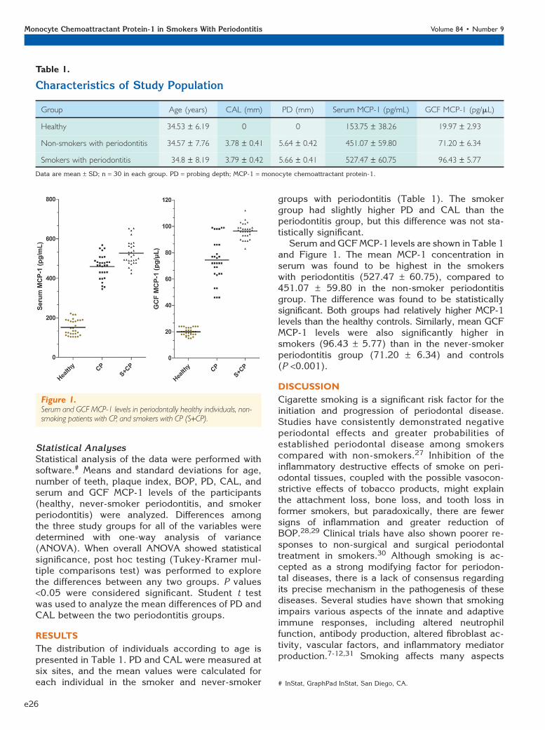
Serum and GCFMCP-1 levels are shown in Table 1and Figure 1. The mean MCP-1 concentration inserum was found to be highest in the smokerswith periodontitis (527.47 – 60.75), compared to451.07 – 59.80 in the non-smoker periodontitisgroup. The difference was found to be statisticallysignificant. Both groups had relatively higher MCP-1levels than the healthy controls. Similarly, mean GCFMCP-1 levels were also significantly higher insmokers (96.43 – 5.77) than in the never-smokerperiodontitis group (71.20 – 6.34) and controls(P <0.001).
DISCUSSION
Cigarette smoking is a significant risk factor for theinitiation and progression of periodontal disease.Studies have consistently demonstrated negativeperiodontal effects and greater probabilities ofestablished periodontal disease among smokerscompared with non-smokers.27 Inhibition of theinflammatory destructive effects of smoke on peri-odontal tissues, coupled with the possible vasocon-strictive effects of tobacco products, might explainthe attachment loss, bone loss, and tooth loss informer smokers, but paradoxically, there are fewersigns of inflammation and greater reduction ofBOP.28,29 Clinical trials have also shown poorer re-sponses to non-surgical and surgical periodontaltreatment in smokers.30 Although smoking is ac-cepted as a strong modifying factor for periodon-tal diseases, there is a lack of consensus regardingits precise mechanism in the pathogenesis of thesediseases. Several studies have shown that smokingimpairs various aspects of the innate and adaptiveimmune responses, including altered neutrophilfunction, antibody production, altered fibroblast ac-tivity, vascular factors, and inflammatory mediatorproduction.7-12,31 Smoking affects many aspects
Table 1.
Characteristics of Study Population
Group Age (years) CAL (mm) PD (mm) Serum MCP-1 (pg/mL) GCF MCP-1 (pg/mL)
Healthy 34.53 – 6.19 0 0 153.75 – 38.26 19.97 – 2.93
Non-smokers with periodontitis 34.57 – 7.76 3.78 – 0.41 5.64 – 0.42 451.07 – 59.80 71.20 – 6.34
Smokers with periodontitis 34.8 – 8.19 3.79 – 0.42 5.66 – 0.41 527.47 – 60.75 96.43 – 5.77
Data are mean – SD; n = 30 in each group. PD = probing depth; MCP-1 = monocyte chemoattractant protein-1.
Figure 1.Serum and GCF MCP-1 levels in periodontally healthy individuals, non-smoking patients with CP, and smokers with CP (S+CP).
# InStat, GraphPad InStat, San Diego, CA.
Monocyte Chemoattractant Protein-1 in Smokers With Periodontitis Volume 84 • Number 9
e26
of the host’s immune response; therefore, it isprobable that these effects on the immune systemcould be the primary contributing factors of smokingto the pathogenesis of periodontal disease.8
Cytokines, such as IL-1, IL-6, IL-8, and TNF-a, areconsidered to be involved in the host response toperiodontal disease as mediators of tissue break-down. Increased levels of these cytokines havebeen observed in the GCF of patients with peri-odontal disease.32,33 The chemokines are a familyof structurally related glycoproteins with potentleukocyte activation and/or chemotactic activity.MCP-1 is chemotactic for monocytes and is knownto regulate the expression of proinflammatory cyto-kines, such as IL-1 and IL-6. MCP-1 is also a potentactivator of human basophils, inducing degranula-tion and the release of histamines and, thus, likelycontributing to the inflammatory responses ob-served in periodontitis.
In the present study, a significant increase isfound in the serum and GCF levels of MCP-1 insmokers and non-smokers with periodontitis. Thisobservation is in agreement with earlier studies thatshowed higher serum23 and GCF MCP-1 levels inpatients with periodontitis compared to healthycontrols.20,21,24 However, a study by Tymkiw et al.34
showed a relatively lower amount of MCP-1 in theGCF of selected sites with periodontitis in smokers.The site selection among smokers might haveinfluenced their results. In the present study, smokersare grouped, and GCF is collected from selectedteeth and pooled to estimate the MCP-1 in smokersand non-smokers with periodontitis and in healthyindividuals.
Although a fourth group of smokers withoutperiodontitis may have been useful as a positivecontrol group, no individuals fit these criteria (20cigarettes per day and 2 years duration). This is onelimitation of the study.
CONCLUSIONS
From the observations of the present study, it canbe concluded that both serum and GCF MCP-1 levelsare significantly increased in smokers with peri-odontitis compared to non-smoking patients withperiodontitis and healthy controls. More longitudinal,prospective studies are necessary to confirm theobservations of the present study. Further researchin this direction could reveal reliable markers toforecast the risk for diseases, such as periodontitis,that lack any sensitive prognostic indicators.
ACKNOWLEDGMENT
The authors report no conflicts of interest relatedto this study.
REFERENCES1. Bergstrom J, Eliasson S. Cigarette smoking and alve-
olar bone height in subjects with a high standard oforal hygiene. J Clin Periodontol 1987;14:466-469.
2. Anil S. Study of the patterns of periodontal destruc-tion in smokers with chronic periodontitis. IndianJ Dent Res 2008;19:124-128.
3. Palmer RM, Scott DA, Meekin TN, Poston RN,Odell EW, Wilson RF. Potential mechanisms of sus-ceptibility to periodontitis in tobacco smokers. J Peri-odontal Res 1999;34:363-369.
4. Baab DA, Oberg PA. The effect of cigarette smokingon gingival blood flow in humans. J Clin Periodontol1987;14:418-424.
5. Haber J. Cigarette smoking: A major risk factor forperiodontitis. Compendium 1994;15:1002, 1004-1008passim, quiz 1014.
6. Johnson JD, Houchens DP, Kluwe WM, Craig DK,Fisher GL. Effects of mainstream and environmentaltobacco smoke on the immune system in animals andhumans: A review. Crit Rev Toxicol 1990;20:369-395.
7. Raulin LA, McPherson JC 3rd, McQuade MJ, HansonBS. The effect of nicotine on the attachment of humanfibroblasts to glass and human root surfaces in vitro.J Periodontol 1988;59:318-325.
8. Al-Ghamdi HS, Anil S. Serumantibody levels in smokerand non-smoker Saudi subjects with chronic peri-odontitis. J Periodontol 2007;78:1043-1050.
9. Andersen P, Pedersen OF, Bach B, Bonde GJ.Serum antibodies and immunoglobulins in smokersand nonsmokers. Clin Exp Immunol 1982;47:467-473.
10. Ryder MI, Fujitaki R, Johnson G, Hyun W. Alterationsof neutrophil oxidative burst by in vitro smoke expo-sure: Implications for oral and systemic diseases. AnnPeriodontol 1998;3:76-87.
11. Sopori M. Effects of cigarette smoke on the immunesystem. Nat Rev Immunol 2002;2:372-377.
12. Stampfli MR, Anderson GP. How cigarette smokeskews immune responses to promote infection, lungdisease and cancer. Nat Rev Immunol 2009;9:377-384.
13. Weiss SJ. Tissue destruction by neutrophils. N EnglJ Med 1989;320:365-376.
14. Mukaida N, Harada A, Yasumoto K, Matsushima K.Properties of pro-inflammatory cell type-specific leu-kocyte chemotactic cytokines, interleukin 8 (IL-8) andmonocyte chemotactic and activating factor (MCAF).Microbiol Immunol 1992;36:773-789.
15. Yu X, Antoniades HN, Graves DT. Expression of mono-cyte chemoattractant protein 1 in human inflamedgingival tissues. Infect Immun 1993;61:4622-4628.
16. Seino Y, Ikeda U, Sekiguchi H, et al. Expressionof leukocyte chemotactic cytokines in myocardialtissue. Cytokine 1995;7:301-304.
17. Villiger PM, Terkeltaub R, Lotz M. Production of mono-cyte chemoattractant protein-1 by inflamed synovialtissue and cultured synoviocytes. J Immunol 1992;149:722-727.
18. Gemmell E, Marshall RI, Seymour GJ. Cytokines andprostaglandins in immune homeostasis and tissue de-struction in periodontal disease. Periodontol 20001997;14:112-143.
19. Tonetti MS, Imboden MA, Gerber L, Lang NP,Laissue J, Mueller C. Localized expression of mRNAfor phagocyte-specific chemotactic cytokines in
J Periodontol • September 2013 Anil, Preethanath, Alasqah, Mokeem, Anand
e27
human periodontal infections. Infect Immun 1994;62:4005-4014.
20. Emingil G, Atilla G, Huseyinov A. Gingival crevicularfluid monocyte chemoattractant protein-1 and RANTESlevels in patients with generalized aggressive peri-odontitis. J Clin Periodontol 2004;31:829-834.
21. Kurtisx B, Tuter G, Serdar M, et al. Gingival crevicularfluid levels of monocyte chemoattractant protein-1 andtumor necrosis factor-alpha in patients with chronicand aggressive periodontitis. J Periodontol 2005;76:1849-1855.
22. Hanazawa S, Kawata Y, Takeshita A, et al. Expressionof monocyte chemoattractant protein 1 (MCP-1) inadult periodontal disease: Increased monocyte che-motactic activity in crevicular fluids and induction ofMCP-1 expression in gingival tissues. Infect Immun1993;61:5219-5224.
23. Pradeep AR, Daisy H, Hadge P. Serum levels of mono-cyte chemoattractant protein-1 in periodontal healthand disease. Cytokine 2009;47:77-81.
24. Pradeep AR, Daisy H, Hadge P. Gingival crevicular fluidlevels of monocyte chemoattractant protein-1 in peri-odontal health and disease. Arch Oral Biol 2009;54:503-509.
25. Armitage GC. Development of a classification systemfor periodontal diseases and conditions. Ann Periodon-tol 1999;4:1-6.
26. Xu L, Loos BG, Craandijk J, Ritsema E, Huffels RA, vander Velden U. Teeth with periodontal bone loss, ciga-rette smoking and plasma cotinine levels. J Int AcadPeriodontol 2002;4:39-43.
27. Ojima M, Hanioka T, Tanaka K, Inoshita E, Aoyama H.Relationship between smoking status and periodontalconditions: Findings from national databases in Japan.J Periodontal Res 2006;41:573-579.
28. Clarke NG, Shephard BC, Hirsch RS. The effects ofintra-arterial epinephrine and nicotine on gingivalcirculation. Oral Surg Oral Med Oral Pathol 1981;52:577-582.
29. Scabbia A, Cho KS, Sigurdsson TJ, Kim CK, TrombelliL. Cigarette smoking negatively affects healing re-sponse following flap debridement surgery. J Periodon-tol 2001;72:43-49.
30. Bergstrom J, Eliasson S, Dock J. Exposure to tobaccosmoking and periodontal health. J Clin Periodontol2000;27:61-68.
31. Barbour SE, Nakashima K, Zhang JB, et al. Tobaccoand smoking: Environmental factors that modify thehost response (immune system) and have an impacton periodontal health. Crit Rev Oral Biol Med 1997;8:437-460.
32. Genco RJ. Host responses in periodontal diseases:Current concepts. J Periodontol 1992; 63(Suppl. 4)338-355.
33. Kamma JJ, Giannopoulou C, Vasdekis VG, Mombelli A.Cytokine profile in gingival crevicular fluid of aggressiveperiodontitis: Influence of smoking and stress. J ClinPeriodontol 2004;31:894-902.
34. Tymkiw KD, Thunell DH, Johnson GK, et al. Influenceof smoking on gingival crevicular fluid cytokines insevere chronic periodontitis. J Clin Periodontol 2011;38:219-228.
Correspondence: Prof. Sukumaran Anil, College of Den-tistry, King Saud University, P.O. Box 60169, Riyadh11545, Saudi Arabia. Fax: +9661-467-9017; e-mail: [email protected].
Submitted November 10, 2012; accepted for publicationDecember 31, 2012.
Monocyte Chemoattractant Protein-1 in Smokers With Periodontitis Volume 84 • Number 9
e28