Human gingival fibroblasts function is stimulated on machined hydrided titanium zirconium dental...
9
Human gingival fibroblasts function is stimulated on machined hydrided titanium zirconium dental implants M. Go ´ mez-Florit a , R. Xing b , J.M. Ramis a , S. Taxt-Lamolle b , H.J. Haugen b , S.P. Lyngstadaas b , M. Monjo a, * a Department of Fundamental Biology and Health Sciences. Research Institute on Health Sciences (IUNICS), University of Balearic Islands, Palma de Mallorca, Spain b Department of Biomaterials, Institute for Clinical Dentistry, University of Oslo, Oslo, Norway 1. Introduction The integration of the dental implant intra-osseous compo- nent and the transmucosal component (abutment) with the hard tissue (bone) and the peri-implant mucosa (gingiva) is essential in order to minimize dental implant failure or complications. While during the past years research and development of dental implant biomaterials has been focused on osseointegration, nowadays soft tissue integration is one of the frontiers in dental implant research, as dental implants require a soft tissue barrier to prevent bacterial penetration 1 j o u r n a l o f d e n t i s t r y 4 2 ( 2 0 1 4 ) 3 0 – 3 8 a r t i c l e i n f o Article history: Received 13 September 2013 Received in revised form 14 October 2013 Accepted 10 November 2013 Keywords: Titanium zirconium Dental implant Abutment Soft tissue Gingival fibroblasts Cathodic polarization a b s t r a c t Objectives: The aim of this study was to evaluate the influence of different titanium zirconium (TiZr) alloy surfaces on primary human gingival fibroblasts (HGF) for improved soft tissue integration of dental implants. Methods: TiZr polished, machined and machined + HCl/H 2 SO 4 acid-etched surfaces were modified by cathodic polarization and/or HNO 3 /HF acid etching. Contact angle of surfaces was measured. The influence of modified TiZr surfaces on HGF was evaluated through the analysis of cell number, morphology, recovery after a wound (wound healing assay) and the expression of several genes, including matrix metalloproteinase-1 (MMP1) and metallopep- tidase inhibitor-1 (TIMP1). Results: Modification of TiZr surfaces decreased its hydrophilicity. Hydride implementation on TiZr surfaces via cathodic polarization increased TIMP1 expression and decreased MMP1/ TIMP1 mRNA ratio. Cathodic polarization of machined surfaces promoted cell attachment. Cells on machined and machined + cathodic polarization surfaces grew aligned to the microgrooves whereas on all polished surfaces they grew randomly. Acid etching of polished and machined surfaces did not improve HGF function. Conclusions: Hydride implementation on TiZr machined surfaces may be used as new dental implant material for improved soft tissue integration. Clinical significance: Enhancing dental implant surfaces’ bioactivity by hydride implementa- tion may promote soft tissue attachment and sealing around the implant and reduce peri- implantitis related to ECM-destruction compared with conventional machined surfaces. # 2013 Elsevier Ltd. All rights reserved. * Corresponding author at: University of Balearic Islands, Cra. de Valldemossa, km 7.5, E-07122 Palma de Mallorca, Spain. Tel.: +34 971259960; fax: +34 971173184. E-mail address: [email protected] (M. Monjo). Available online at www.sciencedirect.com ScienceDirect journal homepage: www.intl.elsevierhealth.com/journals/jden 0300-5712/$ – see front matter # 2013 Elsevier Ltd. All rights reserved. http://dx.doi.org/10.1016/j.jdent.2013.11.003
Transcript of Human gingival fibroblasts function is stimulated on machined hydrided titanium zirconium dental...

Human gingival fibroblasts function is stimulatedon machined hydrided titanium zirconium dentalimplants
M. Gomez-Florit a, R. Xing b, J.M. Ramis a, S. Taxt-Lamolle b,H.J. Haugen b, S.P. Lyngstadaas b, M. Monjo a,*aDepartment of Fundamental Biology and Health Sciences. Research Institute on Health Sciences (IUNICS), University
of Balearic Islands, Palma de Mallorca, SpainbDepartment of Biomaterials, Institute for Clinical Dentistry, University of Oslo, Oslo, Norway
j o u r n a l o f d e n t i s t r y 4 2 ( 2 0 1 4 ) 3 0 – 3 8
a r t i c l e i n f o
Article history:
Received 13 September 2013
Received in revised form
14 October 2013
Accepted 10 November 2013
Keywords:
Titanium zirconium
Dental implant
Abutment
Soft tissue
Gingival fibroblasts
Cathodic polarization
a b s t r a c t
Objectives: The aim of this study was to evaluate the influence of different titanium
zirconium (TiZr) alloy surfaces on primary human gingival fibroblasts (HGF) for improved
soft tissue integration of dental implants.
Methods: TiZr polished, machined and machined + HCl/H2SO4 acid-etched surfaces were
modified by cathodic polarization and/or HNO3/HF acid etching. Contact angle of surfaces
was measured. The influence of modified TiZr surfaces on HGF was evaluated through the
analysis of cell number, morphology, recovery after a wound (wound healing assay) and the
expression of several genes, including matrix metalloproteinase-1 (MMP1) and metallopep-
tidase inhibitor-1 (TIMP1).
Results: Modification of TiZr surfaces decreased its hydrophilicity. Hydride implementation
on TiZr surfaces via cathodic polarization increased TIMP1 expression and decreased MMP1/
TIMP1 mRNA ratio. Cathodic polarization of machined surfaces promoted cell attachment.
Cells on machined and machined + cathodic polarization surfaces grew aligned to the
microgrooves whereas on all polished surfaces they grew randomly. Acid etching of
polished and machined surfaces did not improve HGF function.
Conclusions: Hydride implementation on TiZr machined surfaces may be used as new dental
implant material for improved soft tissue integration.
Clinical significance: Enhancing dental implant surfaces’ bioactivity by hydride implementa-
tion may promote soft tissue attachment and sealing around the implant and reduce peri-
implantitis related to ECM-destruction compared with conventional machined surfaces.
# 2013 Elsevier Ltd. All rights reserved.
Available online at www.sciencedirect.com
ScienceDirect
journal homepage: www.intl.elsevierhealth.com/journals/jden
1. Introduction
The integration of the dental implant intra-osseous compo-
nent and the transmucosal component (abutment) with the
hard tissue (bone) and the peri-implant mucosa (gingiva) is
* Corresponding author at: University of Balearic Islands, Cra. de ValldTel.: +34 971259960; fax: +34 971173184.
E-mail address: [email protected] (M. Monjo).
0300-5712/$ – see front matter # 2013 Elsevier Ltd. All rights reservehttp://dx.doi.org/10.1016/j.jdent.2013.11.003
essential in order to minimize dental implant failure or
complications. While during the past years research and
development of dental implant biomaterials has been focused
on osseointegration, nowadays soft tissue integration is one of
the frontiers in dental implant research, as dental implants
require a soft tissue barrier to prevent bacterial penetration1
emossa, km 7.5, E-07122 Palma de Mallorca, Spain.
d.
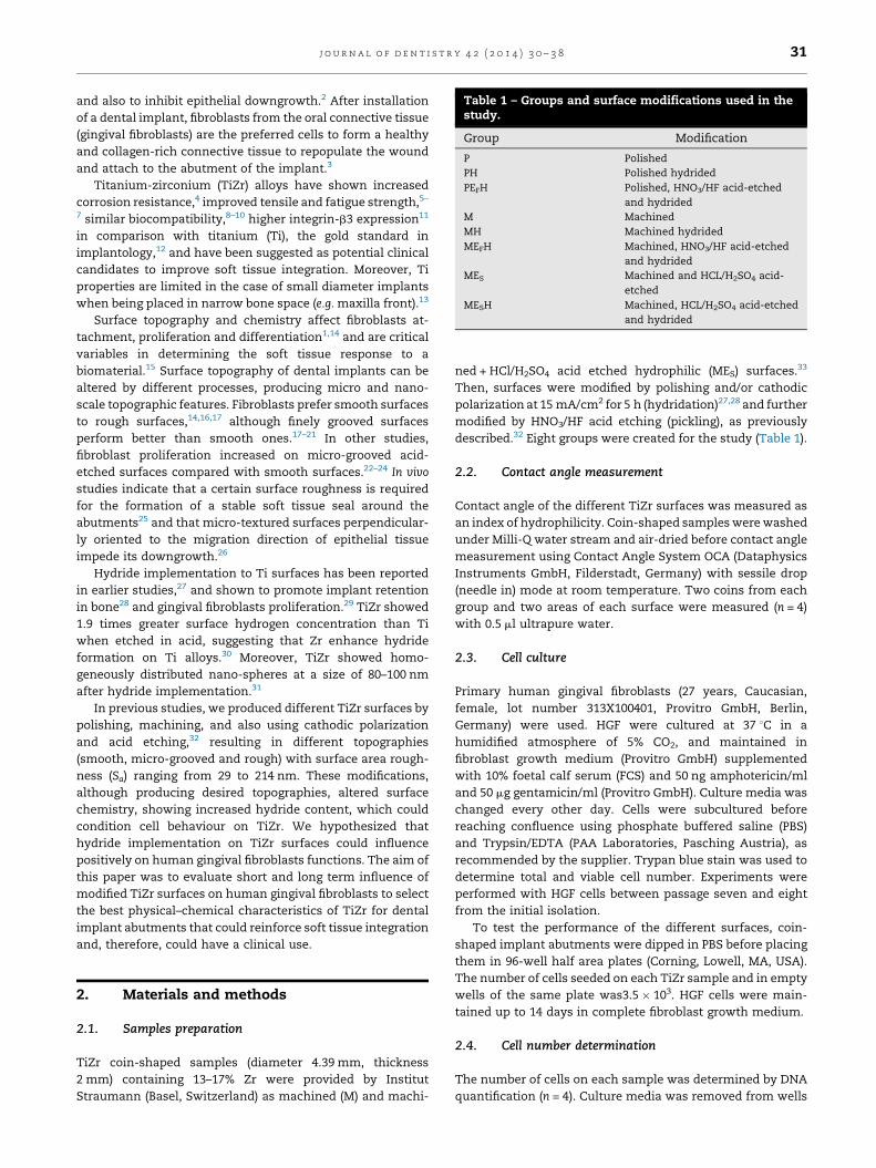
Table 1 – Groups and surface modifications used in thestudy.
Group Modification
P Polished
PH Polished hydrided
PEFH Polished, HNO3/HF acid-etched
and hydrided
M Machined
MH Machined hydrided
MEFH Machined, HNO3/HF acid-etched
and hydrided
MES Machined and HCL/H2SO4 acid-
etched
MESH Machined, HCL/H2SO4 acid-etched
and hydrided
j o u r n a l o f d e n t i s t r y 4 2 ( 2 0 1 4 ) 3 0 – 3 8 31
and also to inhibit epithelial downgrowth.2 After installation
of a dental implant, fibroblasts from the oral connective tissue
(gingival fibroblasts) are the preferred cells to form a healthy
and collagen-rich connective tissue to repopulate the wound
and attach to the abutment of the implant.3
Titanium-zirconium (TiZr) alloys have shown increased
corrosion resistance,4 improved tensile and fatigue strength,5–
7 similar biocompatibility,8–10 higher integrin-b3 expression11
in comparison with titanium (Ti), the gold standard in
implantology,12 and have been suggested as potential clinical
candidates to improve soft tissue integration. Moreover, Ti
properties are limited in the case of small diameter implants
when being placed in narrow bone space (e.g. maxilla front).13
Surface topography and chemistry affect fibroblasts at-
tachment, proliferation and differentiation1,14 and are critical
variables in determining the soft tissue response to a
biomaterial.15 Surface topography of dental implants can be
altered by different processes, producing micro and nano-
scale topographic features. Fibroblasts prefer smooth surfaces
to rough surfaces,14,16,17 although finely grooved surfaces
perform better than smooth ones.17–21 In other studies,
fibroblast proliferation increased on micro-grooved acid-
etched surfaces compared with smooth surfaces.22–24 In vivo
studies indicate that a certain surface roughness is required
for the formation of a stable soft tissue seal around the
abutments25 and that micro-textured surfaces perpendicular-
ly oriented to the migration direction of epithelial tissue
impede its downgrowth.26
Hydride implementation to Ti surfaces has been reported
in earlier studies,27 and shown to promote implant retention
in bone28 and gingival fibroblasts proliferation.29 TiZr showed
1.9 times greater surface hydrogen concentration than Ti
when etched in acid, suggesting that Zr enhance hydride
formation on Ti alloys.30 Moreover, TiZr showed homo-
geneously distributed nano-spheres at a size of 80–100 nm
after hydride implementation.31
In previous studies, we produced different TiZr surfaces by
polishing, machining, and also using cathodic polarization
and acid etching,32 resulting in different topographies
(smooth, micro-grooved and rough) with surface area rough-
ness (Sa) ranging from 29 to 214 nm. These modifications,
although producing desired topographies, altered surface
chemistry, showing increased hydride content, which could
condition cell behaviour on TiZr. We hypothesized that
hydride implementation on TiZr surfaces could influence
positively on human gingival fibroblasts functions. The aim of
this paper was to evaluate short and long term influence of
modified TiZr surfaces on human gingival fibroblasts to select
the best physical–chemical characteristics of TiZr for dental
implant abutments that could reinforce soft tissue integration
and, therefore, could have a clinical use.
2. Materials and methods
2.1. Samples preparation
TiZr coin-shaped samples (diameter 4.39 mm, thickness
2 mm) containing 13–17% Zr were provided by Institut
Straumann (Basel, Switzerland) as machined (M) and machi-
ned + HCl/H2SO4 acid etched hydrophilic (MES) surfaces.33
Then, surfaces were modified by polishing and/or cathodic
polarization at 15 mA/cm2 for 5 h (hydridation)27,28 and further
modified by HNO3/HF acid etching (pickling), as previously
described.32 Eight groups were created for the study (Table 1).
2.2. Contact angle measurement
Contact angle of the different TiZr surfaces was measured as
an index of hydrophilicity. Coin-shaped samples were washed
under Milli-Q water stream and air-dried before contact angle
measurement using Contact Angle System OCA (Dataphysics
Instruments GmbH, Filderstadt, Germany) with sessile drop
(needle in) mode at room temperature. Two coins from each
group and two areas of each surface were measured (n = 4)
with 0.5 ml ultrapure water.
2.3. Cell culture
Primary human gingival fibroblasts (27 years, Caucasian,
female, lot number 313X100401, Provitro GmbH, Berlin,
Germany) were used. HGF were cultured at 37 8C in a
humidified atmosphere of 5% CO2, and maintained in
fibroblast growth medium (Provitro GmbH) supplemented
with 10% foetal calf serum (FCS) and 50 ng amphotericin/ml
and 50 mg gentamicin/ml (Provitro GmbH). Culture media was
changed every other day. Cells were subcultured before
reaching confluence using phosphate buffered saline (PBS)
and Trypsin/EDTA (PAA Laboratories, Pasching Austria), as
recommended by the supplier. Trypan blue stain was used to
determine total and viable cell number. Experiments were
performed with HGF cells between passage seven and eight
from the initial isolation.
To test the performance of the different surfaces, coin-
shaped implant abutments were dipped in PBS before placing
them in 96-well half area plates (Corning, Lowell, MA, USA).
The number of cells seeded on each TiZr sample and in empty
wells of the same plate was3.5 � 103. HGF cells were main-
tained up to 14 days in complete fibroblast growth medium.
2.4. Cell number determination
The number of cells on each sample was determined by DNA
quantification (n = 4). Culture media was removed from wells

Table 2 – Genes studied by real time RT-PCR and itsrelated function.
Related function Gene
ECM component Collagen I a1 (COL1A1)
ECM component Collagen III a1 (COL3A1)
ECM component Collagen V a1 (COL5A1)
ECM component Collagen XII a1 (COL12A1)
ECM component Decorin (DCN)
ECM component Versican (VCAN)
ECM component Osteonectin (SPARC)
ECM turnover Matrix metalloproteinase-1
(MMP1)
ECM turnover Metallopeptidase inhibitor-1
(TIMP1)
ECM component/cell adhesion Fibronectin (FN1)
Cell adhesion Integrin a2 (ITGA2)
Cell adhesion Integrin a8 (ITGA8)
Cell adhesion Integrin b3 (ITGB3)
Cell structure Vimentin (VIM)
Pro-inflammatory cytokine Interleukin-6 (IL6)
Wound healing a-Smooth muscle actin (ACTA2)
Wound healing Transforming growth factor-b1
(TGFB1)
Wound healing Endothelin-1 (EDN1)
Reference gene Beta-actin (ACTBL2)
Reference gene Glyceraldehyde 3-phosphate de-
hydrogenase (GAPDH)
j o u r n a l o f d e n t i s t r y 4 2 ( 2 0 1 4 ) 3 0 – 3 832
after 48 h and plates were frozen at �80 8C. Then, 100 ml
distilled water were added to each well, incubated for 1 h at
room temperature and then frozen at �80 8C in order to lyse
the cells. Later on, plates were thawed until reaching room
temperature and 100 ml of 20 mg/ml Hoechst 33258 in TNE
buffer were added to each well. 200 ml aliquots were
transferred to 96-well fluorescence plates and the intensity
of fluorescence was measured at lexcitation of 356 nm and
lemission of 465 nm using a multifunction microplate reader
(Cary Eclipse fluorescence spectrophotometer, Agilent Tech-
nologies, Santa Clara, USA). Relative fluorescence units were
correlated with cell number using a linear standard curve.
2.5. Cell staining
Cell monolayers grown for 48 h on the implant surfaces were
washed fixed for 15 min with 4% formaldehyde in PBS. Cells
were washed three times and stained with 5 mg/ml of
phalloidin-fluorescein isothiocyanate (Phalloidin-FITC; Sig-
ma–Aldrich, St. Louis, MO, USA) in PBS-Triton X-100 1% for
30 min in dark conditions. Cells were again washed with PBS
and samples were placed on slides. Finally, a drop of
FluoroshieldTM with DAPI (Sigma–Aldrich) was added and
cover glasses were mounted on the implants.
Two implants of each group were used to perform the
experiment (n = 2) and two images of each implant were taken
with the confocal microscope (Leica DMI 4000B equipped with
Leica TCS SPE laser system, Wetzlar, Germany).
2.6. Scanning electron microscopy and wound healingassay
A scanning electron microscope (SEM, Hitachi S-3400N,
Krefeld, Germany) using back scattered electrons, 40 Pa of
pressure and 10 kV of voltage was used to acquire images of
cells grown for 9 days on coin-shaped samples (n = 4). Cells
were washed with PBS and fixed with glutaraldehyde 4% in
PBS for 2 h. The fixative solution was removed, and the cells
were washed with distilled water two times. At 30 min
intervals, the cells were dehydrated by the addition of 50%,
70%, 90% and 100% ethanol solutions. Finally, the ethanol was
removed, and the cells were left at room temperature to
evaporate the remaining ethanol prior to analysis.
Wound healing assay was performed on confluent mono-
layers that were scraped with 10 ml sterile pipette tips in a
straight line to create a scratch (n = 4). Cells on TCP were used
as control to determine confluency. Then, monolayers were
washed once with growth medium to remove cell debris and
were allowed to close the scratch for 2 days at standard cell
culture conditions. Then, samples were processed for SEM as
previously described.
2.7. RNA isolation and real-time RT-PCR analysis
Total RNA was isolated using Tripure1 (Roche Diagnostics),
according to the manufacturer’s protocol from 8 samples
(n = 8). Total RNA was quantified at 260 nm using a
Nanodrop spectrophotometer (NanoDrop Technologies,
Wilmington, DE, USA). The same amount of RNA was
reverse transcribed to cDNA at 42 8C for 60 min using High
Capacity RNA-to-cDNA kit (Applied Biosystems, Foster
City, CA, USA), according to the protocol of the supplier.
Aliquots of each cDNA were frozen (�20 8C) until the PCR
reactions were carried out.
Real-time PCR was performed for two reference genes,
glyceraldehyde-3-phosphate dehydrogenase (GAPDH) and
beta-actin (ACTBL2), and target genes (Table 2). Real-time
PCR was performed in the Lightcycler 4801 (Roche Diagnos-
tics) using SYBR green detection. Primer sequences and real-
time conditions are described elsewhere.11
All samples were normalized by the geometric mean of the
expression levels of ACTBL2 and GAPDH and fold changes
were related to the control groups using the mathematical
model described by Pfaffl34: ratio = EtargetDCp target (mean control–
sample)/EreferenceDCp reference (mean control–sample), where Cp is the
crossing point of the reaction amplification curve as deter-
mined by the LightCycler 480 software. Stability of the
reference genes among samples was lower than 0.56, using
the BestKeeper tool.35 Moreover, a good consistence of the
bestkeeper index was proved as its contributing reference
genes were tightly correlated with it (0.945 < r < 0.952), with a
significance level of p = 0.001 for both genes.
2.8. Statistical analysis
All data are presented as mean values � SD (standard
deviation). The Kolmogorov–Smirnov test was done to assume
parametric or non-parametric distributions for the normality
tests. Differences between groups were assessed by Student t-
test or Mann–Whitney test depending on their normal
distribution. To measure correlations, Spearman correlation
analysis was used. The results were interpreted as follows: no
correlation if r < 0.3, correlation if 0.3 � r < 0.5, and strong
correlation if 0.5 � r � 1. A negative r indicated a negative
Fig. 1 – (A) Contact angle measurement of the different TiZr
surfaces. (B) Cell number quantification on the different
TiZr surfaces. Boxes represent the 25th and 75th
percentiles and the median (line). The whiskers of the
graph show the largest and smallest values. Mean is
plotted with (+) in the box. Dotted-line represent the
number of cells attached to plastic surfaces
(6912 W 4360 cells/well). Significant differences were
assessed by Student t-test: (a) p = 0.05 versus M surfaces;
(b) p = 0.05 PH and PEFH versus P, MESH versus MES; (c)
p = 0.05 versus number of cells on plastic.
j o u r n a l o f d e n t i s t r y 4 2 ( 2 0 1 4 ) 3 0 – 3 8 33
correlation while a positive r indicated a positive correlation.
Results were considered statistically significant at p-
values � 0.05. SPSS1 program for Windows, version 17.0 (SPSS
Inc., Chicago, IL, USA) was used.
3. Results
3.1. Surface hydrophilicity
We found that P, M and MES surfaces were the most
hydrophilic surfaces (contact angle of 65, 69 and 728
respectively) (Fig. 1A). The hydridation and the pickling
processes of P, M and MES surfaces increased surface
hydrophobicity.
3.2. Cell number
Cell number after 48 h of culture (Fig. 1B) remained statistically
unchanged between groups. The number of cells attached to
surfaces after 48 h of culture only increased on MH surfaces,
compared with the initial cell number.
3.3. Cell morphology
Confocal images (Fig. 2, left column) show large numbers of
cells attached to P, M and MH surfaces after 2 days of cell
culture; few cells attached to PH, MEFH and MES surfaces with
a weakly stained cytoskeleton; only easy-stained nuclei with
abnormal margin cells attached to PEFH and MESH surfaces.
Cells grew aligned to each other with parallel thick stress
fibres on M and MH surfaces, while on P surfaces cells had
round morphology with few stress fibres growing without
clear orientation; on PH, MEFH and MES, cells were either
extended or round without clear stress fibres and orientation,
except on MES where they grew mainly aligned; on PEFH and
MESH surfaces, cytoskeleton almost disappeared.
After 9 days (Fig. 2, central column) cell number and
morphology on P, PH, M and MH surfaces was comparable. Cell
number on MEFH and PEFH after 9 days was higher than after 2
days. In contrast, few cells grew on MES and MESH surfaces.
At high magnification (Fig. 2, right column), images show
spindle-shaped cell morphology on P, PH, M and MH surfaces.
However, cells on M and MH were longer and thinner than on P
and PH surfaces; on M and MH surfaces, cells aligned
themselves along the groove axis of these TiZr surfaces,
while on P and PH, no clear orientation was observed.
Moreover, small and round cells grew on PEFH and MEFH
surfaces without clear orientation. The few remaining cells on
MES and MESH surfaces showed certain degree of alignment
with the grooved surface, but several cells had lost the spindle-
shaped morphology or its cellular extensions grew in any
direction.
3.4. Wound healing assay
HGF healed the wounds created on all the TiZr surfaces after
2 days, except on MES and MESH surfaces (Supplemental Fig.
1). Although we did not quantify cell number, it is worth
highlighting cell number on PH, PEFH and MEFH increased at
this time point compared with 48 h after seeding. In
contrast, cell counts on MES and MESH surfaces were even
lower and a confluent monolayer was never reached before
the scratch.
Supplementary material related to this article can be
found, in the online version, at http://dx.doi.org/10.1016/
j.jdent.2013.11.003.
3.5. Gene expression analysis
We found that hydride implementation to TiZr surfaces
increased TIMP1 expression and decreased MMP1/TIMP1
mRNA ratio (Figs. 3 and 4). Besides, PH performed very similar
to P surfaces, although TIMP1 expression increased and EDN1
expression decreased on PH; PEFH surfaces showed decreased
expression of all the studied genes, except for TIMP1, MMP1,
ITGA2, IL6, ACTA2 and EDN1. Only TIMP1 expression

Fig. 2 – Cell morphology on the different TiZr surfaces. Confocal microscopy was used to visualize actin immunostaining
(green) and nuclei staining (blue), bar scale = 100 mm (left column). Scanning electron microscopy was used to visualize HGF
on the surfaces at an original magnification 100T, bar scale = 500 mm (central column) and at an original magnification
500T, bar scale = 100 mm (right column). (For interpretation of the references to color in this figure legend, the reader is
referred to the web version of this article.)
j o u r n a l o f d e n t i s t r y 4 2 ( 2 0 1 4 ) 3 0 – 3 834
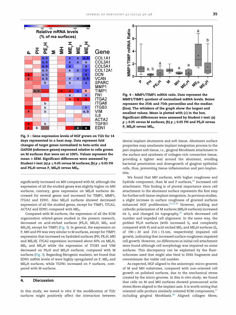
Fig. 3 – Gene expression levels of HGF grown on TiZr for 14
days represented in a heat map. Data represent fold
changes of target genes normalized to beta-actin and
GAPDH (reference genes) expressed relative to cells grown
on M surfaces that were set at 100%. Values represent the
mean W SEM. Significant differences were assessed by
Student t-test: (a) p = 0.05 versus M surfaces; (b) p = 0.05 PH
and PEFH versus P, MESH versus MES.
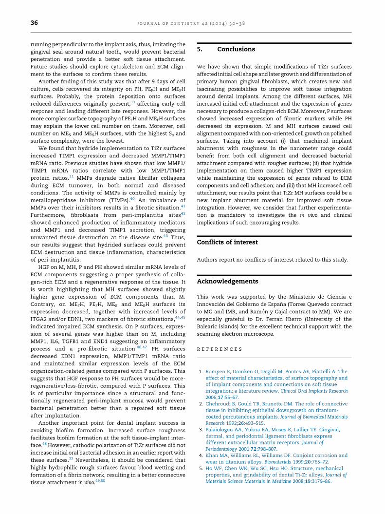
Fig. 4 – MMP1/TIMP1 mRNA ratio. Data represent the
MMP1/TIMP1 quotient of normalized mRNA levels. Boxes
represent the 25th and 75th percentiles and the median
(line). The whiskers of the graph show the largest and
smallest values. Mean is plotted with (+) in the box.
Significant differences were assessed by Student t-test: (a)
p = 0.05 versus M surfaces; (b) p = 0.05 PH and PEFH versus
P, MESH versus MES.
j o u r n a l o f d e n t i s t r y 4 2 ( 2 0 1 4 ) 3 0 – 3 8 35
significantly increased on MH compared with M, although the
expression of all the studied genes was slightly higher on MH
surfaces; contrary, gene expression on MEFH surfaces de-
creased for several genes and increased for TIMP1, MMP1,
ITGA2 and EDN1. Also MESH surfaces showed decreased
expression of all the studied genes, except for TIMP1, ITGA2,
ACTA2 and EDN1 compared with MES.
Compared with M surfaces, the expression of all the ECM
organization related-genes studied in the present research,
decreased on acid-etched surfaces (PEFH, MEFH, MES and
MESH), except for TIMP1 (Fig. 3). In general, the expression on
P, MH and PH was very similar to M surfaces, except for TIMP1
expression that increased on hydrided surfaces (PH, PEFH, MH
and MEFH). ITGA2 expression increased about 60% on MEFH,
MES and MESH while the expression of ITGB3 and VIM
decreased on PEFH and MESH surfaces, compared with M
surfaces (Fig. 3). Regarding fibrogenic markers, we found that
EDN1 mRNA levels of were highly upregulated on P, MES and
MESH surfaces, while TGFB1 increased on P surfaces, com-
pared with M surfaces.
4. Discussion
In this study, we tested in vitro if the modification of TiZr
surfaces might positively affect the interaction between
dental implant abutments and soft tissue. Abutment surface
properties may ameliorate implant integration process to the
peri-implant soft tissue, i.e., gingival fibroblasts attachment to
the surface and synthesis of collagen-rich connective tissue,
providing a tighter seal around the abutment, avoiding
bacterial penetration and downgrowth of gingival epithelial
cells, thus, preventing tissue inflammation and peri-implan-
titis.
We found that MH surfaces, with higher roughness and
hydride component, than M and P surfaces,32 increased cell
attachment. This finding is of pivotal importance since cell
attachment to the abutment surface represents the first step
for further soft tissue-implant integration. In previous studies,
a slight increase in surface roughness of grooved surfaces
enhanced HGF proliferation.22,23,36 However, pickling and
cathodic polarization of M surfaces (MEFH surfaces) increased
its Sa and changed its topography,32 which decreased cell
number and impeded cell alignment. In the same way, the
pickled PEFH surfaces (with increased Sa and complexity
compared with P) and acid-etched MES and MESH surfaces (Saof 190 � 20 and 214 � 15 nm, respectively) impaired cell
growth, indicating that increased surface roughness impaired
cell growth. However, no differences in initial cell attachment
were found although cell morphology was impaired on some
surfaces. This discrepancy can be explained by the fluor-
ochromes used that might also bind to DNA fragments and
overestimate the viable cell number.
As expected, HGF aligned to the anisotropic micro-grooves
of M and MH substrates, compared with non-oriented cell
growth on polished surfaces, due to the mechanical stress
created by the micro-grooves. In this in vitro study, we found
that cells on M and MH surfaces showed pronounced actin
stress fibres aligned to the implant axis. It is worth noting that
oriented cells produce similarly oriented ECM components,37
including gingival fibroblasts.38 Aligned collagen fibres,
j o u r n a l o f d e n t i s t r y 4 2 ( 2 0 1 4 ) 3 0 – 3 836
running perpendicular to the implant axis, thus, imitating the
gingival seal around natural tooth, would prevent bacterial
penetration and provide a better soft tissue attachment.
Future studies should explore cytoskeleton and ECM align-
ment to the surfaces to confirm these results.
Another finding of this study was that after 9 days of cell
culture, cells recovered its integrity on PH, PEFH and MEFH
surfaces. Probably, the protein deposition onto surfaces
reduced differences originally present,39 affecting early cell
response and leading different late responses. However, the
more complex surface topography of PEFH and MEFH surfaces
may explain the lower cell number on them. Moreover, cell
number on MES and MESH surfaces, with the highest Sa and
surface complexity, were the lowest.
We found that hydride implementation to TiZr surfaces
increased TIMP1 expression and decreased MMP1/TIMP1
mRNA ratio. Previous studies have shown that low MMP1/
TIMP1 mRNA ratios correlate with low MMP1/TIMP1
protein ratios.11 MMPs degrade native fibrillar collagens
during ECM turnover, in both normal and diseased
conditions. The activity of MMPs is controlled mainly by
metallopeptidase inhibitors (TIMPs).40 An imbalance of
MMPs over their inhibitors results in a fibrotic situation.41
Furthermore, fibroblasts from peri-implantitis sites42
showed enhanced production of inflammatory mediators
and MMP1 and decreased TIMP1 secretion, triggering
unwanted tissue destruction at the disease site.43 Thus,
our results suggest that hydrided surfaces could prevent
ECM destruction and tissue inflammation, characteristics
of peri-implantitis.
HGF on M, MH, P and PH showed similar mRNA levels of
ECM components suggesting a proper synthesis of colla-
gen-rich ECM and a regenerative response of the tissue. It
is worth highlighting that MH surfaces showed slightly
higher gene expression of ECM components than M.
Contrary, on MEFH, PEFH, MES and MESH surfaces its
expression decreased, together with increased levels of
ITGA2 and/or EDN1, two markers of fibrotic situations,44,45
indicated impaired ECM synthesis. On P surfaces, expres-
sion of several genes was higher than on M, including
MMP1, IL6, TGFB1 and END1 suggesting an inflammatory
process and a pro-fibrotic situation.46,47 PH surfaces
decreased EDN1 expression, MMP1/TIMP1 mRNA ratio
and maintained similar expression levels of the ECM
organization-related genes compared with P surfaces. This
suggests that HGF response to PH surfaces would be more-
regenerative/less-fibrotic, compared with P surfaces. This
is of particular importance since a structural and func-
tionally regenerated peri-implant mucosa would prevent
bacterial penetration better than a repaired soft tissue
after implantation.
Another important point for dental implant success is
avoiding biofilm formation. Increased surface roughness
facilitates biofilm formation at the soft tissue–implant inter-
face.48 However, cathodic polarization of TiZr surfaces did not
increase initial oral bacterial adhesion in an earlier report with
these surfaces.32 Nevertheless, it should be considered that
highly hydrophilic rough surfaces favour blood wetting and
formation of a fibrin network, resulting in a better connective
tissue attachment in vivo.49,50
5. Conclusions
We have shown that simple modifications of TiZr surfaces
affected initial cell shape and later growth and differentiation of
primary human gingival fibroblasts, which creates new and
fascinating possibilities to improve soft tissue integration
around dental implants. Among the different surfaces, MH
increased initial cell attachment and the expression of genes
necessary to produce a collagen-rich ECM. Moreover, P surfaces
showed increased expression of fibrotic markers while PH
decreased its expression. M and MH surfaces caused cell
alignment compared with non-oriented cell growth on polished
surfaces. Taking into account (i) that machined implant
abutments with roughness in the nanometer range could
benefit from both cell alignment and decreased bacterial
attachment compared with rougher surfaces; (ii) that hydride
implementation on them caused higher TIMP1 expression
while maintaining the expression of genes related to ECM
components and cell adhesion; and (iii) that MH increased cell
attachment, our results point that TiZr MH surfaces could be a
new implant abutment material for improved soft tissue
integration. However, we consider that further experimenta-
tion is mandatory to investigate the in vivo and clinical
implications of such encouraging results.
Conflicts of interest
Authors report no conflicts of interest related to this study.
Acknowledgements
This work was supported by the Ministerio de Ciencia e
Innovacion del Gobierno de Espana (Torres Quevedo contract
to MG and JMR, and Ramon y Cajal contract to MM). We are
especially grateful to Dr. Ferran Hierro (University of the
Balearic Islands) for the excellent technical support with the
scanning electron microscope.
r e f e r e n c e s
1. Rompen E, Domken O, Degidi M, Pontes AE, Piattelli A. Theeffect of material characteristics, of surface topography andof implant components and connections on soft tissueintegration: a literature review. Clinical Oral Implants Research2006;17:55–67.
2. Chehroudi B, Gould TR, Brunette DM. The role of connectivetissue in inhibiting epithelial downgrowth on titanium-coated percutaneous implants. Journal of Biomedical MaterialsResearch 1992;26:493–515.
3. Palaiologou AA, Yukna RA, Moses R, Lallier TE. Gingival,dermal, and periodontal ligament fibroblasts expressdifferent extracellular matrix receptors. Journal ofPeriodontology 2001;72:798–807.
4. Khan MA, Williams RL, Williams DF. Conjoint corrosion andwear in titanium alloys. Biomaterials 1999;20:765–72.
5. Ho WF, Chen WK, Wu SC, Hsu HC. Structure, mechanicalproperties, and grindability of dental Ti-Zr alloys. Journal ofMaterials Science Materials in Medicine 2008;19:3179–86.

j o u r n a l o f d e n t i s t r y 4 2 ( 2 0 1 4 ) 3 0 – 3 8 37
6. Kobayashi E, Matsumoto S, Doi H, Yoneyama T, HamanakaH. Mechanical properties of the binary titanium-zirconiumalloys and their potential for biomedical materials. Journal ofBiomedical Materials Research 1995;29:943–50.
7. Thoma DS, Jones AA, Dard M, Grize L, Obrecht M, CochranDL. Tissue integration of a new titanium-zirconium dentalimplant: a comparative histologic and radiographic study inthe canine. Journal of Periodontology 2011;82:1453–61.
8. Grandin HM, Berner S, Dard M. A review of titaniumzirconium (TiZr) alloys for use in endosseous dentalimplants. Materials 2012;5:1348–60.
9. Linkevicius T, Apse P. Influence of abutment material onstability of peri-implant tissues: a systematic review.International Journal of Oral and Maxillofacial Implants2008;23:449–56.
10. Pae A, Lee H, Kim HS, Kwon YD, Woo YH. Attachment andgrowth behaviour of human gingival fibroblasts on titaniumand zirconia ceramic surfaces. Biomedical Materials2009;4:025005.
11. Gomez-Florit M, Ramis JM, Xing R, Taxt-Lamolle S, HaugenHJ, Lyngstadaas SP, et al. Differential response of humangingival fibroblasts to titanium- and titanium-zirconium-modified surfaces. Journal of Periodontal Research 2013. http://dx.doi.org/10.1111/jre.12121.
12. Steinemann SG. Titanium—the material of choice?Periodontology 2000 1998;17:7–21.
13. Saulacic N, Bosshardt DD, Bornstein MM, Berner S, Buser D.Bone apposition to a titanium-zirconium alloy implant, ascompared to two other titanium-containing implants.European Cells and Materials 2012;23:273–86. [discussion86–8].
14. Kononen M, Hormia M, Kivilahti J, Hautaniemi J, Thesleff I.Effect of surface processing on the attachment, orientation,and proliferation of human gingival fibroblasts on titanium.Journal of Biomedical Materials Research 1992;26:1325–41.
15. Taylor SR, Gibbons DF. Effect of surface texture on the softtissue response to polymer implants. Journal of BiomedicalMaterials Research 1983;17:205–27.
16. Ponsonnet L, Comte V, Othmane A, Lagneau C, CharbonnierM, Lissac M, et al. Effect of surface topography andchemistry on adhesion, orientation and growth offibroblasts on nickel–titanium substrates. Materials Scienceand Engineering C 2002;21:157–65.
17. Wieland M, Chehroudi B, Textor M, Brunette DM. Use of Ti-coated replicas to investigate the effects on fibroblast shapeof surfaces with varying roughness and constant chemicalcomposition. Journal of Biomedical Materials Research2002;60:434–44.
18. Kokubu E, Hamilton DW, Inoue T, Brunette DM. Modulationof human gingival fibroblast adhesion, morphology,tyrosine phosphorylation, and ERK 1/2 localization onpolished, grooved and SLA substratum topographies. Journalof Biomedical Materials Research Part A 2009;91:663–70.
19. den Braber ET, Jansen HV, de Boer MJ, Croes HJ, ElwenspoekM, Ginsel LA, et al. Scanning electron microscopic,transmission electron microscopic, and confocal laserscanning microscopic observation of fibroblasts cultured onmicrogrooved surfaces of bulk titanium substrata. Journal ofBiomedical Materials Research 1998;40:425–33.
20. Guillem-Marti J, Delgado L, Godoy-Gallardo M, Pegueroles M,Herrero M, Gil FJ. Fibroblast adhesion and activation ontomicro-machined titanium surfaces. Clinical Oral ImplantsResearch 2013;24:770–80.
21. Chou L, Firth JD, Uitto VJ, Brunette DM. Substratum surfacetopography alters cell shape and regulates fibronectinmRNA level, mRNA stability, secretion and assembly inhuman fibroblasts. Journal of Cell Science 1995;108:1563–73.
22. Lee SW, Kim SY, Lee MH, Lee KW, Leesungbok R, Oh N.Influence of etched microgrooves of uniform dimension on
in vitro responses of human gingival fibroblasts. Clinical OralImplants Research 2009;20:458–66.
23. Kim SY, Oh N, Lee MH, Kim SE, Leesungbok R, Lee SW.Surface microgrooves and acid etching on titaniumsubstrata alter various cell behaviors of cultured humangingival fibroblasts. Clinical Oral Implants Research2009;20:262–72.
24. Guida L, Oliva A, Basile MA, Giordano M, Nastri L,Annunziata M. Human gingival fibroblast functions arestimulated by oxidized nano-structured titanium surfaces.Journal of Dentistry 2013;41:900–7.
25. Brunette DM, Chehroudi B. The effects of the surfacetopography of micromachined titanium substrata on cellbehavior in vitro and in vivo. Journal of BiomechanicalEngineering 1999;121:49–57.
26. Chehroudi B, Gould TR, Brunette DM. Titanium-coatedmicromachined grooves of different dimensions affectepithelial and connective-tissue cells differently in vivo.Journal of Biomedical Materials Research 1990;24:1203–19.
27. Videm K, Lamolle S, Monjo M, Ellingsen JE, Lyngstadaas SP,Haugen HJ. Hydride formation on titanium surfaces bycathodic polarization. Applied Surface Science 2008;255:3011–5.
28. Lamolle SF, Monjo M, Lyngstadaas SP, Ellingsen JE, HaugenHJ. Titanium implant surface modification by cathodicreduction in hydrofluoric acid: surface characterization andin vivo performance. Journal of Biomedical Materials ResearchPart A 2009;88:581–8.
29. Xing R, Salou L, Taxt-Lamolle S, Reseland JE, LyngstadaasSP, Haugen HJ. Surface hydride on titanium by cathodicpolarization promotes human gingival fibroblast growth.Journal of Biomedical Materials Research Part A 2013. http://dx.doi.org/10.1002/jbm.a.34819.
30. Frank MJ, Walter MS, Lyngstadaas SP, Wintermantel E,Haugen HJ. Hydrogen content in titanium and a titanium–zirconium alloy after acid etching. Materials Science andEngineering C 2013;33:1282–8.
31. Frank MJ, Walter MS, Bucko MM, Pamula E, Lyngstadaas SP,Haugen HJ. Polarization of modified titanium and titanium–zirconium creates nano-structures while hydride formationis modulated. Applied Surface Science 2013;282:7–16.
32. Xing R, Taxt-Lamolle S, Ellingsen JE, Lyngstadaas SP,Haugen HJ. Effect of dental abutment surface modificationson in vivo initial bacterial adhesion. Clinical Oral ImplantsResearch 2013. [submitted for publication].
33. Ferguson SJ, Broggini N, Wieland M, de Wild M, Rupp F, Geis-Gerstorfer J, et al. Biomechanical evaluation of theinterfacial strength of a chemically modified sandblastedand acid-etched titanium surface. Journal of BiomedicalMaterials Research Part A 2006;78:291–7.
34. Pfaffl MW. A new mathematical model for relativequantification in real-time RT-PCR. Nucleic Acids Research2001;29:e45.
35. Pfaffl MW, Tichopad A, Prgomet C, Neuvians TP.Determination of stable housekeeping genes, differentiallyregulated target genes and sample integrity: bestkeeper—Excel-based tool using pair-wise correlations. BiotechnologyLetters 2004;26:509–15.
36. Mustafa K, Oden A, Wennerberg A, Hultenby K, Arvidson K.The influence of surface topography of ceramic abutmentson the attachment and proliferation of human oralfibroblasts. Biomaterials 2005;26:373–81.
37. Manwaring ME, Walsh JF, Tresco PA. Contact guidanceinduced organization of extracellular matrix. Biomaterials2004;25:3631–8.
38. Kearns VR, Williams RL, Mirvakily F, Doherty PJ, Martin N.Guided gingival fibroblast attachment to titanium surfaces:an in vitro study. Journal of Clinical Periodontology 2013;40:99–108.

j o u r n a l o f d e n t i s t r y 4 2 ( 2 0 1 4 ) 3 0 – 3 838
39. Grossner-Schreiber B, Herzog M, Hedderich J, Duck A, HannigM, Griepentrog M. Focal adhesion contact formation byfibroblasts cultured on surface-modified dental implants: anin vitro study. Clinical Oral Implants Research 2006;17:736–45.
40. Sakagami G, Sato E, Sugita Y, Kosaka T, Kubo K, Maeda H,et al. Effects of nifedipine and interleukin-1alpha on theexpression of collagen, matrix metalloproteinase-1, andtissue inhibitor of metalloproteinase-1 in human gingivalfibroblasts. Journal of Periodontal Research 2006;41:266–72.
41. Soell M, Elkaim R, Tenenbaum H, Cathepsin C. Matrixmetalloproteinases, and their tissue inhibitors in gingivaand gingival crevicular fluid from periodontitis-affectedpatients. Journal of Dental Research 2002;81:174–8.
42. Bordin S, Flemmig TF, Verardi S. Role of fibroblastpopulations in peri-implantitis. International Journal of Oraland Maxillofacial Implants 2009;24:197–204.
43. Sorsa T, Tjaderhane L, Salo T. Matrix metalloproteinases(MMPs) in oral diseases. Oral Diseases 2004;10:311–8.
44. Guo F, Carter DE, Mukhopadhyay A, Leask A. Gingivalfibroblasts display reduced adhesion and spreading onextracellular matrix: a possible basis for scarless tissuerepair? PLoS ONE 2011;6:e27097.
45. Shephard P, Martin G, Smola-Hess S, Brunner G, Krieg T,Smola H. Myofibroblast differentiation is induced inkeratinocyte-fibroblast co-cultures and is antagonistically
regulated by endogenous transforming growth factor-betaand interleukin-1. American Journal of Pathology2004;164:2055–66.
46. Birkedal-Hansen H. Role of matrix metalloproteinases inhuman periodontal diseases. Journal of Periodontology1993;64:474–84.
47. Kubota T, Nomura T, Takahashi T, Hara K. Expression ofmRNA for matrix metalloproteinases and tissue inhibitorsof metalloproteinases in periodontitis-affected humangingival tissue. Archives of Oral Biology 1996;41:253–62.
48. Subramani K, Jung RE, Molenberg A, Hammerle CH. Biofilmon dental implants: a review of the literature. InternationalJournal of Oral and Maxillofacial Implants 2009;24:616–26.
49. Schwarz F, Ferrari D, Herten M, Mihatovic I, Wieland M,Sager M, et al. Effects of Surface hydrophilicity andmicrotopography on early stages of soft and hard tissueintegration at non-submerged titanium implants: animmunohistochemical study in dogs. Journal ofPeriodontology 2007;78:2171–84.
50. Schwarz F, Herten M, Sager M, Wieland M, Dard M, Becker J.Histological and immunohistochemical analysis of initialand early subepithelial connective tissue attachment atchemically modified and conventional SLA1 titaniumimplants. A pilot study in dogs. Clinical Oral Investigations2007;11:245–55.


![Synthesis, crystal structure and charge transport properties of one-electron oxidized zirconium diphthalocyanine, [ZrPc2]IBr2](https://static.fdokumen.com/doc/165x107/633691caa1ced1126c0b4678/synthesis-crystal-structure-and-charge-transport-properties-of-one-electron-oxidized.jpg)