Laser Structured Dental Zirconium for Soft Tissue Cell ... - MDPI
16
Citation: Staehlke, S.; Oster, P.; Seemann, S.; Kruse, F.; Brief, J.; Nebe, B. Laser Structured Dental Zirconium for Soft Tissue Cell Occupation— Importance of Wettability Modulation. Materials 2022, 15, 732. https://doi.org/10.3390/ma15030732 Academic Editor: Bruno Chrcanovic Received: 20 December 2021 Accepted: 16 January 2022 Published: 19 January 2022 Publisher’s Note: MDPI stays neutral with regard to jurisdictional claims in published maps and institutional affil- iations. Copyright: © 2022 by the authors. Licensee MDPI, Basel, Switzerland. This article is an open access article distributed under the terms and conditions of the Creative Commons Attribution (CC BY) license (https:// creativecommons.org/licenses/by/ 4.0/). materials Article Laser Structured Dental Zirconium for Soft Tissue Cell Occupation—Importance of Wettability Modulation Susanne Staehlke 1, * , Philip Oster 2 , Susanne Seemann 1 , Fabian Kruse 2 , Jakob Brief 3 and Barbara Nebe 1,4 1 Department of Cell Biology, Rostock University Medical Center, 18057 Rostock, Germany; [email protected] (S.S.); [email protected] (B.N.) 2 Pulsar Photonics GmbH, 52134 Herzogenrath, Germany; [email protected] (P.O.); [email protected] (F.K.) 3 VITA Zahnfabrik H. Rauter GmbH & Co. KG, 79713 Bad Säckingen, Germany; [email protected] 4 Department Science and Technology of Life, Light and Matter, University of Rostock, 18059 Rostock, Germany * Correspondence: [email protected]; Tel.: +49-381-494-7770 Abstract: Various approaches are being pursued to physico-chemically modify the zirconia neck region of dental implants to improve the integration into the surrounding soft tissue. In this study, polished zirconia discs were laser microstructured with periodic cavities and convex waves. These zirconia samples were additionally activated by argon plasma using the kINPen ® 09. The surface topography was characterized by scanning electron microscopy and the surface wettability by water contact angle. The in vitro study with human gingival fibroblasts (HGF-1) was focused on cell spreading, morphology, and actin cytoskeleton organization within the first 24 h. The laser-induced microstructures were originally hydrophobic (e.g., 60 μm cavities 138.4 ◦ ), but after argon plasma activation, the surfaces switched to the hydrophilic state (60 μm cavities 13.7 ◦ ). HGF-1 cells adhered flatly on the polished zirconia. Spreading is hampered on cavity structures, and cells avoid the holes. However, cells on laser-induced waves spread well. Interestingly, argon plasma activation for only 1 min promoted adhesion and spreading of HGF-1 cells even after 2 h cultivation. The cells crawl and grow into the depth of the cavities. Thus, a combination of both laser microstructuring and argon plasma activation of zirconia seems to be optimal for a strong gingival cell attachment. Keywords: zirconium; laser micro-structures; cold atmospheric pressure plasma; water contact angle; scanning electron microscopy; in vitro; human gingival cells; cell morphology; actin cytoskeleton; spreading 1. Introduction Dental implants are a widely accepted and valuable treatment option to replace missing teeth. In terms of esthetic aspects, zirconia implants can be regarded as a viable alternative to the well-proven titanium implants [1,2]. For many years, the dental implant neck region was machined or highly polished to create a plaque-free area or to meet hygienic conditions [3,4]. For ceramic implants, this practice was adopted from titanium materials [5]. In preclinical studies with dental implants, similar qualitative soft tissue integration was reported by Thoma et al. for zirconia and titanium materials [6]. However, oral and maxillofacial surgeons assumed the maturation processes of the epithelial and connective tissues around zirconia implants were faster, as described by Roehling et al. [7]. The zirconia material alone seems to be preferable for gingival cells, which is an important aspect as gingival tissues around implants have a barrier function and the soft tissue integration is as important as bone integration [8,9]. The implant neck region should also provide topographical and/or chemical charac- teristics which facilitate mechanically stable and dense soft tissue attachment to promote cell-mediated tissue integration [10]. The function of cells surrounding implants at the im- plant–tissue interface can be influenced by modifying the material surface properties [3,4]. Materials 2022, 15, 732. https://doi.org/10.3390/ma15030732 https://www.mdpi.com/journal/materials
-
Upload
khangminh22 -
Category
Documents
-
view
4 -
download
0
Transcript of Laser Structured Dental Zirconium for Soft Tissue Cell ... - MDPI

�����������������
Citation: Staehlke, S.; Oster, P.;
Seemann, S.; Kruse, F.; Brief, J.; Nebe,
B. Laser Structured Dental Zirconium
for Soft Tissue Cell Occupation—
Importance of Wettability
Modulation. Materials 2022, 15, 732.
https://doi.org/10.3390/ma15030732
Academic Editor: Bruno Chrcanovic
Received: 20 December 2021
Accepted: 16 January 2022
Published: 19 January 2022
Publisher’s Note: MDPI stays neutral
with regard to jurisdictional claims in
published maps and institutional affil-
iations.
Copyright: © 2022 by the authors.
Licensee MDPI, Basel, Switzerland.
This article is an open access article
distributed under the terms and
conditions of the Creative Commons
Attribution (CC BY) license (https://
creativecommons.org/licenses/by/
4.0/).
materials
Article
Laser Structured Dental Zirconium for Soft Tissue CellOccupation—Importance of Wettability ModulationSusanne Staehlke 1,* , Philip Oster 2, Susanne Seemann 1, Fabian Kruse 2, Jakob Brief 3 and Barbara Nebe 1,4
1 Department of Cell Biology, Rostock University Medical Center, 18057 Rostock, Germany;[email protected] (S.S.); [email protected] (B.N.)
2 Pulsar Photonics GmbH, 52134 Herzogenrath, Germany; [email protected] (P.O.);[email protected] (F.K.)
3 VITA Zahnfabrik H. Rauter GmbH & Co. KG, 79713 Bad Säckingen, Germany; [email protected] Department Science and Technology of Life, Light and Matter, University of Rostock, 18059 Rostock, Germany* Correspondence: [email protected]; Tel.: +49-381-494-7770
Abstract: Various approaches are being pursued to physico-chemically modify the zirconia neckregion of dental implants to improve the integration into the surrounding soft tissue. In this study,polished zirconia discs were laser microstructured with periodic cavities and convex waves. Thesezirconia samples were additionally activated by argon plasma using the kINPen®09. The surfacetopography was characterized by scanning electron microscopy and the surface wettability by watercontact angle. The in vitro study with human gingival fibroblasts (HGF-1) was focused on cellspreading, morphology, and actin cytoskeleton organization within the first 24 h. The laser-inducedmicrostructures were originally hydrophobic (e.g., 60 µm cavities 138.4◦), but after argon plasmaactivation, the surfaces switched to the hydrophilic state (60 µm cavities 13.7◦). HGF-1 cells adheredflatly on the polished zirconia. Spreading is hampered on cavity structures, and cells avoid the holes.However, cells on laser-induced waves spread well. Interestingly, argon plasma activation for only1 min promoted adhesion and spreading of HGF-1 cells even after 2 h cultivation. The cells crawl andgrow into the depth of the cavities. Thus, a combination of both laser microstructuring and argonplasma activation of zirconia seems to be optimal for a strong gingival cell attachment.
Keywords: zirconium; laser micro-structures; cold atmospheric pressure plasma; water contact angle;scanning electron microscopy; in vitro; human gingival cells; cell morphology; actin cytoskeleton;spreading
1. Introduction
Dental implants are a widely accepted and valuable treatment option to replacemissing teeth. In terms of esthetic aspects, zirconia implants can be regarded as a viablealternative to the well-proven titanium implants [1,2]. For many years, the dental implantneck region was machined or highly polished to create a plaque-free area or to meethygienic conditions [3,4]. For ceramic implants, this practice was adopted from titaniummaterials [5]. In preclinical studies with dental implants, similar qualitative soft tissueintegration was reported by Thoma et al. for zirconia and titanium materials [6]. However,oral and maxillofacial surgeons assumed the maturation processes of the epithelial andconnective tissues around zirconia implants were faster, as described by Roehling et al. [7].The zirconia material alone seems to be preferable for gingival cells, which is an importantaspect as gingival tissues around implants have a barrier function and the soft tissueintegration is as important as bone integration [8,9].
The implant neck region should also provide topographical and/or chemical charac-teristics which facilitate mechanically stable and dense soft tissue attachment to promotecell-mediated tissue integration [10]. The function of cells surrounding implants at the im-plant–tissue interface can be influenced by modifying the material surface properties [3,4].
Materials 2022, 15, 732. https://doi.org/10.3390/ma15030732 https://www.mdpi.com/journal/materials

Materials 2022, 15, 732 2 of 16
The initial quality of spreading on a biomaterial is a decisive factor determining the en-suing cell behavior, for example, proliferation, differentiation, and cell type dependentfunctions [11]. The surface topography of zirconia can be generated by polishing, sandblast-ing, etching, or laser treatment [2,12,13]. Laser structuring is suitable for creating definedtopographies (dimension and shape) on zirconia, and for optimizing the surface charac-teristics [13,14]. In previous studies with human osteoblasts, we observed improved celladhesion and growth on femtosecond laser nanostructured and sinusoidal microstructuredtitanium alloys, regardless of hydrophilicity of the titanium surface [15]. What kind oftopography should the neck region of dental zirconia implants have?
To enable sufficient soft tissue attachment to the neck region of the implants andsoft tissue integration, smooth surfaces are considered favorable [5,16]. Pacha-Olivenzaet al. [17] recognized that the overall “race for the surface” [18] between primary humangingival fibroblasts and bacteria is disadvantageous for the fibroblasts on the rougher ones(acid-etched, sandblasted/acid-etched) compared with the smoother surfaces (machined,slightly acid-etched). More specifically, increasing surface roughness reduced fibroblastproliferation and increased the absolute bacterial adhesion [17], suggesting that smoothsurfaces could be preferable in the soft tissue region.
The implant neck region should also provide chemical characteristics to facilitate softtissue growth. Therefore, different chemical modifications and physical treatments havebeen developed to enhance cell acceptance and improve support of dental implants, thatis, ultraviolet light and cold physical plasma [3,10,19]. Physical plasma is an ionized gasdue to high energy supply consisting of ions, electrons, radicals, ultraviolet photons, anduncharged atoms or molecules. Cold physical plasma can be formed under atmosphericor low pressure. These plasmas are used for decontamination/sterilization in dentistry,wound healing, tissue regeneration in medicine [20], or for targeted modification of surfaceproperties of biomaterials [21]. The experimental application of non-thermal plasma forimplant material surface modifications is not quite new, thus titanium [19] and its alloys,polyetheretherketone (PEEK) [19], polycarbonate [22], and also zirconia surfaces [19] weretreated with argon, oxygen, Ar/1% oxygen [23] or ammonia plasmas for several minutesto obtain improved cell attachment, migration, and growth.
So far, there are few analyses available that focus on how physico-chemically char-acteristics of the implant influence the cell behavior after the first contact with fibroblastshave been performed [19,24]. Further knowledge of the responses of gingival fibroblaststo the modified surfaces of zirconia would make it possible to specify the manufacturingsteps required in the fabrication of dental implants.
The oral neck region of dental implants must be optimally integrated into the adjacentsoft tissues to provide long-term clinical implant survival [4]. To achieve this requirement,we combined the influence of surface topography and chemistry on soft tissue cell attach-ment in vitro. We investigated how HGF-1 cells behave on defined laser microstructureswith either various cavity dimensions (concave topology) or waves (convex topology).Additional zirconia surface functionalization was performed with argon plasma (withoutoxygen) using the kINPen®09 plasma jet [25]. The HGF-1 cell response was investigated interms of cell morphology, cell shape, spreading and actin organization in an early period,within the first 24 h.
The purpose of our work was to find a laser-induced micro-nanostructure for theceramic surface neck region suitable for human gingival fibroblast on growth. A combinedplasma-chemical modification should provide hints about the importance of additionalwettability modulation on the gingival cell response.
2. Materials and Methods2.1. Zirconia Samples and Laser Structuring
Yttria-stabilized zirconia discs were used with a diameter of 12 mm and a thickness of1.5 mm. The bulk material, polished zirconia (Control), was further micro-structured withthe laser microprocessing system RDX1000 and the software Machine control: Photonic
Materials 2022, 15, 732 3 of 16
Elements (Pulsar Photonics GmbH, Herzogenrath, Germany). The following geometricmicro-patterns were created: (i) concave structures: cavity 10/20 with a depth of 10 µm,pitch of 20 µm; cavity 60/120 with a depth of 60 µm, a pitch of 120 µm; cavity 180/360 witha depth of 180 µm, a pitch of 360 µm; and (ii) convex structures (Waves): with a depth of10–20 µm, a pitch of 30 µm and a width of 20 µm (plateau of 12 µm in width/length). TheTruMicro2030 laser worked with a pulse duration of <400 fs at a wavelength of 1030 nm (IR)with a raw beam diameter of ~5 mm. The beam was focused by F-Theta optics with a focusdiameter of 16 µm. The ablation file is based on CAD/CAM (Computer-Aided Designand Computer-Aided Manufacturing): To create circles, the template was a circle withcorresponding dimensions, for the convex structures according to a Waves pattern. Laserablation was performed along set path vectors (CAM data) at a feed speed of 1000 mm/s.The laser fluence to create Wave structures was 3.37 J/cm2 und thus slightly higher thanfor cavities with 2.9 J/cm2. The software Photonic Elements was used for machine control.The process strategy was scanner-based, that is, the laser beam is moved via the scanningsystem and positioned on the component (component is stationary).
2.2. Wettability of Surfaces
The water contact angle (WCA) and the surface-free energy (SFE) of the substrate/airinterface were analyzed using the sessile drop method and the Drop Shape Analyzer DSA25(Krüss, Hamburg, Germany) [26]. One drop (1 µL) of distilled water or diiodo-methane(Sigma-Aldrich, Munich, Germany) was deposited onto the sample surface. Three to fivedrops per sample, if procurable, were measured regarding the hydrophilicity of the samples.Drop images were acquired with the digital camera of the DSA25. Wettability values wereevaluated with the supplied software (ADVANCE, V.1.7.2.1, Krüss, Hamburg, Germany):for WCA the optimal fit method (ellipse, tangent, circle, height/width manual) accordingto the curvature of the drop shape was used; SFE, dispersive, and polar components, werecalculated according to Owens, Wendt, Rabel und Kaelble (OWRK).
2.3. Cold Argon (Ar-) Plasma Activation
To modulate the wettability of the polished and laser structured ceramic, the surfaceswere activated with the cold atmospheric pressure plasma jet kINPen®09 (Neoplas ToolsGmbH, Greifswald, Germany) [25,27]. The argon plasma source includes a quartz capillarywith a high-frequency (HF) electrode (diameter of 1 mm). An HF voltage (1.1 MHz/2–6 kV)was applied at this electrode. The gas flow of the feed gas argon (99.99%) was 1.9 slm. Thesettings at the power supply were 60.0 V and 0.05 A. The plasma flame was clearly visiblehad a length of 12–14 mm and a width about 1 mm. The temperature at the tip of theplasma flame did not exceed 50 ◦C and was characterized by Weltmann’s group [27]. Forthe activation of the surfaces, the plasma jet was guided vertically with the quartz capillary1 cm above the surface in a meandering manner for 60 s, so that the plasma had immediatecontact with the whole interface.
2.4. Gingival Cell Culture, Morphology, and Spreading
Cell culture: Gingival fibroblasts are able to produce the extracellular matrix molecules,for example, collagen type I and III, fibronectin as well as proteoglycans (including decorin,biglycan, versican, syndecan, perlecan) for maintenance, wound healing, and regenerationof gingival connective tissues [28,29]. Therefore, we used human gingival fibroblasts ascompetent representatives (HGF-1, ATCC®, CRL-2014™, Manassas, VA, USA), whichwere cultured in Dulbecco’s modified Eagle’s medium (DMEM; high glucose, GlutaMAX;Thermo Fisher Scientific, Gibco, Paisley, UK) containing 10% fetal bovine serum (FBSPremium, South America origin, 0.2 µm sterile-filtered; PAN Biotech, Aidenbach, Germany)and 1% antibiotic-antimycotic (penicillin, streptomycin, and amphotericin B; Anti-Anti100×, Thermo Fisher Scientific, Gibco) (complete medium). To detach the cells, HGF-1 cells were treated with trypsin/ethylenediaminetetraacetic acid (0.25% trypsin/0.38%EDTA; Invitrogen, Gibco, Paisley, UK) for 5 min. The trypsinization was stopped by the

Materials 2022, 15, 732 4 of 16
addition of complete medium, and the cell number was measured by NucleoCounter®
NC-3000™ (ChemoMetec A/S, Allerod, Denmark). The appropriate number of cells for theexperiments was placed in a meandering pattern on the surface, located in a 24-well plate(Greiner Bio-One, Kremsmünster, Austria), and cultured at 37 ◦C and 5% CO2 up to 72 h.
Cell morphology and spreading: The morphology of HGF-1 gingival fibroblasts wasanalyzed after 2, 24, or 72 h by field emission scanning electron microscopy (FE-SEM, 5 kV;Merlin VP compact, Carl Zeiss, Oberkochen, Germany). For this purpose, the cells werewashed with phosphate buffer solution (PBS, Sigma-Aldrich, St Louis, MO, USA), fixedwith 2.5% glutaraldehyde (GA, Merck, Darmstadt, Germany), and dehydrated through anascending ethanol series (30%, 50%, 75%, 90%, and 100%). The samples were dried in theK850 critical point dryer (Emitech, Taunusstein, Germany), and a final step vaporized withcarbon under a vacuum (EM SCD 500, Co. Leica, Bensheim, Germany). To illustrate thecells, a high-efficiency secondary electron detector (HE-SE) was used, and an InlenseDuodetector added to image the structures. Subsequently, the FE-SEM images were analyzedby ImageJ (Version 1.51f, Wayne Rasband, National Institutes of Health, Bethesda, MD,USA). For this purpose, the pixels were first converted into µm using the bar of the FE-SEMimages (all with the same magnification). After that, the cells are manually marked andthen the area, length and width can be analyzed. The quantification of morphometricdata—cell area [µm2] and length to width ratio—of 40 cells per surface was performed.
Actin cytoskeleton and vinculin: For actin and vinculin staining, gingiva fibroblastswere cultured for 24 or 72 h, respectively, washed with PBS, fixed with 4% paraformalde-hyde (PFA, Sigma Aldrich), and further permeabilized with 0.1% Triton X-100 (MerckKGaA, Darmstadt, Germany) at room temperature (RT) for 10 min. For immunolabeling,the primary antibody vinculin-mouse-anti-human (Sigma Aldrich; 1:50 diluted in PBS)was used at RT for 1 h. The secondary antibody anti-rabbit-IgG-AF488 (Invitrogen AG,Carlsbad, CA, USA; diluted 1:100 in PBS) was added at RT in the dark for an additional1 h. For actin staining, fibroblasts were incubated afterward with phalloidine tetramethyl-rhodamine (TRITC, 1:15 in PBS, Sigma Aldrich) at RT in the dark for 30 min. In the finalstep, the samples were embedded with FluoroshieldTM with 4′,6-diamidino-2-phenylindole(DAPI, Sigma-Aldrich).
Hyaluronan (HA): HGF-1 gingival fibroblasts were cultured on a cover slip for 24 hand fixed with 4% PFA at RT for 10 min. Subsequently, the biotinylated HA binding protein(2 mg; Calbiochem, San Diego, CA, USA) was applied for 120 min, followed by incubationwith streptavidin-fluorescein isothiocyanate (FITC, 0.5 mg; Becton Dickinson, San Diego,CA, USA) at RT for 30 min in the dark [30]. Afterward, the cells were embedded withDAPI-FluoroshieldTM.
2.5. Confocal Laser Scanning Microscopy
Cell images were recorded on an inverted confocal laser scanning microscope LSM 780(Carl Zeiss AG, Oberkochen, Germany). For image acquisition, the ZEISS oil immersion63× objective (C-Apochromat) and the ZEN 2011 (black version) software (Carl ZeissAG) were used. The images of micro-structures were displayed as three-dimensional (3D)z-stacks. The processing of the images, insertion of the bar, and overlay of the z-stack, weredone using the ZENblue software.
2.6. Statistic
Statistical analysis was performed with the software GraphPad PRISM Version 7.02for Windows (GraphPad Software Inc., La Jolla, CA, USA). Data were presented asmean ± standard error of the mean (s.e.m.), or median ± interquartile range (IQR, forwettability). Data analysis was conducted after normal distribution analysis as follows:Ordinary one-way ANOVA post hoc Bonferroni (unpaired, for wettability) or Friedman testpost hoc uncorrected Dunn’s test (paired, for spreading analyses). At the level of * p < 0.05,differences were considered statistically significant.

Materials 2022, 15, 732 5 of 16
3. Results
We aimed to elucidate the combinatory influence of laser microstructures and anoptional argon plasma activation of zirconia surfaces on human gingival fibroblast (HGF-1)morphology, spreading and growth. Polished zirconia served as controls for the laser-mediated concave cavities in different dimensions as well as convex waves. We used theargon gas-based cold atmospheric pressure plasma jet kINPen®09 to additionally activatethe ceramic surfaces and determine the importance of the wettability modulation.
3.1. Characterization of Human Gingival Fibroblasts
The human gingival fibroblast (HGF-1) cells are suitable cell models for in vitro ap-proaches on dental materials destine for soft tissue contact. After 48 h, the cells expressa pronounced actin cytoskeleton network with long fibers throughout the cell and wellpronounced vinculin contacts as adaptor proteins, important for bridging the integrinreceptors with actin fibers (Figure 1). However, due to their origin and fibroblast cellfunction, HGF-1 cells do not organize tight junctions in cell–cell contacts (not shown).As extracellular matrix producing cells, HGF-1 cells express, i.a., the glycosaminoglycanhyaluronan in higher amount.
Materials 2022, 15, x FOR PEER REVIEW 5 of 16
2.6. Statistic Statistical analysis was performed with the software GraphPad PRISM Version 7.02
for Windows (GraphPad Software Inc., La Jolla, CA, USA). Data were presented as mean ± standard error of the mean (s.e.m.), or median ± interquartile range (IQR, for wettability). Data analysis was conducted after normal distribution analysis as follows: Ordinary one-way ANOVA post hoc Bonferroni (unpaired, for wettability) or Friedman test post hoc uncorrected Dunn’s test (paired, for spreading analyses). At the level of * p < 0.05, differ-ences were considered statistically significant.
3. Results We aimed to elucidate the combinatory influence of laser microstructures and an op-
tional argon plasma activation of zirconia surfaces on human gingival fibroblast (HGF-1) morphology, spreading and growth. Polished zirconia served as controls for the laser-mediated concave cavities in different dimensions as well as convex waves. We used the argon gas-based cold atmospheric pressure plasma jet kINPen®09 to additionally activate the ceramic surfaces and determine the importance of the wettability modulation.
3.1. Characterization of Human Gingival Fibroblasts The human gingival fibroblast (HGF-1) cells are suitable cell models for in vitro ap-
proaches on dental materials destine for soft tissue contact. After 48 h, the cells express a pronounced actin cytoskeleton network with long fibers throughout the cell and well pro-nounced vinculin contacts as adaptor proteins, important for bridging the integrin recep-tors with actin fibers (Figure 1). However, due to their origin and fibroblast cell function, HGF-1 cells do not organize tight junctions in cell–cell contacts (not shown). As extracel-lular matrix producing cells, HGF-1 cells express, i.a., the glycosaminoglycan hyaluronan in higher amount.
Figure 1. Gingival HGF-1 cellular structures and matrix components: (a) The vinculin adaptor pro-teins (green) co-localize with the fiber ends of the well-organized actin cytoskeleton (red) after 72 h growth (scale bars 5 µm). (b) The extracellular matrix molecule glycosaminoglycan hyaluronan is produced by HGF-1 cells in higher amounts (green, nucleus in blue) after 24 h growth (scale bar 10 µm). (LSM 780, Carl Zeiss, Oberkochen, Germany).
Figure 1. Gingival HGF-1 cellular structures and matrix components: (a) The vinculin adaptorproteins (green) co-localize with the fiber ends of the well-organized actin cytoskeleton (red) after72 h growth (scale bars 5 µm). (b) The extracellular matrix molecule glycosaminoglycan hyaluronanis produced by HGF-1 cells in higher amounts (green, nucleus in blue) after 24 h growth (scale bar10 µm). (LSM 780, Carl Zeiss, Oberkochen, Germany).
3.2. Material Surface Characterization3.2.1. Profiles of Zirconia Samples
Since the manufacturing processes of the laser structures investigated followed differ-ent protocols, we analyzed the resulting topographies by FE-SEM. Regarding the polishedzirconia (Control), the FE-SEM images showed that grinding grooves without orientationwere detectable (Figure 2). After laser structuring, two types of periodic microstructurescan be observed: concave cavities 10/20 holes with a dimension of 10 µm in width anddepth, 30 µm pitch; 60/120 holes with a dimension of 60 µm in width and depth, 120 µmpitch; 180/360 holes with a dimension of 180 µm in width and depth, 360 µm pitch, and asinusoidal, convex microstructure, like pyramid stumps (Waves) with the dimension of10–20 µm in height and 30 µm pitch (Figures 2 and 3). As the cavities became wider anddeeper, the laser had to process the surface more intensively, which was also evident fromthe nanoporous surface texture of the zirconia (grains) at the marginal areas and in the
Materials 2022, 15, 732 6 of 16
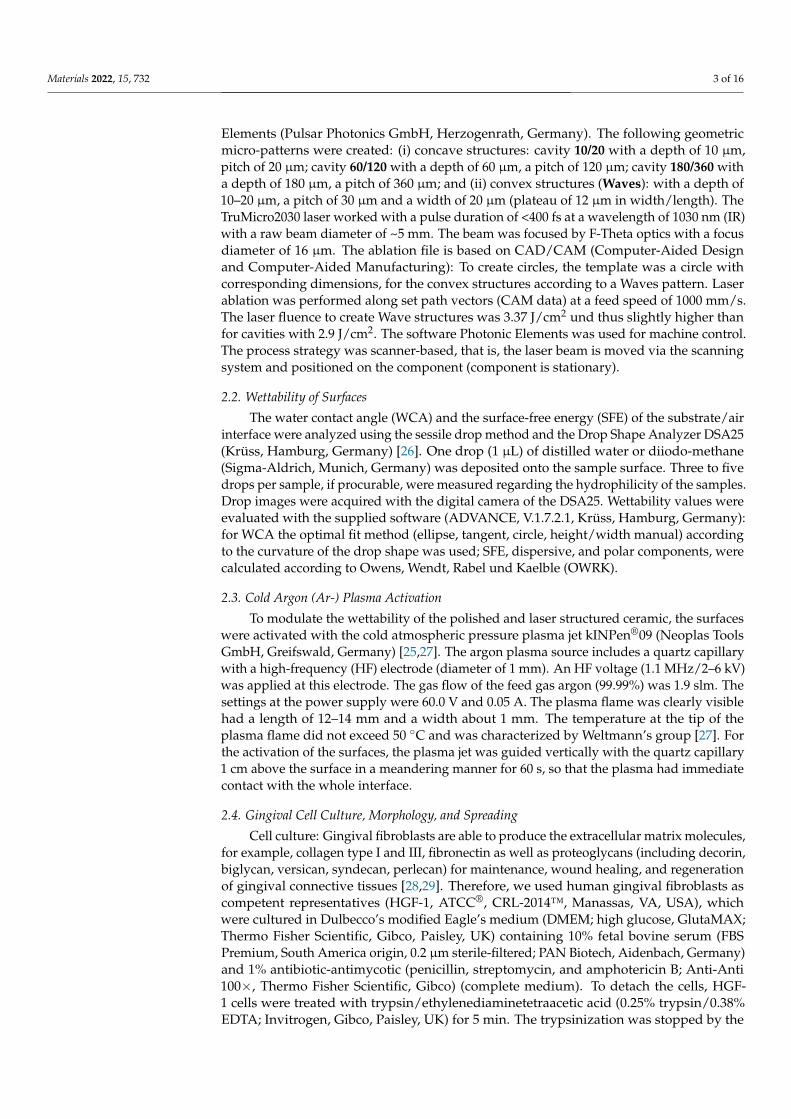
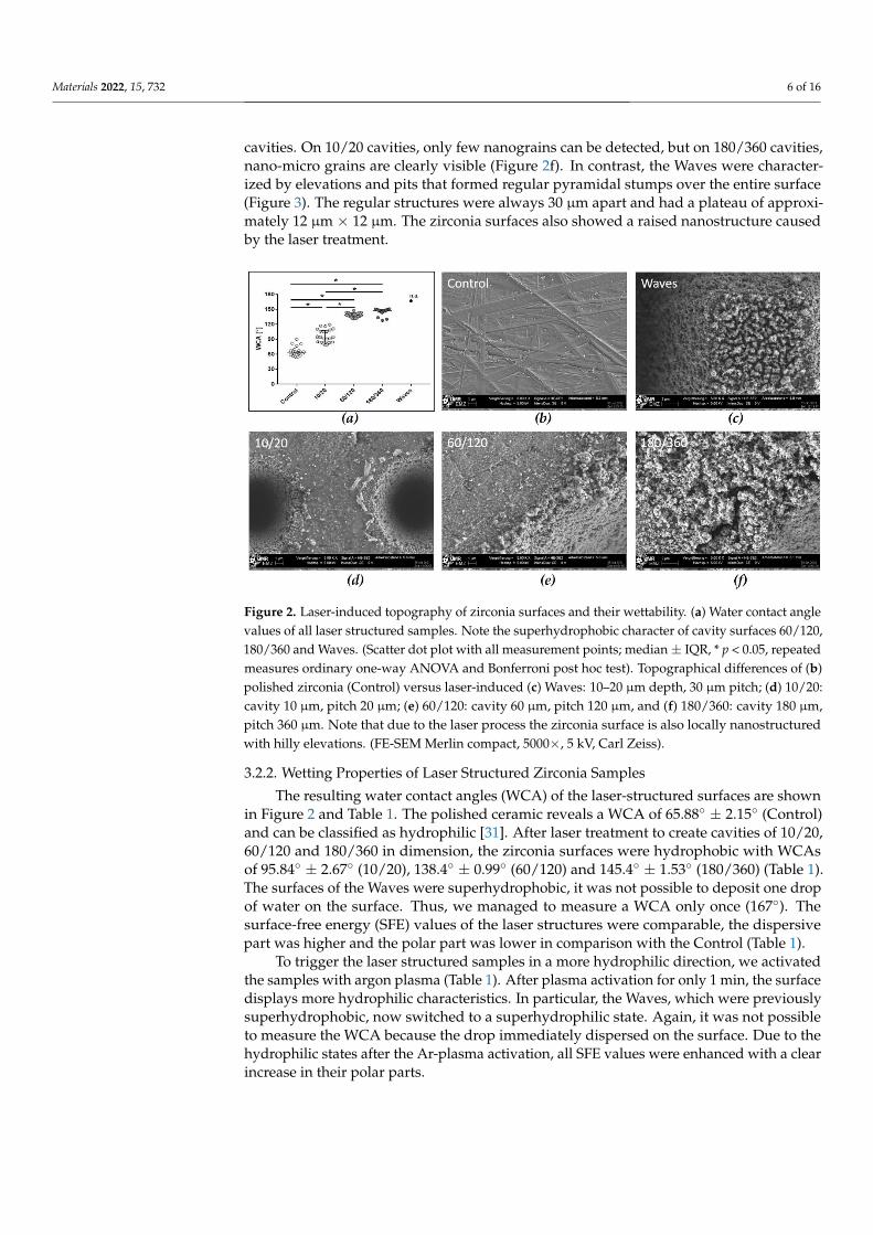
cavities. On 10/20 cavities, only few nanograins can be detected, but on 180/360 cavities,nano-micro grains are clearly visible (Figure 2f). In contrast, the Waves were character-ized by elevations and pits that formed regular pyramidal stumps over the entire surface(Figure 3). The regular structures were always 30 µm apart and had a plateau of approxi-mately 12 µm × 12 µm. The zirconia surfaces also showed a raised nanostructure causedby the laser treatment.
Materials 2022, 15, x FOR PEER REVIEW 6 of 16
3.2. Material Surface Characterization 3.2.1. Profiles of Zirconia Samples
Since the manufacturing processes of the laser structures investigated followed dif-ferent protocols, we analyzed the resulting topographies by FE-SEM. Regarding the pol-ished zirconia (Control), the FE-SEM images showed that grinding grooves without ori-entation were detectable (Figure 2). After laser structuring, two types of periodic micro-structures can be observed: concave cavities 10/20 holes with a dimension of 10 µm in width and depth, 30 µm pitch; 60/120 holes with a dimension of 60 µm in width and depth, 120 µm pitch; 180/360 holes with a dimension of 180 µm in width and depth, 360 µm pitch, and a sinusoidal, convex microstructure, like pyramid stumps (Waves) with the dimen-sion of 10–20 µm in height and 30 µm pitch (Figures 2 and 3). As the cavities became wider and deeper, the laser had to process the surface more intensively, which was also evident from the nanoporous surface texture of the zirconia (grains) at the marginal areas and in the cavities. On 10/20 cavities, only few nanograins can be detected, but on 180/360 cavi-ties, nano-micro grains are clearly visible (Figure 2f). In contrast, the Waves were charac-terized by elevations and pits that formed regular pyramidal stumps over the entire sur-face (Figure 3). The regular structures were always 30 µm apart and had a plateau of ap-proximately 12 µm × 12 µm. The zirconia surfaces also showed a raised nanostructure caused by the laser treatment.
Figure 2. Laser-induced topography of zirconia surfaces and their wettability. (a) Water contact an-gle values of all laser structured samples. Note the superhydrophobic character of cavity surfaces 60/120, 180/360 and Waves. (Scatter dot plot with all measurement points; median ± IQR, * p < 0.05, repeated measures ordinary one-way ANOVA and Bonferroni post hoc test). Topographical differ-ences of (b) polished zirconia (Control) versus laser-induced (c) Waves: 10–20 µm depth, 30 µm pitch; (d) 10/20: cavity 10 µm, pitch 20 µm; (e) 60/120: cavity 60 µm, pitch 120 µm, and (f) 180/360: cavity 180 µm, pitch 360 µm. Note that due to the laser process the zirconia surface is also locally nanostructured with hilly elevations. (FE-SEM Merlin compact, 5000×, 5 kV, Carl Zeiss).
3.2.2. Wetting Properties of Laser Structured Zirconia Samples The resulting water contact angles (WCA) of the laser-structured surfaces are shown
in Figure 2 and Table 1. The polished ceramic reveals a WCA of 65.88° ± 2.15° (Control) and can be classified as hydrophilic [31]. After laser treatment to create cavities of 10/20, 60/120 and 180/360 in dimension, the zirconia surfaces were hydrophobic with WCAs of 95.84° ± 2.67° (10/20), 138.4° ± 0.99° (60/120) and 145.4° ± 1.53° (180/360) (Table 1). The surfaces of the Waves were superhydrophobic, it was not possible to deposit one drop of
Figure 2. Laser-induced topography of zirconia surfaces and their wettability. (a) Water contact anglevalues of all laser structured samples. Note the superhydrophobic character of cavity surfaces 60/120,180/360 and Waves. (Scatter dot plot with all measurement points; median± IQR, * p < 0.05, repeatedmeasures ordinary one-way ANOVA and Bonferroni post hoc test). Topographical differences of (b)polished zirconia (Control) versus laser-induced (c) Waves: 10–20 µm depth, 30 µm pitch; (d) 10/20:cavity 10 µm, pitch 20 µm; (e) 60/120: cavity 60 µm, pitch 120 µm, and (f) 180/360: cavity 180 µm,pitch 360 µm. Note that due to the laser process the zirconia surface is also locally nanostructuredwith hilly elevations. (FE-SEM Merlin compact, 5000×, 5 kV, Carl Zeiss).
3.2.2. Wetting Properties of Laser Structured Zirconia Samples
The resulting water contact angles (WCA) of the laser-structured surfaces are shownin Figure 2 and Table 1. The polished ceramic reveals a WCA of 65.88◦ ± 2.15◦ (Control)and can be classified as hydrophilic [31]. After laser treatment to create cavities of 10/20,60/120 and 180/360 in dimension, the zirconia surfaces were hydrophobic with WCAsof 95.84◦ ± 2.67◦ (10/20), 138.4◦ ± 0.99◦ (60/120) and 145.4◦ ± 1.53◦ (180/360) (Table 1).The surfaces of the Waves were superhydrophobic, it was not possible to deposit one dropof water on the surface. Thus, we managed to measure a WCA only once (167◦). Thesurface-free energy (SFE) values of the laser structures were comparable, the dispersivepart was higher and the polar part was lower in comparison with the Control (Table 1).
To trigger the laser structured samples in a more hydrophilic direction, we activatedthe samples with argon plasma (Table 1). After plasma activation for only 1 min, the surfacedisplays more hydrophilic characteristics. In particular, the Waves, which were previouslysuperhydrophobic, now switched to a superhydrophilic state. Again, it was not possibleto measure the WCA because the drop immediately dispersed on the surface. Due to thehydrophilic states after the Ar-plasma activation, all SFE values were enhanced with a clearincrease in their polar parts.

Materials 2022, 15, 732 7 of 16
Table 1. Surface characteristics of laser structured zirconia surfaces before (w/o plasma) and afterargon plasma activation (+Ar plasma). Note the extreme switch from the superhydrophobic to thehydrophilic state of convex Wave structures.
Surface Method Control 10/20 60/120 180/360 Waves
w/o plasma
Materials 2022, 15, x FOR PEER REVIEW 7 of 16
water on the surface. Thus, we managed to measure a WCA only once (167°). The surface-free energy (SFE) values of the laser structures were comparable, the dispersive part was higher and the polar part was lower in comparison with the Control (Table 1).
To trigger the laser structured samples in a more hydrophilic direction, we activated the samples with argon plasma (Table 1). After plasma activation for only 1 min, the sur-face displays more hydrophilic characteristics. In particular, the Waves, which were pre-viously superhydrophobic, now switched to a superhydrophilic state. Again, it was not possible to measure the WCA because the drop immediately dispersed on the surface. Due to the hydrophilic states after the Ar-plasma activation, all SFE values were enhanced with a clear increase in their polar parts.
Table 1. Surface characteristics of laser structured zirconia surfaces before (w/o plasma) and after argon plasma activation (+ Ar plasma). Note the extreme switch from the superhydrophobic to the hydrophilic state of convex Wave structures.
Surface Method Control 10/20 60/120 180/360 Waves
w/o plasma
WCA [°] 65.88 ± 2.15 95.84 ± 2.67 138.4 ± 0.99 145.4 ± 1.53 ≥150
SFE [mN/m]
dispersive polar
46.82 ± 4.29 31.21 ± 1.62 15.61 ± 2.67
48.12 ± 1.45 47.34 ± 1.45 0.78 ± 0.89
54.84 ± 3.09 45.51 ± 1.53 9.32 ± 1.56
43.13 ± 3.60 33.89 ± 2.52 9.24 ± 1.08
+ Ar-plasma
WCA [°] 17.27 ± 1.35 7.34 ± 2.41 13.67 ± 1.13 10.25 ± 1.12 ≥0
SFE [mN/m]
dispersive polar
72.46 ± 6.21 35.26 ± 3.35 37.20 ± 2.86
80.61 ± 0.4 50.56 ± 0.26 30.05 ± 0.13
77.65 ± 1.75 46.39 ± 0.71 31.25 ± 1.06
79.96 ± 1.05 49.68 ± 0.34 30.28 ± 0.71
3.3. Cellular Behavior 3.3.1. Morphology and Spreading on Laser Structured Zirconia
The different laser structures affected the morphology, spreading and growth of HGF-1 fibroblasts after 24 h (Figure 3). The FE-SEM images showed that HGF-1 cells at-tached very flatly on polished zirconia (Control). On the laser-induced cavities, cells also spread on the planar areas next to the cavities. The HGF-1 cells were able to span the cavities even over longer distances up to 180 µm (Figure 3c–e). Cells partially avoid grow-ing into the holes of the laser structures with the smaller diameters 10/20 and 60/120, per-haps due to hydrophobic surface characteristics (Figure 3c,d). Increasing the size of the hole up to 180 µm (Figure 3e) does not substantially improve the cell ingrowth. Interest-ingly, the Wave structures allow for alignment of the gingival fibroblasts in the grooved space (Figure 3b). On the Waves, the cells did not elongate as on the other surfaces, a polygonal cell shape was evident. The HGF-1 fibroblasts were able to become very thin and, above all, to grow into the interstices with their filopodia (Figure 3b).
The quantification of morphometric data—cell area and length to width ratio—was performed from the FE-SEM images using ImageJ (Version 1.51f) (Figure 3f,g). Interest-ingly, the dimensions of the cavities seem to influence cell spreading. For the smallest cavity 10/20, we could detect significantly increased spreading compared with all other surfaces. Concerning the length to width ratio, we could detect a slight stretching. The spreading decreased significantly on 60/120 cavities compared with the polished zirconia (Control). Here, the lowest L/W ratio could be detected. Interestingly, the spreading on the 180/360 cavity structure was significantly reduced compared with all other surfaces,
WCA [◦] 65.88 ± 2.15 95.84 ± 2.67 138.4 ± 0.99 145.4 ± 1.53 ≥150SFE [mN/m]
dispersivepolar
46.82 ± 4.2931.21 ± 1.6215.61 ± 2.67
48.12 ± 1.4547.34 ± 1.450.78 ± 0.89
54.84 ± 3.0945.51 ± 1.539.32 ± 1.56
43.13 ± 3.6033.89 ± 2.529.24 ± 1.08
+Ar-plasma
Materials 2022, 15, x FOR PEER REVIEW 7 of 16
water on the surface. Thus, we managed to measure a WCA only once (167°). The surface-free energy (SFE) values of the laser structures were comparable, the dispersive part was higher and the polar part was lower in comparison with the Control (Table 1).
To trigger the laser structured samples in a more hydrophilic direction, we activated the samples with argon plasma (Table 1). After plasma activation for only 1 min, the sur-face displays more hydrophilic characteristics. In particular, the Waves, which were pre-viously superhydrophobic, now switched to a superhydrophilic state. Again, it was not possible to measure the WCA because the drop immediately dispersed on the surface. Due to the hydrophilic states after the Ar-plasma activation, all SFE values were enhanced with a clear increase in their polar parts.
Table 1. Surface characteristics of laser structured zirconia surfaces before (w/o plasma) and after argon plasma activation (+ Ar plasma). Note the extreme switch from the superhydrophobic to the hydrophilic state of convex Wave structures.
Surface Method Control 10/20 60/120 180/360 Waves
w/o plasma
WCA [°] 65.88 ± 2.15 95.84 ± 2.67 138.4 ± 0.99 145.4 ± 1.53 ≥150
SFE [mN/m]
dispersive polar
46.82 ± 4.29 31.21 ± 1.62 15.61 ± 2.67
48.12 ± 1.45 47.34 ± 1.45 0.78 ± 0.89
54.84 ± 3.09 45.51 ± 1.53 9.32 ± 1.56
43.13 ± 3.60 33.89 ± 2.52 9.24 ± 1.08
+ Ar-plasma
WCA [°] 17.27 ± 1.35 7.34 ± 2.41 13.67 ± 1.13 10.25 ± 1.12 ≥0
SFE [mN/m]
dispersive polar
72.46 ± 6.21 35.26 ± 3.35 37.20 ± 2.86
80.61 ± 0.4 50.56 ± 0.26 30.05 ± 0.13
77.65 ± 1.75 46.39 ± 0.71 31.25 ± 1.06
79.96 ± 1.05 49.68 ± 0.34 30.28 ± 0.71
3.3. Cellular Behavior 3.3.1. Morphology and Spreading on Laser Structured Zirconia
The different laser structures affected the morphology, spreading and growth of HGF-1 fibroblasts after 24 h (Figure 3). The FE-SEM images showed that HGF-1 cells at-tached very flatly on polished zirconia (Control). On the laser-induced cavities, cells also spread on the planar areas next to the cavities. The HGF-1 cells were able to span the cavities even over longer distances up to 180 µm (Figure 3c–e). Cells partially avoid grow-ing into the holes of the laser structures with the smaller diameters 10/20 and 60/120, per-haps due to hydrophobic surface characteristics (Figure 3c,d). Increasing the size of the hole up to 180 µm (Figure 3e) does not substantially improve the cell ingrowth. Interest-ingly, the Wave structures allow for alignment of the gingival fibroblasts in the grooved space (Figure 3b). On the Waves, the cells did not elongate as on the other surfaces, a polygonal cell shape was evident. The HGF-1 fibroblasts were able to become very thin and, above all, to grow into the interstices with their filopodia (Figure 3b).
The quantification of morphometric data—cell area and length to width ratio—was performed from the FE-SEM images using ImageJ (Version 1.51f) (Figure 3f,g). Interest-ingly, the dimensions of the cavities seem to influence cell spreading. For the smallest cavity 10/20, we could detect significantly increased spreading compared with all other surfaces. Concerning the length to width ratio, we could detect a slight stretching. The spreading decreased significantly on 60/120 cavities compared with the polished zirconia (Control). Here, the lowest L/W ratio could be detected. Interestingly, the spreading on the 180/360 cavity structure was significantly reduced compared with all other surfaces,
WCA [◦] 17.27 ± 1.35 7.34 ± 2.41 13.67 ± 1.13 10.25 ± 1.12 ≥0SFE [mN/m]
dispersivepolar
72.46 ± 6.2135.26 ± 3.3537.20 ± 2.86
80.61 ± 0.450.56 ± 0.2630.05 ± 0.13
77.65 ± 1.7546.39 ± 0.7131.25 ± 1.06
79.96 ± 1.0549.68 ± 0.3430.28 ± 0.71
3.3. Cellular Behavior3.3.1. Morphology and Spreading on Laser Structured Zirconia
The different laser structures affected the morphology, spreading and growth of HGF-1fibroblasts after 24 h (Figure 3). The FE-SEM images showed that HGF-1 cells attached veryflatly on polished zirconia (Control). On the laser-induced cavities, cells also spread onthe planar areas next to the cavities. The HGF-1 cells were able to span the cavities evenover longer distances up to 180 µm (Figure 3c–e). Cells partially avoid growing into theholes of the laser structures with the smaller diameters 10/20 and 60/120, perhaps dueto hydrophobic surface characteristics (Figure 3c,d). Increasing the size of the hole up to180 µm (Figure 3e) does not substantially improve the cell ingrowth. Interestingly, the Wavestructures allow for alignment of the gingival fibroblasts in the grooved space (Figure 3b).On the Waves, the cells did not elongate as on the other surfaces, a polygonal cell shapewas evident. The HGF-1 fibroblasts were able to become very thin and, above all, to growinto the interstices with their filopodia (Figure 3b).
The quantification of morphometric data—cell area and length to width ratio—wasperformed from the FE-SEM images using ImageJ (Version 1.51f) (Figure 3f,g). Interestingly,the dimensions of the cavities seem to influence cell spreading. For the smallest cavity10/20, we could detect significantly increased spreading compared with all other surfaces.Concerning the length to width ratio, we could detect a slight stretching. The spreadingdecreased significantly on 60/120 cavities compared with the polished zirconia (Control).Here, the lowest L/W ratio could be detected. Interestingly, the spreading on the 180/360cavity structure was significantly reduced compared with all other surfaces, as indicatedby a significantly increased elongation (vs. control and 60/120) especially over the holes(180 µm). The evaluation of the Waves showed a cell area and shape comparable tothose of the control. The cell area on Waves was significantly larger than on 180/360cavity. However, the spreading on Waves could not be detected because a part of the cellsdisappeared in the grooves beside the pyramid stumps.

Materials 2022, 15, 732 8 of 16
Materials 2022, 15, x FOR PEER REVIEW 8 of 16
as indicated by a significantly increased elongation (vs. control and 60/120) especially over the holes (180 µm). The evaluation of the Waves showed a cell area and shape comparable to those of the control. The cell area on Waves was significantly larger than on 180/360 cavity. However, the spreading on Waves could not be detected because a part of the cells disappeared in the grooves beside the pyramid stumps.
Figure 3. Scanning electron microscopy of HGF-1 gingival cells on various laser structured zirco-nia surfaces after 24 h growth. Micro-topographical dimensions: (a) polished zirconia as control;(b) waves: 10–20 µm depth, 30 µm pitch, note that cells grow on and in between the waves; (c) 10/20:cavity 10 µm, pitch 20 µm, note that the cells ignore the holes and avoid ingrowth; (d) 60/120:cavity 60 µm, pitch 120 µm, note that cells avoid growing into the holes but rather span the holes;(e) 180/360: cavity 180 µm, pitch 360 µm, note that cells span the holes but partially migrate also into

Materials 2022, 15, 732 9 of 16
the depth. (FE-SEM Merlin compact, Carl Zeiss; false colored by PowerPoint software; upper row200×, bar = 20 µm; lower row 1000×, bar = 10 µm). (f) Quantitative data of HGF-1 cell area after 24 hon the laser structures, and (g) cell ratio of length to width (L/W). (mean ± s.e.m., Friedman test posthoc uncorrected Dunn’s test; * p < 0.05, n = 40 cells).
3.3.2. Actin Cytoskeleton on Convex Waves
Another cell morphological aspect was the organization of the actin cytoskeleton,especially on the Waves (Figure 4). The confocal microscopic images of actin filamentsafter 24 h of cultivation showed long filaments exactly as on the Controls. No influenceof the nano-microtopography on filament length or orientation could be detected. Longactin filaments were detectable both on the plateau of the Waves and between the pyramid-like stumps. On polished zirconia, HGF-1 fibroblasts formed a well-developed actincytoskeleton with long filaments passing through the entire cell body.
Materials 2022, 15, x FOR PEER REVIEW 9 of 16
Figure 3. Scanning electron microscopy of HGF-1 gingival cells on various laser structured zirconia surfaces after 24 h growth. Micro-topographical dimensions: (a) polished zirconia as control; (b) waves: 10–20 µm depth, 30 µm pitch, note that cells grow on and in between the waves; (c) 10/20: cavity 10 µm, pitch 20 µm, note that the cells ignore the holes and avoid ingrowth; (d) 60/120: cavity 60 µm, pitch 120 µm, note that cells avoid growing into the holes but rather span the holes; (e) 180/360: cavity 180 µm, pitch 360 µm, note that cells span the holes but partially migrate also into the depth. (FE-SEM Merlin compact, Carl Zeiss; false colored by PowerPoint software; upper row 200×, bar = 20 µm; lower row 1000×, bar = 10 µm). (f) Quantitative data of HGF-1 cell area after 24 h on the laser structures, and (g) cell ratio of length to width (L/W). (mean ± s.e.m., Friedman test post hoc uncorrected Dunn´s test; * p < 0.05, n = 40 cells).
3.3.2. Actin Cytoskeleton on Convex Waves Another cell morphological aspect was the organization of the actin cytoskeleton, es-
pecially on the Waves (Figure 4). The confocal microscopic images of actin filaments after 24 h of cultivation showed long filaments exactly as on the Controls. No influence of the nano-microtopography on filament length or orientation could be detected. Long actin filaments were detectable both on the plateau of the Waves and between the pyramid-like stumps. On polished zirconia, HGF-1 fibroblasts formed a well-developed actin cytoskel-eton with long filaments passing through the entire cell body.
Figure 4. Organization of the actin cytoskeleton in HGF-1 cells after 24 h. (a) On polished surfaces (Control), gingival cells formed a well-developed actin cytoskeleton with long filaments which span through the entire cell body. (b) On Waves (pyramid stumps: 10–20 µm height, 30 µm pitch), cells grow on the pyramid stumps as well as in the grooves between them (dotted lines, see also Figure 3); long actin filaments are visible in these areas (arrow). (LSM 780, Carl Zeiss; bar = 10 µm, red: actin, white dotted line: plateau of Waves).
3.3.3. Cell Spreading after Wettability Modulation Atmospheric pressure argon plasma (Ar plasma) makes the surface more hydrophilic
(Table 1). As an illustration, we present the FE-SEM images of HGF-1 cells after 2 h culti-vation on the cavity structure 60/120 and on the Waves (Figure 5) after Ar plasma treat-ment compared with untreated laser structured surfaces (w/o plasma). After plasma treat-ment with a jet, the surfaces provide better wettability conditions resulting in better cell growth and spreading. Due to Ar plasma activation, a significantly increased cell area could be seen on all surfaces after 2 h compared with zirconia w/o plasma. Only a slightly decreased cell area on all laser structures was detectable compared with the plasma-acti-vated zirconia controls, that is, the chemistry seems to be dominant over the topography and cells are able to spread optimally.
In the FE-SEM images, it could be observed that the cell growth on 60/120 cavity is more concentrated in and around the holes after only a 2 h cell culture following the plasma treatment (Figure 5b). Thus, after plasma activation, the fibroblasts are able to oc-cupy the entire surface, including the niches/holes. This contrasts with the pure, laser-
Figure 4. Organization of the actin cytoskeleton in HGF-1 cells after 24 h. (a) On polished surfaces(Control), gingival cells formed a well-developed actin cytoskeleton with long filaments which spanthrough the entire cell body. (b) On Waves (pyramid stumps: 10–20 µm height, 30 µm pitch), cellsgrow on the pyramid stumps as well as in the grooves between them (dotted lines, see also Figure 3);long actin filaments are visible in these areas (arrow). (LSM 780, Carl Zeiss; bar = 10 µm, red: actin,white dotted line: plateau of Waves).
3.3.3. Cell Spreading after Wettability Modulation
Atmospheric pressure argon plasma (Ar plasma) makes the surface more hydrophilic(Table 1). As an illustration, we present the FE-SEM images of HGF-1 cells after 2 hcultivation on the cavity structure 60/120 and on the Waves (Figure 5) after Ar plasmatreatment compared with untreated laser structured surfaces (w/o plasma). After plasmatreatment with a jet, the surfaces provide better wettability conditions resulting in bettercell growth and spreading. Due to Ar plasma activation, a significantly increased cellarea could be seen on all surfaces after 2 h compared with zirconia w/o plasma. Onlya slightly decreased cell area on all laser structures was detectable compared with theplasma-activated zirconia controls, that is, the chemistry seems to be dominant over thetopography and cells are able to spread optimally.
In the FE-SEM images, it could be observed that the cell growth on 60/120 cavity ismore concentrated in and around the holes after only a 2 h cell culture following the plasmatreatment (Figure 5b). Thus, after plasma activation, the fibroblasts are able to occupy theentire surface, including the niches/holes. This contrasts with the pure, laser-structuredceramic surface. Here, cells grow more between the holes and avoid the ingrowth intothe depth of the holes. On the Ar plasma functionalized Wave structures, the HGF-1 cellswere huge in the cell area and fuse with the laser structure, especially into the valleys ofthe Waves. This contrasts with untreated Waves, where the cells after 2 h sit on top of thestructure with a small cell area, limited to four pyramid stump-plateaus.

Materials 2022, 15, 732 10 of 16
Materials 2022, 15, x FOR PEER REVIEW 10 of 16
structured ceramic surface. Here, cells grow more between the holes and avoid the in-growth into the depth of the holes. On the Ar plasma functionalized Wave structures, the HGF-1 cells were huge in the cell area and fuse with the laser structure, especially into the valleys of the Waves. This contrasts with untreated Waves, where the cells after 2 h sit on top of the structure with a small cell area, limited to four pyramid stump-plateaus.
Figure 5. Topography and chemistry—combinatory influence of laser microstructures on zirconia and atmospheric pressure plasma on HGF-1 gingival cell growth. (a) Morphology of HGF-1 on
Figure 5. Topography and chemistry—combinatory influence of laser microstructures on zirconiaand atmospheric pressure plasma on HGF-1 gingival cell growth. (a) Morphology of HGF-1 on60/120 (cavity 60 µm, pitch 120 µm) after 2 h growth. Note that cells on Ar plasma-activated cavitiesconcentrate their growth more around and in the cavities (arrows), thereby finding a niche. Thiscontrasts with untreated 60/120 surfaces, where cells avoid growing into the depth of the holes(dotted lines around the 60 µm holes). (b) Morphology of HGF-1 on Waves (pyramid stumps:10–20 µm height, 30 µm pitch) after 2 h. Cells on Ar plasma-activated zirconia melt into the valley ofthe Waves with a large cell area (arrow). This contrasts with untreated Waves, where cells sit on top

Materials 2022, 15, 732 11 of 16
of the pyramid stumps, remaining small in cell size. (FE-SEM Merlin compact, Carl Zeiss; indicatedmagnifications 200×, 500×; mode: InlenseDuo). (c) Cell spreading after 2 h. Note the significantlyincreased cell areas due to the additional Ar plasma activation resulting in higher hydrophilicity (seeTable 1). (mean ± s.e.m.; * untreated vs. Ar plasma, paired t-test, * p < 0.05; # compared to control,Friedman test post hoc uncorrected Dunn´s test, # p < 0.05; n = 40 cells).
4. Discussion
The present study provides insights into how the surface nano-microtopography incombination with plasma-chemistry enables a strong attachment of human gingival HGF-1fibroblasts on zirconia surfaces. The laser-induced microstructures were originally hy-drophobic, but after argon plasma activation, the surfaces switched to the hydrophilic state.
It has been postulated in the literature that physico-chemical surface characteristicssuch as wettability or roughness influence cell behavior, for example, adhesion, proliferationand differentiation [11,32,33]. It has been found that the cell spreading was improved onsmooth surfaces than on moderately rough surfaces [34,35], but roughening the topographyleads to a better anchorage of cells following implant integration and final success [4,16,36].Although the currently available titanium implants are polished on the neck area andare moderately roughened on the endosseous part, the influence of surface topographyon implant survival is not yet clearly understood [37]. Other studies have measuredthe cell behavior of gingiva fibroblasts on zirconia that was either machined [38], etchedor sandblasted [35], polished, and/or atmospheric plasma treated [39,40]. Our studyintroduces a new aspect: defined laser-structured zirconia surfaces.
The laser-structured samples exhibited four different periodic structures that wouldbe applicable to the neck region of zirconia dental implants. We fabricated periodic concavecavities of different widths and depths in the micrometer range—holes with diametersof 10, 60 and 180 µm (see method part) as well as a periodic convex microstructure withrectangular pyramid stumps 10–20 µm in height (Waves). The base material was polishedzirconia, which exhibits gridding grooves made by FE-SEM. Enlarged and deeper cavities,as in cavity 180/360, resulted from a more intense laser irradiation, which led to theroughening of the structure and thus to additional nano-microstructures at the edge areasof the holes. The intense laser irradiation results in a significantly increased heat input.Additionally, Rohr et al. [35] were able to show that a graining (comprising tetragonalstructures) was detectable by heat treatment of the zirconia.
Another characteristic surface aspect—wettability—is influenced by microstructuresand surface roughness, among other factors [3,24,31]. Different studies postulated thatsurface roughness leads to increased hydrophobic behavior [24]. The cause is the inclusionof air in surface pores, which forms a heterogeneous solid/air surface that impairs spon-taneous wetting of the surface (Cassie-Baxter wetting condition) [41]. We could directlydemonstrate this with our laser-induced structures 10/20, 60/120, 180/360 concave cavitiesand the convex Waves. Interestingly, it was not possible to deposit the water dropleton the laser-produced Waves. Thus, the Waves were even superhydrophobic due to thenanostructures on the microstructures (like lotus-leaves). Wang et al. [41] defined thesuperhydrophobic stages (>150◦) and described that the cohesive forces between the waterin the needle and the water droplet were stronger than the adhesive forces between thedroplet and the ceramic surface.
Functionalization of dental implant surfaces with gas plasmas before clinical use isconsidered a potential surface modification method for improved implant integration intothe tissue [19,24,42–45]. To investigate the possible biological efficacy of plasma-activatedzirconia on samples, the laser-structured surfaces were treated with argon plasma usingthe kINPen®09 for only 1 min. Studies proved that plasma treatment had no effect onthe surface topography of the implant materials, but significantly increased the wettabil-ity [21,24,46]. In contrast to the laser treatment, plasma activation induced a higher surfacefree energy (SFE). This rise of SFE with an increase in polar parts was associated with thelower WCA values (hydrophilic characteristic). The effects of plasma activation on WCA

Materials 2022, 15, 732 12 of 16
and SFE, as well as the negative correlation between both parameters, have already beendiscussed in the literature [47]. Gentlemen et al. [48] reviewed that the SFE is a relevantfeature for cell response. The increase in wettability can be attributed to the removal ofcarbon contaminants on the surface by the plasma process [42,43,46,49]. Studies coulddemonstrate that the reduction of WCA due to plasma activation correlated with the initialsurface wettability, i.e., as the initial contact angle was higher, the reduction in WCA causedby functionalization was more pronounced [24,49]. This was also observed in our study,the superhydrophobic (>150◦) wave structure became superhydrophilic (>0◦) after theplasma treatment.
Surmeneva et al. [50] showed that a deposition of nano-hydroxyapatite layers on lasermicro-textured titanium resulted in a nano-micro-structured surface topography, whichimproved surface characteristics such as wettability and SFE.
Human gingival fibroblasts (HGF-1) were used in the present in vitro study to observethe soft tissue response to the different zirconia surfaces more closely. HGF-1 is the main celltype involved in the formation of the mucosal barrier around the implant, which enablessuccessful implant integration [4,38]. Characterization of HGF-1 cells revealed strong actinfilaments spanning the entire cells and vinculin contacts at the focal adhesion points after48 h of cultivation. A pronounced hyaluronan coat was also detected after 24 h of culture,which is important for migration [51], and cell adhesion [30,51].
In their review, Rompen et al. [4] determined that epithelial cells adhered, as well asspread more strongly on metallic surfaces (titanium or gold alloy) than on zirconia surfaces,and modified ceramics promoted cell proliferation to a greater extent than smooth controls.HGF-1 cells on the polished zirconia (Control) spread and exhibited a large cell area thatwas close to the material surface. On the laser structured cavity 10/20, the cells were able tospread well and spanned the small holes (~10 µm) with their large cell body. The study ofSchnell et al. [15] indicated that the size, depth dimension as well as roughness (Sa) of thesurface affected osteoblast behavior. Kunzler et al. [16] demonstrated a higher proliferationrate of osteoblasts on rough surfaces, whereas the proliferation of HGF decreased withincreasing roughness. We also showed in our study that with increasing pattern dimensionsof the cavities, the spreading of HGF-1 was significantly reduced. On cavity 180/360 withthe roughest nano-microstructure, spreading was significantly inhibited compared with allother surfaces. Belaud et al. [52] showed in their work that the combination of nanoscaleand curved structures stimulated stem cell adhesion and migration. We also observedon the convex microstructure (Waves) only a slight impairment of cell spreading, themorphology and spreading were unaffected and comparable to the polished control. Cellsexpressed long filopodia that could be observed in the space between the pyramid stumps,that is, in the grooves. Bartold et al. [28] previously observed that fibroblasts expandedduring migration and formed filopodia that adhered to the substrate. We could also showthis topography-dependent influence of the morphology in previous studies on osteoblastsusing defined titanium microstructures [15,53].
Studies suggested that the micro-roughened implant surface has so far served mainlyto secure the implant in position, but not to promote initial fibroblast adhesion and prolifer-ation [13,35,54]. A further surface modification—plasma activation of dental surfaces—isalready used in practice today, but only for the decontamination of dental implants [21,42].Concerning the initial spreading of HGF-1 cells on the Ar plasma-activated surfaces, wefound significantly increased spreading for all structures compared with untreated ma-terials after 2 h cell culture. Moreover, the FE-SEM images revealed that the cell bodyof the fibroblasts was closely fused with the surface structure. Most interesting was thatthe fibroblasts were able to grow into the cavities (60/120) after plasma activation of zir-conia, thereby finding a niche. This demonstrates clearly that even inaccessible areas ofsurfaces can be seeded by cells after plasma functionalization. In other studies, enhancedand improved fibroblast attachment was found on titanium and zirconia surfaces whenfunctionalized with atmospheric plasma [19,55]. Guo et al. [19] compared biomaterialfunctionalization and found that oxygen plasma supported gingival fibroblast attachment

Materials 2022, 15, 732 13 of 16
as well as proliferation better than argon plasma activation. Rabel et al. [24] indicated intheir proliferation analysis (up to 7 d) no significant differences between HGF on oxygenplasma-treated and untreated control surfaces. Canullo et al. [55] also demonstrated in theirstudy that cleaning with argon plasma accelerated the initial adhesion of L929 fibroblasts,but this effect disappeared after 48 h as a result of saturation.
Discussions of other studies must consider choice of materials, topography, and/orplasma sources. In this respect, Guo et al. [19] found that the effects of plasma func-tionalization on cell response varied depending on the type of material treated (titaniumvs. zirconia).
In this study, the limitation from the cell biology point of view is the usage of only onehuman cell type as representative of the soft tissue. This should be expanded in furtherexperiments. Ingrowth of cells and the acceptance of the material topography is a precondi-tion for the cell physiology and function. However, in the following experiments it shouldbe observed how HGF-1 cells synthesize extracellular matrix molecules (e.g., collagen I,fibronectin, hyaluronan). This preliminary study was limited to the morphological behaviorof HGF-1 gingival cells. Our research raises new questions, and further studies are neededto understand cell behavior at the interface of differently modified zirconia surfaces moreprecisely. The present results suggest that, in the development of laser structured dentalimplant surfaces for improved tissue integration, argon plasma activation would be apossible compatible method for advanced modification approaches [24,42]. In particular,the plasma treatment with a plasma jet enables the activation of dental implants directlybefore insertion in clinical practice.
5. Conclusions
Laser-induced nano-micro texturing of zirconia surfaces seems to be a good tool forproducing defined topographic patterns to control cell growth. However, micro-roughsurfaces caused by the laser process showed decreased surface wettability accompanied byreduced cell spreading. An activation of the laser-structured surface using argon plasmacan equalize these effects. The zirconia surfaces switched to the hydrophilic state with ahigher SFE and the cell areas significantly increased. In addition, cells could find a niche inthe laser-induced holes and the grooves. Within the limits of the present study, it can beconcluded that the argon plasma activation of zirconia is a potential method for increasingthe wettability of structured zirconia with its defined laser-induced patterns and providinga stimulus for improved HGF-1 cell attachment and ingrowth.
Author Contributions: Conceptualization, B.N., J.B., S.S. (Susanne Staehlke); methodology, S.S.(Susanne Staehlke), S.S. (Susanne Seemann), B.N.; software, S.S. (Susanne Staehlke), S.S. (SusanneSeemann); validation, S.S. (Susanne Staehlke), S.S. (Susanne Seemann), P.O., F.K.; formal analysis, S.S.(Susanne Staehlke), S.S. (Susanne Seemann); investigation, S.S. (Susanne Staehlke), S.S. (Susanne See-mann), P.O., F.K.; resources, J.B., B.N.; data curation, S.S. (Susanne Staehlke), S.S. (Susanne Seemann),P.O., F.K.; writing—original draft preparation, B.N., S.S. (Susanne Staehlke), S.S. (Susanne Seemann),P.O.; writing—review and editing, B.N., J.B., S.S. (Susanne Staehlke), S.S. (Susanne Seemann), P.O.;visualization, S.S. (Susanne Staehlke), S.S. (Susanne Seemann), B.N.; supervision, B.N.; project admin-istration, B.N., J.B.; funding acquisition, J.B., B.N. All authors have read and agreed to the publishedversion of the manuscript.
Funding: This research was funded by the PROSEC gGmbH (Progress in Science and Education withCeramics, grant contract no. 2020-02003).
Institutional Review Board Statement: Not applicable.
Informed Consent Statement: Not applicable.
Data Availability Statement: The datasets generated during and/or analysed during the currentstudy are available from the corresponding author on reasonable request. The dataset is stored on thelocal UMR server.

Materials 2022, 15, 732 14 of 16
Acknowledgments: The disks used for laser structuring were donated by VITA Zahnfabrik H. RauterGmbH & Co. KG. The authors thank Marcus Frank and Armin Springer (Electron Microscopic Center,UMR) for helpful assistance in SEM-imaging. We thank Finja Hildebrandt for argon plasma treatmentof samples. We thank the Leibniz Institute for Plasma Science and Technology (INP) Greifswald forthe support concerning the plasma source kINPen®09. We are very thankful to André Schoene for hisinspiring thoughts to laser structuring of dental ceramic for soft tissue regeneration.
Conflicts of Interest: The authors declare no conflict of interest.
References1. Roehling, S.; Schlegel, K.A.; Woelfler, H.; Gahlert, M. Performance and outcome of zirconia dental implants in clinical studies: A
meta-analysis. Clin. Oral Implant. Res. 2018, 29, 135–153. [CrossRef]2. Balmer, M.; Spies, B.C.; Kohal, R.J.; Hämmerle, C.H.; Vach, K.; Jung, R.E. Zirconia implants restored with single crowns or fixed
dental prostheses: 5-year results of a prospective cohort investigation. Clin. Oral Implant. Res. 2020, 31, 452–462. [CrossRef]3. Rupp, F.; Liang, L.; Geis-Gerstorfer, J.; Scheideler, L.; Hüttig, F. Surface characteristics of dental implants: A review. Dent. Mater.
2018, 34, 40–57. [CrossRef]4. Rompen, E.; Domken, O.; Degidi, M.; Farias Pontes, A.E.; Piattelli, A. The effect of material characteristics, of surface topography
and of implant components and connections on soft tissue integration: A literature review. Clin. Oral Implant. Res. 2006, 17 (Suppl.S2), 55–67. [CrossRef] [PubMed]
5. Hämmerle, C.H.; Brägger, U.; Bürgin, W.; Lang, N.P. The effect of subcrestal placement of the polished surface of ITI implants onmarginal soft and hard tissues. Clin. Oral Implant. Res. 1996, 7, 111–119. [CrossRef]
6. Thoma, D.S.; Ioannidis, A.; Cathomen, E.; Hämmerle, C.H.F.; Hüsler, J.; Jung, R.E. Discoloration of the peri-implant mucosacaused by zirconia and titanium implants. Int. J. Periodontics Restor. Dent. 2016, 36, 39–45. [CrossRef]
7. Roehling, S.; Schlegel, K.A.; Woelfler, H.; Gahlert, M. Zirconia compared to titanium dental implants in preclinical studies—Asystematic review and meta-analysis. Clin. Oral Implant. Res. 2019, 30, 365–395. [CrossRef] [PubMed]
8. Linkevicius, T.; Apse, P. Biologic width around implants. An evidence-based review. Stomatologija 2008, 10, 27–35. [PubMed]9. Cochran, D.L.; Hermann, J.S.; Schenk, R.K.; Higginbottom, F.L.; Buser, D. Biologic width around titanium implants. A histometric
analysis of the implanto-gingival junction around unloaded and loaded nonsubmerged implants in the canine mandible. J.Periodontol. 1997, 68, 186–198. [CrossRef] [PubMed]
10. Mandracci, P.; Mussano, F.; Rivolo, P.; Carossa, S. Surface treatments and functional coatings for biocompatibility improvementand bacterial adhesion reduction in dental implantology. Coatings 2016, 6, 7. [CrossRef]
11. Anselme, K. Osteoblast adhesion on biomaterials. Biomaterials 2000, 21, 667–681. [CrossRef]12. Fischer, J.; Schott, A.; Märtin, S. Surface micro-structuring of zirconia dental implants. Clin. Oral Implant. Res. 2016, 27, 162–166.
[CrossRef] [PubMed]13. Schünemann, F.H.; Galárraga-Vinueza, M.E.; Magini, R.; Fredel, M.; Silva, F.; Souza, J.C.M.; Zhang, Y.; Henriques, B. Zirconia
surface modifications for implant dentistry. Mater. Sci. Eng. C 2019, 98, 1294–1305. [CrossRef]14. Han, J.; Zhang, F.; Van Meerbeek, B.; Vleugels, J.; Braem, A.; Castagne, S. Laser surface texturing of zirconia-based ceramics for
dental applications: A review. Mater. Sci. Eng. C Mater. Biol. Appl. 2021, 123, 112034. [CrossRef]15. Schnell, G.; Staehlke, S.; Duenow, U.; Nebe, J.B.; Seitz, H. Femtosecond laser nano/micro textured Ti6Al4V surfaces–effect on
wetting and MG-63 cell adhesion. Materials 2019, 12, 2210. [CrossRef] [PubMed]16. Kunzler, T.P.; Drobek, T.; Schuler, M.; Spencer, N.D. Systematic study of osteoblast and fibroblast response to roughness by means
of surface-morphology gradients. Biomaterials 2007, 28, 2175–2182. [CrossRef]17. Pacha-Olivenza, M.A.; Tejero, R.; Fernández-Calderón, M.C.; Anitua, E.; Troya, M.; González-Martín, M.L. Relevance of
Topographic Parameters on the Adhesion and Proliferation of Human Gingival Fibroblasts and Oral Bacterial Strains. Biomed.Res. Int. 2019, 2019, 8456342. [CrossRef]
18. Gristina, A.G.; Naylor, P.T.; Myrvik, Q. The Race for the Surface: Microbes, Tissue Cells, and Biomaterials. In Molecular Mechanismsof Microbial Adhesion; Switalski, L., Höök, M., Beachey, E., Eds.; Springer: New York, NY, USA, 1989. [CrossRef]
19. Guo, L.; Smeets, R.; Kluwe, L.; Hartjen, P.; Barbeck, M.; Cacaci, C.; Gosau, M.; Henningsen, A. Cytocompatibility of Titanium,Zirconia and Modified PEEK after Surface Treatment Using UV Light or Non-Thermal Plasma. Int. J. Mol. Sci. 2019, 20, 5596.[CrossRef] [PubMed]
20. von Woedtke, T.; Reuter, S.; Masur, K.; Weltmann, K.D. Plasmas for medicine. Phys. Rep. 2013, 530, 291–320. [CrossRef]21. Piest, C.; Wille, S.; Strunskus, T.; Polonskyi, O.; Kern, M. Efficacy of Plasma Treatment for Decontaminating Zirconia. J. Adhes
Dent. 2018, 20, 289–297. [CrossRef]22. Bergemann, C.; Quade, A.; Kunz, F.; Ofe, S.; Klinkenberg, E.-D.; Laue, M.; Schroeder, K.; Weissmann, V.; Hansmann, H.; Weltmann,
K.-D.; et al. Ammonia Plasma Functionalized Polycarbonate Surfaces Improve Cell Migration Inside an Artificial 3D Cell CultureModule. Plasma Process. Polym. 2012, 9, 261–272. [CrossRef]
23. Duske, K.; Jablonowski, L.; Koban, I.; Matthes, R.; Holtfreter, B.; Sckell, A.; Nebe, J.B.; von Woedtke, T.; Weltmann, K.-D.; Kocher,T. Cold atmospheric plasma in combination with mechanical treatment improves osteoblast growth on biofilm covered titaniumdiscs. Biomaterials 2015, 52, 327–334. [CrossRef]

Materials 2022, 15, 732 15 of 16
24. Rabel, K.; Kohal, R.J.; Steinberg, T.; Rolauffs, B.; Adolfsson, E.; Altmann, B. Human osteoblast and fibroblast response to oralimplant biomaterials functionalized with non-thermal oxygen plasma. Sci. Rep. 2021, 11, 17302. [CrossRef] [PubMed]
25. Hoentsch, M.; von Woedtke, T.; Weltmann, K.-D.; Nebe, J.B. Time-dependent effects of low-temperature atmospheric-pressureargon plasma on epithelial cell attachment, viability and tight junction formation in vitro. J. Phys. D Appl. Phys. 2012, 45, 025206.[CrossRef]
26. Staehlke, S.; Rebl, H.; Finke, B.; Mueller, P.; Gruening, M.; Nebe, J.B. Enhanced calcium ion mobilization in osteoblasts on aminogroup containing plasma polymer nanolayer. Cell Biosci. 2018, 8, 22. [CrossRef]
27. Weltmann, K.-D.; Kindel, E.; Brandenburg, R.; Meyer, C.; Bussiahn, R.; Wilke, C.; von Woedtke, T. Atmospheric pressure plasmajet for medical therapy: Plasma parameters and risk estimation. Contrib. Plasma Phys. 2009, 49, 631–640. [CrossRef]
28. Bartold, P.M.; Walsh, L.J.; Narayanan, A.S. Molecular and cell biology of the gingiva. Periodontology 2000, 24, 28–55. [CrossRef]29. Oksala, O.; Salo, T.; Tammi, R.; Hakkinen, L.; Jalkanen, M.; Inki, P.; Larjava, H. Expression of proteoglycans and hyalu-ronan
during wound healing. J. Histochem. Cytochem. 1995, 43, 125–135. [CrossRef]30. Bergemann, C.; Waldner, A.-C.; Emmert, S.; Nebe, J.B. The Hyaluronan pericellular coat and cold atmospheric plasma treatment
of cells. Appl. Sci. 2020, 10, 5024. [CrossRef]31. Law, K.-Y. Definitions for hydrophilicity, hydrophobicity, and superhydrophobicity: Getting the basics right. J. Phys. Chem. Lett.
2014, 5, 686–688. [CrossRef]32. Anselme, K.; Ponche, A.; Bigerelle, M. Relative influence of surface topography and surface chemistry on cell response to bone
implant materials. Part 2: Biological aspects. Proc. Inst. Mech. Eng. H 2010, 224, 1487–1507. [CrossRef]33. Oliveira, S.M.; Alves, N.M.; Mano, J.F. Cell interactions with superhydrophilic and superhydrophobic surfaces. J. Adhes. Sci.
Technol. 2012, 28, 843–863. [CrossRef]34. Bergemann, C.; Duske, K.; Nebe, J.B.; Schöne, A.; Bulnheim, U.; Seitz, H.; Fischer, J. Microstructured zirconia surfaces modulate
osteogenic marker genes in human primary osteoblasts. J. Mater. Sci. Mater. Med. 2015, 26, 26. [CrossRef]35. Rohr, N.; Zeller, B.; Matthisson, L.; Fischer, J. Surface structuring of zirconia to increase fibroblast viability. Dent. Mater. 2020, 36,
779–786. [CrossRef]36. Staehlke, S.; Springer, A.; Freitag, T.; Brief, J.; Nebe, J.B. The Anchorage of Bone Cells onto an Yttria-Stabilized Zirconia Surface
with Mild Nano-Micro Curved Profiles. Dent. J. 2020, 8, 127. [CrossRef]37. Albrektsson, T.; Wennerberg, A. On osseointegration in relation to implant surfaces. Clin. Implant. Dent. Relat. Res. 2019, 21, 4–7.
[CrossRef] [PubMed]38. Pandoleon, P.; Bakopoulou, A.; Papadopoulou, L.; Koidis, P. Evaluation of the biological behaviour of various dental implant
abutment materials on attachment and viability of human gingival fibroblasts. Dent. Mater. 2019, 35, 1053–1063. [CrossRef][PubMed]
39. Yang, Y.; Zheng, M.; Liao, Y.; Zhou, J.; Li, H.; Tan, J. Different behavior of human gingival fibroblasts on surface modified zirconia:A comparison between ultraviolet (UV) light and plasma. Dent. Mater. J. 2019, 38, 756–763. [CrossRef]
40. Zheng, M.; Yang, Y.; Liu, X.-Q.; Liu, M.-Y.; Zhang, X.-F.; Wang, X.; Li, H.-P.; Tan, J.-G. Enhanced biological behavior of in vitrohuman gingivalfibroblasts on cold plasma-treated zirconia. PLoS ONE 2015, 10, e0140278. [CrossRef]
41. Wang, S.; Jiang, L. Definition of Superhydrophobic States. Adv. Mater. 2007, 19, 3423–3424. [CrossRef]42. Cha, S.; Park, Y.-S. Plasma in dentistry. Clin. Plasma Med. 2014, 2, 4–10. [CrossRef] [PubMed]43. Duske, K.; Koban, I.; Kindel, E.; Schröder, K.; Nebe, B.; Holtfreter, B.; Jablonowski, L.; Weltmann, K.D.; Kocher, T. Atmospheric
plasma enhances wettability and cell spreading on dental implant metals. J. Clin. Periodontol. 2012, 39, 400–407. [CrossRef]44. Canullo, L.; Genova, T.; Wang, H.-L.; Carossa, S.; Mussano, F. Plasma of argon increases cell attachment and bacterial decontami-
nation on different implant surfaces. Int. J. Oral Maxillofac. Surg. 2017, 32, 1315–1323. [CrossRef] [PubMed]45. Weltmann, K.D.; Von Woedtke, T. Plasma medicine—Current state of research and medical application. Plasma Phys. Contr. Fusion
2017, 59, 014031. [CrossRef]46. Henningsen, A.; Smeets, R.; Heuberger, R.; Jung, O.T.; Hanken, H.; Hiland, M.; Cacaci, C.; Precht, C. Changes in surface
characteristics of titanium and zirconia after surface treatment with ultraviolet light or non-thermal plasma. Eur. J. Oral Sci. 2018,126, 126–134. [CrossRef] [PubMed]
47. Sevilla, P.; Lopez-Suarez, C.; Pelaez, J.; Tobar, C.; Rodriguez-Alonso, V.; Suarez, M.J. Influence of Low-Pressure Plasma on theSurface Properties of CAD-CAM Leucite-Reinforced Feldspar and Resin Matrix Ceramics. Appl. Sci. 2020, 10, 8856. [CrossRef]
48. Gentleman, M.M.; Gentleman, E. The role of surface free energy in osteoblast-biomaterial interactions. Int. Mater. Rev. 2014, 59,417–429. [CrossRef]
49. Noro, A.; Kaneko, M.; Murata, I.; Yoshinari, M. Influence of surface topography and surface physicochemistry on wettability ofzirconia (tetragonal zirconia polycrystal). J. Biomed. Mater. Res. Part B Appl. Biomater. 2013, 101, 355–363. [CrossRef] [PubMed]
50. Surmeneva, M.; Nikityuk, P.; Hans, M.; Surmenev, R. Deposition of Ultrathin Nano-Hydroxyapatite Films on Laser Micro-Textured Titanium Surfaces to Prepare a Multiscale Surface Topography for Improved Surface Wettability/Energy. Materials 2016,9, 862. [CrossRef] [PubMed]
51. Dicker, K.T.; Gurski, L.A.; Pradhan-Bhatt, S.; Witt, R.L.; Farach-Carson, M.C.; Jia, X. Hyaluronan: A simple polysaccharide withdiverse biological functions. Acta Biomater. 2014, 10, 1558–1570. [CrossRef]
52. Belaud, V.; Petithory, T.; Ponche, A.; Mauclair, C.; Donnet, C.; Pieuchot, L.; Benayoun, S.; Anselme, K. Influence of multiscale andcurved structures on the migration of stem cells. Biointerphases 2018, 13, 06D408. [CrossRef]

Materials 2022, 15, 732 16 of 16
53. Staehlke, S.; Koertge, A.; Nebe, B. Intracellular calcium dynamics in dependence on topographical features of titanium. Biomaterials2015, 46, 48–57. [CrossRef]
54. Abrahamsson, I.; Zitzmann, N.U.; Berglundh, T.; Linder, E.; Wennerberg, A.; Lindhe, J. The mucosal attachment to titaniumimplants with different surface characteristics: An experimental study in dogs. J. Clin. Periodontol. 2002, 29, 448–455. [CrossRef]
55. Canullo, L.; Cassinelli, C.; Götz, W.; Tarnow, D. Plasma of argon accelerates murine fibroblast adhesion in early stages of titaniumdisk colonization. Int. J. Oral Maxillofac. Implant. 2013, 28, 957–962. [CrossRef] [PubMed]