A Thiol Peroxidase Is an H2O2 Receptor and Redox-Transducer in Gene Activation
Upload
independentCategory
view
0download
0
Antioxidant Systems and O2.�/H2O2 Production in the
Apoplast of Pea Leaves. Its Relation with Salt-InducedNecrotic Lesions in Minor Veins1
Jose A. Hernandez, Maria Angeles Ferrer, Ana Jimenez, Alfonso Ros Barcelo, and Francisca Sevilla*
Department of Nutrition and Plant Physiology, Centro de Edafologıa y Biologıa Aplicada del Segura-ConsejoSuperior de Investigaciones Cientıficas, Campus de Espinardo, E–30100 Murcia, Spain (J.A.H., A.J., F.S.); andDepartment of Plant Biology (Plant Physiology), University of Murcia, E–30100 Murcia, Spain (M.A.F., A.R.B.)
The present work describes, for the first time, the changes that take place in the leaf apoplastic antioxidant defenses inresponse to NaCl stress in two pea (Pisum sativum) cultivars (cv Lincoln and cv Puget) showing different degrees ofsensitivity to high NaCl concentrations. The results showed that only superoxide dismutase, and probably dehydroascorbatereductase (DHAR), were present in the leaf apoplastic space, whereas ascorbate (ASC) peroxidase, monodehydroascorbatereductase (MDHAR), and glutathione (GSH) reductase (GR) seemed to be absent. Both ASC and GSH were detected in theleaf apoplastic space and although their absolute levels did not change in response to salt stress, the ASC/dehydroascorbateand GSH to GSH oxidized form ratios decreased progressively with the severity of the stress. Apoplastic superoxidedismutase activity was induced in NaCl-treated pea cv Puget but decreased in NaCl-treated pea cv Lincoln. An increase inDHAR and GR and a decrease in ASC peroxidase, MDHAR, ASC, and GSH levels was observed in the symplast fromNaCl-treated pea cv Lincoln, whereas in pea cv Puget an increase in DHAR, GR, and MDHAR occurred. The results suggesta strong interaction between both cell compartments in the control of the apoplastic ASC content in pea leaves. However,this anti-oxidative response does not seem to be sufficient to remove the harmful effects of high salinity. This finding is moreevident in pea cv Lincoln, which is characterized by a greater inhibition of the growth response and by a higher rise in theapoplastic hydrogen peroxide content, O2
.� production and thiobarbituric acid-reactive substances, and CO protein levels.This NaCl-induced oxidative stress in the apoplasts might be related to the appearance of highly localized O2
.�/H2O2-induced necrotic lesions in the minor veins in NaCl-treated pea plants. It is possible that both the different anti-oxidativecapacity and the NaCl-induced response in the apoplast and in the symplast from pea cv Puget in comparison with pea cvLincoln contributes to a better protection of pea cv Puget against salt stress.
NaCl stress is a major factor limiting crop produc-tion because it affects almost all plant functions(Bohnert and Jensen, 1996). Therefore, it is importantto understand how plants respond and adapt to suchstress. Adaptation of the plant cells to high salinityinvolves osmotic adjustment and the compartmenta-tion of toxic ions, whereas an increasing body ofevidence suggests that high salinity also induces ox-idative stress (Hernandez et al., 1993, 1995, 1999;Gosset et al., 1996; Gomez et al., 1999; Savoure et al.,1999). Therefore, antioxidant resistance mechanismsmay provide a strategy to enhance salt tolerance, andprocesses underlying antioxidant responses to saltstress must be clearly understood. In previous stud-ies, we have suggested a pivotal role for subcellular
compartmentation in antioxidant defense mecha-nisms under stress conditions, including senescenceand NaCl stress (Jimenez et al., 1997, 1998a; del Rıo etal., 1998; Gomez et al., 1999).
In pea (Pisum sativum) plants, it has been demon-strated that the metabolism of chloroplasts and mi-tochondria under NaCl stress favored the formationof O2
.� and H2O2 in two pea cultivars differing inNaCl sensitivity (Hernandez et al., 1993, 1995). Whenthe effect of salt stress on the antioxidant defenseswas examined at the transcript levels of mitochon-drial, chloroplast, and cytosolic enzymes, an increasein all the components of the ascorbate (ASC)-glutathione (GSH) cycle and superoxide dismutase(SOD) isozymes was only found in tolerant peaplants (Gomez et al., 1999; Hernandez et al., 2000).Transcript levels for mitochondrial Mn-SOD, chloro-plastic CuZn-SOD and phospholipid hydroperoxideGSH peroxidase, and cytosolic GSH reductase (GR)and ASC peroxidase (APX) were strongly induced inthe salt-tolerant variety but not in the salt-sensitiveone, suggesting that the induction of antioxidant de-fenses is one component of the tolerance mechanismsof peas to long-term salt treatment (Hernandez et al.,2000).
1 This work was supported by the Direccion General de Ensen-nanza Superior e Investigacion Cientıfica, Spain (project no. PB95–004 – 02), by the Ministerio de Educacion y Cultura, Spain (projectno. PB–97–1042), by the European Union (project no. FAIR–CT–98 –5020), and by the University of Murcia (postdoctoral fellow-ship to M.A.F.).
* Corresponding author; e-mail [email protected];fax 34 –968 –396213.
Article, publication date, and citation information can be foundat www.plantphysiol.org/cgi/doi/10.1104/pp.010188.
Plant Physiology, November 2001, Vol. 127, pp. 817–831, www.plantphysiol.org © 2001 American Society of Plant Biologists 817

The effects of both biotic and abiotic stress on theantioxidant systems of the apoplastic space havebeen studied by some authors, and results suggestthat this compartment could be important in theplant cell response to SO2, O3, and pathogens (Dietz,1996; Luwe, 1996; Vanacker et al., 1998a, 1998b). Like-wise, it is known that the above-described elicitorsare capable of inducing the synthesis of activatedoxygen species (AOS) in the apoplast of plant cellsand also modulate the level of cellular antioxidantsas well as the levels of apoplastic antioxidant en-zymes (Luwe, 1996; Ranieri et al., 1996; Blinda et al.,1997; Thordal-Christensen et al., 1997; Schraudner etal., 1998; Vanacker et al., 1998a, 1998b; Piqueras et al.,1999; Hernandez et al., 2001). However, little isknown about the capacity of salt to induce the syn-thesis of AOS in the apoplast and, in this case, therole played by apoplastic antioxidants. This informa-tion is important because in plant cells subject to saltstress, initial events most likely occur externally inthe apoplasm-cell membrane space.
Nevertheless, relatively little information is avail-able regarding the presence of the antioxidant en-zymes and low-Mr antioxidants, such as ASC andGSH, in the apoplast of plant tissues (Vanacker etal., 1998a), and the results found are often contra-dictory. Thus, although previous studies have failedto detect the antioxidant enzymes of the ASC-GSHcycle in the apoplast (Castillo and Greppin, 1988;Polle et al., 1990; Luwe, 1996) Vanacker et al. (1998a,1998b) recently described the presence not only ofSOD activity, but also APX, monodehydroascorbatereductase (MDHAR), dehydroascorbate (DHA) re-ductase (DHAR), GR, and catalase in the apoplastfrom both barley (Hordeum vulgare) and oat (Avenasativa) leaves. In regard to the antioxidants ASC andGSH, it should be pointed out that although bothASC and DHA are apparently present in high con-centrations in the leaf apoplast (Polle et al., 1990;Luwe, 1996; Ranieri et al., 1996; Vanacker et al.,1998a, 1998b), the presence of GSH is still uncertain,and little or no GSH has been found in the apoplastof plant cells (Luwe, 1996; Vanacker et al., 1998a,1998b).
In this work, the presence of SOD and of the en-zymes of the ASC-GSH cycle (APX, MDHAR, GR,and DHAR), as well as the ASC and GSH contents,were studied in the leaf apoplastic space of two peacultivars, one sensitive (cv Lincoln) and the otherrelatively tolerant (cv Puget) to 70 mm NaCl. Thisstudy was carried out to characterize the antioxidantcapacity in this cell compartment and also to observeits behavior in the face of possible oxidative pertur-bation induced by an NaCl stress situation. In bothpea cultivars, salt stress induces oxidative effects inthe apoplasts, although such effects are lower in therelatively NaCl-tolerant pea (cv Puget) cultivar thanin the NaCl-sensitive (cv Lincoln) plants. Oxidativedamage was shown by the appearance under high
NaCl stress of highly localized O2.�/H2O2-induced
necrotic lesions in minor veins, which resemble themicroburst observed by other authors in response topathogenic (biotic) stress situations.
RESULTS
The growth of pea cv Lincoln plants, estimated asshoot fresh weight (Fig. 1A) and shoot dry weight(Fig. 1B), was not affected by 50 mm NaCl in thenutrient medium, but was reduced by about 30% and50% in the presence of 70 and 90 mm NaCl, respec-tively, demonstrating that this pea cultivar is tolerantto 50 mm NaCl but sensitive to higher NaCl concen-trations. In a previous study, the growth response ofpea cv Puget, also estimated as shoot fresh and dryweight, among others parameters, seemed to not besignificantly reduced by 70 and 90 mm NaCl (Her-nandez et al., 1999; Fig. 1, C and D), indicating thatthis pea cultivar is relatively NaCl tolerant.
In pea cv Lincoln, the H2O2 content of the intercel-lular washing fluid (IWF) from NaCl-treated plantsincreased 4.8-, 10.0-, and 11.6-fold in plants treatedwith 50, 70, and 90 mm NaCl, respectively (Fig. 2A).The concentration of H2O2 in the apoplastic fluidranged from 0.06 �m in control leaves to 0.70 �m in90 mm NaCl-stressed pea leaves. The rise in H2O2 in70 and 90 mm NaCl-treated plants was matched byan increase in lipid peroxidation (Fig. 2B), and pro-tein oxidation at the higher NaCl concentration (Fig.2C). In pea cv Puget plants, the H2O2 content in theIWF also increased with the severity of the stress butwas limited to only 2.7-fold at 90 mm NaCl. Thus, theconcentration of H2O2 in the apoplastic fluid ranged
Figure 1. Plant growth response from pea cv Lincoln (A and B) andpea cv Puget (C and D) plants to increasing salinity, expressed asshoot fresh weight and shoot dry weight after 15 d of growth inhydroponic culture. Differences from control values were significantat: (a) P � 0.05 and (b) P � 0.01 according to Duncan’s MultipleRange Test.
Hernandez et al.
818 Plant Physiol. Vol. 127, 2001

from 0.06 �m in control leaves to 0.20 �m in 90 mmNaCl-stressed plants (Fig. 2D). In pea cv Pugetplants, 90 mm NaCl produced an increase in lipidperoxidation and protein oxidation (Fig. 2, E and F),although not so high as in pea cv Lincoln.
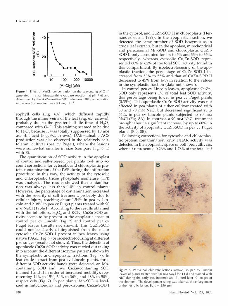
The treatment of pea cv Lincoln plants with 90 mmNaCl also produced the appearance of necrotic leaflesions localized initially on minor veins. These ne-crotic lesions can be easily monitored during the firststages of injury by the bleaching of chlorophylls (Fig.3A, arrows). As the lesions advance, they turn brownand their size becomes visible to the naked eye. Nonecrotic lesions were observed in control plants atany time of the culture period. When these salt-induced necrotic lesions (SINLs) of minor veins werestained with Nitroblue tetratolium (NBT) during thefirst stages of development, a blue/red wine stainingappeared on the cell walls of mesophyll cells (Fig.3B). This staining was due to the production of O2
.�
because it was totally suppressed by SOD (Fig. 3C,arrows), and by 10 mm MnCl2, a highly effectivedismutating catalyst agent of O2
.� (Fig. 4).When O2
.� production, as monitored by NBT, wasfollowed with the degree of severity (judging fromincreased lesion size) of the SINLs (Fig. 5, A–C) inpea leaves, the formation of an O2
.�-generating frontwas observed on the outer edge of the necrotic lesion
(Fig. 5C, arrows). This O2.�-generating front was
clearly distinguishable from the strictly local produc-tion of O2
.�, which takes place during the first stagesof lesion development (Fig. 5A). These results sug-gest that the SINLs advance as the O2
.� productionfront advances, suggesting a key role for O2
.� in thedevelopment of these SINLs.
When stained with the diaminebenzydine (DAB)reagent to locate H2O2, the SINLs also showed ared-brown staining localized on the cell walls of me-
Figure 3. Periveinal chlorotic lesions (arrows) in pea cv Lincolnleaves of plants treated with 90 mM NaCl for 14 d (A). Periveinalchlorotic lesions (arrows) in pea leaves of plants treated with 90 mM
NaCl for 14 d, and stained with NBT in the absence (B) and in thepresence (C) of 100 units mL�1 SOD. Bars � 250 �m.
Figure 2. Effects of salinity on the levels of apoplastic H2O2, lipidperoxidation estimated as apoplastic thiobarbituric acid-reactivesubstances (TBARS), and apoplastic carbonyl (CO)-proteins in pealeaves cv Lincoln (A, B, and C) and cv Puget (D, E, and F). Differencesfrom control values were significant at: (a) P � 0.01 and (b) P �0.001 according to Duncan’s Multiple Range Test.
Salt Stress and Antioxidant System in Pea Leaf Apoplast
Plant Physiol. Vol. 127, 2001 819
sophyll cells (Fig. 6A), which diffused rapidlythrough the minor veins of the leaf (Fig. 6B, arrows),probably due to the greater half-life time of H2O2compared with O2
.�. This staining seemed to be dueto H2O2 because it was totally suppressed by 10 mmascorbic acid (Fig. 6C, arrows). DAB-stainable AOSproduction was also observed in the relatively salt-tolerant cultivar (pea cv Puget), where the lesionswere somewhat smaller in size (compare Fig. 6, Dwith E).
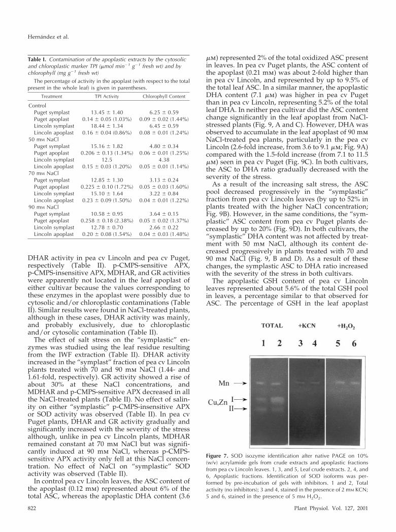
The quantification of SOD activity in the apoplastof control and salt-stressed pea plants took into ac-count corrections for cytosolic and chloroplastic pro-tein contamination of the IWF during the infiltrationprocedure. In this way, the activity of the cytosolicand chloroplastic triose phosphate isomerase (TPI)was analyzed. The results showed that contamina-tion was always less than 1.0% in control plants.However, the percentage of contamination increasedwith the severity of salt treatment, probably due tocellular injury, reaching about 1.54% in pea cv Lin-coln and 2.38% in pea cv Puget plants treated with 90mm NaCl (Table I). According to the results obtainedwith the inhibitors, H2O2 and KCN, CuZn-SOD ac-tivity seems to be present in the apoplastic space ofcontrol pea cv Lincoln (Fig. 7) and control pea cvPuget leaves (results not shown). This CuZn-SODcould not be clearly distinguished from the majorcytosolic CuZn-SOD I present in pea leaves usingnative PAGE (Fig. 7) or isoelectrofocusing at differentpH ranges (results not shown). Thus, the detection ofapoplastic CuZn-SOD activity was carried out takinginto account the different isozyme patterns shown bythe symplastic and apoplastic fractions (Fig. 7). Inleaf crude extract from pea cv Lincoln plants, threedifferent SOD activity bands were detected, an Mn-containing SOD and two CuZn-containing SOD(named I and II in order of increased mobility), rep-resenting 14% to 15%, 34% to 36%, and 48% to 50%,respectively (Fig. 7). In pea plants, Mn-SOD is local-ized in mitochondria and peroxisomes, CuZn-SOD I
in the cytosol, and CuZn-SOD II in chloroplasts (Her-nandez et al., 1999). In the apoplastic fraction, wedetected the same number of SOD isozymes as incrude leaf extracts, but in the apoplast, mitochondrialand peroxisomal Mn-SOD and chloroplastic CuZn-SOD II only accounted for 4% to 5% and 33% to 35%,respectively, whereas cytosolic Cu,Zn-SOD repre-sented 60% to 62% of the total SOD activity found inthis compartment. By isoelectrofocusing of the apo-plastic fraction, the percentage of CuZn-SOD I in-creased from 53% to 55% and that of CuZn-SOD IIdecreased to 45% from 47% in relation to the valuesin the symplastic fraction (data not shown).
In control pea cv Lincoln leaves, apoplastic CuZn-SOD only represents 1% of total leaf SOD activity,this percentage being lower in pea cv Puget plants(0.35%). This apoplastic CuZn-SOD activity was notaffected in pea plants of either cultivar treated with50 and 70 mm NaCl but decreased significantly, to54%, in pea cv Lincoln plants subjected to 90 mmNaCl (Fig. 8A). In contrast, a 90-mm NaCl treatmentbrought about a significant increase, by up to 60%, inthe activity of apoplastic CuZn-SOD in pea cv Pugetplants (Fig. 8B).
Following corrections for cytosolic and chloroplas-tic protein contamination, only DHAR activity wasdetected in the apoplastic space of both pea cultivars,where it represented 0.26% and 1.78% of the total leaf
Figure 5. Periveinal chlorotic lesions (arrows) in pea cv Lincolnleaves of plants treated with 90 mM NaCl for 14 d and stained withNBT during the early (A), intermediate (B), and later (C) stages ofdevelopment. The development rating was taken as the enlargementof the necrotic lesion. Bars � 250 �m.
Figure 4. Effect of MnCl2 concentration on the scavenging of O2.�
generated in a xanthine/xanthine oxidase reaction (at pH 7.6) anddetermined by the SOD-sensitive NBT reduction. NBT concentrationin the reaction medium was 0.1 mg mL�1.
Hernandez et al.
820 Plant Physiol. Vol. 127, 2001
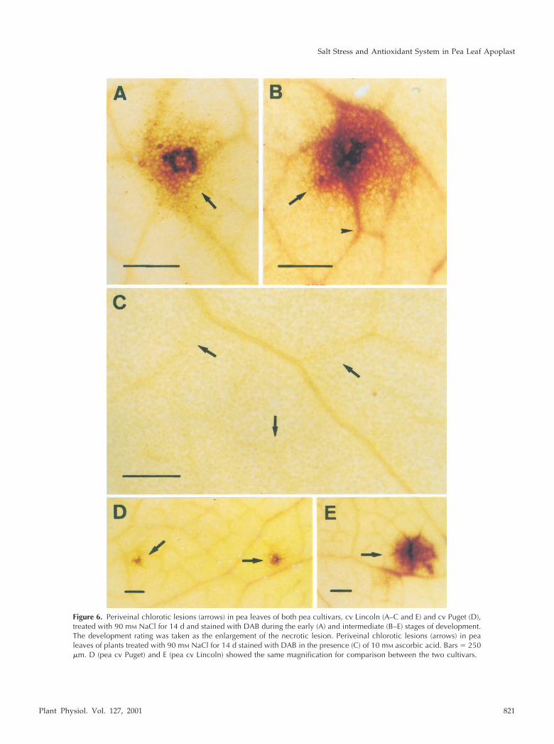
Figure 6. Periveinal chlorotic lesions (arrows) in pea leaves of both pea cultivars, cv Lincoln (A–C and E) and cv Puget (D),treated with 90 mM NaCl for 14 d and stained with DAB during the early (A) and intermediate (B–E) stages of development.The development rating was taken as the enlargement of the necrotic lesion. Periveinal chlorotic lesions (arrows) in pealeaves of plants treated with 90 mM NaCl for 14 d stained with DAB in the presence (C) of 10 mM ascorbic acid. Bars � 250�m. D (pea cv Puget) and E (pea cv Lincoln) showed the same magnification for comparison between the two cultivars.
Salt Stress and Antioxidant System in Pea Leaf Apoplast
Plant Physiol. Vol. 127, 2001 821
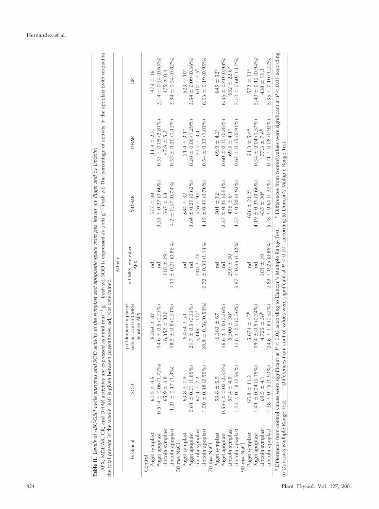
DHAR activity in pea cv Lincoln and pea cv Puget,respectively (Table II). p-CMPS-sensitive APX,p-CMPS-insensitive APX, MDHAR, and GR activitieswere apparently not located in the leaf apoplast ofeither cultivar because the values corresponding tothese enzymes in the apoplast were possibly due tocytosolic and/or chloroplastic contaminations (TableII). Similar results were found in NaCl-treated plants,although in these cases, DHAR activity was mainly,and probably exclusively, due to chloroplasticand/or cytosolic contamination (Table II).
The effect of salt stress on the “symplastic” en-zymes was studied using the leaf residue resultingfrom the IWF extraction (Table II). DHAR activityincreased in the “symplast” fraction of pea cv Lincolnplants treated with 70 and 90 mm NaCl (1.44- and1.61-fold, respectively). GR activity showed a rise ofabout 30% at these NaCl concentrations, andMDHAR and p-CMPS-sensitive APX decreased in allthe NaCl-treated plants (Table II). No effect of salin-ity on either “symplastic” p-CMPS-insensitive APXor SOD activity was observed (Table II). In pea cvPuget plants, DHAR and GR activity gradually andsignificantly increased with the severity of the stressalthough, unlike in pea cv Lincoln plants, MDHARremained constant at 70 mm NaCl but was signifi-cantly induced at 90 mm NaCl, whereas p-CMPS-sensitive APX activity only fell at this NaCl concen-tration. No effect of NaCl on “symplastic” SODactivity was observed (Table II).
In control pea cv Lincoln leaves, the ASC content ofthe apoplast (0.12 mm) represented about 6% of thetotal ASC, whereas the apoplastic DHA content (3.6
�m) represented 2% of the total oxidized ASC presentin leaves. In pea cv Puget plants, the ASC content ofthe apoplast (0.21 mm) was about 2-fold higher thanin pea cv Lincoln, and represented by up to 9.5% ofthe total leaf ASC. In a similar manner, the apoplasticDHA content (7.1 �m) was higher in pea cv Pugetthan in pea cv Lincoln, representing 5.2% of the totalleaf DHA. In neither pea cultivar did the ASC contentchange significantly in the leaf apoplast from NaCl-stressed plants (Fig. 9, A and C). However, DHA wasobserved to accumulate in the leaf apoplast of 90 mmNaCl-treated pea plants, particularly in the pea cvLincoln (2.6-fold increase, from 3.6 to 9.1 �m; Fig. 9A)compared with the 1.5-fold increase (from 7.1 to 11.5�m) seen in pea cv Puget (Fig. 9C). In both cultivars,the ASC to DHA ratio gradually decreased with theseverity of the stress.
As a result of the increasing salt stress, the ASCpool decreased progressively in the “symplastic”fraction from pea cv Lincoln leaves (by up to 52% inplants treated with the higher NaCl concentration;Fig. 9B). However, in the same conditions, the “sym-plastic” ASC content from pea cv Puget plants de-creased by up to 20% (Fig. 9D). In both cultivars, the“symplastic” DHA content was not affected by treat-ment with 50 mm NaCl, although its content de-creased progressively in plants treated with 70 and90 mm NaCl (Fig. 9, B and D). As a result of thesechanges, the symplastic ASC to DHA ratio increasedwith the severity of the stress in both cultivars.
The apoplastic GSH content of pea cv Lincolnleaves represented about 5.6% of the total GSH poolin leaves, a percentage similar to that observed forASC. The percentage of GSH in the leaf apoplast
Figure 7. SOD isozyme identification after native PAGE on 10%(w/v) acrylamide gels from crude extracts and apoplastic fractionsfrom pea cv Lincoln leaves. 1, 3, and 5, Leaf crude extracts. 2, 4, and6, Apoplastic fractions. Identification of SOD isoforms was per-formed by pre-incubation of gels with inhibitors. 1 and 2, Totalactivity (no inhibitors); 3 and 4, stained in the presence of 2 mM KCN;5 and 6, stained in the presence of 5 mM H2O2.
Table I. Contamination of the apoplastic extracts by the cytosolicand chloroplastic marker TPI (�mol min�1 g�1 fresh wt) and bychlorophyll (mg g�1 fresh wt)
The percentage of activity in the apoplast (with respect to the totalpresent in the whole leaf) is given in parentheses.
Treatment TPI Activity Chlorophyll Content
ControlPuget symplast 13.45 � 1.40 6.25 � 0.59Puget apoplast 0.14 � 0.05 (1.03%) 0.09 � 0.02 (1.44%)Lincoln symplast 18.44 � 1.34 6.45 � 0.59Lincoln apoplast 0.16 � 0.04 (0.86%) 0.08 � 0.01 (1.24%)
50 mM NaClPuget symplast 15.16 � 1.82 4.80 � 0.34Puget apoplast 0.206 � 0.13 (1.34%) 0.06 � 0.01 (1.25%)Lincoln symplast 12.5 4.38Lincoln apoplast 0.15 � 0.03 (1.20%) 0.05 � 0.01 (1.14%)
70 mM NaClPuget symplast 12.85 � 1.30 3.13 � 0.24Puget apoplast 0.225 � 0.10 (1.72%) 0.05 � 0.03 (1.60%)Lincoln symplast 15.10 � 1.64 3.22 � 0.84Lincoln apoplast 0.23 � 0.09 (1.50%) 0.04 � 0.01 (1.22%)
90 mM NaClPuget symplast 10.58 � 0.95 3.64 � 0.15Puget apoplast 0.258 � 0.18 (2.38%) 0.05 � 0.02 (1.37%)Lincoln symplast 12.78 � 0.70 2.66 � 0.22Lincoln apoplast 0.20 � 0.08 (1.54%) 0.04 � 0.03 (1.48%)
Hernandez et al.
822 Plant Physiol. Vol. 127, 2001

increased by up to 8.7% of the total GSH present inthe leaf with the 50 mm NaCl treatment, but showedno statistical changes at higher salt concentration(Fig. 10A). The percentage for glutathione oxidizedform (GSSG) in the leaf apoplast from pea cv Lincolnplants was about 18%, and only increased signifi-cantly (2.55-fold) in the leaf apoplast of pea plantstreated with 90 mm NaCl (Fig. 10A). A higher per-centage of GSH was found in the apoplast of pea cvPuget plants, ranging from 16% to 22%, and thevalues were nearly three times higher than in pea cvLincoln (Fig. 10C). The percentage of GSSG in theapoplast of control pea cv Puget plants was lower(only 5.2%) than in pea cv Lincoln. However, at 70and 90 mm NaCl, similar percentages were found inboth cultivars, and a significant increase of GSSG wasfound in the apoplast of pea cv Puget plants treatedwith these salt levels (Fig. 10C).
The GSH content decreased significantly in the“symplast” from both pea cultivars only in plantstreated with 90 mm NaCl (Fig. 10, B and D). How-ever, an increase in the GSSG content was invariablyobserved in the “symplast” from pea cv Lincolnplants subjected to salt stress (as much as 2.52-fold at90 mm NaCl; Fig. 10B), so that the “symplast” GSH toGSSG ratio decreased in NaCl-treated plant of thiscultivar. However, no significant GSSG accumulationwas observed in the symplast from pea cv Pugetplants (Fig. 10D) and the GSH to GSSG ratio slightlydecreased.
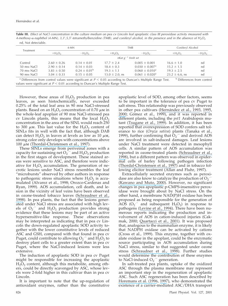
When the effect of salt stress on apoplasticp-CMPS-insensitive (class III) peroxidases was stud-ied, the results showed that such activity in pea cvLincoln plants, measured with both non-physiological(4-methoxy-�-naphthol and 3,3�,5,5�-tetra methylben-zidine) and physiological (coniferyl alcohol) sub-strates, increased in response to salt stress, althoughthese changes were not statistically significant (TableIII). The highest stimulation was attained with 70-and 90-mm NaCl treatments when 4-MN and co-niferyl alcohol, respectively, were used as substrates.Similar results were obtained in the relatively salt-tolerant pea cultivar (data not shown). Isoenzymepatterns of apoplastic class III peroxidase onlyshowed minor (nonsignificant) changes in response
to salt treatment, and the effect seemed to be generalfor all the isoenzymes (results not shown).
DISCUSSION
A comparison of the growth response to salt stressshows that pea cv Lincoln is tolerant to 50 mm NaCl,although it is more sensitive to higher NaCl concen-trations than pea cv Puget. The latter was previouslydesignated as moderately tolerant to NaCl stress,when its growth response was compared with thatreported for other leguminous plants, including dif-ferent pea cultivars (Hernandez et al., 1995, 1999;Gomez et al., 1999).
Evidence for the effects of salt stress-inducingchanges in plant metabolism is well documented(Greenway and Munns, 1980). Salt stress, in additionto the known components of osmotic stress and iontoxicity, is also manifested as an oxidative stress, andall of these contribute to its deleterious effect (Gueta-Dahan et al., 1997). However, ion content and salttolerance are not often correlated, and several studiesindicate that acquisition of salt tolerance may also bea consequence of improving resistance to oxidativestress (Hernandez et al., 1993, 1995, 1999; Gosset etal., 1996; Streb and Feierabend, 1996; Gueta-Dahan etal., 1997; Gomez et al., 1999).
Very few enzymes and metabolites have beenshown unequivocally to be either absent or present inthe apoplast (Dietz, 1996). Moreover, unavoidablecontamination by cytosolic and chloroplastic compo-nents occurs. In this work, TPI was used both as acytosolic and a chloroplastic marker to determine thedegree of contamination of the apoplastic space bycomponents originating in the symplast of plant cellsbecause SODs and the enzymes of the ASC-GSHcycle are mainly accumulated in those subcellularcompartments (Jimenez et al., 1997, 1998a; Ogawa etal., 1997; Foyer and Mullineaux, 1998; Gomez et al.,1999; Hernandez et al., 2000).
In pea apoplast, values found for SOD activityagree with data for Scots pine (Pinus sylvestris) nee-dles and oat and barley leaves (0.1%–2.5% of totalactivity; Streller and Winsgle, 1994; Vanacker et al.,1998a, 1998b). In agreement with reports for apoplas-
Figure 8. Effect of NaCl concentration in theculture medium on the level of apoplastic SODactivity in pea cv Lincoln (A) and cv Puget (B)leaves, after corrections for chloroplastic andcytosolic contaminations. Differences from con-trol values were significant at: (a) P � 0.05according to Duncan’s Multiple Range Test.
Salt Stress and Antioxidant System in Pea Leaf Apoplast
Plant Physiol. Vol. 127, 2001 823
Tabl
eII
.Le
vels
ofA
SC-G
SHcy
cle
enzy
mes
and
SOD
activ
ityin
the
sym
plas
tan
dap
opla
stic
spac
efr
ompe
ale
aves
(cv
Puge
tan
dcv
Linc
oln)
APX
,M
DH
AR
,G
R,
and
DH
AR
activ
ities
are
expr
esse
das
nmol
min
�1
g�1
fres
hw
t.SO
Dis
expr
esse
das
units
g�1
fres
hw
t.Th
epe
rcen
tage
ofac
tivity
inth
eap
opla
st(w
ithre
spec
tto
the
tota
lpr
esen
tin
the
who
lele
af)
isgi
ven
betw
een
pare
nthe
ses.
nd,
Not
dete
rmin
ed.
Act
ivity
Trea
tmen
tSO
Dp-
Chl
orom
ercu
rphe
nyl
sulfo
nic
acid
(p-C
MPS
)-se
nsiti
veA
PX
p-C
MPS
-ins
ensi
tive
APX
MD
HA
RD
HA
RG
R
Con
trol
Puge
tsy
mpl
ast
61.5
�4.
56,
264
�82
nd52
7�
3011
.4�
2.5
473
�16
Puge
tap
opla
st0.
514
�0.
06(1
.72%
)14
.6�
0.5
(0.2
3%)
nd3.
53�
0.27
(0.6
6%)
0.33
�0.
05(2
.81%
)3.
14�
0.34
(0.6
5%)
Linc
oln
sym
plas
t65
.9�
4.8
6,35
2�
320
330
�29
567
�18
47.9
�5.
247
5�
0.4
Linc
oln
apop
last
1.21
�0.
17(1
.8%
)18
.5�
0.8
(0.3
1%)
3.11
�0.
21(0
.86%
)4.
2�
0.37
(0.7
4%)
0.55
�0.
20(1
.12%
)3.
94�
0.54
(0.8
2%)
50m
MN
aCl
Puge
tsy
mpl
ast
63.8
�7.
96,
454
�51
nd58
4�
3221
.4�
3.1a
523
�10
a
Puge
tap
opla
st0.
81�
0.01
(1.8
5%)
21.7
�0.
5(0
.33%
)nd
3.64
�0.
21(0
.62%
)0.
28�
0.06
(1.2
9%)
3.54
�0.
09(0
.36%
)Li
ncol
nsy
mpl
ast
67.1
�2.
25,
445
�11
7a24
0�
2354
6�
9453
.7�
3.1
630
�2.
5b
Linc
oln
apop
last
1.50
�0.
38(2
.59%
)28
.8�
0.56
(0.5
3%)
2.72
�0.
30(1
.13%
)4.
15�
0.41
(0.7
6%)
0.54
�0.
32(1
.03%
)6.
03�
0.19
(0.9
5%)
70m
MN
aCl
Puge
tsy
mpl
ast
53.8
�5.
96,
363
�67
nd50
1�
1269
.9�
4.3c
645
�32
b
Puge
tap
opla
st0.
595
�0.
02(2
.35%
)16
.6�
1.0
(0.2
6%)
nd2.
57�
0.31
(0.5
1%)
0.60
�0.
10(0
.85%
)6.
36�
0.40
(0.9
8%)
Linc
oln
sym
plas
t57
.4�
4.9
5,50
0�
50a
299
�30
496
�6a
69.3
�4.
1a63
2�
22.6
b
Linc
oln
apop
last
1.53
�0.
18(2
.59%
)31
.6�
2.0
(0.5
6%)
3.97
�0.
36(1
.32%
)4.
57�
0.50
(0.9
2%)
0.67
�0.
15(0
.91%
)7.
10�
0.60
(1.1
2%)
90m
MN
aCl
Puge
tsy
mpl
ast
65.8
�11
.25,
674
�45
bnd
626
�21
.2a
21.3
�5.
4a57
2�
33a
Puge
tap
opla
st1.
41�
0.04
(3.1
5%)
19.4
�0.
9(0
.34%
)nd
4.19
�0.
31(0
.66%
)0.
34�
0.04
(1.5
7%)
5.40
�0.
12(0
.94%
)Li
ncol
nsy
mpl
ast
69.5
�4.
14,
725
�58
b30
1�
3943
5�
30a
77.3
�7.
4b43
8�
11.3
Linc
oln
apop
last
1.38
�0.
19(1
.93%
)24
.6�
1.8
(0.5
2%)
2.83
�0.
25(0
.86%
)5.
78�
0.61
(1.3
2%)
0.71
�0.
08(0
.92%
)5.
35�
0.10
(1.2
2%)
aD
iffer
ence
sfr
omco
ntro
lval
ues
wer
esi
gnifi
cant
atP
�0.
05ac
cord
ing
toD
unca
n’s
Mul
tiple
Ran
geTe
st.
bD
iffer
ence
sfr
omco
ntro
lval
ues
wer
esi
gnifi
cant
atP
�0.
01ac
cord
ing
toD
unca
n’s
Mul
tiple
Ran
geTe
st.
cD
iffer
ence
sfr
omco
ntro
lva
lues
wer
esi
gnifi
cant
atP
�0.
001
acco
rdin
gto
Dun
can’
sM
ultip
leR
ange
Test
.
Hernandez et al.
824 Plant Physiol. Vol. 127, 2001

tic CuZn-SOD from spinach leaves (Ogawa et al.,1996, 1997), the apoplastic CuZn-SOD isozyme de-tected in both pea cultivars here seems to be indis-tinguishable from the main cytosolic CuZn-SOD, atleast under our experimental conditions.
The absence of APX activity in the apoplast of bothpea cultivars agrees with what has been also reportedfor different plants (Polle et al., 1990; Duran Carriland Rodriguez-Bujan, 1999). Our results differ fromthose reported by Vanacker et al. (1998), who de-scribed the presence not only of SOD and APX activ-ity in the apoplast of both barley and oat leaves, butalso of MDHAR, DHAR, GR, and catalase. In our peacultivars, similar percentages for these enzymes wereobserved in the apoplast (Table II). However, theseactivities were apparently due to cytosolic and/orchloroplastic contaminations, which ranged from0.86% to 1.03% in control leaves to 1.54% to 2.38% in90 mm NaCl-treated pea plants (cv Lincoln and cvPuget, respectively).
It is necessary to point out that the presence ofDHAR in the apoplast of control pea leaves is stilluncertain in both cultivars, if the total chlorophyllcontent of the apoplastic solution was considered asa symplastic marker instead of TPI activity. This factis also supported by the absence of GR activity in theapoplast. No DHAR activity could be measured inthe apoplastic space of beech (Fagus sylvatica) leaves(Luwe, 1996) and, recently, Morell et al. (1997) havereported that a certain pseudo-DHAR activity foundin plants may be due to side reactions of proteinscontaining redox-active di-Cys sites. However, Foyerand Mullineaux (1998) expressed several consistentarguments in favor of the presence of DHA andDHAR activity in plant tissues. Moreover, the pres-ence of a plasma membrane-associated MDHAR,
which may play a role in the reduction of ASC inboth cytosol and apoplast, has been described (Navasand Gomez-Dıaz, 1995; Berczi and Moller, 1998). Allthese results suggest that the presence of a “DHARenzyme” in the apoplast fraction of pea leaves shouldbe regarded with caution.
The ASC levels in the apoplast of spinach (0.65mm), barley (0.016 mm), and oat leaves (0.01 mm;Takahama and Okini, 1992; Vanacker et al., 1998a,1998b) agree well with the ASC concentration presentin the pea leaf apoplast, which ranged from 0.12 to0.22 mm. A different picture exists regarding GSHbecause little or no GSH has been detected in theapoplast of certain plant cells. This has been found inthe apoplast from Picea abies needles (Polle et al.,1990) and in the apoplast of barley leaves (Vanackeret al., 1998b), where its concentration ranged from 6to 6.5 �m. GSH concentration in pea leaf apoplastwas lower and ranged from 0.8 to 1.2 �m for pea cvLincoln and from 2.0 to 2.4 �m from pea cv Puget.
In response to NaCl stress conditions, the appear-ance of SINLs in the minor veins of 90 mm NaCl-treated plants supports the establishment of an oxi-dative stress situation in the apoplasts of both peacultivars. The NaCl-induced oxidative stress situa-tion is observed as highly localized areas of AOS(O2
.� and H2O2) production in minor veins in bothpea cultivars and is also manifested by increases inlipid peroxidation, H2O2 content, and protein oxida-tion. However, some differences between both peacultivars were found because all the above oxidativeeffects were less pronounced in pea cv Puget (Fig.6D) than in pea cv Lincoln (Fig. 6E) in 90 mm NaClstress conditions, as reflected by the smaller necroticlesions in pea cv Puget plants.
Figure 10. Effect of NaCl concentration in the culture medium onapoplastic and “symplastic” GSH and GSSG contents of pea cvLincoln (A and B) and cv Puget (C and D) leaves. Differences fromcontrol values were significant at: (a) P � 0.05 and (b) P � 0.01according to Duncan’s Multiple Range Test.
Figure 9. Effect of NaCl concentration in the culture medium onapoplastic and “symplastic” ASC and DHA contents of pea cv Lin-coln (A and B) and cv Puget (C and D) leaves. Differences fromcontrol values were significant at: (a) P � 0.05, (b) P � 0.01, and (c)P � 0.001 according to Duncan’s Multiple Range Test.
Salt Stress and Antioxidant System in Pea Leaf Apoplast
Plant Physiol. Vol. 127, 2001 825
However, these areas of H2O2 production in pealeaves, as seen histochemically, never exceeded0.25% of the total leaf area in 90 mm NaCl-stressedplants. Based on an H2O2 concentration of 0.70 �m inthe whole-leaf apoplast of 90 mm NaCl-stressed peacv Lincoln plants, this means that the local H2O2concentration in the area of the SINL would reach 250to 300 �m. This last value for the H2O2 content ofSINLs fits in well with the fact that, although DABcan detect H2O2 in leaves at levels as low as 10 �m,strong color only develops with concentrations above100 �m (Thordal-Christensen et al., 1997).
These SINLs emerge from periveinal zones with acapacity for sustaining net O2
.� and H2O2 productionin the first stages of development. These stained ar-eas were sensitive to ASC, and therefore were indic-ative for H2O2 accumulation. The generation of ne-crotic lesions under NaCl stress resembles the leaf“microbursts” observed by other authors in responseto pathogenic stress situations where H2O2 is accu-mulated (Alvarez et al., 1998; Orozco-Cardenas andRyan, 1999). AOS accumulation, cell death, and le-sion in the vicinity of leaf veins have been observedin ozone-treated tobacco leaves (Schraudner et al.,1998). In pea plants, the fact that the lesions gener-ated under NaCl stress are associated with high lev-els of O2
.� and H2O2 production provides strongevidence that these lesions may be part of an activehypersensitive-like response. These observationsmay be interpreted as indicating that in pea cv Lin-coln the down-regulated apoplastic SOD activity to-gether with the lower constitutive levels of reducedASC and GSH, compared with that found in pea cvPuget, could contribute to allowing O2
.� and H2O2 todestroy plant cells to a greater extent than in pea cvPuget, where the NaCl-induced lesions were lessextensive.
The induction of apoplastic SOD in pea cv Pugetmight be responsible for increasing the apoplasticH2O2 content, although this metabolite, among oth-ers, could be directly scavenged by ASC, whose lev-els were 2-fold higher in this cultivar than in pea cvLincoln.
It is important to note that the up-regulation ofantioxidant enzymes, rather than the constitutive
apoplastic level of SOD, among other factors, seemsto be important in the tolerance of pea cv Puget tosalt stress. This relationship was previously observedin other pea cultivars (Hernandez et al., 1993, 1995,2000; Gomez et al., 1999), and it was reported indifferent plants, including the pst1 Arabidopsis mu-tant (Tsugane et al., 1999). In addition, it has beenreported that overexpression of SOD confers salt tol-erance to rice (Oryza sativa) plants (Tanaka et al.,1999), further confirming that O2
.� and derived AOSare involved in salt-induced damages. Leaf lesionsunder NaCl treatment were detected in mesophyllcells. A similar pattern of AOS accumulation wasreported in ozone-treated plants (Schraudner et al.,1998), but a different pattern was observed in epider-mal cells of barley following pathogen infection(Thordal-Christensen et al., 1997) and in tobacco fol-lowing elicitor treatment (Allan and Fluhr, 1997).
Extracellularly secreted enzymes such as peroxi-dase are also know to catalyze the production of O2
.�
(Kawano and Muto, 2000). However, no significantchanges in pea apoplastic p-CMPS-insensitive perox-idase were brought about by NaCl stress. On theother hand, a membrane NAD(P)H oxidase has beenproposed as being responsible for the generation ofAOS (O2
.� and subsequent H2O2) in response topathogens (Levine et al., 1994). There have been nu-merous reports indicating the production and in-volvement of AOS in cation-induced injuries (Cak-mak, 2000; Quartacci et al., 2001). It was proposedthat, analogous to the mammalian enzyme, it is likelythat NADPH oxidase can be activated by cations(Cross et al., 1999). This enzyme, together with ox-alate oxidase in the apoplast, could be the enzymaticsource participating in AOS accumulation duringNaCl stress, similar to that suggested under ozonestress (Schraudner et al., 1998). Further studieswould determine the contribution of these enzymesto NaCl-induced O2
.� generation.In salt-treated pea plants, uptake of the oxidized
ASC through the plasma membrane may representan important step in the regeneration of apoplasticASC. Such ASC regeneration has been described byHoremans et al. (1996, 1997), who demonstrated theexistence of a carrier-mediated ASC/DHA transport
Table III. Effect of NaCl concentration in the culture medium on pea cv Lincoln leaf apoplastic class III peroxidase activity measured with4-methoxy-�-naphthol (4-MN), 3,3�,5,5�-tetramethylbenzidine (TMB), and coniferyl alcohol, in the presence and in the absence of H2O2
nd, Not detectable.
Treatment4-MN TMB Coniferyl Alcohol
�H2O2 �H2O2 �H2O2 �H2O2 �H2O2 �H2O2
nKat g�1 fresh wt
Control 2.60 � 0.26 0.14 � 0.01 17.7 � 2.4 0.005 � 0.001 16.6 � 1.0 nd50 mM NaCl 2.90 � 0.14 0.14 � 0.03 18.4 � 0.3 0.030 � 0.007a 15.2 � 1.5 nd70 mM NaCl 3.83 � 0.50 0.24 � 0.01b 19.3 � 1.1 0.060 � 0.010a 19.3 � 2.5 nd90 mM NaCl 3.04 � 0.33 0.15 � 0.05 13.0 � 2.0, ns 0.061 � 0.020a 23.2 � 6.6, ns nda Differences from control values were significant at P � 0.05 according to Duncan’s Multiple Range Test. b Differences from control
values were significant at P � 0.01 according to Duncan’s Multiple Range Test.
Hernandez et al.
826 Plant Physiol. Vol. 127, 2001

system that preferentially translocates DHA from theapoplast to the cytosol and that probably exists toreduce this molecule in this cell compartment(Castillo and Greppin, 1988; Luwe et al., 1993; Hore-mans et al., 1997). This mechanism could be occur-ring in both pea cultivars, although its efficacy seemsto be rather low in NaCl-stressed plants, particularlyin pea cv Lincoln. Thus, in both cultivars, the lack ofvariation in total apoplastic ASC under NaCl stresswas accompanied by a decrease in both the apoplas-tic ASC to DHA ratio and the ASC redox state, inboth cases mainly due to the increase in the apoplas-tic DHA content. However, all these changes wereless pronounced in pea cv Puget that in pea cv Lin-coln, in which the increase in apoplastic DHA con-tent was much higher under NaCl stress. Changes inthe redox balance in the apoplastic antioxidant sys-tem have also been reported in response to ozone(Schraudner et al., 1998), pathogens (Thordal-Christensen et al., 1997), and heavy metals (Piqueraset al., 1999).
This suggested mechanism for apoplastic regener-ation fits with the induction found in the symplasticDHAR and GR activities under NaCl stress, showingthat “symplastic” ASC is regenerated using the GSHpool, although in pea cv Puget its regeneration couldalso involve MDHAR activity. This is in agreementwith reports for other pea cultivars (Moran et al.,1994; Hernandez et al., 2000). Thus, it seems thatneither MDHAR nor DHAR limited the ASC contentof the symplast or affected its reduction state, both ofwhich seem to be determined by its rate of synthesisand degradation and/or its transport to other cellcompartments. Similar to our findings in other peacultivars (Hernandez et al., 2000), it should be notedthat symplastic DHA accumulation does not occur ineither pea cv Lincoln or pea cv Puget.
GSH oxidation seems to occur at a higher rate inthe symplast of pea cv Lincoln under NaCl stressbecause in such conditions a progressive increase inthe symplastic GSSG content was observed. This factcould be related to a higher degree of induction ofDHAR than of GR in the symplast, especially at 90mm NaCl. Like ASC, constitutive apoplastic GSHcontent was much more elevated in pea cv Pugetthan in pea cv Lincoln plants, and showed no signif-icant variations at higher salt concentrations, atwhich a decrease in both the GSH to GSSG ratio andthe GSH redox state in this compartment occurred.The presence of GSSG in the apoplast may be theresult of the oxidizing conditions existing in the cellwall, and of the interaction between GSH and thedisulfides from membrane and apoplastic enzymes.This could explain why the GSSG level was higher inthe apoplast than in the symplast. GSH synthesisseems to take place in the cytosol and in chloroplasts(Noctor et al., 1998), but the presence of a GSH trans-porter at the plasma membrane has been described(Jamaı et al., 1996). Therefore, this GSH transporter
would retrieve GSSG for its reduction and recyclingto the cytoplasm (Jamaı et al., 1996) and it seems to besimilar to the previously described ASC/DHA trans-porter (Horemans et al., 1996).
In conclusion, salt stress produced an O2.�- and
H2O2-mediated oxidative stress in the apoplast,which brought about visible NaCl injuries. A dualfunction of AOS, including O2
.� and H2O2, in exac-erbating damage and signaling the activation ofdefense responses has been described, firstly inpathogenesis (Dat et al., 2000), and more recentlyduring plant responses to several abiotic stresses,including salt stress (Gueta-Dahan et al., 1997; Me-neguzzo et al., 1999). Our results are in agreementwith those previously reported, in which a lethallevel of AOS damages the cell, whereas a moderatelevel of AOS enhances the adaptation to salt stress(Hernandez et al., 1995; Gueta-Dahan et al., 1997). Wesuggest that NaCl is an abiotic elicitor of phytopatho-logical and anti-oxidative defenses. In fact, togetherwith AOS production and cell death, overlap in in-duced gene expression after pathogen and NaCl ex-posure has been observed (Knight et al., 1997; De-lumeau et al., 2000). As has been reported underozone stress, it seems that parts of the mechanismsinvolved in plant pathogen responses are also in-duced by NaCl stress, although in this case the ap-pearance of necrotic leaf lesions is associated withsensitivity.
MATERIALS AND METHODS
Plant Material and Growth Conditions
Seeds of pea (Pisum sativum L. cv Lincoln and cv Puget)plants supplied by Arnedo (Navarra, Spain) and SharpesInternational Seeds Ltd. (Sleaford, UK), respectively, weresurface sterilized and germinated in vermiculite with 0.5mm CaSO4 for 1 week. Seedlings were grown in potscontaining aerated nutrient solution in a growth chamber(ASL, Madrid) under optimal conditions for 7 d, as de-scribed by Hernandez et al. (1999). After that time, plantswere grown in a similar medium supplemented by differ-ent NaCl concentrations (0, 50, 70, and 90 mm) for 14 d.
Extraction of the IWF
For the recovery of the IWF, 20 g of pea leaves wassoaked in deionized water, and subsequently vacuum in-filtrated for 3 min at 1.0 KPa and 4°C with 50 mmK-phosphate buffer (pH 6.5) containing 0.2 m KCl and 0.1mm CaCl2. For APX activity, 5 mm sodium ASC was added.Leaves were then quickly dried and centrifuged at 1,000gfor 5 min at 4°C in a 25-mL syringe barrel placed in acentrifuge tube.
To assay enzymatic activities and carbonyl-protein con-tent, the IWF fraction was concentrated about 10-fold usingCentripep 10 tubes (Amicon, Bedford, MA) and prepuri-fied by chromatography on Sephadex G-50 MPD10 col-umns (Pharmacia Biotech AB, Uppsala) equilibrated with
Salt Stress and Antioxidant System in Pea Leaf Apoplast
Plant Physiol. Vol. 127, 2001 827
50 mm K-phosphate buffer (pH 6.5) with or without 5 mmsodium ASC. To determine H2O2 and lipid peroxidation,the IWF fraction obtained after centrifugation at 1,000g wasused directly. For ASC and GSH analysis, leaves wereinfiltrated with cold metaphosphoric acid (2%, w/v), con-taining 0.1 mm bathophenanthroline-disulfonic acid andcentrifuged as above. Apoplastic concentrations for H2O2
and other apoplastic metabolites are given without correc-tion for any dilution due to infiltration of the air spaces.
Contamination by cytoplasmic and chloroplastic constit-uents was assessed by measuring the levels of TPI (Feiera-bend, 1975) and chlorophylls (Arnon, 1949). Glc-6-phosphate dehydrogenase was not used as cytosolicmarker because it has been reported that only 10% of thisenzyme is present in the cytosol fraction from pea plants(Corpas et al., 1998).
Leaf Enzyme Extraction
All operations were performed at 4°C. Leaf residues (2g), which resulted from IWF extraction, were homogenizedwith a mortar and pestle in 4 mL of ice-cold 50 mmK-phosphate buffer, pH 7.8, 0.1 mm EDTA containing 5 mmCys, 1% (w/v) polyvinyl pyrrolidone, 0.1 mm phenylmeth-ylsulfonic fluoride, and 0.2% (v/v) Triton X-100. For APXactivity, 20 mm sodium ASC was added. The “symplastic”homogenate was centrifuged at 14,000g for 20 min and thesupernatant fraction was filtered through Sephadex G-50PD10 columns (Pharmacia Biotech AB), equilibrated withthe same buffer used for the homogenization, with orwithout 5 mm sodium ASC.
Determination of H2O2, Lipid Peroxidation, andProtein Oxidation
The H2O2 concentration in IWF was determined imme-diately after isolation by a peroxidase-coupled assay using4-aminoantipyrine and phenol as donor substrates (Frew etal., 1983). The carbonyl content in oxidatively modifiedproteins was quantified using the 2,4-dinitrophenylhy-drazone assay procedure (Levine et al., 1990). The extent oflipid peroxidation in apoplastic fluid was estimated bydetermining the concentration of substances reacting tothiobarbituric acid-reactive substances (Buege and Aust,1978).
Enzymatic Activities of the ASC-GSH Cycle
APX, DHAR, MDHAR, and GR activities were assayedaccording to previously published protocols, as describedby Jimenez et al. (1997). Enzyme activities were correctedfor nonenzymatic rates and for interfering oxidations(Jimenez et al., 1997). For APX, the oxidation rate of ASCwas estimated between 1 and 60 s after starting the reactionby the addition of H2O2. Correction was made for the lownonenzymatic oxidation of ASC by H2O2. APX was mea-sured in the presence and absence of the specific inhibitorpCMPS (0.5 mm). pCMPS-sensitive APX activity was con-sidered as being due to class I APX (EC 1.11.1.11), whereas
pCMPS-insensitive APX activity was considered as due toclass APX (EC 1.11.1.7), i.e. APX activity due to substrate-unspecific (guaiacol) class III peroxidases (Jimenez et al.,1998b). To determine MDHAR activity, monodehydroascor-bate was generated by the ASC/ASC oxidase system. Therate of monodehydroascorbate-independent NADH oxida-tion (without ASC and ASC oxidase) was subtracted fromthe initial monodehydroascorbate-dependent NADH oxida-tion rate (with ASC and ASC oxidase). For DHAR activity,the reaction rate was corrected for the nonenzymatic reduc-tion of DHA by GSH. A factor of 0.98 was considered toaccount for the small contribution to the absorbance byGSSG. Values due to GR activity were corrected for thesmall, nonenzymatic oxidation of NADPH by GSSG (Jime-nez et al., 1997). The values of the ASC-GSH cycle enzymesdetermined in the apoplast fractions were corrected by thepercentage of contamination caused by the cytosolic andchloroplastic marker, TPI.
Determination of the ASC/GSH Pool
Apoplastic ASC and DHA and apoplastic GSH andGSSG contents were directly determined in the IWF byHPLC, according to Jimenez et al. (1997). Reduced andoxidized ASC and GSH were extracted with 2% (w/v)metaphosphoric acid from the leaf material resulting fromthe IWF extraction, and measured as described by Jimenezet al. (1997).
SOD Activity
Total SOD activity was assayed, in the absence and in thepresence of KCN, by the ferricytochrome c method usingxanthine/xanthine oxidase as the source of O2
.� radicals(McCord and Fridovich, 1969). SOD activity was correctedby the percentage of contamination caused by the cytosolicand chloroplastic marker, TPI. To separate SOD isozymes,non-denaturing PAGE and isoelectrofocusing were per-formed on 10% and 6% (w/v) acrylamide gels, respec-tively, using a mini protean II dual slab cell (Bio-Rad,Hercules, CA). The range of ampholytes (Pharmacia) usedwere: pH 3.5 to 5, pH 4.2 to 5.4, and pH 3.5 to 7.0. Sampleswere prefocused at 150 V for 30 min, and then focused at250 V for 1 h 30 min. SOD isozymes were localized by thephotochemical method of Weissiger and Fridovich (1973).Isoenzyme identification was performed by selective inhi-bition with KCN or H2O2 (Hernandez et al., 1999). Thepercentage of activity for the different SODs was quantifiedby recording the transmittance of gels in a CS-9000 densi-tometer (Shimadzu, Kyoto).
Class III Peroxidase Activity and Isoenzyme Analysis
Class III peroxidase activity in IWF was determined inassays containing 50 mm Tris-acetate buffer (pH 5.0) and0.5 mm H2O2, using the following electron donors: 0.1 mmconiferyl alcohol (�262 � 9,600 m�1 cm�1), 0.1 mg mL�1
tetramethylbenzidine-HCl (�652 � 39,000 m�1 cm�1), and1.0 mm 4-methoxy-�-naphtol (�595 � 21,600 m�1 cm�1; Ros
Hernandez et al.
828 Plant Physiol. Vol. 127, 2001
Barcelo, 1998). The reaction was initiated by adding en-zyme. Controls were carried out in the absence of H2O2.
Class III peroxidase isoenzymes were separated by iso-electric focusing in 3.5 to 10.0 pH gradients as described byFerrer and Ros Barcelo (1999), and stained with 4-methoxy-�-naphtol for 1 h at 25°C using a reaction medium identicalto that described above. Controls were carried out in theabsence of H2O2.
Determination of Protein Content
Protein content was estimated according to Bradford(1976) using bovine serum albumin as standard.
Histochemical Detection of H2O2 and O2.� in
Pea Leaves
The histochemical detection of H2O2 and O2.� in pea
leaves was performed as described by Schraudner et al.(1998) with minor modifications. In the case of H2O2 weused an endogenous peroxidase-dependent in situ histo-chemical staining, in which leaf quarters were vacuuminfiltrated with 0.1 mg mL�1 3,3�-diaminobenzidine in 50mm Tris-acetate buffer (pH 5.0) and incubated at 25°C inthe dark for 24 h. Controls were performed in the presenceof 10 mm ascorbic acid.
The histochemical detection of O2.� was performed by
infiltrating leaf quarters directly with 0.1 mg mL�1 NBT in25 mm K-HEPES [4-(2-hydroxyethyl)-1-piperazineethane-sulfonic acid] buffer (pH 7.6) and incubating at 25°C in thedark for 2 h. Controls in the presence of both 10 mm MnCl2and/or 100 units mL�1 SOD were performed. The O2
.�-removing compounds (MnCl2 and SOD) were directlyadded to the infiltration buffer.
In both cases, leaf quarters were rinsed in 80% (v/v)ethanol for 10 min at 70°C, mounted in lactic acid:phenol:water (1:1:1, v/v), and photographed directly using an SZX12 microscope (Olympus. Kyoto).
Received February 21, 2001; returned for revision April 28,2001; accepted July 17, 2001.
LITERATURE CITED
Allan AC, Fluhr R (1997) Two distinct sources of elicitedreactive oxygen species in tobacco epidermal cells. PlantCell 9: 1559–1572
Alvarez ME, Pennell RI, Meijer PJ, Ishikawa A, DixonRA, Lamb C (1998) Reactive oxygen intermediates me-diate a systemic signal network in the establishment ofplant immunity. Cell 92: 773–784
Arnon DI (1949) Copper enzymes in isolated chloroplasts:polyphenoloxidase in Beta vulgaris. Plant Physiol 24:1–15
Berczi A, Moller IM (1998) NADH-monodehydroascorbateoxidoreductase is one of the redox enzymes in spinachleaf plasma membranes. Plant Physiol 116: 1029–1036
Blinda A, Koch B, Ramanjulu S, Dietz KJ (1997) De novosynthesis and accumulation of apoplastic proteins in
leaves of heavy metal-exposed barley seedlings. PlantCell Environ 20: 969–981
Bohnert HJ, Jensen RG (1996) Metabolic engineering forincreased salt tolerance: the next step. Aust J PlantPhysiol 23: 661–667
Bradford MM (1976) A rapid and sensitive method for thequantitation of microgram quantities of protein utilizingthe principle of protein-dye binding. Anal Biochem 72:248–254
Buege JA, Aust SD (1978) Microsomal lipid peroxidation.Methods Enzymol 52: 302–310
Cakmak I (2000) Possible roles of zinc in protecting plantcells from damage by reactive oxygen species. New Phy-tol 146: 185–205
Castillo FJ, Greppin H (1988) Extracellular ascorbic acidand enzyme activities related to ascorbic acid metabo-lism in Sedum album L. after ozone exposure. EnvironExp Bot 28: 231–238
Corpas FJ, Barroso JB, Sandalio LM, Distefano S, PalmaJM, Lupianez JA, del Rıo LA (1998) A dehydrogenase-mediated recycling system of NADPH in plant peroxi-somes. Biochem J 330: 777–784
Cross AR, Erichson R, Ellis BA, Curnutte JT (1999) Spon-taneous activation of NADPH oxidase in cell-free sys-tem: unexpected multiple effects of magnesium ion con-centrations. Biochem J 338: 229–233
Dat J, Vandenabeele S, Vranova E, Van Montagu M, InzeD, Van Breusegem F (2000) Dual action of the activeoxygen species during plant stress responses. Cell MolLife Sci 57: 779–795
del Rıo LA, Pastori GM, Palma JM, Sandalio LM, SevillaF, Corpas FJ, Jimenez A, Lopez-Huertas E, HernandezJA (1998) The activated oxygen role of peroxisomes insenescence. Plant Physiol 116: 1195–1200
Delumeau O, Morere-Le Paven MC, Montrichard F,Laval-Martin DL (2000) Effects of short-term NaCl stresson calmodulin transcript levels and calmodulin-dependent NAD kinase activity in two species of tomato.Plant Cell Environ 23: 329–336
Dietz KJ (1996) Functions and responses of the leaf apo-plast under stress. Prog Bot 58: 221–254
Duran-Carril MV, Rodriguez Bujan C (1998) Antioxidantsystems in the leaf apoplast compartment of Pinus pin-aster Ait. and Pinus radiata D. Don. plants exposed to SO2.Ann Appl Biol 133: 455–466
Feierabend J (1975) Developmental studies on microbodiesin wheat leaves: III. On the photocontrol of microbodydevelopment. Planta 123: 63–77
Ferrer MA, Ros Barcelo A (1999) Differential effects ofnitric oxide on peroxidase and H2O2 production by thexylem of Zinnia elegans. Plant Cell Environ 22: 891–897
Foyer CH, Mullineaux P (1998) The presence of dehy-droascorbate and dehydroascorbate reductase in planttissues. FEBS Lett 425: 528–529
Frew J, Jones P, Scholes G (1983) Spectophotometric de-termination of hydrogen peroxide and organic hydroper-oxides at low concentrations in aqueous solution. AnalChim Acta 155: 130–150
Gomez JM, Hernandez JA, Jimenez A, del Rıo LA, SevillaF (1999) Differential response of antioxidative enzymes
Salt Stress and Antioxidant System in Pea Leaf Apoplast
Plant Physiol. Vol. 127, 2001 829
of chloroplasts and mitochondria to long-term NaClstress of pea plants. Free Radic Res 31: S11–S18
Gosset DR, Banks SW, Millhollon EP, Lucas MC (1996)Antioxidant response to NaCl stress in a control and anNaCl-tolerant cotton cell line grown in the presence ofparaquat, buthionine sulfoximine, and exogenous gluta-thione. Plant Physiol 112: 803–809
Greenway H, Munns R (1980) Mechanisms of salt toler-ance innonhalophytes. Annu Rev Plant Physiol 31:149–190
Gueta-Dahan Y, Yaniv Z, Zilinskas BA, Ben-Hayyim G(1997) Salt and oxidative stress: similar and specific re-sponses and their relation to salt tolerance in Citrus.Planta 203: 460–469
Hernandez JA, Campillo A, Jimenez A, Alarcon JJ,Sevilla F (1999) Response of antioxidant systems andleaf water relations to NaCl stress in pea plants. NewPhytol 141: 241–251
Hernandez JA, Corpas FJ, Gomez M, del Rıo LA, SevillaF (1993) Salt-induced oxidative stress mediated by acti-vated oxygen species in pea leaf mitochondria. PhysiolPlant 89: 103–110
Hernandez JA, Jimenez A, Mullineaux PM, Sevilla F(2000) Tolerance of pea (Pisum sativum L.) to long-termsalt stress is associated with induction of antioxidantdefenses. Plant Cell Environ 23: 853–862
Hernandez JA, Olmos E, Corpas FJ, Sevilla F, del Rıo LA(1995) Salt-induced oxidative stress in chloroplast of peaplants. Plant Sci 105: 151–167
Hernandez JA, Talavera JM, Martınez-Gomez P, DicentaF, Sevilla F (2001) Response of antioxidant enzymes toplum pox virus in two apricot cultivars. Physiol Plant111: 313–321
Horemans N, Asard H, Caubergs RJ (1996) Transport ofascorbate into plasma membrane vesicles of Phaseolusvulgaris L. Protoplasma 194: 177–185
Horemans N, Asard H, Caubergs RJ (1997) The ascorbatecarrier of higher plant plasma membranes preferentiallytranslocates the fully oxidized (dehidroascorbate) mole-cule. Plant Physiol 114: 1247–1253
Jamaı A, Tommasini R, Martinoia E, Delrot S (1996) Char-acterization of glutathione uptake in broad bean leafprotoplasts. Plant Physiol 111: 1145–1152
Jimenez A, Hernandez JA, del Rıo LA, Sevilla F (1997)Evidence for the presence of the ascorbate-glutathionecycle in mitochondria and peroxisomes of pea (Pisumsativum L.) leaves. Plant Physiol 114: 275–284
Jimenez A, Hernandez JA, Pastori GM, del Rıo LA,Sevilla F (1998a) On the role of the ascorbate-glutathionecycle of mitochondria and peroxisomes in the senescenceof pea leaves. Plant Physiol 118: 1327–1335
Jimenez A, Hernandez JA, Ros Barcelo A, Sandalio LM,del Rıo LA, Sevilla F (1998b) Characterization of mito-chondrial and peroxisomal ascorbate peroxidase of pealeaves. Physiol Plant 104: 687–692
Kawano T, Muto S (2000) Mechanism of peroxidase ac-tions for salicylic acid-induced generation of active oxy-gen species and an increase in cytosolic calcium in to-bacco cell suspension culture. J Exp Bot 51: 685–693
Knight H, Trewavas AJ, Knight MR (1997) Calcium sig-naling in Arabidopsis thaliana responding to drought andsalinity. Plant Cell 12: 1067–1078
Levine A, Tenhaken R, Dixon R, Lamb C (1994) H2O2
from the oxidative burst orchestrates the plant hypersen-sitive disease resistance response. Cell 79: 583–593
Levine RL, Garland D, Oliver CN, Amici A, Climent I,Lenz AG, Ahn B, Shaltiel S, Stadtman ER (1990) Deter-mination of carbonyl content in oxidatively modifiedproteins. Methods Enzymol 186: 464–478
Luwe M (1996) Antioxidants in the apoplast and symplastof beech (Fagus sylvatica L.) leaves: seasonal variationsand responses to changing ozone concentration in air.Plant Cell Environ 19: 321–328
Luwe M, Takahama U, Heber U (1993) Role of ascorbate indetoxifying ozone in the apoplast of spinach (Spinaceaoleracea L.) leaves. Plant Physiol 101: 969–976
McCord JM, Fridovich I (1969) Superoxide dismutase: anenzymic function for erythrocuprein. J Biol Biochem 244:6049–6055
Meneguzzo S, Navari-Izzo F, Izzo R (1999) Antioxidativeresponses of shoots and roots of wheat to increasingNaCl concentrations. J Plant Physiol 155: 274–280
Moran JF, Becana M, Iturbe-Ormaetxe I, Freschilla S,Klucas RV, Aparicio-Tejo P (1994) Drought induces ox-idative stress in pea plants. Planta 194: 346–352
Morell S, Follmann H, De Tullio M, Haberlein I (1997)Dehydroascorbate and dehydroascorbate reductase arephantom indicators of oxidative stress in plants. FEBSLett 414: 567–570
Navas P, Gomez-Dıaz C (1995) Ascorbate free radical andits role in growth control. Protoplasma 184: 8–13
Noctor G, Arisi ACM, Jouanin L, Kunert KJ, RennenbergH, Foyer CH (1998) Glutathione biosynthesis, metabo-lism and relationship to stress tolerance explored intransformed plants. J Exp Bot 49: 623–647
Ogawa K, Kanematsu S, Asada K (1996) Intra- and extra-cellular localization of “cytosolic” Cu,Zn-superoxide dis-mutase in spinach leaf and hypocotyl. Plant Cell Physiol37: 790–799
Ogawa K, Kanematshu S, Asada K (1997) Generation ofsuperoxide anion and localization of CuZn-superoxidedismutase in the vascular tissue of spinach hypocotyls:their association with lignification. Plant Cell Physiol 38:1118–1126
Orozco-Cardenas M, Ryan CA (1999) Hydrogen peroxideis generated systemically in plant leaves by woundingand systemic via the octadecanoid pathway. Proc NatlAcad Sci USA 96: 6553–6557
Piqueras A, Olmos E, Martınez-Solano JR, Hellın E (1999)Cd-induced oxidative burst in tobacco BY2 cells: timecurse, subcellular location and antioxidant response.Free Radic Res 31: S33–S38
Polle A, Chakrabarti K, Schumann W, Rennenbreg H(1990) Composition and properties of hydrogen peroxidedecomposing systems in extracellular and total extractsfrom needles of Norway Spruce (Picea abies L., Karst.).Plant Physiol 94: 312–319
Quartacci MF, Cosi E, Navari-Izzo F (2001) Lipids and
Hernandez et al.
830 Plant Physiol. Vol. 127, 2001
NADPH-dependent superoxide production in plasmamembrane vesicles from roots of wheat grown undercopper deficiency or excess. J Exp Bot 52: 77–84
Ranieri A, D’Urso G, Nali C, Lorenzini G, Soldatini GF(1996) Ozone stimulates apoplastic antioxidant systemsin pumpkin leaves. Physiol Plant 97: 381–387
Ros Barcelo A (1998) The generation of H2O2 in the xylemof Zinnia elegans is mediated by an NADPH-oxidase-likeenzyme. Planta 207: 207–216
Savoure A, Thorin D, Davey M, Hua XJ, Mauro S, VanMontagu M, Inze D, Verbruggen N (1999) NaCl andCuZnSO4 treatments trigger distinct oxidative defensemechanism in Nicotiana plumbaginifolia L. Plant Cell En-viron 22: 387–396
Schraudner M, Moeder W, Wiese C, Van Camp W, InzeD, Langebartels C, Sandermann H Jr (1998) Ozone-induced oxidative burst in the ozone biomonitor plant,tobacco Bel W3. Plant J 16: 235–245
Streb P, Feierabend J (1996) Oxidative stress-responsesaccompanying photoinactivation of catalase in NaCl-treated rye leaves. Bot Acta 109: 125–132
Streller S, Wingsle G (1994) Pinus sylvestris L. needlescontain extracellular CuZn-superoxide dismutase. Planta192: 195–201
Takahama U, Okini T (1992) Regulation of peroxidase-dependent oxidation of phenolic in the apoplast of spin-ach leaves by ascorbate. Plant Cell Physiol 33: 379–387
Tanaka Y, Hibino T, Hayashi Y, Tanaka A, Kishitani S,Takabe T, Yokota S, Takabe T (1999) Salt tolerance oftransgenic rice overexpresing yeast mitochondrial Mn-SOD in chloroplasts. Plant Sci 148: 131–138
Thordal-Christensen H, Zhang Z, Wei Y, Collinge DB(1997) Subcellular localization of H2O2 in plants: H2O2
accumulation in papillae and hypersensitive responseduring the barley-powdery mildew interaction. Plant J 11:1187–1194
Tsugane K, Kobayashi K, Niwa Y, Ohba Y, Wada KA(1999) Recessive Arabidopsis mutant that grows photo-autotrophically under salt stress shows enhanced activeoxygen detoxification. Plant Cell 11: 1195–1206
Vanacker H, Carver TLW, Foyer CH (1998a) Pathogen-induced changes in the antioxidant status of the apoplastin barley leaves. Plant Physiol 117: 1103–1114
Vanacker H, Harbinson J, Carver TLW, Foyer CH (1998b)Antioxidant defenses of the apoplast. Protoplasma 205:129–140
Weissiger RA, Fridovich I (1973) Superoxide dismutase:organelle specifity. J Biol Chem 248: 3582–3592
Salt Stress and Antioxidant System in Pea Leaf Apoplast
Plant Physiol. Vol. 127, 2001 831
Copyright © 2022 FDOKUMEN