![Effects of recombinant drug-specific single chain antibody Fv fragment on [ 3H]-desipramine distribution in rats](https://static.fdokumen.com/doc/165x107/631cd3f3d5372c006e04a0a0/effects-of-recombinant-drug-specific-single-chain-antibody-fv-fragment-on-3h-desipramine.jpg)
Selection of Beet Necrotic Yellow Vein Virus Specific Single-chain Fv Antibodies from a...
10
European Journal of Plant Pathology 105: 147–156, 1999. © 1999 Kluwer Academic Publishers. Printed in the Netherlands. Selection of beet necrotic yellow vein virus specific single-chain Fv antibodies from a semi-synthetic combinatorial antibody library Remko A. Griep, Charlotte van Twisk and Arjen Schots * Laboratory for Monoclonal Antibodies, Wageningen Agricultural University, P.O. Box 8123, 6700 ES Wageningen, the Netherlands. * Author for correspondence (E-mail: [email protected]; Fax: +31317-485267) Accepted 24 November 1998 Key words: BNYVV, ELISA, phage-antibodies, phage display, ScFv, ScFv-AP/S, sugar beet Abstract Methods for the generation of monoclonal antibodies against plant viruses are limited because current hybridoma techniques do not allow efficient exploitation of the immune repertoire. Moreover, the immunization procedures often lead to a bias towards an immunodominant contaminant in the immunogen preparation and not to the plant virus itself. The selection of six different single-chain antibody variable fragments (scFv) against beet necrotic yellow vein virus from a semi-synthetic human combinatorial antibody library showed the feasibility of the phage display system. No bias towards minor contaminants in the purified virus preparation was observed in ELISA, as all the selected scFvs reacted only with beet necrotic yellow vein virus infected plant homogenates. In addition, two of the isolated beet necrotic yellow vein virus-specific scFvs could be produced in E. coli as a scFv fusion protein with alkaline phosphatase, and were applied in ELISA as specific ready to use antibody-enzyme conjugates. Because of their specificity, these antibodies have potential to be used as reagents in sensitive diagnostic assays for routine testing for beet necrotic yellow vein virus in sugar beets. Abbreviations: BNYVV – beet necrotic yellow vein virus; MAbs – Monoclonal Antibodies; PAbs – Polyclonal antibodies; PhAbs – phage-antibodies; scFv – single-chain variable fragment; scFv-AP/S – single-chain variable fragment genetically fused to alkaline phosphatase. Introduction Most diagnostic assays for plant viruses utilize poly- clonal antibodies (PAbs) or monoclonal antibodies (MAbs). MAbs do have some advantages over PAbs. They are homogeneous and well defined reagents which allow standardization of assays between dif- ferent laboratories (Regenmortel, 1986; Regenmortel, 1990). Due to the fact that MAbs recognize single epi- topes they may give less cross-reactions, discriminate between viral strains (Torrance et al., 1986; Torrance, 1995) and MAbs directed against conserved epitopes may allow grouping of virus isolates for epidemiolog- ical and taxonomical studies. It is difficult to raise MAbs to some plant viruses because minor contaminants in the isolated virus frac- tion exhibit immunodominance (George and Converse, 1988). This was shown to be the case for beet necrotic yellow vein virus (BNYVV), a furovirus causing rhizomania (Tamada and Baba, 1973). During purifi- cation the virus aggregates with host cell constituents (Putz and Kuszala, 1978) and only a few MAbs were obtained reacting specifically with BNYVV, while the majority of the MAbs reacted with healthy plant anti- gens. It is noteworthy that polyclonal antisera collected at the time of spleen removal had high virus-specific and low anti-healthy plant sap titers. The same phe- nomenon has been observed in developing MAbs to DISK EJPP931 PIPS 201113
-
Upload
independent -
Category
Documents
-
view
1 -
download
0
Transcript of Selection of Beet Necrotic Yellow Vein Virus Specific Single-chain Fv Antibodies from a...

European Journal of Plant Pathology105:147–156, 1999.© 1999Kluwer Academic Publishers. Printed in the Netherlands.
Selection of beet necrotic yellow vein virus specific single-chain Fv antibodiesfrom a semi-synthetic combinatorial antibody library
Remko A. Griep, Charlotte van Twisk and Arjen Schots∗
Laboratory for Monoclonal Antibodies, Wageningen Agricultural University, P.O. Box 8123, 6700 ES Wageningen,the Netherlands.∗Author for correspondence (E-mail: [email protected]; Fax: +31 317-485267)
Accepted 24 November 1998
Key words:BNYVV, ELISA, phage-antibodies, phage display, ScFv, ScFv-AP/S, sugar beet
Abstract
Methods for the generation of monoclonal antibodies against plant viruses are limited because current hybridomatechniques do not allow efficient exploitation of the immune repertoire. Moreover, the immunization proceduresoften lead to a bias towards an immunodominant contaminant in the immunogen preparation and not to the plantvirus itself. The selection of six different single-chain antibody variable fragments (scFv) against beet necroticyellow vein virus from a semi-synthetic human combinatorial antibody library showed the feasibility of the phagedisplay system. No bias towards minor contaminants in the purified virus preparation was observed in ELISA, as allthe selected scFvs reacted only with beet necrotic yellow vein virus infected plant homogenates. In addition, two ofthe isolated beet necrotic yellow vein virus-specific scFvs could be produced inE. colias a scFv fusion protein withalkaline phosphatase, and were applied in ELISA as specific ready to use antibody-enzyme conjugates. Becauseof their specificity, these antibodies have potential to be used as reagents in sensitive diagnostic assays for routinetesting for beet necrotic yellow vein virus in sugar beets.
Abbreviations:BNYVV – beet necrotic yellow vein virus; MAbs – Monoclonal Antibodies; PAbs – Polyclonalantibodies; PhAbs – phage-antibodies; scFv – single-chain variable fragment; scFv-AP/S – single-chain variablefragment genetically fused to alkaline phosphatase.
Introduction
Most diagnostic assays for plant viruses utilize poly-clonal antibodies (PAbs) or monoclonal antibodies(MAbs). MAbs do have some advantages over PAbs.They are homogeneous and well defined reagentswhich allow standardization of assays between dif-ferent laboratories (Regenmortel, 1986; Regenmortel,1990). Due to the fact that MAbs recognize single epi-topes they may give less cross-reactions, discriminatebetween viral strains (Torrance et al., 1986; Torrance,1995) and MAbs directed against conserved epitopesmay allow grouping of virus isolates for epidemiolog-ical and taxonomical studies.
It is difficult to raise MAbs to some plant virusesbecause minor contaminants in the isolated virus frac-tion exhibit immunodominance (George and Converse,1988). This was shown to be the case for beet necroticyellow vein virus (BNYVV), a furovirus causingrhizomania (Tamada and Baba, 1973). During purifi-cation the virus aggregates with host cell constituents(Putz and Kuszala, 1978) and only a few MAbs wereobtained reacting specifically with BNYVV, while themajority of the MAbs reacted with healthy plant anti-gens. It is noteworthy that polyclonal antisera collectedat the time of spleen removal had high virus-specificand low anti-healthy plant sap titers. The same phe-nomenon has been observed in developing MAbs to
DISK EJPP931 PIPS 201113
148
cauliflower mosaic virus (George and Converse, 1988)and blueberry red ringspot virus (Hepp and Converse,1986).
Various methods have been described to reduce theimmune response to plant contaminants to enhancethe yield of virus-specific MAbs. For instance, theinduction of immunological tolerance in neonatal mice(Hsu et al., 1990), the use of immunosuppressiveagents like cyclophosphamide (Matthew and Sandrock,1987), or use of antigen based B-cell selection systems(Parks et al., 1979). The common drawback of thesemethods is that the conditions have to be establishedfor each virus.
Current hybridoma methods (Kohler and Milstein,1975) for the generation of MAbs against plantpathogens do not satisfactorily exploit the immunerepertoire. While the antibody repertoire is estimatedto consist of over 108 different antibodies, only a fewthousand different hybridoma clones are obtained, ofwhich on average less then 1% produce antigen bindingantibodies. Thus, the chance that a useful hybridomaderived MAb will be obtained is low. This diversity and
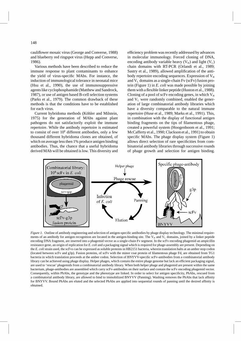
Figure 1. Outline of antibody engineering and selection of antigen-specific antibodies by phage display technology. The minimal require-ments of an antibody for antigen recognition are located in the antigen-binding site. The VH and VL domains, joined by a linker peptideencoding DNA fragment, are inserted into a phagemid vector as a single-chain Fv segment. In the scFv encoding phagemid an ampicillinresistance gene, an origin of replication forE. coliand a packaging signal which is required for phage-assembly are present. Depending ontheE. colistrain used, the scFvs can be expressed as soluble proteins in HB2151 bacteria, wherein translation halts at an amber stop codon(located between scFv and g3p). Fusion proteins, of scFv with the minor coat protein of filamentous phage Fd, are obtained from TG1bacteria in which translation proceeds at the amber codon. Selection of BNYVV-specific scFv-antibodies from a combinatorial antibodylibrary can be achieved using phage display. Helper phages, which contain the entire phage genome but lack an efficient packaging signal,are used to ‘rescue’ phagemids from a combinatorial antibody library. When both helper phage and phagemid are present within the samebacterium, phage-antibodies are assembled which carry scFv-antibodies on their surface and contain the scFv encoding phagemid vector.Consequently, within PhAbs, the genotype and the phenotype are linked. In order to select for antigen specificity, PhAbs, rescued froma combinatorial antibody library, are allowed to bind to immobilized BNYVV (Panning). Washing removes the PhAbs that lack affinityfor BNYVV. Bound PhAbs are eluted and the selected PhAbs are applied into sequential rounds of panning until the desired affinity isobtained.
efficiency problem was recently addressed by advancesin molecular immunology. Forced cloning of DNA,encoding antibody variable heavy (VH) and light (VL)chain domains with RT-PCR (Orlandi et al., 1989;Sastry et al., 1989), allowed amplification of the anti-body repertoire encoding sequences. Expression of VH
and VL domains as a single-chain Fv (scFv) fusion pro-tein (Figure 1) inE. coli was made possible by joiningthem with a flexible linker peptide (Huston et al., 1988).Cloning of a pool of scFv encoding genes, in which VH
and VL were randomly combined, enabled the gener-ation of large combinatorial antibody libraries whichhave a diversity comparable to the natural immunerepertoire (Huse et al., 1989; Marks et al., 1991). This,in combination with the display of functional antigenbinding fragments on the tips of filamentous phage,created a powerful system (Hoogenboom et al., 1991;McCafferty et al., 1990; Clackson et al., 1991) to obtainspecific MAbs. The phage display system (Figure 1)allows direct selection of rare specificities from com-binatorial antibody libraries through successive roundsof phage growth and selection for antigen binding.

149
For instance, Bradbury et al. (1993) showed the fea-sibility of this technique by selecting antigen bind-ing phages from a pool of non-binding phages, evenat a ratio of one binding in 109 irrelevant phages. Inaddition, the phage display system may also prove tobe a more universal method to obtain specific MAbs,since the antibody specificities are not biased towardsimmunodominant epitopes.
To study the versatility of the phage display system,we have applied it to BNYVV. This virus was chosenas a model system because it is a classic example ofthe difficulties outlined in raising specific MAbs. Weexamined the feasibility of the phage display system inisolating a diverse repertoire of BNYVV-specific anti-body fragments from a semi-synthetic combinatorialantibody library (Nissim et al., 1994).
Materials and methods
Bacterial strains. E. colistrains used for the isola-tion of recombinant antibodies, were TG1 (K12,1(lac-pro), supE, thi, hsdD5/F′traD36, proA+B+, lacIq,lacZ1M15) for selection of specific phage-antibodiesand HB2151 (K12,ara, 1lac-pro), thi/F′/proA+B+,lacIqZ1M15) for expression of soluble scFv-antibodyfragments. HB2151 bacteria were grown at 30◦C in2TY containing 100µg ml−1 ampicillin and 2% (w/v)glucose (Sambrook et al., 1990). When the OD600
reached 0.5, the bacteria were pelleted and the super-natant was discarded. The bacteria were resuspendedin 2TY+AMP (100µg/ml), induced with 1 mM IPTGand grown for 18 h at 16◦C.
Purification of beet necrotic yellow vein virions.Beetnecrotic yellow vein virus (Dutch isolate) was purifiedfrom mechanically inoculatedChenopodium quinoaaccording to a slightly modified protocol from Putzand Kuszala (1978). InfectedC. quinoaleaves (200 g)were homogenized in a mixture of 400 ml CCl4 and800 ml extraction buffer (0.1 M Tris/HCl buffer pH 9containing 0.14 M NaCl; 5% (v/v) ethanol) and 2%(w/v) polyvinylpyrrolidon. The mixture was stirred for15 min at room temperature (RT) and then centrifugedfor 15 min at 5,000× g (GSA rotor, Sorvall). To thecollected supernatant, 5% (w/v) PEG-8000 was addedand stirred at RT for 15 min and subsequently at 4◦C for90 min. The virions were pelleted at 16,000× g (GSA,Sorvall) for 20 min. After resuspending the pellets in100 ml extraction buffer and stirring overnight at 4◦C,
sucrose was added to 3.3% (w/v) and the mixture wascentrifuged at 27,500× g for 20 min (SW28, Beckman)to remove large aggregates. Subsequently the virionswere pelleted at 110,000× g (SW28, Beckman) for 2 hthrough a 3 cm layer of 20% (w/v) sucrose in extractionbuffer. The pellets were resuspended in 50 ml extrac-tion buffer and, after stirring for 30 min at RT, cen-trifuged for 10 min at 1,600× g to remove insolubleprecipitates. Half of the supernatant was adjusted toa density of 1.1 g/cm3, half to 1.3 g/cm3 with CsCl2.The two halves were layered 1 : 1 and centrifuged at154,000× g (SW41Ti, Beckman) for 18 h. The virionscontaining band was collected and dialyzed overnightagainst 0.1 M Tris/HCl buffer pH 9 containing 0.14 MNaCl and then centrifuged for 5 min at 12,000× g toremove aggregates. The virion concentration was deter-mined with a Beckman photospectrometer using a for-mula [(1552×A280)+ (−757.3×A260)], described byWarburg and Christian (1942), which gives the proteinconcentration inµg ml−1. The concentration wasadjusted to 1 mg ml−1 with 0.1 M Tris/HCl buffer pH 9containing 0.14 M NaCl and 1 ml aliquots were frozenin liquid nitrogen and stored at−74◦C until use.
Expansion of the antibody library.From a stock ofthe combinatorial library, which was constructed byNissim et al. (1994), 50µl was plated on a 240×240 mm minimal plate (Sambrook et al., 1990) to which1 mM thiamin was added. The bacteria were grownovernight at 30◦C, scraped from the plate and resus-pended in 50 ml of 2TY containing 100µg ml−1 ampi-cillin and 1% (w/v) glucose. A freezer stock (Sambrooket al., 1990) was prepared from 10 ml of these bacteria.One bacterial OD600 unit was taken from the remain-ing 40 ml, the volume adjusted to 10 ml by adding of2TY containing 100µg ml−1 ampicillin and 1% (w/v)glucose, and the bacteria were grown at 37◦C whileshaking (250 rpm). When an OD600 of 0.5 was reached,1011 helper phages (M13K07, Pharmacia, Uppsala,Sweden) were added. The 15 ml tube containing themixture was put in a waterbath without shaking toallow for optimal infection. After 30 min the bacteriawere pelleted (2,100× g, 10 min) and resuspendedin 25 ml 2TY containing 100µg ml−1 ampicillin and25µg ml−1 kanamycin. The bacteria were transferred toa 1 liter Erlenmeyer, with 225 ml of prewarmed (37◦C)2TY containing 100µg ml−1 ampicillin and 25µg ml−1
kanamycin, and were grown for 18 h at 30◦C whileshaking (250 rpm).

150
Preparation of phage-antibodies for panning (large-scale production). The 250 ml overnight culture washarvested and the bacteria were removed by cen-trifugation (10,000× g, 20 min, GSA, Sorvall). Thephages in the supernatant were precipitated by adding50 ml of 20% (w/v) PEG-6000/2.5 M NaCl and mixedthoroughly for 1 h at 4◦C. The precipitated phageswere pelleted (8,000× g, 40 min) and resuspended in20 ml of sterile H2O. Precipitation was repeated byadding 4 ml of PEG/NaCl and mixed for 20 min at4 ◦C. The phages were pelleted (17,000× g, 10 min;SS34, Beckman), resuspended in 2.5 ml sterile H2Oand the stock was stored at 4◦C for further use. Usu-ally 5 × 10¹³ phages were produced, as was estab-lished by plating TG1 bacteria on selective ampicillinplates after infection with serial dilutions of the phagesuspension.
Panning procedure. Immunosorbent tubes (Maxi-sorb, Nunc) were coated with 40µg BNYVV(10µg ml−1 in 50 mM NaHCO3, pH 9.5) for 2 h ona rollerbench at 25◦C and for another 18 h at 4◦C.The tubes were washed 3 times with PBS and blockedwith PBM (PBS containing 2% (w/v) skimmed milkpowder) for 30 min. Simultaneously, 2 ml of phage-antibodies was mixed with 2 ml of 2× PBM andpreincubated for 30 min. After removing the blockingsolution from the tubes, 4 ml of the phage-antibodiescontaining PBM was added to an antigen-coated tube.Phage-antibodies were allowed to bind to BNYVV for30 min on a roller bench and for another 90 min with-out rotation. Free phages were removed by washing thetubes 20 times with 4 ml PBS containing 0.1% (v/v)Tween-20 and for another 20 times with 4 ml PBS toremove the detergent. After washing the tubes wererinsed with 4 ml 0.1 M Tris/HCl pH 8.9 containing0.14 M NaCl. Bound phages were eluted by adding 1 mlof a 0.5 mg ml−1 BNYVV solution in 0.1 m Tris/HClpH 8.9 containing 0.14 m NaCl to the tube and incu-bated for 45 min with rotation, followed by a wash with0.75 ml PBS. The BNYVV-eluted phages and phagesin the PBS washing were pooled. One ml aliquot, of theBNYVV-eluted phages, was used to infect 9 ml ofE.coli TG1 bacteria, for 30 min at 37◦C in a water bath.The infectedE. coli cells were pelleted (2, 100× g,10 min) and subsequently resuspended in 1 ml of 2TY,containing 100µg ml−1 ampicillin and 1% (w/v) glu-cose. From these suspensions 50µl aliquots were takenand plated in serial dilutions on 2TY agar plates con-taining 100µg ml−1 ampicillin and 1% (w/v) glucose
to establish the number of eluted phages and to makefreezer stocks (Sambrook et al., 1990) of TG1 bacteriawhich were growing in single colonies. The remaining950µl was plated separately on 240× 240 mm min-imal plates (Sambrook et al., 1990) to which 1 mMthiamin was added, and grown for 18 h at 30◦C. Furtherenrichment of BNYVV-specific phage-antibodies wasachieved by three additional panning rounds, whichwere performed according to Figure 1.
Preparation of phage-antibodies for ELISA (medium-scale production). From a streak of the bacterialfreezer stocks, prepared during the panning procedure,single colonies were picked and plated on minimalplates (Sambrook et al., 1990) to which 1 mM thi-amin was added. The bacteria were grown for 18 hat 30◦C, scraped from the plate and resuspended in2 ml of 2TY containing 100µg ml−1 ampicillin and 1%(w/v) glucose. One bacterial OD600 unit was adjusted to1 ml by adding of 2TY containing 100µg ml−1 ampi-cillin and 1% (w/v) glucose, and 2× 10¹⁰ helperphages (M13K07, Pharmacia). The 1.5 ml Eppendorftube containing the mixture was put in a waterbathfor 30 min without shaking to allow for optimal infec-tion at 37◦C. The bacteria were pelleted (8,000× g,10 min) and resuspended in 750µl 2TY containing100µg ml−1 ampicillin and 50µg ml−1 kanamycin. Thebacteria were transferred to a 24-well culture plate andgrown for 18 h at 30◦C while shaking (100 rpm). The750µl overnight culture was harvested and the bacteriawere removed by centrifugation (8,000× g, 10 min). Ifnecessary, the phages in the supernatant were precipi-tated by adding 150µl of 20% (w/v) PEG-6000/2.5 MNaCl and mixing well for 1 h at 4◦C. The precipitatedphage-antibodies were pelleted (8,000× g, 10 min),resuspended in 250µl of PBM and stored at 4◦C untilfurther use.
Preparation of phage-antibodies for ELISA (small-scale production). After each panning round, 100 sin-gle colonies were picked and inoculated in 100µl of2TY containing 100µg ml−1 ampicillin and 1% (w/v)glucose in a well of a 96-well culture plate. To studythe effect of the panning also 100 single colonieswere picked from the original library before anyselection was carried out (Panning round 0). Thebacteria were grown shaking (200 rpm) for 18 h at30◦C. From these overnight cultures 5µl was taken,inoculated in a new 96-well plate well, containing100µl of 2TY with 100µg ml−1 ampicillin and 1%

151
(w/v) glucose and grown shaking for 1 h at 37◦C.To allow super-infection, 50µl aliquots of 2TY con-taining 100µg ml−1 ampicillin, 1% (w/v) glucose and2.5× 109 helper phages (M13K07, Pharmacia) wereadded to each well and incubated for 30 min with-out shaking and for 1 h with shaking (200 rpm) at37◦C. The bacteria were pelleted (2, 800× g, 10 min)and resuspended in 200µl 2TY containing 100µg ml−1
ampicillin and 50µg ml−1 kanamycin. The bacteriawere grown shaking (200 rpm) at 30◦C for 18 h,removed by centrifugation (2,800× g, 10 min) and thephage-antibody containing supernatant was stored at4 ◦C until further use.
Phage-ELISA. The phage-ELISA was essentiallycarried out according to Clark and Adams (1977)and to Tijsen (1985). Briefly, ELISA plates (Labstar,Costar, Cambridge, UK) were coated overnight with100µl of polyclonal rabbit-anti-BNYVV antibodies(3µg IgG ml−1 in 50 mM NaHCO3, pH 9.5) at 4◦C.The plates were washed with PBST (PBS contain-ing 0.1% (v/v) Tween-20) and blocked with PBM for30 min at 37◦C at 200µl per well. The microtiterplates were washed twice with PBST and incubatedfor 1 h at RT with 100µl/well of a C. quinoa leafhomogenate (50 mg dry leaf material ml−1 in PBM),with or without BNYVV lesions. After each incu-bation step the microtiter plates were washed fourtimes with PBST. The plates were subsequently incu-bated for 1 h at RT with phage samples, diluted 1 : 1in PBM; with polyclonal mouse-anti-M13 antibod-ies, diluted 1 : 10,000 in PBM; and finally with poly-clonal rat-anti-mouse antibodies conjugated to alkalinephosphatase (Jackson Immuno-Research Laboratories,Inc., Westgrove, PA) diluted 1 : 2500 in PBM at100µl per well. The ELISA was developed by adding100µl p-nitrophenylphosphate substrate per well andabsorbance readings were made at 405 nm, usuallywithin 60 min. A reaction in phage-ELISA was consid-ered to be positive if the threshold value (mean of thebackground+ three times the standard deviation) wasexceeded.
MvaI-fingerprinting. Fingerprinting was performedon PCR amplified scFv-DNA using the restrictionenzymeMvaI. Single colonies were tooth picked andgrown for 4 h in 2TY containing 100µg/µl ampicillinand 1% (w/v) glucose. The bacteria were boiled for3 min and pelleted at 14,000× g for 10 min. Fromthe supernatant 2µl was added to a 48µl PCR mix
containing 2.5µM dNTPs; 0.25 U Super Taq DNApolymerase (HT Biotechnology, Cambridge, UK);10µM Forward primer (5′-AGG AAA CAG CTA TGACCA TGA TTA CGC CAA G-3′) and 10µM Backwardprimer (5′-GCC CAA TAG GAA CCC ATG TAC CGTAAC ACT G-3′); 2 mM MgCl2 and 50 mM Tris/HCl,pH 8 and 25 cycles (1 min 94◦C; 2 min 72◦C) were per-formed on a thermal cycler (Perkin Elmer). From thePCR mix, 20µl was added to 36.5µl H2O and 6,5µlof buffer H (Boehringer, Mannheim, Germany). Aftermixing 2µl (1U/µl) MvaI (Boehringer) was added andincubated 18 h at 37◦C. TheMvaI digestion patternswere analyzed on a 3% FMC Metaphor agarose gel(Epicentre Technologies, Madison, USA).
Cloning of scFvs into pDAP2/S.The genes encodingthe scFvs, BNY-8 and BNY-10, were PCR amplifiedusing the forward and backward primers,HindIII andNotI digested, gel purified, ligated intoHindIII/NotIdigested pDAP2/S vector (Kerschbaumer et al., 1997)and transfected toE. coli TG1 bacteria. Transformedbacteria were picked and tested for the expressionof scFv-alkaline phosphatase fusion-proteins throughincubations withp-nitrophenylphosphate.
Production of scFv-AP/S fusion-proteins.Cultureswere grown and induced as described by Kerschbaumeret al. (1997). Pelleted bacteria (5,000 rpm in a GSArotor during 20 min at 4◦C) were resuspended in 1/20volume (referring to the original culture size) of a50 mM Tris/HCl pH 8 buffer (containing 30% sucroseand 1 mM EDTA). After incubation for 5 min at 0◦Cthe bacteria were pelleted (10,000 rpm in a GSA rotorduring 20 min at 4◦C) and the produced proteins wereextracted by an osmotic shock from the periplasm with1/20 volume (referring to the original culture size) of5 mM MgSO4 and incubation for 45 min at 0◦C. Thebacterial debris was removed by centrifugation.
ELISA. An antigen coated plate (ACP)-ELISA wasused to assess the specificity of the recombinant scFv-AP2/S fusion proteins in the osmotic shock fractionsaccording to standard methods, in which the plates werewashed four times between each incubation. Briefly,the wells of a 96-well microtiter plate were coated withthe appropriate antigen in 0.1 M NaCO3 (pH 9.6) for2 h at 37◦C. After blocking with PBMT-5% (PBS con-taining 5% skimmed milk powder and 0.1% Tween-20)at 200µl/well for 30 min at 37◦C, the wells were incu-bated with scFv-AP2/S diluted in 100µl of PBMT-1%

152
for 1 h at 37◦C. The reaction was visualized by a sub-sequent incubation withp-nitrophenylphosphate.
Results
Selection of BNYVV binding clones from a phage-antibody library. To select BNYVV-specificmonoclonal antibodies, scFv expressing phages wererescued from a semi-synthetic human antibody librarycontaining 108 different antibody encoding phagemids(Nissim et al., 1994), and subjected to panning(Figure 1). The rescued phages, containing 1013 phage-antibodies (PhAbs), were allowed to bind to immobi-lized BNYVV particles. Bound PhAbs, were eluted byspecific elution with competing antigen. To monitorthe efficiency of selection during the panning exper-iments, the phage recovery was measured after eachpanning round. Determination of the phage recovery(Figure 2A) showed a clear enhancement. Especiallyin the third round where a strong increase in phagerecovery, about 1,000 fold, was found. Because thefourth panning round did not show a further increase inphage recovery, a fifth selection round was not applied.
To verify if any of the selected phage-antibodieswere BNYVV-specific, a polyclonal sandwich phage-ELISA was carried out with samples taken from thestocks of phage-antibodies which were prepared bylarge-scale production for each panning round. In thisBNYVV-specific phage-ELISA, the polyclonal PhAbsshowed increasing signals for subsequent rounds ofpanning (data not shown). The same stocks of poly-clonal PhAbs were also tested for their reactivity usinga phage-ELISA format in which purified BNYVV wascoated directly to the plate. Although the signals fromthe sandwich ELISA were stronger than those obtainedfrom the ACP phage-ELISA (data not shown), bothELISAs showed a similar increase. To ensure that dif-ferences in ELISA signals could be contributed toenhancements in specificity and not to degradation ofthe oldest samples during storage, and to allow thePhAbs obtained from the fourth selection round to beincluded in the ELISA as well, PhAbs were rescuedsimultaneously from the freezer stocks of the bacte-ria. When comparable amounts of PhAbs were testedin ELISA, again an increase in BNYVV binding wasobserved up to the third panning round (Figure 2B).This seemed to be a superlative level, as the signalshowed no further increase after the fourth round ofselection.
Figure 2. Recovery and specificity of BNYVV-binding phage-antibodies in four sequential rounds of panning. (A) The numberof recovered PhAbs was counted and the recovery was plotted foreach subsequent panning round. (B) Phage-antibodies were pro-duced (medium-scale production) and comparable amounts weretested in polyclonal phage-ELISA. Phage-antibodies that wererescued from a stock of the antibody library before any selectionhad been applied served as negative control (panning round 0). (C)In addition the percentage of BNYVV-specific phage-antibodyproducing clones (small-scale production) was shown by a mono-clonal phage-ELISA. Colonies were counted positive in case thederived signal exceeded the mean (n = 3) of the negative controlpHen (non-scFv expressing phage) plus three times the standarddeviation.
153
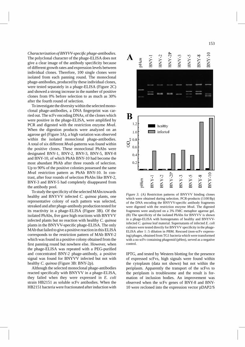
Characterization of BNYVV-specific phage-antibodies.The polyclonal character of the phage-ELISA does notgive a clear image of the antibody specificity becauseof different growth rates and expression levels betweenindividual clones. Therefore, 100 single clones wereisolated from each panning round. The monoclonalphage-antibodies, produced by these individual clones,were tested separately in a phage-ELISA (Figure 2C)and showed a strong increase in the number of positiveclones from 0% before selection to as much as 30%after the fourth round of selection.
To investigate the diversity within the selected mono-clonal phage-antibodies, a DNA fingerprint was car-ried out. The scFv encoding DNAs, of the clones whichwere positive in the phage-ELISA, were amplified byPCR and digested with the restriction enzymeMvaI.When the digestion products were analyzed on anagarose gel (Figure 3A), a high variation was observedwithin the isolated monoclonal phage-antibodies.A total of six differentMvaI-patterns was found withinthe positive clones. These monoclonal PhAbs weredesignated BNY-1, BNY-2, BNY-3, BNY-5, BNY-8and BNY-10, of which PhAb BNY-10 had become themost abundant PhAb after three rounds of selection.Up to 90% of the positive colonies possessed the sameMvaI restriction pattern as PhAb BNY-10. In con-trast, after four rounds of selection PhAbs like BNY-2,BNY-3 and BNY-5 had completely disappeared fromthe antibody pool.
To study the specificity of the selected MAbs towardshealthy and BNYVV infectedC. quinoaplants, onerepresentative colony of each pattern was selected,streaked and after phage-antibody production tested forits reactivity in a phage-ELISA (Figure 3B). Of theisolated PhAbs, five gave high reactions with BNYVVinfected plants but no reaction with healthyC. quinoaplants in the BNYVV-specific phage-ELISA. The onlyMAb that failed to give a positive reaction in this ELISAcorresponds to the restriction pattern of MAb BNY-2which was found in a positive colony obtained from thefirst panning round but nowhere else. However, whenthe phage-ELISA was repeated with a PEG-purifiedand concentrated BNY-2 phage-antibody, a positivesignal was found for BNYVV infected but not withhealthyC. quinoa(Figure 3B: BNY-2p).
Although the selected monoclonal phage-antibodiesreacted specifically with BNYVV in a phage-ELISA,they failed when they were expressed inE. colistrain HB2151 as soluble scFv antibodies. When theHB2151 bacteria were fractionated after induction with
Figure 3. (A) Restriction patterns of BNYVV binding cloneswhich were obtained during selection. PCR-products (1100 Bp)of the DNA encoding the BNYVV-specific antibody fragmentswere digested with the restriction enzymeMvaI. The digestedfragments were analyzed on a 3% FMC metaphor agarose gel.(B) The specificity of the isolated PhAbs for BNYVV is shownin a phage-ELISA with homogenates of healthy and BNYVV-infectedC. quinoaleaf material. Supernatants of infectedE. colicultures were tested directly for BNYVV specificity in the phage-ELISA after 1 : 5 dilution in PBM. Rescued (non-scFv express-ing) phages, obtained from TG1 bacteria which were transformedwith a no scFv containing phagemid (pHen), served as a negativecontrol.
IPTG, and tested by Western blotting for the presenceof expressed scFvs, high signals were found withinthe cytoplasm (data not shown) but not within theperiplasm. Apparently the transport of the scFvs tothe periplasm is troublesome and the result is for-mation of inclusion bodies. An improvement wasobserved when the scFv genes of BNY-8 and BNY-10 were recloned into the expression vector pDAP2/S
154
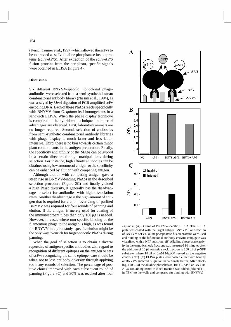
(Kerschbaumer et al., 1997) which allowed the scFvs tobe expressed as scFv-alkaline phosphatase fusion pro-teins (scFv-AP/S). After extraction of the scFv-AP/Sfusion proteins from the periplasm, specific signalswere obtained in ELISA (Figure 4).
Discussion
Six different BNYVV-specific monoclonal phage-antibodies were selected from a semi-synthetic humancombinatorial antibody library (Nissim et al., 1994), aswas assayed byMvaI digestion of PCR amplified scFvencoding DNA. Each of these PhAbs reacts specificallywith BNYVV from C. quinoaleaf homogenates in asandwich ELISA. When the phage display techniqueis compared to the hybridoma technique a number ofadvantages are observed. First, laboratory animals areno longer required. Second, selection of antibodiesfrom semi-synthetic combinatorial antibody librarieswith phage display is much faster and less labor-intensive. Third, there is no bias towards certain minorplant contaminants in the antigen preparation. Finally,the specificity and affinity of the MAbs can be guidedin a certain direction through manipulations duringselection. For instance, high affinity antibodies can beobtained using low amounts of antigen or the specificitycan be enhanced by elution with competing antigen.
Although elution with competing antigen gave asteep rise in BNYVV-binding PhAbs in the describedselection procedure (Figure 2C) and finally yieldeda high PhAb diversity, it generally has the disadvan-tage to select for antibodies with high dissociationrates. Another disadvantage is the high amount of anti-gen that is required for elution: over 2 mg of purifiedBNYVV was required for four rounds of panning andelution. If the antigen is merely used for coating ofthe immunosorbent tubes then only 160µg is needed.However, in cases where non-specific binding of thefilamentous phage to the antigen is high, as was foundfor BNYVV in a pilot study, specific elution might bethe only way to enrich for target-specific PhAbs duringpanning.
When the goal of selection is to obtain a diverserepertoire of antigen-specific antibodies with regard torecognition of different epitopes on the antigen or setsof scFvs recognizing the same epitope, care should betaken not to lose antibody diversity through applyingtoo many rounds of selection. The percentage of pos-itive clones improved with each subsequent round ofpanning (Figure 3C) and 30% was reached after four
Figure 4. (A) Outline of BNYVV-specific ELISA. The ELISAplate was coated with the target antigen BNYVV. For detectionof BNYVV, scFv alkaline phosphatase fusion proteins were usedand binding of the bifunctional antibody-enzyme conjugate wasvisualized withp-NPP substrate. (B) Alkaline phosphatase activ-ity in the osmotic shock fractions was measured 10 minutes afterthe addition of 10µl osmotic shock fraction to 100µl of p-NPPsubstrate, where 10µl of 5mM MgSO4 served as the negativecontrol (NC). (C) ELISA plates were coated either with healthyor BNYVV infectedC. quinoain carbonate buffer. After block-ing, 100µl of the alkaline phosphatase, BNY8-AP/S or BNY10-AP/S containing osmotic shock fraction was added (diluted 1 : 1in PBM) to the wells and compared for binding with BNYVV.

155
rounds of selection. When more subsequent rounds ofselection would have been performed the 100% wouldprobably have been reached. However, a decrease in thediversity was observed after the third round of selec-tion. This indicates that too many rounds of selectionresults either in antibodies with the highest affinity forthe most abundant epitope or in antibodies which areproduced at a faster rate and which are not necessarilythe most specific.
The inability of E. coli to export the selectedBNYVV-specific scFvs to the periplasm was observed.Several mutations are known which can enhance thetransport of scFvs to the periplasm (Nieba et al., 1996)and it might be worthwhile to introduce these muta-tions into the BNYVV-specific antibodies. The pres-ence of anE. coli protein domain (AP/S) on theC-terminus of the scFv molecule already seemed toincrease the stability and the folding efficiency of thesoluble scFv. In addition, the dimeric character of alka-line phosphatase may also add an avidity factor to theaffinity of the scFv-AP/S fusion protein for the antigen.Moreover, production of MAbs inE. coli offers sev-eral important features for the standardization of detec-tion assays. Production of scFv-enzyme fusion proteinsmight give a better reproducibility of the detectionof antigens in various immunoassays since the highlyvariable step of antibody enzyme conjugation is omit-ted. The recombinant scFv antibodies can be purifiedvia co-expressed affinity tags (Casey et al., 1995; Neriet al., 1995) to yield ultra pure, ready to use alkalinephosphatase conjugated reagents.
It is interesting that all the scFv expressing clonesisolated with the phage display procedure are BNYVV-specific and do not react with antigens from theC. quinoahomogenate. Phage display rather allowsPhAbs to be selected on the presence of a specific epi-tope than on the basis of immunogenicity. For exam-ple, glycoproteins are highly immunogenic. Therefore,the immune response elicited by a virus preparationthat is slightly contaminated with a plant glycoproteinwill be biased to that particular glycoprotein. Theresult is a high proportion of host-specific MAbs. Ifthe same virus preparation would be used to selectPhAbs from a combinatorial antibody library, spe-cific clones are obtained against the most abundantepitope present; i.e., the virus. This was what wasobserved for the BNYVV virus preparation, the virusis almost pure but still contains some plant contam-inants, yet the PhAbs obtained with this method areBNYVV-specific. It would be worthwhile to eval-uate their use in a routinely applied standardized
detection assay for BNYVV in sugar beets. They cer-tainly have potential, as they are highly BNYVV-specific.
Acknowledgments
This work was supported by a grant from the ProgramCommittee on Agricultural Biotechnology (PcLB) andby E.C. grant AIR3-CT94-1046. We thank prof. dr.W.B. van Muiswinkel (Department of Experimen-tal Animal Morphology and Cell Biology (EDC),Wageningen Agricultural University, The Netherlands)for critically reading the manuscript, dr. ir. D. Peters(Department of Virology, Wageningen AgriculturalUniversity, The Netherlands) for his kind gift of poly-clonal Rabbit anti-BNYVV serum and G. Winter(MRC Laboratory of Molecular Biology, Cambridge,UK.) for the permission to use the Nissim library.
References
Bradbury A, Persic L, Werge T and Cattaneo A (1993) Use of liv-ing columns to select specific phage antibodies. Biotechnology11: 1565–1596
Casey JL, Keep PA, Chester KA and Robson L (1995) Purificationof bacterially expressed single-chain Fv antibodies for clinicalapplications using metal chelate chromatography. J ImmunolMethods 179: 105–116
Clackson T, Hoogenboom HR, Griffiths AD and Winter G (1991)Making antibodies using phage display libraries. Nature 352:624–628
Clark MF and Adams AN (1977) Characteristics of the microplatemethod of enzyme-linked immunosorbent assay for the detec-tion of plant viruses. J Gen Virol 34: 475–483
George RA and Converse RH (1988) Methods for the enrichmentof desired B-cell populations before anti-cauliflower mosaicvirus hybridoma formation. Phytopathology 78: 1631–1636
Hepp RF and Converse RH (1986) Blueberry ringspot detectionin crude sap of highbush blueberry plants. Plant Disease 71:536–539
Hoogenboom HR, Griffiths AD, Johnson KS, Chiswell DJ,Hudson P and Winter G (1991) Multi-subunit proteins on thesurface of filamentous phage: methodologies for displayingantibody (Fab) heavy and light chains. Nucleic Acids Res 15:4133–4137
Hsu HT, Wang YC, Lawson RH, Wang M and Gonsalves D(1990) Splenocytes of mice with induced immunological toler-ance to plant antigens for construction of hybridomas secretingtomato spotted wilt virus-specific antibodies. Phytopathology80: 158–162
Huse WD, Sastry L, Iverson SA, Kang AS, Alting-Mees M,Burton DR, Benkovic SJ and Lerner RA (1989) Generation ofa large combinatorial library of the immunoglobulin repertoirein phage lambda. Science 246: 1275–1281

156
Huston JS, Levinson D, Mudgett-Hunter M, Tai M, Novotny J,Margolies MN, Ridge RJ, Bruccoleri RE, Haber E, Crea R andOppermann H (1988) Protein engineering of antibody bindingsites: Recovery of specific activity in an anti-digoxin single-chain Fv analogue produced inEscherichia coli. Proc Natl AcadSci U.S.A. 85: 5879–5883
Kerschbaumer RJ, Hirschl S, Kaufman A, Ibl M, Koenig R andHimmler G (1997) Single-chain Fv fusion proteins suitableas coating and detecting reagents in a double antibody sand-wich enzyme-linked immunosorbent assay. Anal Biochem 249:219–227
Kohler G and Milstein C (1975) Continuous cultures of fusedcells secreting antibody of predefined specificity. Nature 256:495–497
Marks JD, Hoogenboom HR, Bonnert TP, McCafferty J,Griffiths AD and Winter G (1991) By-passing immunization:Human antibodies from V-gene libraries displayed on phage.J Mol Biol 222: 581–597
Matthew, WD and AW Sandrock, Jr. 1987. Cyclophosphamidetreatment used to manipulate the immune response for the pro-duction of monoclonal antibodies. J Immunol Methods 100:73–82
McCafferty J, Griffiths AD, Winter G and Chiswell DJ (1990)Phage-antibodies: filamentous phage displaying antibody vari-able domains. Nature 348: 552–554
Neri D, Delalla C, Petrul H, Neri P and Winter G (1995) Calmod-ulin as a versatile tag for antibody fragments. Biotechnology13: 373–377
Nieba L, Honegger A, Krebber C and Pluckthun A (1997) Disrupt-ing the hydrophobic patches at the antibody variable/constantdomain interface: improved in vivo folding and physical char-acterization of an engineered scFv fragment. Protein-Eng 10:435–440
Nissim A, Hoogenboom HR, Tomlinson IM, Flynn G, MidgleyC, Lane D and Winter G (1994) Antibody fragments from asingle pot phage display library as immunochemical reagents.EMBO J 13: 692–698
Orlandi O, Gussow DH, Jones PT and Winter G (1989) Cloningimmunoglobulin variable domains for the expression by thepolymerase chain reaction. Proc Natl Acad Sci USA 86:3833–3837
Parks DR, Bryan VM, Oi VT and Herzenberg LA (1979)Antigen-specific identification and cloning of hybridomas witha fluorescence-activated cell sorter. Proc Natl Acad Sci USA76: 1962–1966
Putz C and Kuszala M (1978) La rhizomanie de la betteravesucriere en Alsace. Recherche d’une nouvelle methode depurification du “Beet Necrotic Yellow Vein Virus”. Ann Phy-topathol 10: 247–262
Sambrook J, Fritch EF and Maniatis T (1990) Molecularcloning: A laboratory manual. Cold Spring Harbour Labora-tory, New York
Sastry L, Alting-Mees M, Huse WD, Short JM, Sorge JA, Hay BH,Janda KD, Benkovic SJ and Lerner RA (1989) Cloning of theimmunological repertoire inEscherichia colifor generation ofmonoclonal catalytic antibodies: Construction of a heavy chainvariable region-specific cDNA library. Proc Natl Acad Sci USA86: 5728–5732
Tamada T and Baba T (1973) Beet necrotic yellow vein virusfrom rhizomania-affected sugar beet in Japan. Ann PhytopathSoc Japan 39: 325–332
Tijsen P (1985) Preparation of enzyme-antibody or otherenzyme-macromolecule conjugates. Laboratory Techniques inBiochemistry and Molecular biology. Vol 15. Practice andTheory of Enzyme Immunoassays. R.H. Burdon and P.H. vanKnippenberg (eds.), Elsevier Publishing, Amsterdam
Torrance L, Larkins AP and Butcher GW (1986) Characterizationof monoclonal antibodies against potato virus X and compari-son of serotypes with resistance groups. J Gen Virol 67: 57–67
Torrance L (1995) Use of monoclonal antibodies in plant pathol-ogy. Eur J of Plant Path 101: 351–363
Van Regenmortel MHV (1986) The potential for using mon-oclonal antibodies in the detection of plant viruses. In:Jones RAC and Torrance L (eds) Developments and applica-tions in virus testing (pp 89–101) Wellesbourne: Associationof Applied Biologists
Van Regenmortel MHV (1990) Plant viruses. In: VanRegenmortel MHV and Neurath AR (eds) Immunochemistry ofViruses, II. The basis for Serodiagnosis and Vaccines (pp 505–515) Elsevier, Amsterdam, The Netherlands
Warburg O and Christian W (1942) Isolierung und Kristallisationdes Garungsferments Enolase. Biochem Z 310: 384–421