Innate Response to Focal Necrotic Injury Inside the Blood-Brain Barrier1

RESEARCH ARTICLE
Connexin32 hemichannels contribute to the apoptotic-to-necrotictransition during Fas-mediated hepatocyte cell death
Mathieu Vinken • Elke Decrock • Elke De Vuyst • Marijke De Bock •
Roosmarijn E. Vandenbroucke • Bruno G. De Geest • Joseph Demeester •
Niek N. Sanders • Tamara Vanhaecke • Luc Leybaert • Vera Rogiers
Received: 20 May 2009 / Revised: 16 November 2009 / Accepted: 18 November 2009
� Birkhauser Verlag, Basel/Switzerland 2009
Abstract The present study was set up to investigate the
fate of connexin32 and its channels in hepatocellular
apoptosis. Primary hepatocyte cultures were exposed to Fas
ligand and cycloheximide, and modifications in connex-
in32 expression and localization, and gap junction
functionality were studied. We found that gap junction
functionality rapidly declined upon progression of cell
death, which was associated with a decay of the gap
junctional connexin32 protein pool. Simultaneously, levels
of newly synthesized connexin32 protein increased and
gathered in a hemichannel configuration. This became
particularly evident towards the end stages of the cell death
process and was not reflected at the transcriptional level.
We next either silenced connexin32 expression or inhibited
connexin32 hemichannel activity prior to cell death
induction. Both approaches resulted in a delayed termina-
tion of the cell death response. We conclude that
connexin32 hemichannels facilitate the apoptotic-to-
necrotic transition, which typically occurs in the final stage
of hepatocellular apoptosis.
Keywords Apoptosis � Primary hepatocyte �Connexin32 � Gap junction � Hemichannel
Abbreviations
Ac-DEVD-AFC Acetyl-Asp-Glu-Val-Asp-7-amino-4-
trifluoromethylcoumarin
ATP Adenosine triphosphate
CHX Cycloheximide
Cx Connexin
FasL Fas ligand
FRAP Fluorescence recovery after
photobleaching
GAPDH Glyceraldehyde-3-phosphate
dehydrogenase
GJIC Gap junctional intercellular
communication
HBSS–Hepes Hank’s balanced salt solution
supplemented with Hepes
LDH Lactate dehydrogenase
PbAE1 1,4-Butanediol diacrylate-based
poly-beta-aminoester
PBS Phosphate-buffered saline solution
M. Vinken (&) � T. Vanhaecke � V. Rogiers
Department of Toxicology, Faculty of Medicine and Pharmacy,
Vrije Universiteit Brussel, Laarbeeklaan 103,
1090 Brussels, Belgium
e-mail: [email protected]
E. Decrock � E. De Vuyst � M. De Bock � L. Leybaert
Physiology Group, Department of Basic Medical Sciences,
Faculty of Medicine and Health Sciences, Ghent University,
De Pintelaan 185, 9000 Ghent, Belgium
R. E. Vandenbroucke
Department for Molecular Biomedical Research,
Ghent University-VIB, Technologiepark 927,
9052 Ghent, Belgium
B. G. De Geest � J. Demeester � N. N. Sanders
Laboratory of General Biochemistry and Physical Pharmacy,
Faculty of Pharmaceutical Sciences, Ghent University,
Harelbekestraat 72, 9000 Ghent, Belgium
B. G. De Geest
Laboratory of Pharmaceutical Technology,
Faculty of Pharmaceutical Sciences, Ghent University,
Harelbekestraat 72, 9000 Ghent, Belgium
N. N. Sanders
Laboratory of Gene Therapy, Faculty of Veterinary Medicine,
Ghent University, Heidestraat 19, 9820 Merelbeke, Belgium
Cell. Mol. Life Sci.
DOI 10.1007/s00018-009-0220-2 Cellular and Molecular Life Sciences

PBSD? Divalent ion-supplemented PBS
qRT-PCR Quantitative real-time reverse
transcriptase-polymerase chain reaction
siRNA Small interfering RNA
TBS Tris-buffered saline solution
Introduction
Liver homeostasis basically relies on the critical balance
between cell growth, cellular differentiation and cell death.
The latter mainly occurs through apoptosis, a well-
orchestrated process that depends on the molecular actions
of caspase proteins [1]. One of the most prominent sig-
naling pathways that drives apoptosis in liver is initiated by
binding of a specific subset of ligands, such as the Fas
ligand (FasL), to their corresponding receptors at the cell
membrane surface. Hepatocytes strongly express the Fas
receptor, and its ligand binding results in the induction of
caspase 8. Activated caspase 8 then triggers caspase 3,
which subsequently cleaves a broad spectrum of cellular
proteins, giving rise to the apoptotic phenotype [2, 3]. In
normal liver, the prevalence of apoptosis is very low,
affecting 0.05–0.1% of all liver cells in rodents [4]. During
a variety of liver pathologies as well as upon exposure to
hepatotoxicants, however, apoptotic activity drastically
increases, which is frequently associated with elevated
FasL levels [2, 3].
Gap junctions are indispensable gatekeepers of liver
homeostasis. These communicating cell junctions mediate
the direct intercellular exchange of small and hydrophilic
molecules (e.g., second messengers), a flux called gap
junctional intercellular communication (GJIC). Gap junc-
tions are composed of two hemichannels, so-called
connexons, of neighboring cells, which in turn are built up
by six connexin (Cx) subunits. More than 20 mammalian
connexins have so far been characterized and they are
named according to their molecular weight. Connexin
species known to be expressed in human and rodent liver
include Cx32, Cx26, Cx43, Cx40, Cx37, Cx39, and
Cx31.9/Cx30.2. Among these, Cx32 constitutes about 90%
of the total hepatic connexin amount and is abundantly
expressed by hepatocytes. Cx32-based gap junctions have
been repeatedly found to act as key players in charge of
hepatocyte proliferation and functioning [5, 6]. Their
contribution in the occurrence of hepatocyte cell death,
however, has so far been poorly documented [6, 7].
Overall, conflicting results have been published with
respect to the role of gap junctions in apoptosis, as some
groups observed a positive correlation between GJIC and
apoptotic activity, whereas others showed that gap junctions
rather counteract this process [5, 8, 9]. To make the picture
even more complicated, accumulating evidence points to
the participation of hemichannels in apoptotic cell death.
Hemichannels have long been considered as merely struc-
tural precursors for gap junctions, but a large body of
compelling evidence now clearly demonstrates that
connexons themselves also provide a pathway for com-
munication, albeit between the intracellular compartment
and the extracellular environment [5, 9–11]. Although a
limited number of reports have described cytoprotective
functions for hemichannels [11, 12], the vast majority of
currently available data indicate that connexons are actively
involved in the spread of apoptosis [9–11, 13–17].
The present study was set up to investigate the role of
Cx32 and its channels in Fas-mediated hepatocyte cell
death. We addressed primary hepatocyte cultures as an
experimental setting, as this system allows the monitoring
of the entire time course of apoptosis. Indeed, apoptotic
hepatocytes are barely detectable in vivo, as they are too
rapidly engulfed by neighboring phagocytes [18–20]. We
provide an in-depth scenario of the FasL-induced changes
in hepatocellular Cx32 expression, localization, and func-
tion. We demonstrate for the first time the presence of
Cx32 hemichannels in hepatocytes, and we concomitantly
show that they are actively involved in the late phases of
Fas-mediated cell death.
Materials and methods
Chemicals and reagents
FasL (produced in HEK293 cells and similar to natural
human FasL) and cycloheximide (CHX) came from Alexis
(Switzerland) and Sigma (Belgium), respectively. Acetyl-
Asp-Glu-Val-Asp-7-amino-4-trifluoromethylcoumarin
(Ac-DEVD-AFC) was purchased from Merck (Belgium).
Propidium iodide, Hoechst 33342 and calcein–acetoxy-
methyl ester were from Invitrogen (Belgium), whereas
Annexin V Fluos and Annexin V Alexa 568 were obtained
from Roche (Germany). The 32Gap27 peptide
(SRPTEKTVFT) was synthesized by Thermo Fisher Sci-
entific (Germany). The 1,4-butanediol diacrylate-based
poly-beta-aminoester (PbAE1) was synthesized as descri-
bed elsewhere [21]. All other chemicals were commercially
available products of analytical grade and were supplied by
Sigma, unless specified otherwise.
Hepatocyte cultivation and cell death induction
Procedures for the housing of rats, and isolation and cul-
tivation of hepatocytes were approved by the local ethical
committee of the Vrije Universiteit Brussel (Belgium).
Male outbred Sprague–Dawley rats (Charles River
M. Vinken et al.

Laboratories, Belgium) were kept under controlled envi-
ronmental conditions with free access to food and water.
Hepatocytes were isolated by use of a two-step collagenase
method, and cell viability was assessed by trypan blue
exclusion [22]. Viable (C85%) hepatocytes were plated at
a density of 0.56 9 105 cells/cm2 in William’s medium E
(Invitrogen, Belgium) supplemented with 7 ng/ml gluca-
gon, 292 mg/ml L-glutamine, antibiotics (7.33 I.E./ml
sodium benzyl penicillin, 50 lg/ml kanamycin monosul-
fate, 10 lg/ml sodium ampicillin, 50 lg/ml streptomycin
sulfate) and 10% v/v fetal bovine serum. After 4 and 24 h,
the cell culture medium was removed and replaced by
serum-free medium supplemented with 25 lg/ml hydro-
cortisone sodium hemisuccinate and 0.5 lg/ml insulin
(hepatocyte culture medium). Cell death was induced 44 h
post-plating by renewal with hepatocyte culture medium
containing 200 ng/ml FasL and 2 lg/ml CHX. Sampling
was performed at the start of cell death induction, and at 2,
4, and 6 h thereafter.
Gene silencing by small interfering RNA (siRNA)
Cx32 expression was suppressed by siRNA treatment with
On-target plus smart pool siRNA (accession number
NM_017251) from Dharmacon (Belgium) containing a
mixture of four siRNA duplexes directed against rat gjb1.
Preparation of PbAE1/siRNA complexes (N:P ratio 50:1)
was carried out as previously described [21]. The siRNA
transfection was started 4 h after cell seeding by replacing
the initial culture medium by hepatocyte culture medium
containing the PbAE1/siRNA complexes (final siRNA
concentration 100 nM). After 4 h, the culture medium was
removed and replaced by regular hepatocyte culture med-
ium. The hepatocyte culture medium was renewed once
more (16 h post-transfection) prior to cell death induction
(44 h post-plating). Experiments using non-targeting
siRNA (Dharmacon, Belgium) were performed in parallel.
Determination of the apoptotic and necrotic indices
Hepatocyte cultures were washed twice with phosphate-
buffered saline solution (PBS) containing 1.2 mM CaCl2and 340 lM MgCl2.6H2O (PBSD?). Subsequently, cells
were stained with 2% v/v Annexin V Fluos, 3 lg/ml
Hoechst 33342 and 1 lg/ml Propidium iodide in Annexin
V buffer (140 mM NaCl, 5 mM CaCl2, 10 mM Hepes) for
15 min at room temperature. Culture dishes were thereafter
rinsed with PBSD? and subjected to fluorescence micros-
copy (Nikon, Japan). At least five images per culture dish
were taken. The number of cells, positive for the concerned
marker, was counted in each image and expressed relative
to the total number of nuclei present.
Measurement of caspase 3-like activity
Caspase 3-like activity in primary hepatocyte cultures was
measured fluorometrically by using the Ac-DEVD-AFC
substrate as described elsewhere [23]. The enzyme activity
was expressed as nmol AFC/min 9 lg protein.
Measurement of lactate dehydrogenase (LDH) leakage
Hepatocyte membrane damage was evaluated by determi-
nation of the LDH index [24], using a commercially
available kit (Merck, Germany). The LDH index was cal-
culated by the following equation: (100 9 LDH activity in
supernatant)/[LDH activity in (supernatant ? cells)].
Fluorescence recovery after photobleaching (FRAP)
For FRAP analysis, cultured hepatocytes were loaded
with 10 lM calcein–acetoxymethyl ester in Hank’s bal-
anced salt solution buffered with Hepes (HBSS–Hepes;
0.81 mM MgSO4.7H2O, 0.95 mM CaCl2.2H2O, 137 mM
NaCl, 0.18 mM Na2HPO4.2H2O, 5.36 mM KCl,
0.44 mM KH2PO4, 5.55 mM D-glucose, 25 mM Hepes)
for 30 min at room temperature. In order to detect and
thus to specifically study apoptotic cells, hepatocytes
were stained with 2% v/v Annexin V Alexa 568 in
Annexin V buffer for 15 min at room temperature. After
extensive rinsing, cultures were kept for 10 min in
HBSS–Hepes. Fluorescence within a single cell was
photobleached by 1 s spot exposure to 488 nm Argon
laser light, and dye influx from neighboring cells was
recorded over the next 5 min with a 940 water immer-
sion objective (Nikon, Japan). Fluorescence in the
bleached cell was expressed as the percentage recovery
relative to the prebleach level. At least six cells per
culture dish were examined.
Measurement of extracellular adenosine triphosphate
(ATP)
ATP was measured using a commercial luciferin/luciferase
assay kit (Sigma). Briefly, cultured hepatocytes were
washed with divalent-free buffer (137 mM NaCl, 0.18 mM
Na2HPO4.2H2O, 5.36 mM KCl, 0.44 mM KH2PO4, 4 mM
ethylene glycol tetra-acetic acid, 5.55 mM D-glucose,
25 mM Hepes) and incubated for 2.5 min with divalent-
free buffer at room temperature. For baseline measure-
ments, divalent-free buffer was replaced by HBSS–Hepes.
After 2.5 min, ATP assay mix in HBSS–Hepes was added
and luminescence was measured. ATP release was
expressed as the percentage of ATP release triggered by
divalent-free medium.
Connexin32 and hepatocellular apoptosis
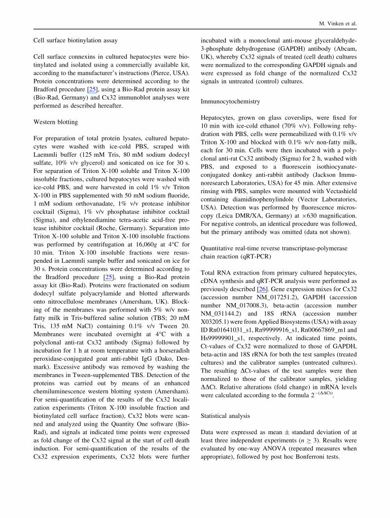
Cell surface biotinylation assay
Cell surface connexins in cultured hepatocytes were bio-
tinylated and isolated using a commercially available kit,
according to the manufacturer’s instructions (Pierce, USA).
Protein concentrations were determined according to the
Bradford procedure [25], using a Bio-Rad protein assay kit
(Bio-Rad, Germany) and Cx32 immunoblot analyses were
performed as described hereafter.
Western blotting
For preparation of total protein lysates, cultured hepato-
cytes were washed with ice-cold PBS, scraped with
Laemmli buffer (125 mM Tris, 80 mM sodium dodecyl
sulfate, 10% v/v glycerol) and sonicated on ice for 30 s.
For separation of Triton X-100 soluble and Triton X-100
insoluble fractions, cultured hepatocytes were washed with
ice-cold PBS, and were harvested in cold 1% v/v Triton
X-100 in PBS supplemented with 50 mM sodium fluoride,
1 mM sodium orthovanadate, 1% v/v protease inhibitor
cocktail (Sigma), 1% v/v phosphatase inhibitor cocktail
(Sigma), and ethylenediamine tetra-acetic acid-free pro-
tease inhibitor cocktail (Roche, Germany). Separation into
Triton X-100 soluble and Triton X-100 insoluble fractions
was performed by centrifugation at 16,060g at 4�C for
10 min. Triton X-100 insoluble fractions were resus-
pended in Laemmli sample buffer and sonicated on ice for
30 s. Protein concentrations were determined according to
the Bradford procedure [25], using a Bio-Rad protein
assay kit (Bio-Rad). Proteins were fractionated on sodium
dodecyl sulfate polyacrylamide and blotted afterwards
onto nitrocellulose membranes (Amersham, UK). Block-
ing of the membranes was performed with 5% w/v non-
fatty milk in Tris-buffered saline solution (TBS; 20 mM
Tris, 135 mM NaCl) containing 0.1% v/v Tween 20.
Membranes were incubated overnight at 4�C with a
polyclonal anti-rat Cx32 antibody (Sigma) followed by
incubation for 1 h at room temperature with a horseradish
peroxidase-conjugated goat anti-rabbit IgG (Dako, Den-
mark). Excessive antibody was removed by washing the
membranes in Tween-supplemented TBS. Detection of the
proteins was carried out by means of an enhanced
chemiluminescence western blotting system (Amersham).
For semi-quantification of the results of the Cx32 locali-
zation experiments (Triton X-100 insoluble fraction and
biotinylated cell surface fraction), Cx32 blots were scan-
ned and analyzed using the Quantity One software (Bio-
Rad), and signals at indicated time points were expressed
as fold change of the Cx32 signal at the start of cell death
induction. For semi-quantification of the results of the
Cx32 expression experiments, Cx32 blots were further
incubated with a monoclonal anti-mouse glyceraldehyde-
3-phosphate dehydrogenase (GAPDH) antibody (Abcam,
UK), whereby Cx32 signals of treated (cell death) cultures
were normalized to the corresponding GAPDH signals and
were expressed as fold change of the normalized Cx32
signals in untreated (control) cultures.
Immunocytochemistry
Hepatocytes, grown on glass coverslips, were fixed for
10 min with ice-cold ethanol (70% v/v). Following rehy-
dration with PBS, cells were permeabilized with 0.1% v/v
Triton X-100 and blocked with 0.1% w/v non-fatty milk,
each for 30 min. Cells were then incubated with a poly-
clonal anti-rat Cx32 antibody (Sigma) for 2 h, washed with
PBS, and exposed to a fluorescein isothiocyanate-
conjugated donkey anti-rabbit antibody (Jackson Immu-
noresearch Laboratories, USA) for 45 min. After extensive
rinsing with PBS, samples were mounted with Vectashield
containing diamidinophenylindole (Vector Laboratories,
USA). Detection was performed by fluorescence micros-
copy (Leica DMR/XA, Germany) at 9630 magnification.
For negative controls, an identical procedure was followed,
but the primary antibody was omitted (data not shown).
Quantitative real-time reverse transcriptase-polymerase
chain reaction (qRT-PCR)
Total RNA extraction from primary cultured hepatocytes,
cDNA synthesis and qRT-PCR analysis were performed as
previously described [26]. Gene expression mixes for Cx32
(accession number NM_017251.2), GAPDH (accession
number NM_017008.3), beta-actin (accession number
NM_031144.2) and 18S rRNA (accession number
X03205.1) were from Applied Biosystems (USA) with assay
ID Rn01641031_s1, Rn99999916_s1, Rn00667869_m1 and
Hs99999901_s1, respectively. At indicated time points,
Ct-values of Cx32 were normalized to those of GAPDH,
beta-actin and 18S rRNA for both the test samples (treated
cultures) and the calibrator samples (untreated cultures).
The resulting DCt-values of the test samples were then
normalized to those of the calibrator samples, yielding
DDCt. Relative alterations (fold change) in mRNA levels
were calculated according to the formula 2-(DDCt).
Statistical analysis
Data were expressed as mean ± standard deviation of at
least three independent experiments (n C 3). Results were
evaluated by one-way ANOVA (repeated measures when
appropriate), followed by post hoc Bonferroni tests.
M. Vinken et al.

Results
Characterization of the cell death response induced
by FasL/CHX
To induce cell death, cultures of hepatocytes were exposed
for 6 h to FasL (200 ng/ml) and CHX (2 lg/ml), a potent
inhibitor of protein synthesis which has been shown to
potentiate Fas-mediated cellular responses [27, 28]. Char-
acterization of the cell death response was carried out by
combined in situ stainings with Annexin V, Hoechst 33342
and Propidium iodide (Fig. 1a–c). An early event during
the commitment of cells to apoptosis is the externalization
of phosphatidyl serine in the cell plasma membrane, a
process that can be monitored by the phospholipid binding
protein Annexin V [29]. In the current experimental
setting, however, the number of Annexin V-positive
hepatocytes only significantly (p \ 0.05) increased after
6 h of exposure to FasL/CHX (Fig. 1a). In a subsequent
stage of apoptosis, cells display chromatin fragmentation
and condensation, which can be detected by the DNA
intercalating dye Hoechst 33342 [19, 29]. These hallmarks
progressively appeared in our experimental setting, thus
demonstrating the deleterious outcome of FasL/CHX
(Fig. 1b). Late apoptotic cells typically switch to a rather
necrotic appearance, which is associated with disruption of
the cell plasma membrane. Propidium iodide only enters
cells that have lost cell plasma membrane integrity and
thus specifically stains cells undergoing the apoptotic-to-
necrotic transition [29]. In line with this, Propidium iodide-
positive counts significantly (p \ 0.001) peaked after 6 h
of exposure of hepatocytes to FasL/CHX (Fig. 1c).
Fig. 1 Characterization of the cell death response induced by FasL/
CHX in hepatocytes. Primary hepatocytes were cultivated and cell
death was induced as specified in ‘‘Materials and methods’’. In a
parallel set of experiments, cells were treated with Cx32 siRNA or32Gap27 prior to cell death induction. At indicated time points, cells
were stained with Annexin V, Hoechst 33342, and Propidium iodide,
and subjected to light microscopy and fluorescence microscopy. At
least five images per culture dish were taken. The number of cells
positive for a Annexin V b Hoechst 33342 and c Propidium iodide
was counted in each image and expressed relative to the total number
of nuclei present. Data are expressed as mean ± standard deviation of
six independent experiments. Results were evaluated by one-way
ANOVA followed by post hoc Bonferroni tests. Asterisks indicate
significant differences compared with the corresponding ‘‘control’’
(untreated) condition per indicated time point (*p \ 0.05,
**p \ 0.01, ***p \ 0.001). Number signs indicate significant differ-
ences compared with the corresponding ‘‘200 ng/ml FasL ? 2 lg/ml
CHX’’ (cell death) condition per indicated time point (##p \ 0.01,###p \ 0.001)
Connexin32 and hepatocellular apoptosis

To further quantify the cell death response, both apop-
tosis and necrosis were recorded at the activity level.
Apoptosis was measured by using Ac-DEVD-AFC, a pro-
totypical, though not exclusive, caspase 3 substrate [19],
whereas necrotic activity was assessed by determination of
the LDH index, a cytosolic enzyme that leaks into the cell
culture medium upon disrupture of the cell plasma mem-
brane [24] (Fig. 2a–b). Caspase 3 activation was previously
reported to be a reliable and sensitive marker of apoptosis
in primary hepatocytes [18]. Indeed, this parameter already
significantly (p \ 0.05) increased after 2 h and reached a
maximum after 4 h (Fig. 2a). By contrast, the LDH index
only significantly (p \ 0.001) increased 6 h after cell death
induction (Fig. 2b).
Effects of FasL/CHX on Cx32 expression,
localization and function
We used FRAP analysis to analyze the functional state of
the gap junctions between the hepatocytes following the
induction of Fas-mediated cell death (Fig. 3a–b). Photo-
bleaching was directed in these experiments to Annexin
V-positive hepatocytes. We found a continuous decrease in
cell–cell dye coupling during the 6 h observation period,
becoming statistically significant (p \ 0.01) after 4 h
(Fig. 3b).
Since alterations in GJIC are frequently accompanied by
similar changes in connexin expression [6], we next mon-
itored hepatocellular Cx32 protein levels during the time
course of FasL/CHX-induced cell death. To this end,
immunoblotting and subsequent densitometric analyses
were performed (Fig. 4a–b). Unexpectedly, the Cx32 pro-
tein amount progressively increased, reaching a maximum
level after 6 h of cell death induction (Fig. 4b).
We then investigated whether the increased Cx32 pro-
tein levels were associated with alterations in its cellular
localization. For this purpose, we prepared Triton X-100
insoluble protein fractions and biotinylated cell surface
protein fractions prior to Cx32 immunoblot analyses
(Fig. 5a–b). The former is known to contain gap junctional
Cx32 whereas the latter indicates Cx32 present in non-
junctional plasma membrane regions, i.e., in a hemichannel
configuration [30]. As depicted in Fig. 5b, gap junctional
Cx32 gradually decreases upon exposure of hepatocytes to
FasL/CHX, a finding that could explain the deterioration of
GJIC under these conditions (Fig. 3a–b). By contrast, the
Cx32 amount in the biotinylated cell surface protein frac-
tion was elevated during cell death, reaching a peak level
Fig. 2 Modulation of apoptotic and necrotic activities during cell
death induced by FasL/CHX in hepatocytes. Primary hepatocytes
were cultivated and cell death was induced as specified in ‘‘Materials
and methods’’. In a parallel set of experiments, cells were treated with
Cx32 siRNA or 32Gap27 prior to cell death induction. At indicated
time points, a caspase 3-like activity and b LDH leakage were
measured. Caspase 3-like activity is expressed as nmol AFC/
min 9 lg protein. The LDH index was calculated by the equation
(100 9 LDH activity in supernatant)/[LDH activity in (supernatant ?
cells)]. Data are expressed as mean ± standard deviation of six
independent experiments. Results were evaluated by one-way
ANOVA followed by post hoc Bonferroni tests. Asterisks indicate
significant differences compared with the corresponding ‘‘control’’
(untreated) condition per indicated time point (*p \ 0.05,
***p \ 0.001). Number signs indicate significant differences com-
pared with the corresponding ‘‘200 ng/ml FasL ? 2 lg/ml CHX’’
(cell death) condition per indicated time point (##p \ 0.01,###p \ 0.001)
M. Vinken et al.

after 6 h (Fig. 5b). Immunocytochemistry analysis further
confirmed the increased presence of Cx32 at unapposed
cell plasma membrane regions at the end of the cell death
process (Fig. 5c). This observation may underlie the
overall augmentation in de novo synthesized Cx32 protein
in our experimental setting (Fig. 4a–b).
To examine whether the elevated hepatocellular Cx32
production during Fas-mediated cell death is also reflected
at the transcriptional level, we performed qRT-PCR anal-
yses, using three housekeeping genes (GAPDH, 18S rRNA,
and beta-actin) (Fig. 6a–c). We consistently observed
strongly decreased Cx32 gene transcription, whereby Cx32
mRNA levels drastically declined after 2 h of exposure of
the hepatocytes to FasL/CHX and remained low until the
end of the cell death process (Fig. 6a–c).
Effects of Cx32 siRNA on cell death induced
by FasL/CHX
To unravel the relevance of the augmentation in hepato-
cellular Cx32 protein production, especially towards the
late phases of Fas-mediated cell death, we used a mixture
of four Cx32 siRNA duplexes (100 nM). Following siRNA
treatment, the Cx32 protein amount declined to
30.9 ± 7.5% of the control (untreated) level (Fig. 7a–b).
The Cx32 siRNA-treated hepatocytes displayed signifi-
cantly (p \ 0.05) decreased FRAP recovery (21.6 ± 6.9%
vs 58.2 ± 14.4%) (Fig. 7c), thus confirming that gap
junctions composed of Cx32 account for the vast majority
of GJIC between hepatocytes. The Cx32 siRNA duplexes
also significantly (p \ 0.05) lowered extracellular ATP
release by hepatocytes to 65.1 ± 7.5% of the control level
following withdrawal of divalent ions from the cell culture
medium (Fig. 7d), which is a well-known protocol to
Fig. 3 Effects of FasL/CHX on GJIC between hepatocytes. Primary
hepatocytes were cultivated and cell death was induced as specified in
‘‘Materials and methods’’. At indicated time points, cells were
subjected to FRAP analysis, whereby a total of 72 pictures were
taken, starting 1 min before photobleaching and ending 5 min after
photobleaching. a Example pictures (magnification 9400) taken at
the start of procedure (prebleach), just after photobleaching (0 s) and
at the end of the registration period (300 s) are shown. Bleached cells
are delineated by dashed lines. b At least six cells per culture dish
were examined. Fluorescence in the bleached cells is expressed as the
percentage recovery relative to the prebleach level. Data are
expressed as mean ± standard deviation of four independent exper-
iments. Results were evaluated by one-way ANOVA followed by post
hoc Bonferroni tests. Asterisks indicate significant differences com-
pared with the corresponding ‘‘control’’ (untreated) condition per
indicated time point (**p \ 0.01, ***p \ 0.001)
Fig. 4 Effects of FasL/CHX on Cx32 protein levels in hepatocytes.
Primary hepatocytes were cultivated and cell death was induced as
specified in ‘‘Materials and methods’’. a At indicated time points,
western blot analysis of Cx32 was performed. b For semi-quantifi-
cation of the results, densitometric analysis was performed. Cx32
signals of cell death-induced cultures (?) were normalized to the
corresponding GAPDH signals and are expressed as fold change of
the normalized Cx32 signals in control (-) cultures. Data are
expressed as mean ± standard deviation of four independent exper-
iments. Results were evaluated by one-way ANOVA followed by post
hoc Bonferroni tests. Asterisks indicate significant differences com-
pared with the corresponding control (untreated) condition per
indicated time point (**p \ 0.01)
Connexin32 and hepatocellular apoptosis

trigger hemichannel opening and thus to measure their
functionality [30–32].
When Cx32 gene silencing was performed prior to cell
death induction, no changes in the Annexin V-based
apoptotic index and its Hoechst 33342-based counterpart
were observed (Fig. 1a–b). Caspase 3-like activity also
remained unchanged under these conditions (Fig. 2a).
However, both the number of Propidium iodide-positive
counts (Fig. 1c) and the LDH index (Fig. 2b) were sig-
nificantly (p \ 0.001) reduced after 6 h. Experiments using
non-targeting siRNA were performed in parallel, whereby
no effects on all parameters measured were observed (data
not shown). These results indicate that Cx32 plays a role in
the late phases of Fas-mediated hepatocellular cell death.
Effects of Cx32 mimetic peptide on cell death induced
by FasL/CHX
The increased presence of Cx32 in non-junctional cell
plasma membrane regions after 6 h of cell death induc-
tion in primary hepatocytes (Fig. 5a–c) as well as the
strongly decreased gap junction activity at this time point
(Fig. 3a–b), combined with the results obtained by the
Fig. 5 Effects of FasL/CHX on the localization of Cx32 in hepato-
cytes. Primary hepatocytes were cultivated and cell death was induced
as specified in ‘‘Materials and methods’’. a At indicated time points,
Triton X-100 insoluble Cx32 fractions and biotinylated Cx32 cell
surface fractions were prepared, and were subsequently subjected to
Cx32 Western blot analysis. b For semi-quantification of the results,
densitometric analysis was performed. Data are expressed as
mean ± standard deviation of three independent experiments and
represent fold change of the Cx32 signals at the start of cell death
induction. Results were evaluated by one-way ANOVA followed by
post hoc Bonferroni tests. Asterisks indicate significant differences
compared with the corresponding Cx32 signal at the start of cell death
induction (**p \ 0.01). c After 6 h of cell death induction, hepato-
cyte cultures were subjected to immunocytochemistry analysis, using
a primary antibody directed against Cx32 and a fluorescein
isothiocyanate-conjugated secondary antibody (green; white arrows),
as specified in ‘‘Materials and methods’’. Nuclear counterstaining was
performed with diamidinophenylindole (blue). Samples were ana-
lyzed by fluorescence microscopy at 9630 magnification. Untreated
(control) cultures and treated (cell death) cultures are shown in the leftpanel and right panel, respectively, and are representative of three
independent experiments
Fig. 6 Effects of FasL/CHX on Cx32 mRNA levels in hepatocytes.
Primary hepatocytes were cultivated and cell death was induced as
specified in ‘‘Materials and methods’’. At indicated time points, qRT-
PCR analysis was performed, using a GAPDH, b 18S rRNA, and
c beta-actin as housekeeping genes. Relative alterations (fold change)
in Cx32 mRNA levels were calculated according to the 2-(DDCt)
method. Data are expressed as mean ± standard deviation of four
independent experiments. Results were evaluated by one-way
ANOVA followed by post hoc Bonferroni tests. Asterisks indicate
significant differences compared with the corresponding control
(untreated) condition per indicated time point (*p \ 0.05,
**p \ 0.01, ***p \ 0.001)
M. Vinken et al.

Cx32 siRNA experiments (Figs. 1a–c and 2a–b), suggest
that Cx32-based hemichannels, but not their full channel
counterparts, are involved in the control of the terminal
stages of Fas-mediated hepatocellular cell death. To
substantiate this anticipated role for hepatocellular Cx32-
originating connexons, we used 32Gap27, a short peptide
that mimics a sequence in the second extracellular loop of
the Cx32 subunit. Previous research showed that short-
term incubations (0.5–6.5 h) with connexin mimetic
peptides are sufficient to inhibit hemichannel-related
responses, i.e., low extracellular divalent ion-triggered
cellular ATP release, in a connexin-specific way, whereas
longer exposure regimes (24 h) also result in the reduc-
tion of corresponding GJIC [31, 32]. In agreement with
this finding, 6.5 h exposure (i.e., 0.5 h pre-incubation
followed by 6 h exposure, as required to complete the cell
death response) of the hepatocytes to 32Gap27 (0.25 mg/
ml) significantly (p \ 0.05) reduced the ATP release
triggered by divalent ion depletion to 62.4 ± 4.8% of the
initial level (Fig. 8a). FRAP recovery, reflecting GJIC,
remained unchanged under these conditions (Fig. 8b).
At all time points measured, the apoptotic markers
Annexin V and Hoechst 33342 were left unaffected when32Gap27 treatment was initiated 30 min before inducing
cell death in primary hepatocytes (Fig. 1a–b). This also
held true for the caspase 3-like activity (Fig. 2a). Upon
counteracting Cx32 hemichannel-related functioning,
however, the peaks in both the Propidium iodide-based
necrotic index (Fig. 1c) and the LDH index (Fig. 2b),
typically culminating after 6 h of cell death induction, were
abolished. These findings are identical to the outcome of
the Cx32 siRNA experiments and point to an active role for
Cx32 hemichannels in the termination of Fas-mediated
hepatocellular cell death.
Fig. 7 Efficiency of Cx32 siRNA duplexes in hepatocytes. Primary
hepatocytes were cultivated and siRNA-mediated Cx32 gene silenc-
ing was performed as outlined in ‘‘Materials and methods’’ section.
a Cx32 western blot analysis was carried out, followed by densito-
metric analysis. b Cx32 signals of Cx32 siRNA-treated cells and non-
targeting siRNA-treated cells were normalized to the corresponding
GAPDH signals and are expressed as fold change of the normalized
Cx32 signals in control (untreated) cultures. Data are expressed as
mean ± standard deviation of four independent experiments. Results
were evaluated by one-way ANOVA followed by post hoc Bonferroni
tests. Asterisks indicate significant differences compared with the
control (untreated) condition (***p \ 0.001). c FRAP analysis was
performed. At least six cells per culture dish were examined.
Fluorescence in the bleached cells is expressed as the percentage
recovery relative to the prebleach level. Data are expressed as
mean ± standard deviation of four independent experiments. Results
were evaluated by one-way ANOVA followed by post hoc Bonferroni
tests. Asterisks indicate significant differences compared with the
control (untreated) condition (*p \ 0.05). d The release of ATP was
measured under basal (baseline) and induced (divalent-free medium)
conditions, and is expressed as the percentage of ATP release
triggered by divalent-free medium in untreated cultures (control).
Data are expressed as mean ± standard deviation of three indepen-
dent experiments. Results were evaluated by one-way ANOVA
followed by post hoc Bonferroni tests. Asterisks indicate significant
differences between the ‘‘divalent-free medium’’ condition and the
corresponding ‘‘baseline’’ condition (**p \ 0.01, ***p \ 0.001).
Number signs indicate significant differences between the ‘‘diva-
lent-free medium’’ condition of ‘‘Cx32 siRNA’’ cultures and the
‘‘divalent-free medium’’ condition of ‘‘control’’ cultures (#p \ 0.05)
Connexin32 and hepatocellular apoptosis

Discussion
The aim of this study was to examine the fate of gap
junctions in hepatocyte apoptosis. We found that GJIC
between cultured hepatocytes drastically decreases upon
progression of the FasL/CHX-induced cell death response.
This outcome is in line with the recent study of Theiss et al.
[33], showing strongly reduced cell–cell coupling between
primary lens epithelial cells upon exposure to a number of
apoptosis-inducing chemicals, including CHX. Kalvelyte
et al. [17] reported that abrogation of GJIC during apop-
tosis results from the removal of gap junctions and not
from their functional closure. Our results can be reconciled
with this finding, as we observed a progressive diminution
of the gap junctional Cx32 protein pool. Simultaneously,
de novo synthesized Cx32 gathered at non-junctional areas
of the cell plasma membrane surface, particularly towards
the final stages of the cell death process. This modification
was not reflected at the transcriptional level. In fact, Cx32
protein and mRNA levels behaved in opposite ways, as the
latter rapidly declined during progression of apoptosis.
Such disconnection between connexin gene transcription
and translation was also observed in apoptosis-primed
human osteoblasts and was thought to represent a reflexive
adaptation of the connexin mRNA machinery to changes
occurring at the downstream protein level [34].
While Cx32 gap junctions seem to play only a minor
role in hepatocellular cell death, our data suggest that Cx32
hemichannels could fulfill an important function, especially
during the final stage of this process. Such distinctive roles
for these Cx32-based channels may suggest differential and
even opposite regulation, a feature that has previously been
reported for gap junctions and hemichannels composed of
Cx43 [35, 36]. The involvement of Cx32 hemichannels in
our experimental setting was investigated via four different
strategies. Indeed, we applied siRNA technology to silence
Cx32 protein synthesis, we performed cell surface biotin-
ylation studies, we used a mimetic peptide to interfere with
Cx32 hemichannel functioning and we applied low extra-
cellular divalent ion-triggered cellular ATP release. With
respect to that latter, we cannot rule out the participation of
alternative ATP release pathways known to be present in
hepatocytes, including P2X7 receptors [37] and pannexin-
containing hemichannels [38], which may explain the
incomplete inhibition of ATP release by 32Gap27 in our
experiments. Both the inhibition of Cx32 hemichannel-
related cellular responses by 32Gap27 and the siRNA-
mediated silencing of Cx32 expression had an identical
outcome, namely a drop in the LDH index and a reduction
of the number of Propidium iodide-positive cells at the end
of the cell death process, whereas early apoptotic markers
were not affected. Our data are thus consistent with the
scenario that Cx32 hemichannels mediate the late phases of
FasL/CHX-induced cell death in hepatocytes, by promot-
ing the transition from an apoptotic to a necrotic
phenotype. This finding supports the research of Kalvelyte
and his group, performed on Cx32-transfected HeLa cells,
showing that Cx32 accelerates the transformation of
apoptotic cells into a necrotic state, partly depending on the
ability to form functional hemichannels [17]. In the same
experimental setting, metabolic inhibition, an ischemia-like
condition leading to cell death, was recently reported to be
associated with the increased formation and activity of
Cx32 hemichannels [39]. In a similar way, connexons
composed of Cx43 [13, 14, 16, 17] have been reported to
propagate apoptotic cell death in a number of cell types.
Fig. 8 Efficiency of 32Gap27 in hepatocytes. Primary hepatocytes
were cultivated and cells were treated with 32Gap27 (30 min pre-
incubation followed by 6 h of exposure) as outlined in ‘‘Materials and
methods’’. a The release of ATP was measured under basal (baseline)
and induced (divalent-free medium) conditions, and is expressed as
the percentage of ATP release triggered by divalent-free medium in
untreated cultures (control) at 30 min. Data are expressed as
mean ± standard deviation of three independent experiments. Results
were evaluated by one-way ANOVA followed by post hoc Bonferroni
tests. Asterisks indicate significant differences between the ‘‘divalent-
free medium’’ condition and the corresponding ‘‘baseline’’ condition
(***p \ 0.001). Number signs indicate significant differences
between the ‘‘divalent-free medium’’ condition of ‘‘32Gap27’’ cultures
after 6.5 h and the ‘‘divalent-free medium’’ condition of ‘‘control’’
cultures after 30 min (#p \ 0.05). b FRAP analysis was performed.
At least six cells per culture dish were examined. Fluorescence in the
bleached cells is expressed as the percentage recovery relative to the
prebleach level. Data are expressed as mean ± standard deviation of
three independent experiments. Results were evaluated by one-way
ANOVA followed by post hoc Bonferroni tests
M. Vinken et al.

In conclusion, the current study is the first to demon-
strate the presence of functional Cx32 hemichannels in
hepatocytes. We showed that these particular cell plasma
membrane channels are actively involved in the termina-
tion of Fas-mediated cell death in primary hepatocyte
cultures. Further research is required to investigate the
relevance of these findings in vivo and to examine the
involvement of Cx32 hemichannels in other cellular events
that constitute the hepatocyte’s life cycle known to be
governed by Cx32-based gap junctions, including prolif-
eration and differentiation [6].
Acknowledgments The authors wish to thank Mr. Bart Degreef,
Mr. Roel Fiey and Miss Sofie Wijthouck for their excellent technical
assistance. This work was supported by grants from the Fund for
Scientific Research Flanders (FWO-Vlaanderen), the Interuniversity
Attraction Poles Program (Belgian Science Policy), the Research
Council of the Vrije Universiteit Brussel (OZR-VUB) and the Euro-
pean Union (FP6 projects CARCINOGENOMICS and LIINTOP).
References
1. Maeda S (2000) Mechanisms of active cell death in isolated
hepatocytes. In: Berry MN, Edwards AM (eds) The hepatocyte
review. Kluwer, Norwell, pp 281–300
2. Malhi H, Gores GJ, Lemasters JJ (2006) Apoptosis and necrosis
in the liver: a tale of two deaths? Hepatology 43:S31–S44
3. Malhi H, Gores GJ (2008) Cellular and molecular mechanisms of
liver injury. Gastroenterology 134:1641–1654
4. Qiao L, Farrell GC (1999) The effects of cell density, attachment
substratum and dexamethasone on spontaneous apoptosis of rat
hepatocytes in primary culture. In Vitro Cell Dev Biol Anim
35:417–424
5. Vinken M, Vanhaecke T, Papeleu P, Snykers S, Henkens T,
Rogiers V (2006) Connexins and their channels in cell growth
and cell death. Cell Signal 18:592–600
6. Vinken M, Henkens T, De Rop E, Fraczek J, Vanhaecke T,
Rogiers V (2008) Biology and pathobiology of gap junctional
channels in hepatocytes. Hepatology 47:1077–1088
7. Albright CD, Kuo J, Jeong S (2001) cAMP enhances Cx43 gap
junction formation and function and reverses choline deficiency
apoptosis. Exp Mol Pathol 71:34–39
8. Krysko DV, Leybaert L, Vandenabeele P, D’Herde K (2005) Gap
junctions and the propagation of cell survival and cell death
signals. Apoptosis 10:459–469
9. Contreras JE, Sanchez HA, Veliz LP, Bukauskas FF, Bennett
MV, Saez JC (2004) Role of connexin-based gap junction
channels and hemichannels in ischemia-induced cell death in
nervous tissue. Brain Res Brain Res Rev 47:290–303
10. Evans WH, De Vuyst E, Leybaert L (2006) The gap junction
cellular internet: connexin hemichannels enter the signaling
limelight. Biochem J 397:1–14
11. Rodriguez-Sinovas A, Cabestrero A, Lopez D, Torre I, Morente
M, Abellan A, Miro E, Ruiz-Meana M, Garcia-Dorado D (2007)
The modulatory effects of connexin 43 on cell death/survival
beyond cell coupling. Prog Biophys Mol Biol 94:219–232
12. Plotkin LI, Manolagas SC, Bellido T (2002) Transduction of cell
survival signals by connexin-43 hemichannels. J Biol Chem
277:8648–8657
13. Decrock E, De Vuyst E, Vinken M, Van Moorhem M, Vranck K,
Wang N, Van Laeken L, De Bock M, D’Herde K, Lai CP,
Rogiers V, Evans WH, Naus CC, Leybaert L (2009) Connexin 43
hemichannels contribute to the propagation of apoptotic cell
death in a rat C6 glioma cell model. Cell Death Differ 16:151–
163
14. Ramachandran S, Xie LH, John SA, Subramaniam S, Lal R
(2007) A novel role for connexin hemichannel in oxidative stress
and smoking-induced cell injury. PLoS ONE 2:e712
15. Takeuchi H, Jin S, Wang J, Zhang G, Kawanokuchi J, Kuno R,
Sonobe Y, Mizuno T, Suzumura A (2006) Tumor necrosis factor-
alpha induces neurotoxicity via glutamate release from hemi-
channels of activated microglia in an autocrine manner. J Biol
Chem 281:21362–21368
16. Hur KC, Shim JE, Johnson RG (2003) A potential role for Cx43-
hemichannels in staurosporin-induced apoptosis. Cell Commun
Adhes 10:271–277
17. Kalvelyte A, Imbrasaite A, Bukauskiene A, Verselis VK,
Bukauskas FF (2003) Connexins and apoptotic transformation.
Biochem Pharmacol 66:1661–1672
18. Gomez-Lechon MJ, O’Connor JE, Lahoz A, Castell JV, Donato
MT (2008) Identification of apoptotic drugs: multiparametric
evaluation in cultured hepatocytes. Curr Med Chem 15:2071–
2085
19. Bai L, Wang J, Yin XM, Dong Z (2003) Analysis of apoptosis:
basic principles and procedures. In: Yin XM, Dong Z (eds)
Essentials of apoptosis: a guide for basic and clinical research.
Humana, Totowa, NJ, pp 239–251
20. Gill GH, Dive D (2000) Apoptosis: basic mechanisms and rele-
vance to toxicology. In: Roberts R (ed) Apoptosis in toxicology.
Taylor & Francis, London, pp 1–20
21. Vandenbroucke RE, De Geest BG, Bonne S, Vinken M, Van-
haecke T, Heimberg H, Wagner E, Rogiers V, De Smedt SC,
Demeester J, Sanders NN (2008) Prolonged gene silencing in
hepatoma cells and primary hepatocytes after small interfering
RNA delivery with biodegradable poly(beta-amino esters).
J Gene Med 10:783–794
22. Papeleu P, Vanhaecke T, Henkens T, Elaut G, Vinken M, Sny-
kers S, Rogiers V (2006) Isolation of rat hepatocytes. Methods
Mol Biol 320:229–237
23. Fraczek J, Deleu S, Lukaszuk A, Doktorova T, Tourwe D, Geerts
A, Vanhaecke T, Vanderkerken K, Rogiers V (2009) Screening of
amide analogues of Trichostatin A in cultures of primary rat
hepatocytes: search for potent and safe HDAC inhibitors. Invest
New Drugs 27:338–346
24. Bergmeyer HU (1974) Lactate dehydrogenase. In: Bergmeyer
HU (ed) Methods of enzymatic analysis. Academic, New York,
pp 574–579
25. Bradford MM (1976) A rapid and sensitive method for the
quantitation of microgram quantities of protein utilizing the
principle of protein-dye binding. Anal Biochem 72:248–254
26. Vinken M, Henkens T, Vanhaecke T, Papeleu P, Geerts A, Van
Rossen E, Chipman JK, Meda P, Rogiers V (2006) Trichostatin
A enhances gap junctional intercellular communication in
primary cultures of adult rat hepatocytes. Toxicol Sci 91:484–
492
27. Rouquet N, Carlier K, Briand P, Wiels J, Joulin V (1996) Mul-
tiple pathways of Fas-induced apoptosis in primary culture of
hepatocytes. Biochem Biophys Res Commun 229:27–35
28. Ni R, Tomita Y, Matsuda K, Ichihara A, Ishimura K, Ogasawara
J, Nagata S (1994) Fas-mediated apoptosis in primary cultured
mouse hepatocytes. Exp Cell Res 215:332–337
29. Foster JR (2000) Detection and biomarkers of apoptosis. In:
Roberts R (ed) Apoptosis in toxicology. Taylor & Francis,
London, pp 213–232
30. Schalper KA, Palacios-Prado N, Orellana JA, Saez JC (2008)
Currently used methods for identification and characterization of
hemichannels. Cell Commun Adhes 15:207–218
Connexin32 and hepatocellular apoptosis

31. Leybaert L, Braet K, Vandamme W, Cabooter L, Martin PE,
Evans WH (2003) Connexin channels, connexin mimetic pep-
tides and ATP release. Cell Commun Adhes 10:251–257
32. De Vuyst E, Decrock E, Cabooter L, Dubyak GR, Naus CC,
Evans WH, Leybaert L (2006) Intracellular calcium changes
trigger connexin 32 hemichannel opening. EMBO J 25:34–44
33. Theiss C, Mazur A, Meller K, Mannherz HG (2007) Changes in
gap junction organization and decreased coupling during induced
apoptosis in lens epithelial and NIH-3T3 cells. Exp Cell Res
313:38–52
34. Sharrow AC, Li Y, Micsenyi A, Grisworld RD, Wells A, Monga
SS, Blair HC (2008) Modulation of osteoblast gap junction
connectivity by serum, TNFalpha, and TRAIL. Exp Cell Res
314:297–308
35. De Vuyst E, Decrock E, De Bock M, Yamasaki H, Naus CC,
Evans WH, Leybaert L (2007) Connexin hemichannels and gap
junction channels are differentially influenced by lipopolysac-
charide and basic fibroblast growth factor. Mol Biol Cell 18:34–46
36. Retamal MA, Froger N, Palacios-Prado N, Ezan P, Saez PJ, Saez
JC, Giaume C (2007) Cx43 hemichannels and gap junction
channels in astrocytes are regulated oppositely by proinflamma-
tory cytokines released from activated microglia. J Neurosci
27:13781–13792
37. Gonzales E, Prigent S, Abou-Lovergne A, Boucherie S, Tjord-
mann T, Jacquemin E, Combettes L (2007) Rat hepatocytes
express functional P2X receptors. FEBS Lett 581:3260–3266
38. Bruzzone R, Hormuzdi SG, Barbe MT, Herb A, Monyer H (2003)
Pannexins, a family of gap junction proteins expressed in brain.
Proc Natl Acad Sci USA 100:13644–13649
39. Sanchez HA, Orellana JA, Verselis VK, Saez JC (2009) Meta-
bolic inhibition increases activity of connexin-32 hemichannels
permeable to Ca2? in transfected HeLa cells. Am J Physiol Cell
Physiol 197:C665–C678
M. Vinken et al.
Copyright © 2022 FDOKUMEN