Hepatocyte growth factor and its receptor are required for malaria infection
Upload
independentCategory
view
1download
0
JPP 2009, 61: 767–774� 2009 The AuthorsReceived January 04, 2009Accepted March 02, 2009DOI 10.1211/jpp/61.06.0009ISSN 0022-3573
Correspondence: Prof. Dr KexinLiu, College of Pharmacy, DalianMedical University, 9 WesternSection, Lvshun South Street,Dalian, 116044, P.R. China.E-mail: [email protected]
Research Paper
Protective effect of JBP485 on concanavalin A-induced
liver injury in mice
Tao Yanga, Jingjing Wua, Changyuan Wanga, Qi Liua,
Xiaochi Maa, Jinyong Penga, Taiichi Kakub and Kexin Liua
aCollege of Pharmacy, Dalian Medical University, Dalian, Liaoning, China and bJapan Bioproducts Co. Ltd,Tomigaya, Shibuya-ku, Tokyo
Abstract
Objectives Cyclo-trans-4-L-hydroxyprolyl-L-serine (JBP485) was first isolated fromLaennec (hydrolysate of human placenta). We thought it valuable to clarify the anti-hepatitis molecular mechanism of JBP485 to develop a new oral anti-hepatitis drug.Methods We investigated the hepatoprotective effect of JBP485 on immune-mediated,concanavalin A (Con A)-induced liver injury in mice. Mice were administered JBP485before and after injection of Con A (10 mg/kg). Eight hours after Con A, the cytosolicenzyme activity (alanine aminotransferase, lactate dehydrogenase) in serum, and theenzyme activity or concentration (superoxide dismutase, maleic dialdehyde, myeloperox-idase, nitric oxide) in liver homogenate were determined. The liver slices were investigatedto observe changes in histology. The effect of JBP485 on level of tumour necrosis factor-a(TNF-a) and intercellular adhesion molecule-1 (ICAM-1) in liver were detected byimmunohistochemistry. Hepatocyte DNA fragmentation was assayed by agarose gelelectrophoresis and the transcription of the genes bax and bcl-2 in hepatocytes wasdetermined by reverse transcription-polymerase chain reaction.Key findings Con A increased the cytosolic and liver homogenate enzyme activity, andthe concentrations of ICAM-1 and TNF-a, which were significantly inhibited by JBP485administration. Also, the increase in DNA fragmentation and decrease in bcl-2/bax mRNAinduced by Con A administration were significantly inhibited by JBP485.Conclusions These results indicated that immune-mediated liver damage can beprevented by JBP485, and that this is mainly associated with immunomodulatory effectson T cells and adhesion molecules, antioxidation, and inhibition of apoptosis.Keywords apoptosis; concanavalin A; hepatitis; JBP485; Laennec
Introduction
Human placental extract (HPE) has been used to treat a number of liver diseases,including hepatitis and cirrhosis.[1] Laennec (a trade name for hydrolysate of humanplacenta) is produced by Japan Bioproducts Industry Co. Ltd (Tokyo, Japan) bypurification of human placental extracts, and it has been used clinically to treat chronichepatic injury for over 40 years in Japan.[2] JBP485 (Figure 1), a dipeptide, was one ofthe constituents isolated from Laennec, and it has been synthesized enantioselectively bychemical means. From our preliminary experiment, we had shown that JBP485 exhibitedexcellent gastrointestinal absorption[2] and had anti-hepatitis activity in concanavalin A(Con A)-induced hepatocyte toxicity.[3] This suggested that JBP485 was recognized bythe peptide transporter system in the gastrointestinal tract. Therefore, it is valuable toclarify the anti-hepatitis molecular mechanism of JBP485 to develop a new oral anti-hepatitis drug.
A well-described mouse model of T-cell-dependent liver injury is inducible byinjection of the T-cell mitogenic plant lectin concanavalin Con A, which leads tofulminant hepatitis within eight hours.[4] The liver injury model induced by Con Ainjection into mice is regarded as an appropriate model for studying immune-mediatedliver diseases.
In this study, to clarify its anti-hepatitis molecular mechanism we examined theprotective effect of JBP485 on immune-mediated liver injury in mice.
767
Materials and Methods
Reagents
JBP485 was obtained from Japan Bioproducts Industry Co.Ltd (Tokyo, Japan). Con A and collagenase were purchasedfrom Wako Pure Chemical Industries Ltd (Osaka, Japan).Anti-mouse tumour necrosis factor-a (TNF-a) antibody andanti-mouse intercellular adhesion molecule-1 (ICAM-1)antibody were purchased from Boster Biotechnology Co.Ltd (Wuhan, China). DNA fragmentation and reversetranscription-polymerase chain reaction (RT-PCR) reagentswere obtained from TaKaRa Biotechnology (Dalian) Co. Ltd(Dalian, China). Other reagents will be further specified.
Animals
Female BALB/c mice, 20–24 g (Experimental AnimalCenter of Dalian Medical University, Dalian, China), werehoused at 23 ± 2∞C under a 12-h light–dark cycle, at50 ± 5% relative humidity throughout the whole experi-mental period. All mice were allowed free access to waterand chow diet. All experimental protocols were approved bythe Institutional Animal Ethics Committee of Dalian MedicalUniversity, which follows the standards of the Committee forthe Purpose of Control and Supervision of Experimentationon Animals, Government of China.
Concanavalin A-induced hepatitis
Mice were divided randomly into three groups: the Con Agroup, the Con A + JBP485 group and the normal group. Inthe Con A group and Con A + JBP485 group, mouse liverdamage was induced by injection of Con A (10 mg/kg,1.5 mg/ml),[5,6] dissolved in pyrogen-free saline, through thetail vein. JBP485 (25 mg/kg, 2.5 mg/ml) dissolved inpyrogen-free saline was administered orally by gavage tomice 0.5 h before and 2 and 5 h after challenge with Con Ain the Con A + JBP485 group. The Con A group was giventhe same volume of pyrogen-free saline without JBP485. Thenormal group was given the same volume of pyrogen-freesaline instead of Con A and JBP485 at the same times.
Analysis of serum transaminase activity anddetermination of superoxide dismutase,maleic dialdehyde, myeloperoxidase andnitrite in liver tissue
Injection of Con A leads to fulminant hepatitis within 8 h.[4]
Therefore, hepatocyte damage was assessed 8 h after Con Aadministration by measuring the serum enzyme activity ofalanine aminotransferase (ALT) and lactate dehydrogenase
(LDH)[7–9] using commercial kits (Nanjing JianchengBioengineering Institute, Nanjing, China). Mouse liver wasremoved immediately after blood was harvested and waskept at –20∞C. Liver samples were thawed, weighed andhomogenized on ice with phosphate-buffered saline (pH 7.4).The homogenate was used for assays of superoxidedismutase (SOD), maleic dialdehyde (MDA), myeloperox-idase (MPO) and nitrite. The assays were carried out usingassay kits (Nanjing Jiancheng Bioengineering Institute,Nanjing, China) and performed exactly as described by themanufacturer’s instructions. Protein was determined usingthe Lowry method.
Histology and immunohistochemistry
Mouse liver was removed 8 h after Con A injection, fixed in10% phosphate-buffered formaldehyde, embedded in paraffinand then sectioned (4 mm) and stained with haematoxylin–eosin (H&E) for morphological examination. For immunohis-tochemistry studies, sections were incubated overnight at 4∞Cwith 1 : 100 and 1 : 150 dilutions of anti-mouse TNF-a andICAM-1, respectively. Antibody binding was visualized usingbiotinylated rabbit anti-goat IgG, avidin–biotin complex and3.30-diaminobenzidine. In the immunohistochemistry experi-ment, sections in each group were photographed by anOLYMPUS BX51 TR-32000 (Japan) automatic photomicro-graphy system, using Imagepro-plus 4.5 (USA) 13 ¥ 517 Rimage analysis software.
RT-PCR analysis of expression of apoptosis-related genes, bax and bcl-2, in hepatocytes
Total cytoplasmic RNA was extracted from 6.0 ¥ 106 to7.0 ¥ 106 hepatocytes in each group using the TRIzolReagent (Invitrogen, Beijing, China) according to themanufacturer’s instructions. Two-step RT-PCR was per-formed according to the protocol of the kit (Takara, Dalian,China) and amplified in a GeneAmp PCR system (TechneTC512, UK). The concentration of RNA was determined byabsorbance at 260 nm, and its integrity was confirmed bymeans of electrophoresis on 1% agarose gels, and thenstained with 0.1 mg/l ethidium bromide. One microgramof total RNA for each sample was subjected to RT-PCR.The following oligonucleotide pairs were used: reverse(50-CGCCGGGCTGGGGATGACTTCT-30) and forward pri-mer (50-CACTTGTGGCCCAGGTATGC-30) for bcl-2, andreverse (50-GAGCAGCCGCCCCAGGATG-30) and forwardprimer (50-GGTGAGCGAGGCGGTGAGGAC-30) for bax.To verify that equal amounts of cDNA were presented in thePCR reaction, b-actin with the reverse (50-GGGCACAG-TGTGGGTGAC-30) and forward primer (50-CTGGCACCA-CACCTTCTAC-30) was used as an internal control. Forbcl-2, the RT-PCR conditions were as follows: reversetranscription at 42∞C for 30 min, inactivation of the reversetranscriptase at 99∞C for 5 min and 4∞C for 5 min, pre-denaturation at 94∞C for 2 min, 30 cycles at 94∞C for 30 s(denaturation), 61∞C for 30 s (annealing), 72∞C for 1 min(elongation) and, finally, one cycle at 72∞C for 7 min. Forbax RT-PCR, the protocol was similar, but for b-actin, thetemperature used was 53∞C for annealing. The expectedfragment lengths were 428 bp for bcl-2, 417 bp for bax and
HO
C=O
NH
CH2OH
O=C
N
Figure 1 Chemical structure of JBP485
768 Journal of Pharmacy and Pharmacology 2009; 61: 767–774

364 bp for b-actin. The products were electrophoresed on anagarose gel and visualized under UV light. Semiquantitativeevaluation was performed using the UVP Bio SpectrumImaging System (Ultra-Violet Products Ltd, Cambridge,UK). Levels of bcl-2 and bax mRNA were calculated inarbitrary units as the proportion of PCR product intensity tob-actin PCR product intensity from the same RNA sample.
Statistical analysis
Statistical analysis was performed using SPSS (version 9;SPSS, Chicago, USA). All data are expressed as the mean ±SEM of n separate experiments. Groups of data werecompared with analysis of variance followed by Student’st-test (one-tailed). P < 0.05 was regarded as significant.
Results
Decrease in activity of serum enzymesafter oral administration of JBP485in concanavalin A-intoxicated mice
To examine whether JBP485 promotes the repair of liverinjury, we determined the change in the activity of enzymes
such as ALT and LDH in serum from Con A-intoxicatedmice after administration of JBP485. The increase in theactivity of these biochemical markers for liver injury in theserum caused by Con A intoxication was inhibited by oraladministration of JBP485 at a dose of 25 mg/kg (2.5 mg/ml)(Figure 2a, b).
Antioxidative effect of JBP485 in liver tissueof concanavalin A-intoxicated mice
To examine the antioxidative effect of JBP485 on ConA-induced hepatitis, we analysed the SOD activity and MDAproduction in liver tissue. Compared with the normal group,the level of liver SOD in the Con A group was reduced by33.9% (92.3 ± 1.9 U/mg protein vs 61.0 ± 2.1 U/mg protein,P < 0.01). However, after administration of JBP485, the liverSOD activity was replenished to 81.4 ± 1.4 U/mg protein,which was an increase of 33.4% versus the Con A group(P < 0.01) (Figure 2c). By contrast, there was an increasein the MDA level in liver homogenates 8 h after Con Ainjection (2.2 ± 0.05 nmol/g protein in normal mice vs3.6 ± 0.07 nmol/g protein in the Con A alone group,
0
50
100
150
200
250
(a) (b)
Normal Con A Con A + JBP485
ALT
(U
/l)
∗∗
##
∗∗
010000200003000040000500006000070000
Normal Con A Con A + JBP485
LDH
(U
/l)
#
(c) (d)
∗∗
0.0
20.0
40.0
60.0
80.0
100.0
Normal Con A Con A + JBP485
SOD
(U
/mg
pro
tein
)
##∗∗
0.00.51.01.52.02.53.03.54.0
Normal Con A Con A + JBP485
MD
A (
nm
ol/m
g p
rote
in)
##
(e) (f)
∗∗
0.0
0.5
1.0
1.5
2.0
2.5
3.0
Normal Con A Con A + JBP485
MPO
(U
/g) ##
∗∗
0.0
0.2
0.4
0.6
0.8
1.0
Normal Con A Con A + JBP485
Nit
rite
(�
mo
l/g p
rote
in)
#
Figure 2 Effect of JBP485 on markers of liver damage in mice. The effect of JBP485 (25 mg/kg) on serum alanine aminotransferase (ALT) (a),
lactate dehydrogenase (LDH) (b), superoxide dismutase (SOD) (c), maleic dialdehyde (MDA) (d), myeloperoxidase (MPO) (e) and nitrite (f) levels in
BALB/c mice was determined 8 h after intravenous injection of concanavalin A (Con A) (10 mg/kg). Data are expressed as mean ± SEM, n = 8 per
group for ALT and LDH; n = 6 per group for others. **P < 0.01 vs normal group; #P < 0.05, ##P < 0.01 vs Con A group.
Liver protection by JBP485 Tao Yang et al. 769

P < 0.01). After administration of JBP485, the MDA levelwas reduced by 72.2% (2.6 ± 0.19 nmol/g protein, P < 0.01)vs the Con A group (Figure 2d).
Effect of JBP485 on myeloperoxidase activityand nitrite content in liver tissue ofconcanavalin A-intoxicated mice
To examine the effect of JBP485 on Con A-induced liverinjury in response to hepatic neutrophil recruitment and nitricoxide (NO) expression, the MPO content and the levels ofnitrite in liver tissue were measured, respectively, after oraladministration of JBP485 in Con A-treated mice. MPO is anenzyme restricted mainly to neutrophils, which reflectsneutrophil tissue infiltration. The levels of nitrite reflect thelevel of NO in liver tissue indirectly. There was a markedincrease in liver MPO content in the Con A group comparedwith the normal group (2.57 ± 0.06 U/g vs 1.97 ± 0.03 U/g,P < 0.01). However, after administration of JBP485 at a doseof 25 mg/kg, there was a reduction in MPO content comparedwith the Con A group (2.14 ± 0.04 U/g vs 2.57 ± 0.06 U/g,P < 0.01) (Figure 2e). There was a significant increase innitrite levels (183.04%) in the Con A group comparedwith the normal group (0.842 ± 0.111 mmol/g protein vs
0.460 ± 0.032 mmol/g protein, P < 0.01). JBP485 resulted ina marked reduction in nitrite content compared with the Con Agroup (0.478 ± 0.067 mmol/g protein vs 0.842 ± 0.111 mmol/g protein, P < 0.05), to almost the normal level (Figure 2f).
Effect of JBP485 on the histological changesin the liver of concanavalin A-intoxicated mice
To evaluate the effect of JBP485 on hepatitis, we examinedthe histological changes in the liver of mice at 8 h after Con Ainjection. Compared with normal cells (Fiure 3a, d), the portalareas and hepatic lobuli were infiltrated with mononuclear andpolymorphonuclear cells. Massive necrosis with cytoplasmicswelling of most hepatocytes was observed in Con A-treatedmice by microscopic examination (Figure 3b, e). However, theinflammatory infiltration, necrosis and cytoplasmic swellingdecreased in JBP485-treated mice (Figure 3c, f).
Immunohistochemical analysis for liver TNF-aand ICAM-1 in concanavalin A-intoxicated mice
To determine whether JBP485 can affect cytokine andadhesion molecule expression by macrophages and hepato-cytes, we examined the effect of JBP485 on the expression ofTNF-a and ICAM-1 in liver tissue of Con A-treated mice
25 �m
25 �m
(a)
(b)
(c)
(d)
(e)
(f)
25 �m 100 �m
100 �m
100 �m
Figure 3 Effect of JBP485 on hepatocyte necrosis and inflammatory infiltration in mice. The effect of JBP485 on the change in hepatocyte necrosis
(straight arrow) and inflammatory infiltration (curved arrow) was determined 8 h after concanavalin A (Con A)-induced liver injury in BALB/c mice.
(a, d) Liver section from normal group. (b, e) Liver section from mouse 8 h after Con A injection. (c, f) Liver section from mouse treated with JBP485
(25 mg/kg). Inflammation and necrosis were markedly alleviated compared with the Con A group. (n = 6 per group; Hematoxylin-eosin (H&E),
magnification ¥ 100 and ¥ 400, respectively).
770 Journal of Pharmacy and Pharmacology 2009; 61: 767–774

after oral administration of JBP485. The expression of TNF-aand ICAM-1 in liver tissue was examined by immunohis-tochemistry (Figure 4). In the normal group (Figure 4c, f), theexpression of TNF-a and ICAM-1 was visualized as a lightbrown colour following immunostaining. The expressionof these entities was highly upregulated 8 h after Con Aadministration (Figure 4a, d). Compared with the Con Agroup, the increase in TNF-a and ICAM-1 expression wassignificantly suppressed by oral administration of JBP485(Figure 4b, e).
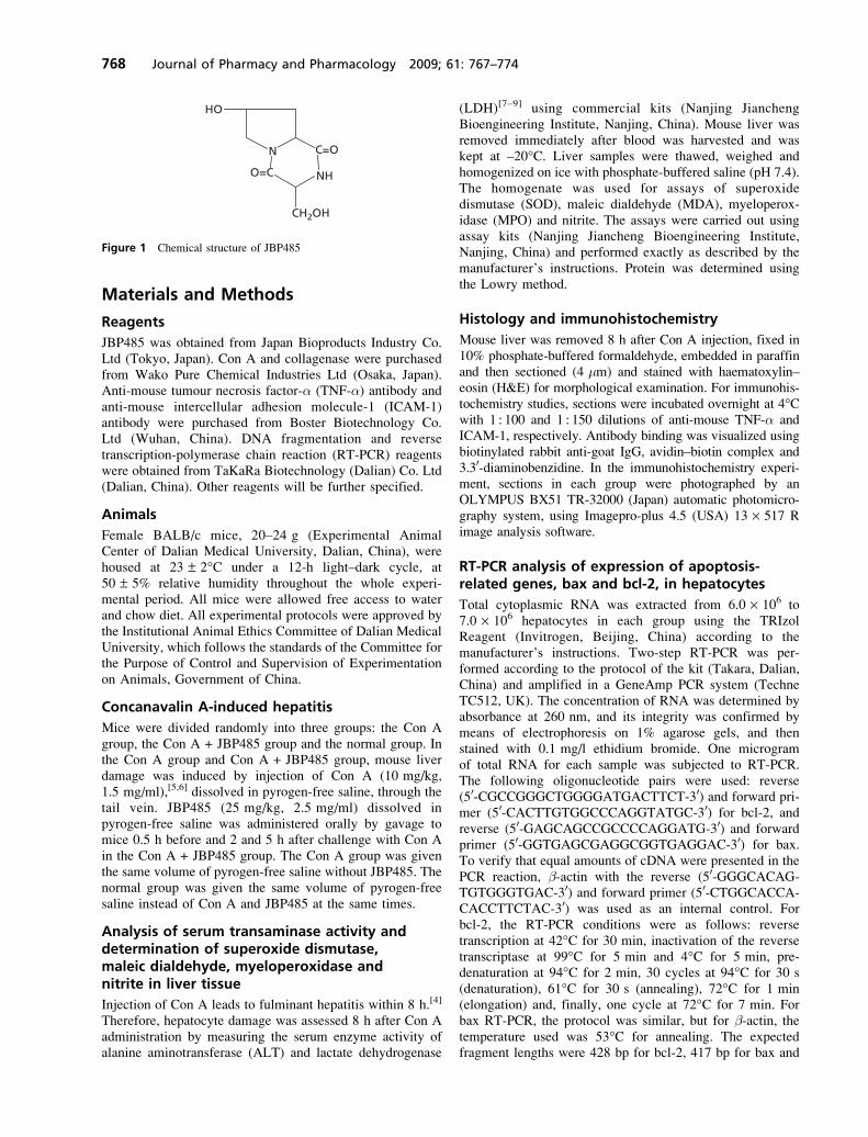
Effect of JBP485 on concanavalin A-inducedDNA fragmentation in mice
To examine whether JBP485 could inhibit apoptosis inhepatocytes, the effect of JBP485 on DNA fragmentationwas investigated. Hepatocyte DNA fragmentation was assayedby agarose gel electrophoresis (Figure 5). The characteristicDNA ladder was found in the Con A group; however, theDNA ladder was significantly attenuated in the JBP485-treated group, which indicated that JBP485 could inhibithepatocyte apoptosis induced by Con A injection in mice.
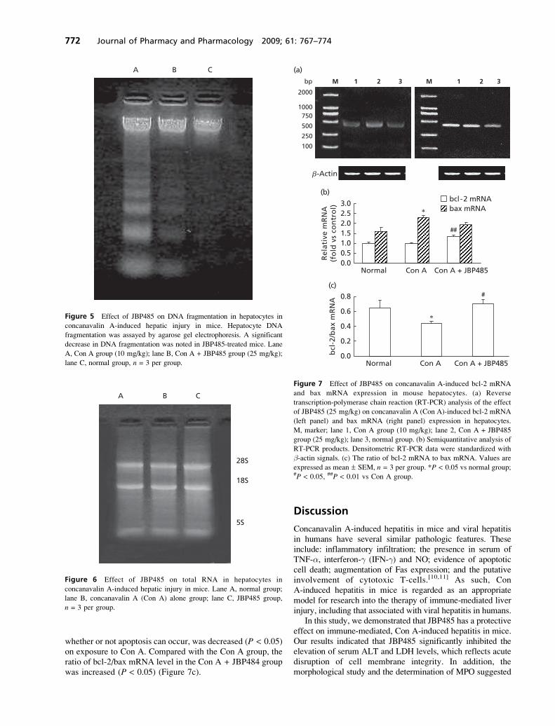
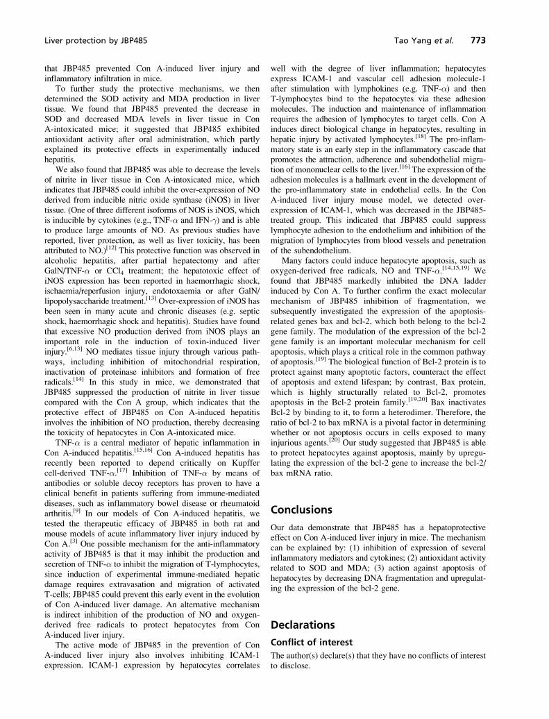
RT-PCR analysis of mRNA of apoptosis-relatedgenes bax and bcl-2
To further confirm the effect of JBP485 on inhibition ofapoptosis in Con A-treated mice, we subsequently investi-gated the expression of the apoptosis-related genes bax andbcl-2 by semiquantitative RT-PCR. The total RNA extractedfrom each group was loaded into 1% agarose gel. Bandsat 28S, 18S and 5S could be clearly observed, whichconfirmed the integrity of the total RNA (Figure 6). In theRT-PCR, 5 ml bax, bcl-2 and b-actin PCR product wereseparated by electrophoresis and revealed by ethidiumbromide staining. The PCR products of bax, bcl-2 and b-actin were 417 bp, 428 bp and 364 bp in length, respectively(Figure 7a). There was no change in the bcl-2 mRNA level8 h after Con A administration compared with the normalgroup. However, after treatment with JBP485, the bcl-2mRNA level increased compared with the Con A group at8 h after Con A administration (P < 0.01) (Figure 7b). Thebax mRNA level in the Con A group increased comparedwith the normal group, but no significant change was foundbetween the Con A and Con A + JBP485 groups (Figure 7b).The ratio of bcl-2/bax mRNA level, which determines
100 �m
(a)
(b)
(c)
(d)
(e)
(f)
100 �m
100 �m 100 �m
100 �m 100 �m
Figure 4 Histopathological changes in liver of mice after induction of liver injury with concanavalin A and treatment with JBP485. Changes in
pathology were observed following immunohistochemical staining for tumour necrosis factor-a (TNF-a) (a, b, c) and intracellular adhesion molecule-
1 (ICAM-1) (d, e, f) in liver tissue 8 h after concanavalin A (Con A) treatment in BALB/c mice with (b, e) or without (a, d) JBP485 (25 mg/kg). In the
Con A group, there was a marked overexpression of TNF-a (straight arrows) and ICAM-1 (curved arrows) in the cytoplasm; these increases were
significantly inhibited by JBP485. (c) and (f) represent the normal group. (n = 6; magnification ¥ 400).
Liver protection by JBP485 Tao Yang et al. 771
whether or not apoptosis can occur, was decreased (P < 0.05)on exposure to Con A. Compared with the Con A group, theratio of bcl-2/bax mRNA level in the Con A + JBP484 groupwas increased (P < 0.05) (Figure 7c).
Discussion
Concanavalin A-induced hepatitis in mice and viral hepatitisin humans have several similar pathologic features. Theseinclude: inflammatory infiltration; the presence in serum ofTNF-a, interferon-g (IFN-g) and NO; evidence of apoptoticcell death; augmentation of Fas expression; and the putativeinvolvement of cytotoxic T-cells.[10,11] As such, ConA-induced hepatitis in mice is regarded as an appropriatemodel for research into the therapy of immune-mediated liverinjury, including that associated with viral hepatitis in humans.
In this study, we demonstrated that JBP485 has a protectiveeffect on immune-mediated, Con A-induced hepatitis in mice.Our results indicated that JBP485 significantly inhibited theelevation of serum ALT and LDH levels, which reflects acutedisruption of cell membrane integrity. In addition, themorphological study and the determination of MPO suggested
A B C
Figure 5 Effect of JBP485 on DNA fragmentation in hepatocytes in
concanavalin A-induced hepatic injury in mice. Hepatocyte DNA
fragmentation was assayed by agarose gel electrophoresis. A significant
decrease in DNA fragmentation was noted in JBP485-treated mice. Lane
A, Con A group (10 mg/kg); lane B, Con A + JBP485 group (25 mg/kg);
lane C, normal group, n = 3 per group.
A B C
28S
18S
5S
Figure 6 Effect of JBP485 on total RNA in hepatocytes in
concanavalin A-induced hepatic injury in mice. Lane A, normal group;
lane B, concanavalin A (Con A) alone group; lane C, JBP485 group,
n = 3 per group.
bp M 1 2 3 M 1 2 3
2000
1000750
500
250
100
(a)
0.00.51.01.52.02.53.0
(b)
Rel
ativ
e m
RN
A(f
old
vs
con
tro
l) ∗
##
Normal Con A Con A + JBP485
(c)
∗
0.0
0.2
0.4
0.6
0.8
bcl
-2/b
ax m
RN
A
#
Normal Con A Con A + JBP485
bcl-2 mRNAbax mRNA
�-Actin
Figure 7 Effect of JBP485 on concanavalin A-induced bcl-2 mRNA
and bax mRNA expression in mouse hepatocytes. (a) Reverse
transcription-polymerase chain reaction (RT-PCR) analysis of the effect
of JBP485 (25 mg/kg) on concanavalin A (Con A)-induced bcl-2 mRNA
(left panel) and bax mRNA (right panel) expression in hepatocytes.
M, marker; lane 1, Con A group (10 mg/kg); lane 2, Con A + JBP485
group (25 mg/kg); lane 3, normal group. (b) Semiquantitative analysis of
RT-PCR products. Densitometric RT-PCR data were standardized with
b-actin signals. (c) The ratio of bcl-2 mRNA to bax mRNA. Values are
expressed as mean ± SEM, n = 3 per group. *P < 0.05 vs normal group;#P < 0.05, ##P < 0.01 vs Con A group.
772 Journal of Pharmacy and Pharmacology 2009; 61: 767–774
that JBP485 prevented Con A-induced liver injury andinflammatory infiltration in mice.
To further study the protective mechanisms, we thendetermined the SOD activity and MDA production in livertissue. We found that JBP485 prevented the decrease inSOD and decreased MDA levels in liver tissue in ConA-intoxicated mice; it suggested that JBP485 exhibitedantioxidant activity after oral administration, which partlyexplained its protective effects in experimentally inducedhepatitis.
We also found that JBP485 was able to decrease the levelsof nitrite in liver tissue in Con A-intoxicated mice, whichindicates that JBP485 could inhibit the over-expression of NOderived from inducible nitric oxide synthase (iNOS) in livertissue. (One of three different isoforms of NOS is iNOS, whichis inducible by cytokines (e.g., TNF-a and IFN-g) and is ableto produce large amounts of NO. As previous studies havereported, liver protection, as well as liver toxicity, has beenattributed to NO.)[12] This protective function was observed inalcoholic hepatitis, after partial hepatectomy and afterGalN/TNF-a or CCl4 treatment; the hepatotoxic effect ofiNOS expression has been reported in haemorrhagic shock,ischaemia/reperfusion injury, endotoxaemia or after GalN/lipopolysaccharide treatment.[13] Over-expression of iNOS hasbeen seen in many acute and chronic diseases (e.g. septicshock, haemorrhagic shock and hepatitis). Studies have foundthat excessive NO production derived from iNOS plays animportant role in the induction of toxin-induced liverinjury.[6,13] NO mediates tissue injury through various path-ways, including inhibition of mitochondrial respiration,inactivation of proteinase inhibitors and formation of freeradicals.[14] In this study in mice, we demonstrated thatJBP485 suppressed the production of nitrite in liver tissuecompared with the Con A group, which indicates that theprotective effect of JBP485 on Con A-induced hepatitisinvolves the inhibition of NO production, thereby decreasingthe toxicity of hepatocytes in Con A-intoxicated mice.
TNF-a is a central mediator of hepatic inflammation inCon A-induced hepatitis.[15,16] Con A-induced hepatitis hasrecently been reported to depend critically on Kupffercell-derived TNF-a.[17] Inhibition of TNF-a by means ofantibodies or soluble decoy receptors has proven to have aclinical benefit in patients suffering from immune-mediateddiseases, such as inflammatory bowel disease or rheumatoidarthritis.[9] In our models of Con A-induced hepatitis, wetested the therapeutic efficacy of JBP485 in both rat andmouse models of acute inflammatory liver injury induced byCon A.[3] One possible mechanism for the anti-inflammatoryactivity of JBP485 is that it may inhibit the production andsecretion of TNF-a to inhibit the migration of T-lymphocytes,since induction of experimental immune-mediated hepaticdamage requires extravasation and migration of activatedT-cells; JBP485 could prevent this early event in the evolutionof Con A-induced liver damage. An alternative mechanismis indirect inhibition of the production of NO and oxygen-derived free radicals to protect hepatocytes from ConA-induced liver injury.
The active mode of JBP485 in the prevention of ConA-induced liver injury also involves inhibiting ICAM-1expression. ICAM-1 expression by hepatocytes correlates
well with the degree of liver inflammation; hepatocytesexpress ICAM-1 and vascular cell adhesion molecule-1after stimulation with lymphokines (e.g. TNF-a) and thenT-lymphocytes bind to the hepatocytes via these adhesionmolecules. The induction and maintenance of inflammationrequires the adhesion of lymphocytes to target cells. Con Ainduces direct biological change in hepatocytes, resulting inhepatic injury by activated lymphocytes.[18] The pro-inflam-matory state is an early step in the inflammatory cascade thatpromotes the attraction, adherence and subendothelial migra-tion of mononuclear cells to the liver.[16] The expression of theadhesion molecules is a hallmark event in the development ofthe pro-inflammatory state in endothelial cells. In the ConA-induced liver injury mouse model, we detected over-expression of ICAM-1, which was decreased in the JBP485-treated group. This indicated that JBP485 could suppresslymphocyte adhesion to the endothelium and inhibition of themigration of lymphocytes from blood vessels and penetrationof the subendothelium.
Many factors could induce hepatocyte apoptosis, such asoxygen-derived free radicals, NO and TNF-a.[14,15,19] Wefound that JBP485 markedly inhibited the DNA ladderinduced by Con A. To further confirm the exact molecularmechanism of JBP485 inhibition of fragmentation, wesubsequently investigated the expression of the apoptosis-related genes bax and bcl-2, which both belong to the bcl-2gene family. The modulation of the expression of the bcl-2gene family is an important molecular mechanism for cellapoptosis, which plays a critical role in the common pathwayof apoptosis.[19] The biological function of Bcl-2 protein is toprotect against many apoptotic factors, counteract the effectof apoptosis and extend lifespan; by contrast, Bax protein,which is highly structurally related to Bcl-2, promotesapoptosis in the Bcl-2 protein family.[19,20] Bax inactivatesBcl-2 by binding to it, to form a heterodimer. Therefore, theratio of bcl-2 to bax mRNA is a pivotal factor in determiningwhether or not apoptosis occurs in cells exposed to manyinjurious agents.[20] Our study suggested that JBP485 is ableto protect hepatocytes against apoptosis, mainly by upregu-lating the expression of the bcl-2 gene to increase the bcl-2/bax mRNA ratio.
Conclusions
Our data demonstrate that JBP485 has a hepatoprotectiveeffect on Con A-induced liver injury in mice. The mechanismcan be explained by: (1) inhibition of expression of severalinflammatory mediators and cytokines; (2) antioxidant activityrelated to SOD and MDA; (3) action against apoptosis ofhepatocytes by decreasing DNA fragmentation and upregulat-ing the expression of the bcl-2 gene.
Declarations
Conflict of interest
The author(s) declare(s) that they have no conflicts of interestto disclose.
Liver protection by JBP485 Tao Yang et al. 773

Funding
This work was supported by a grant from the NationalNatural Science Foundation of China (No. 30672498) andNatural Science Foundation of Liaoning Province technolo-gical office (No. 2004225003-6). Special thanks go to JapanBioproducts Co. Ltd for their fund support.
References
1. Liu KX et al. Human placental extract stimulates liver
regeneration in rats. Biol Pharm Bull 1998; 21: 44–49.
2. Liu KX et al. Hydroxyprolylserine derivatives JBP923 and
JBP485 exhibit the antihepatitis activities after gastrointestinal
absorption in rats. J Pharmacol Exp Ther 2000; 294: 510–515.
3. Wu JJ et al. Protective effect of JBP485 on concanavalin
A-induced hepatocyte toxicity in primary cultured rat hepato-
cytes. Eur J Pharmacol 2008; 589(1–3): 299–305.
4. Tiegs G et al. A T cell-dependent experimental liver injury in mice
inducible by concanavalin A. J Clin Invest 1992; 90: 196–203.
5. Okamoto T et al. Aminoguanidine prevents concanavalin
A-induced hepatitis in mice. Eur J Pharmacol 2000; 396:
125–130.
6. Ksontini R et al. Disparate roles for TNF-a and Fas ligand in
concanavalin A-induced hepatitis. J Immunol 1998; 160: 4082–
4089.
7. Ajuebor MN et al. C-C chemokine ligand 2/monocyte
chemoattratant protein-1 directly inhibits NKT cell IL-4
production and is hepatoprotective in T cell-mediated hepatitis
in the mouse. J Immunol 2003; 170: 5252–5259.
8. Hentze H et al. Depletion of hepatic glutathione prevents death
receptor-dependent apoptotic and necrotic liver injury in mice.
Am J Pathol 2000; 156: 2045–2056.
9. Wolf AM et al. The kinase inhibitor imatinib mesylate inhibits
TNF-a production in vitro and prevents TNF-dependent acute
hepatic inflammation. Proc Natl Acad Sci 2005; 102: 13622–
13627.
10. Kusters S et al. Interferon gamma plays a critical role in T-cell-
dependent liver injury in mice initiated by concanavalin A.
Gastoroenterology 1996; 111: 462–471.
11. Yoneda M et al. A novel therapy for acute hepatitis utilizing
dehydroepiandrosterone in the murine model of hepatitis.
Biochem Pharmacol 2004; 68: 2283–2289.
12. Li J, Billiar TR. Determinants of nitric oxide protection and
toxicity in the liver. Am J Physiol 1999; 276: G1069–G1073.
13. Sass G et al. Inducible nitric oxide synthase is critical for immune-
mediated liver injury in mice. J Clin Invest 2001; 107: 439–447.
14. Marshall HE et al. Nitrosation and oxidant in the regulation of
gene expression. FASEB J 2000; 14: 1889–1900.
15. Mizuhara H et al. T cell activation-associated hepatic injury:
mediation by tumor necrosis factor and protection by inter-
leukin 6. J Exp Med 1994; 179: 1529–1537.
16. Bruck R et al. Allicin, the active component of garlic, prevents
immune-mediated, concanavalin A-induced hepatic injury in
mice. Liver Int 2005; 25: 613–621.
17. Schumann J. Importance of Kupffer cells for T-cell-dependent
liver injury in mice. Am J Pathol 2000; 157: 1671–1683.
18. Wang ZZ et al. Protection of Veratrum nigrum L.var. ussuriense
Nakai alkaloids against ischemia-reperfusion injury of the rat
liver. World J Gastroenterol 2007; 13: 564–571.
19. Gao H, Zhou YW. Inhibitory effect of picroside P on
hepatocyte apoptosis. Acta Pharmacol Sin 2005; 26: 729–736.
20. Cheng BH et al. D-b-Hydroxybutyrate inhibits the apoptosis of
PC12 cells induced by 6-OHDA in relation to up-regulating the
ratio of Bcl-2/Bax mRNA. Auton Neurosci-Basic 2007; 134:
38–44.
774 Journal of Pharmacy and Pharmacology 2009; 61: 767–774
Copyright © 2022 FDOKUMEN