Hepatocyte lesions and cellular immune response in yellow fever infection
8
Transactions of the Royal Society of Tropical Medicine and Hygiene (2007) 101, 161—168 available at www.sciencedirect.com journal homepage: www.elsevierhealth.com/journals/trst Hepatocyte lesions and cellular immune response in yellow fever infection Juarez A.S. Quaresma a,b,∗ , Vera L.R.S. Barros c , Carla Pagliari d , Elaine R. Fernandes d , Heitor F. Andrade Jr d , Pedro F.C. Vasconcelos c , Maria I.S. Duarte d a Tropical Medicine Center, Federal University of Para, Av. Generalissimo Deodoro 92, Belem, 66055-240, Brazil b Department of Pathology, State University of Para, Trav. Perebebui 2623, Belem, 66087-070, Brazil c Evandro Chagas Institute, Ministry of Health, Av. Almirante Barroso 492, Belem, 66090-000, Brazil d Faculty of Medicine, State University of Sao Paulo, Av. Dr. Arnaldo 455, Sala 1118, Sao Paulo, 01246-903, Brazil Received 31 January 2005; received in revised form 23 February 2006; accepted 24 February 2006 Available online 26 July 2006 KEYWORDS Yellow fever; Arbovirus; Flavivirus; Immunologic markers; Apoptosis; Brazil Summary The study of the in-situ cellular immune response is very important for the under- standing of different liver infections. In the present study, 53 liver samples obtained by vis- cerotomy from patients who died during the course of jungle yellow fever were analyzed. The diagnosis was confirmed by serology, viral isolation and virus-specific immunohistochemistry. The specimens were analyzed by immunohistochemistry using specific antibodies for apoptosis, CD45RO, CD4, CD8, CD20, S100, CD57 and CD68. Quantitative analysis of the labeling pattern showed a clear predominance of the different phenotypes in the portal tract and midzone region of the acini. There was a predominance of T CD4+ lymphocytes, accompanied by the presence of T CD8+ lymphocytes, natural killer cells (CD57), macrophages and antigen-presenting cells (S100). The disproportion between the intensity of inflammation and the degree of hepatic injury was probably due to the intense apoptotic component, which classically does not induce an inflammatory response. The present study demonstrates that, despite the disproportion between injury and inflammation, the cellular immune response plays an important role in the pathogenesis of the hepatocytic injury observed in yellow fever, probably as a result of cytolytic actions through mechanisms involving MHC II and the activation of Fas receptors and granzymes/perforins. © 2006 Royal Society of Tropical Medicine and Hygiene. Published by Elsevier Ltd. All rights reserved. ∗ Corresponding author. Tel.: +55 91 3241 4681; fax: +55 91 241 0032. E-mail address: [email protected] (J.A.S. Quaresma). 1. Introduction Yellow fever (YF) is caused by an arbovirus of the genus Fla- vivirus, family Flaviviridae (Westaway et al., 1985). There are two different epidemiologic forms of YF, which differ in 0035-9203/$ — see front matter © 2006 Royal Society of Tropical Medicine and Hygiene. Published by Elsevier Ltd. All rights reserved. doi:10.1016/j.trstmh.2006.02.019
-
Upload
independent -
Category
Documents
-
view
2 -
download
0
Transcript of Hepatocyte lesions and cellular immune response in yellow fever infection

Transactions of the Royal Society of Tropical Medicine and Hygiene (2007) 101, 161—168
avai lab le at www.sc iencedi rec t .com
journa l homepage: www.e lsev ierhea l th .com/ journa ls / t rs t
Hepatocyte lesions and cellular immune responsein yellow fever infection
Juarez A.S. Quaresmaa,b,∗, Vera L.R.S. Barrosc, Carla Pagliari d,Elaine R. Fernandesd, Heitor F. Andrade Jrd,Pedro F.C. Vasconcelosc, Maria I.S. Duarted
a Tropical Medicine Center, Federal University of Para, Av. Generalissimo Deodoro 92, Belem, 66055-240, Brazilb Department of Pathology, State University of Para, Trav. Perebebui 2623, Belem, 66087-070, Brazilc Evandro Chagas Institute, Ministry of Health, Av. Almirante Barroso 492, Belem, 66090-000, Brazild Faculty of Medicine, State University of Sao Paulo, Av. Dr. Arnaldo 455, Sala 1118, Sao Paulo, 01246-903, Brazil
Received 31 January 2005; received in revised form 23 February 2006; accepted 24 February 2006Available online 26 July 2006
KEYWORDSYellow fever;Arbovirus;Flavivirus;Immunologicmarkers;Apoptosis;Brazil
Summary The study of the in-situ cellular immune response is very important for the under-standing of different liver infections. In the present study, 53 liver samples obtained by vis-cerotomy from patients who died during the course of jungle yellow fever were analyzed. Thediagnosis was confirmed by serology, viral isolation and virus-specific immunohistochemistry.The specimens were analyzed by immunohistochemistry using specific antibodies for apoptosis,CD45RO, CD4, CD8, CD20, S100, CD57 and CD68. Quantitative analysis of the labeling patternshowed a clear predominance of the different phenotypes in the portal tract and midzone regionof the acini. There was a predominance of T CD4+ lymphocytes, accompanied by the presenceof T CD8+ lymphocytes, natural killer cells (CD57), macrophages and antigen-presenting cells(S100). The disproportion between the intensity of inflammation and the degree of hepaticinjury was probably due to the intense apoptotic component, which classically does not inducean inflammatory response. The present study demonstrates that, despite the disproportion
between injury and inflammation, the cellular immune response plays an important role inthe pathogenesis of the hepatocytic injury observed in yellow fever, probably as a result ofcytolytic actions through mechanisms involving MHC II and the activation of Fas receptors andgranzymes/perforins.© 2006 Royal Society of Tropica1
reserved.
∗ Corresponding author. Tel.: +55 91 3241 4681;fax: +55 91 241 0032.
E-mail address: [email protected] (J.A.S. Quaresma).
Yva
0035-9203/$ — see front matter © 2006 Royal Society of Tropical Medicindoi:10.1016/j.trstmh.2006.02.019
l Medicine and Hygiene. Published by Elsevier Ltd. All rights
. Introduction
ellow fever (YF) is caused by an arbovirus of the genus Fla-ivirus, family Flaviviridae (Westaway et al., 1985). Therere two different epidemiologic forms of YF, which differ in
e and Hygiene. Published by Elsevier Ltd. All rights reserved.

1 J.A.S. Quaresma et al.
tdaoittwoaAtfncAaoiiha
lisa1tta(cetpasi
2
2i
LofimeBTigmtmu1ta
Table 1 Characteristics of 53 Brazilian yellow feverpatients
State No. (%) Male Female Clinical form
Goias 15 (28) 13 2 HemorrhagicPara 8 (15) 8 0 HemorrhagicMaranhao 7 (13) 4 3 HemorrhagicRoraima 6 (11) 5 1 HemorrhagicMinas Gerais 5 (9) 5 0 HemorrhagicTocantins 4 (8) 4 0 HemorrhagicAmazonas 3 (6) 3 0 Hemorrhagic
To
2
THtaifS
ma(I
2
Tsphalo
2
Atwyf
2
T
62
heir transmission cycle. In urban YF, the virus is transmittedirectly to humans by the mosquito Aedes aegypti in urbanreas, while jungle YF is transmitted by various mosquitoesf the genus Aedes in Africa, and Haemagogus and Sabethesn the Americas. In jungle cycles, non-human primates arehe primary vertebrate hosts on both continents and, asransmission is sylvatic, human infections are incidentalhen humans enter the forest while working, such as whenpening roads and exploring the area, etc. (Degallier etl., 1992). Urban transmission has not been reported in themericas since 1959. The present level of A. aegypti infes-ation in large urban centers near YF endemic areas, therequent migration of populations between endemic andon-endemic areas, and a low YF vaccine coverage in manyountries mean that re-urbanization of the disease in themericas could happen (Vasconcelos, 2003; Vasconcelos etl., 2004). Consequently, YF once again represents a seri-us public health problem in the New World. The dangers particularly great in Brazil, the most populated countryn the endemic area, and increasing figures of YF activityave been recorded over the last 5 years (Vasconcelos etl., 2001, 2004).
Since the classical studies of Councilman, the histopatho-ogic presentation of the liver in YF has been character-zed by the presence of hepatocytic lesions represented byteatosis, necrosis and apoptosis (Councilman, 1890; Klotznd Belt, 1930a, 1930b; Quaresma et al., 2005; Rocha-Lima,912). This intense picture of tissue injury is characteris-ically accompanied by an inflammation that is dispropor-ional to the degree of liver tissue involvement and by thebsence of viral particles in electron microscopy materialQuaresma et al., 2005; Vieira et al., 1983). Despite thisharacteristic picture, no studies are available in the lit-rature regarding the local immune response and its rela-ionship with hepatic lesions in YF. The objective of theresent study was to analyze qualitative and quantitativespects of the cellular immune response and its relation-hip with the occurrence and distribution of hepatic lesionsn YF.
. Materials and methods
.1. Histologic examination andmmunohistochemical detection of YF virus antigen
iver samples obtained from the archives of the Departmentf Pathology, Evandro Chagas Institute (Belem, Brazil), werexed in 10% neutral-buffered formalin, paraffin embedded,icron-thick sectioned and stained with hematoxylin and
osin. All 53 patients, 46 males and 7 females, were fromrazil (Table 1) and ranged in age from 3 to 74 years.he diagnosis was made by serology, viral isolation and
mmunohistochemistry for the detection of specific YF anti-ens using monoclonal antibodies (Hall et al., 1991). Five-icrometer-thick sections were also stained with Masson’s
richrome, reticulin and Perl’s stain for examination by light
icroscopy. The sections were evaluated semiquantitativelysing a subjective scale ranging from 0 to 3, where 0 = none,= mild, 2 = moderate and 3 = severe damage, according to
he degree of inflammatory infiltration, hepatic apoptosisnd steatosis, as previously described (Xiao et al., 2001).
tiici
Mato Grosso do Sul 3 (6) 2 1 HemorrhagicMato Grosso 1 (2) 1 0 HemorrhagicDistrito Federal 1 (2) 1 0 Hemorrhagic
he project was approved by the Ethics and Research boardsf the Evandro Chagas Institute.
.2. Immunologic markers
he immunohistochemical technique originally described bysu et al. (1981) was used to characterize the phenotype ofhe inflammatory cells. Immunostaining for the detection ofpoptosis was carried out according to the manufacturer’snstructions, as previously described (Gold et al., 1994). Theollowing antibodies were used: CD45RO, CD4, CD8, CD20,100, CD57, CD68 and anti-apoptosis (ApopTag).
A grid-scale with 10 × 10 subdivisions in an area of 0.0625m2 was used for quantitative analysis of cell phenotype and
poptosis. Ten fields were counted at large magnification×400) in all three areas of the acini (I = periportal area;I = midzone; III = central vein area).
.3. Negative controls
he following liver samples obtained during routine autop-ies were included as negative controls: five liver sam-les from patients with negative serology for the mainepatotropic viruses, who showed no morphologic alter-tions; and five liver specimens from cases diagnosed aseptospirosis based on clinical presentation, specific serol-gy, histopathology and immunohistochemical analysis.
.4. Statistical analysis
ll data are reported as mean ± SD and were analyzed sta-istically by one-way ANOVA followed by the Bonferroni test,ith the level of significance set at P ≤ 0.05. Statistical anal-sis was performed using the GraphPad Prism 3.0 softwareor Windows (GraphPad Software, San Diego, CA, USA).
.5. Theory
he study of the immune response is of great importance for
he understanding of the evolution of tissue lesions inherentn different infections (Bertolleti and Maini, 2000). In viralnfections affecting the liver, such as hepatitis B and C, theytotoxic cellular response and the action of cytokines aremportant factors involved in the control of viral replication
Hepatocytes and cellular immunity in yellow fever
Figure 1 Semiquantitative analysis of histopathologic eventsin the hepatic acini. AP: apoptosis; NEC: necrosis; ST: steatosis;
st2vmiwiliartoaabraih
3
I: hepatic area 1; II: hepatic area 2; III: hepatic area 3 (*P ≤ 0.05;**P ≤ 0.001; ANOVA followed by Bonferroni).
and hepatocyte death (Willuweit et al., 2001). Indeed, theexistence of clinically asymptomatic patients infected withhepatitis B virus indicates the importance of the immuneresponse in the genesis of tissue lesions and, consequently,
its determining role in the evolution of the disease (Ganemand Prince, 2004). Furthermore, over the last few years, thedifferentiated clinical evolution of individuals in responseto the same etiologic agent has led to increased empha-Hssm
Figure 2 Histopathology and immunohistochemistry of yellow fe(HE, original magnification ×400). (B) Inflammatory portal infiltratImmunohistochemical staining showing large amounts of YF antigenapoptotic hepatocytes (×200).
163
is on the importance of the role of the immune system inhe evolution of hepatic viral infections (Willuweit et al.,001). In addition to these eminently hepatotropic agents,iral infections that progress with important vascular impair-ent have been the target of studies, because they cause
mportant and characteristic hepatic lesions that are still notell understood (Zaki and Peters, 1997). Among these viral
nfections, dengue and YF raise attention due to their pecu-iar pathogenesis, which is characterized by intense hepaticnvolvement (Monath, 1995, 2001). Furthermore, the mech-nisms that induce tissue lesions and the type of immuneesponse in the human host are poorly understood, despitehe classic description of the main histopathologic aspectsf hepatic injury during YF infection more than a centurygo (Councilman, 1890; Rocha-Lima, 1912). Hepatic dam-ge caused by the YF virus is intense and is characterizedy a preferential involvement of the midzone area, whichesults in necrosis and eosinophilic degeneration of the hep-tocytes, associated with a discrete lymphomononuclearnflammatory response disproportionate to the degree ofepatocyte involvement (Vieira et al., 1983).
. Results
istologic analysis confirmed previous findings and demon-trated hepatocytic alterations characterized by lytic necro-is, steatosis and apoptosis, which showed a preferentialidzone distribution and were accompanied by a mild to
ver (YF) in human liver. (A) Councilman bodies and steatosise of mononuclear cells (HE, original magnification ×200). (C)s (in red) (×200). (D) Immunohistochemical staining showing

164 J.A.S. Quaresma et al.
Figure 3 Immunohistochemical staining of immune cells in human liver, using specific antibodies (×400). (A) CD45RO lympho-c imml
moman
sfCo
tfizb
ytes in the portal tract. (B—D) Portal tract, showing intenseymphocytes (D).
oderate inflammatory infiltrate predominantly consistingf mononuclear cells. Semiquantitative evaluation of theorphologic events observed in the present material showedclear predominance of hepatocyte apoptosis over lytic
ecrosis (Figures 1 and 2A, B).Immunohistochemical analysis of the lesions demon-
trated positivity for hepatocyte apoptosis (ApopTag) andor inflammatory cells of the CD45RO, CD4, CD8, CD20, S100,D68 and CD57 type (Figures 2—4). The quantitative analysisf the inflammatory cells is shown in Table 2.
lAah
Table 2 Immunohistochemical analysis of lesions from 53 Brazili
Antibody Mean ± SDa
Area Ib Area IIb
ApopTag 2.51 ± 0.64 16.41 ±CD45RO (Pan T) 2.29 ± 1.09 4.97 ±CD20 (Pan B) 0.52 ± 0.24 1.03 ±CD4 1.70 ± 0.55 3.04 ±CD8 1.20 ± 0.41 2.63 ±CD57 0.46 ± 0.17 1.04 ±S100 0.11 ± 0.08 0.29 ±CD68 2.66 ± 1.09 5.54 ±*P ≤ 0.05; **P ≤ 0.001; ANOVA followed by Bonferronni.a Results per mm2 of tissue of cellular phenotype in the portal tract anb Areas I, II and III: hepatic areas 1, 2 and 3, respectively.
unostaining for CD4 lymphocytes (B); CD8 lymphocytes (C); B
ApopTag-labeled cells were observed in all regions ofhe lobule, mainly including hepatocytes, but also Kupf-er cells and some mononucleated inflammatory cells. Morentense immunolabeling was observed in hepatocytes of mid-one areas (P ≤ 0.001). The generally frequent immunola-eled apoptotic hepatocytes corresponded to the morpho-
ogic aspect characteristic of Councilman bodies (Figure 5A).smaller percentage of these bodies was not immunore-ctive by this technique, and immunolabeling of apoptoticepatocytes in normal controls (Area I = 0.05 ± 0.00; Area
an yellow fever patients
Area IIIb Portal tract
3.07** 1.48 ± 0.73 —1.85* 0.61 ± 0.20 10.26 ± 2.95**
0.28* 0.28 ± 0.14 3.19 ± 1.89**
0.59* 0.81 ± 0.29 11.24 ± 2.93**
0.85* 0.55 ± 0.17 7.52 ± 2.09**
0.32* 0.24 ± 0.24 1.37 ± 0.45**
0.14* 0.08 ± 0.10 1.15 ± 0.49**
1.33* 1.24 ± 0.59 1.30 ± 0.45**
d three acinar areas.

Hepatocytes and cellular immunity in yellow fever
Figure 4 Immunohistochemical staining of immune cells inhuman liver, using specific antibodies (×400). (A) Natural killercells (CD57) stained in the midzone area. (B) Antigen-presenting
lmppottwlt
aouab
tdocf
bti
4
AadiliB(eita
ciiciiK1ttwstninduce gene expression and the synthesis of proteins such as
cells (S100) stained in the portal tract. (C) CD68 cells stained inthe midzone area.
II = 0.07 ± 0.01; Area III = 0.02 ± 0.00) and leptospirosis (AreaI = 0.62 ± 0.05; Area II = 0.98 ± 0.45; Area III = 0.30 ± 0.00)was less and not significant.
Cells immunolabeled with anti-CD45RO antibodies wereobserved in both the acini and in the portal tract. In theacini, immunolabeling was more frequent in the midzone,followed by zones I and III, respectively (P ≤ 0.05). At times,aggregates of immunolabeled T lymphocytes and neutrophilswere observed around areas of lytic hepatocyte necrosis(Figure 5B). In the portal tract, the immunohistochemicalpattern demonstrated a higher density of immunolabeled T
lymphocytes than in the acini (P < 0.001) (Figure 5C, D).CD4+ and CD8+ T cells were observed throughout the hep-atic parenchyma, both in the portal tract (P < 0.001) andin the acini, with a higher concentration in zone II, fol-
cl
t
165
owed by zones I and III (Figure 5C, D). CD4+ T cells wereore frequent than CD8+ T cells, both in the acini andortal tract. Immunolabeled cells exceeding the limitinglaque and arranged in the periportal region were frequentlybserved. Anti-CD20-immunolabeled cells were observed inhe acini and portal tract. In the acini, the frequency ofhese cells accompanied that observed for T cells, i.e. thereas predominance in zone II (P ≤ 0.05). A lower density of B
ymphocytes than T lymphocytes was observed in the portalract (Figure 5E).
Anti-CD68 positivity was detected in Kupffer cells in thecini and in macrophages in the portal tract. The frequencyf immunolabeled cells in the areas of the hepatic lob-les continued to show a midzone predominance (P ≤ 0.05),lthough the quantitative difference in the positivity patternetween zones I, II and III was less significant (Figure 5F).
Anti-CD57-immunolabeled cells were observed both inhe acini and in the portal tract, with their distribution in theifferent zones of the lobule demonstrating a predominancenly in zone II (P ≤ 0.05). Immunolabeled natural killer (NK)ells were frequent in the portal tract and showed a higherrequency per area than in the acini (P ≤ 0.05) (Figure 5G).
Antigen-presenting cells labeled with the anti-S100 anti-ody were present in both acini and portal tract, withheir distribution in the acini demonstrating predominancen zone II (P < 0.05) (Figure 5H).
. Discussion
fact that calls attention to YF, in addition to the intensepoptotic component, is the disproportion between theegree of parenchymal involvement and the scarcity of annflammatory infiltrate, a remarkable characteristic of theiver in this disease. This marked characteristic has beennvestigated in classical studies (Hudson, 1928; Klotz andelt, 1930a, 1930b) and was later confirmed by Smetana1962), Vieira et al. (1983), Branquet (1996) and Quaresmat al. (2005), both in human material and in experimentallynfected monkeys. In the present samples, this characteris-ic poor inflammatory response was observed in the lobulend the portal tract.
Some data are important to an understanding of theauses of this discrete inflammatory component. First, theres the intense degree of apoptosis in hepatocytes thatnduces no important inflammatory response. In this pro-ess, the apoptotic bodies are phagocytosed by neighbor-ng local macrophages and thus do not elicit large regionalnflammatory alterations (Kaufmann and Hengartner, 2001;err et al., 1972; Majno and Joris, 1995; Tran and Miller,999). However, recent studies have demonstrated neu-rophil migration to the hepatic parenchyma in rats duringhe course of apoptosis induced by experimental infectionith Listeria monocytogenes. Park et al. (2002) demon-
trated that the activation of Fas receptors by the adminis-ration of moderate doses of anti-Fas antibodies, accompa-ied by the induction of hepatocyte apoptosis, was able to
hemokines and macrophage inflammatory proteins, whiched to the accumulation of neutrophils.
It should be emphasized that, in the present study, neu-rophils, although rare, were mainly observed adjacent to

166 J.A.S. Quaresma et al.
Figure 5 Quantitative analysis of immunostaining for yellow fever (YF), leptospirosis (LE) and negative controls (NC). In YF,comparison of the different acinar areas shows a predominance of immune response and apoptosis in the midzone area comparedwith other lobular areas. A strong immune response was also observed in the portal tract. (A) Apoptotic hepatocytes (�) andCouncilman bodies (�). (B) Neutrophils. (C) CD4+ lymphocytes. (D) CD8+ lymphocytes. (E) CD20+ cells. (F) CD68+ cells. (G) Naturalkiller cells (CD57). (H) S100 cells (antigen-presenting cells). I: hepatic area 1; II: hepatic area 2; III: hepatic area 3; PT: portal tract(*P ≤ 0.05; **P ≤ 0.001; ANOVA followed by Bonferroni).
mgain1aprTwTTiTgq
aTr(erawdlitHnalehtt
rpmbttit
cpddd
CTr
Hepatocytes and cellular immunity in yellow fever
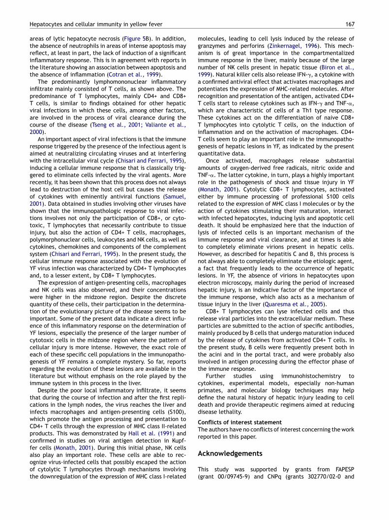
areas of lytic hepatocyte necrosis (Figure 5B). In addition,the absence of neutrophils in areas of intense apoptosis mayreflect, at least in part, the lack of induction of a significantinflammatory response. This is in agreement with reports inthe literature showing an association between apoptosis andthe absence of inflammation (Cotran et al., 1999).
The predominantly lymphomononuclear inflammatoryinfiltrate mainly consisted of T cells, as shown above. Thepredominance of T lymphocytes, mainly CD4+ and CD8+T cells, is similar to findings obtained for other hepaticviral infections in which these cells, among other factors,are involved in the process of viral clearance during thecourse of the disease (Tseng et al., 2001; Valiante et al.,2000).
An important aspect of viral infections is that the immuneresponse triggered by the presence of the infectious agent isaimed at neutralizing circulating viruses and at interferingwith the intracellular viral cycle (Chisari and Ferrari, 1995),inducing a cellular immune response that is classically trig-gered to eliminate cells infected by the viral agents. Morerecently, it has been shown that this process does not alwayslead to destruction of the host cell but causes the releaseof cytokines with eminently antiviral functions (Samuel,2001). Data obtained in studies involving other viruses haveshown that the immunopathologic response to viral infec-tions involves not only the participation of CD8+, or cyto-toxic, T lymphocytes that necessarily contribute to tissueinjury, but also the action of CD4+ T cells, macrophages,polymorphonuclear cells, leukocytes and NK cells, as well ascytokines, chemokines and components of the complementsystem (Chisari and Ferrari, 1995). In the present study, thecellular immune response associated with the evolution ofYF virus infection was characterized by CD4+ T lymphocytesand, to a lesser extent, by CD8+ T lymphocytes.
The expression of antigen-presenting cells, macrophagesand NK cells was also observed, and their concentrationswere higher in the midzone region. Despite the discretequantity of these cells, their participation in the determina-tion of the evolutionary picture of the disease seems to beimportant. Some of the present data indicate a direct influ-ence of this inflammatory response on the determination ofYF lesions, especially the presence of the larger number ofcytotoxic cells in the midzone region where the pattern ofcellular injury is more intense. However, the exact role ofeach of these specific cell populations in the immunopatho-genesis of YF remains a complete mystery. So far, reportsregarding the evolution of these lesions are available in theliterature but without emphasis on the role played by theimmune system in this process in the liver.
Despite the poor local inflammatory infiltrate, it seemsthat during the course of infection and after the first repli-cations in the lymph nodes, the virus reaches the liver andinfects macrophages and antigen-presenting cells (S100),which promote the antigen processing and presentation toCD4+ T cells through the expression of MHC class II-relatedproducts. This was demonstrated by Hall et al. (1991) andconfirmed in studies on viral antigen detection in Kupf-
fer cells (Monath, 2001). During this initial phase, NK cellsalso play an important role. These cells are able to rec-ognize virus-infected cells that possibly escaped the actionof cytolytic T lymphocytes through mechanisms involvingthe downregulation of the expression of MHC class I-relatedA
T(
167
olecules, leading to cell lysis induced by the release ofranzymes and perforins (Zinkernagel, 1996). This mech-nism is of great importance in the compartmentalizedmmune response in the liver, mainly because of the largeumber of NK cells present in hepatic tissue (Biron et al.,999). Natural killer cells also release IFN-�, a cytokine withconfirmed antiviral effect that activates macrophages andotentiates the expression of MHC-related molecules. Afterecognition and presentation of the antigen, activated CD4+cells start to release cytokines such as IFN-� and TNF-�,hich are characteristic of cells of a Th1 type response.hese cytokines act on the differentiation of naive CD8+lymphocytes into cytolytic T cells, on the induction of
nflammation and on the activation of macrophages. CD4+cells seem to play an important role in the immunopatho-
enesis of hepatic lesions in YF, as indicated by the presentuantitative data.
Once activated, macrophages release substantialmounts of oxygen-derived free radicals, nitric oxide andNF-�. The latter cytokine, in turn, plays a highly importantole in the pathogenesis of shock and tissue injury in YFMonath, 2001). Cytolytic CD8+ T lymphocytes, activatedither by immune processing of professional S100 cellselated to the expression of MHC class I molecules or by thection of cytokines stimulating their maturation, interactith infected hepatocytes, inducing lysis and apoptotic celleath. It should be emphasized here that the induction ofysis of infected cells is an important mechanism of themmune response and viral clearance, and at times is ableo completely eliminate virions present in hepatic cells.owever, as described for hepatitis C and B, this process isot always able to completely eliminate the etiologic agent,fact that frequently leads to the occurrence of hepatic
esions. In YF, the absence of virions in hepatocytes uponlectron microscopy, mainly during the period of increasedepatic injury, is an indicative factor of the importance ofhe immune response, which also acts as a mechanism ofissue injury in the liver (Quaresma et al., 2005).
CD8+ T lymphocytes can lyse infected cells and thuselease viral particles into the extracellular medium. Thesearticles are submitted to the action of specific antibodies,ainly produced by B cells that undergo maturation inducedy the release of cytokines from activated CD4+ T cells. Inhe present study, B cells were frequently present both inhe acini and in the portal tract, and were probably alsonvolved in antigen processing during the effector phase ofhe immune response.
Further studies using immunohistochemistry toytokines, experimental models, especially non-humanrimates, and molecular biology techniques may helpefine the natural history of hepatic injury leading to celleath and provide therapeutic regimens aimed at reducingisease lethality.
onflicts of interest statementhe authors have no conflicts of interest concerning the workeported in this paper.
cknowledgements
his study was supported by grants from FAPESPgrant 00/09745-9) and CNPq (grants 302770/02-0 and

1
4A
R
B
B
BC
C
C
D
G
G
H
H
H
K
K
K
K
M
M
M
P
Q
R
S
S
T
T
V
V
V
V
V
W
W
X
68
01349/2004-7). We thank Rosana Cardoso and Marialvaraujo for assistance at the beginning of this study.
eferences
ertolleti, A., Maini, M.K., 2000. Protection or damage: a dual rolefor the virus-specific cytotoxic T lymphocyte response in hepati-tis B and C infections? Curr. Opin. Immunol. 12, 403—408.
iron, C.A., Nguyen, K.B., Pien, G.C., Cousens, L.P., Salazar-Mather,T.P., 1999. Natural killer cells in antiviral defense. Ann. Rev.Immunol. 17, 189—220.
ranquet, D., 1996. A yellow fever liver. Med. Trop. 56, 224—227.hisari, F.V., Ferrari, C., 1995. Hepatitis B virus immunopathogen-
esis. Annu. Rev. Immunol. 13, 29—60.otran, R.S., Kumar, V., Collins, T., 1999. Robbins Pathologic Basics
of Diseases, second ed. W.B. Saunders, Pennsylvania.ouncilman, W.T., 1890. Report on etiology and prevention of yellow
fever. Public Health Bull. 2, 151—159.egallier, N., Travassos da Rosa, A.P.A., Vasconcelos, P.F.C., Travas-
sos da Rosa, E.S., Rodrigues, S.G., Sa Filho, G.C., Travassos daRosa, J.F.S., 1992. New entomological and virological data onthe vectors of sylvatic yellow fever. J. Braz. Assoc. Advanc. Sci.44, 136—142.
anem, D., Prince, A.M., 2004. Mechanisms of disease: hepatitis Bvirus infection — natural history and clinical consequences. N.Engl. J. Med. 350, 1118—1129.
old, R., Schmied, M., Giegerich, G., Breitschopf, H., Hartung, H.P.,Toyka, K.V., Lassmann, H., 1994. Differentiation between cellu-lar apoptosis and necrosis by combined use of in situ tailing andnick translation techniques. Lab. Invest. 71, 219—225.
all, W.C., Crowell, T.P., Watts, D.M., Barros, V.L., Kruger, H.,Pinheiro, F., Peters, C.J., 1991. Demonstration of yellow feverand dengue antigens in formalin-fixed paraffin-embedded humanliver by immunohistochemical analysis. Am. J. Trop. Med. Hyg.45, 408—417.
su, S.M., Raine, L., Fanger, H., 1981. Use of avidin-biotin-peroxidase complex (ABC) in immunoperoxidase technique: acomparison between ABC and unlabeled antibody (PAP) proce-dure. J. Histochem. Cytochem. 29, 577—580.
udson, N.P., 1928. The pathology of experimental yellow fever inthe Macacus rhesus. Am. J. Pathol. 4, 395—429.
aufmann, S.H., Hengartner, M.O., 2001. Programmed cell death:alive and well in the new millennium. Trends Cell Biol. 11,526—534.
err, J.F.R., Wyllie, A.H., Currie, A.R., 1972. Apoptosis: a basicbiological phenomenon with wide-ranging implications in tissuekinetics. Br. J. Cancer 26, 239—257.
lotz, O., Belt, T.H., 1930a. The pathology of liver in yellow fever.Am. J. Pathol. 6, 663—687.
lotz, O., Belt, T.H., 1930b. Regeneration of liver and kidney fol-lowing yellow fever. Am. J. Pathol. 6, 689—697.
ajno, G., Joris, I., 1995. Apoptosis, oncosis, and necrosis. Anoverview of cell death. Am. J. Pathol. 146, 3—15.
onath, T.P., 1995. Flaviviruses, in: Mandell, G.L., Bennett, J.E.,Dolin, R. (Eds.), Principles and Practice of Infectious Disease.Churchill Livingstone, New York, pp. 1465—1473.
onath, T.P., 2001. Yellow fever: an update. Lancet Infect. Dis. 1,11—20.
Z
Z
J.A.S. Quaresma et al.
ark, S., Murray, D., John, B., Crispe, N., 2002. Biology and signif-icance of T-cell apoptosis in the liver. Immunol. Cell Biol. 80,74—83.
uaresma, J.A.S., Barros, V.L.R.S., Fernandes, E.R., Takakura, C.,Pagliari, C., Vasconcelos, P.F.C., De Andrade Jr., H.F., Duarte,M.I.S., 2005. Reconsideration of histopathology and ultrastruc-tural aspects of the human liver in yellow fever. Acta Tropica 94,116—127.
ocha-Lima, H., 1912. Zur pathologischen Anatomie des Gelb-fiebers. Verh. Dtsch. Path. Ges. 15, 163—182.
amuel, C.E., 2001. Antiviral actions of interferons. Clin. Microbiol.Rev. 14, 778—809.
metana, H.F., 1962. The histopathology of experimental yellowfever. Virchows Arch. Pathol. Anat. Physiol. Klin. Med. 335,411—427.
ran, P.B., Miller, R.J., 1999. Aggregates in neurodegenerative dis-ease: crowds and power? Trends Neurosci. 22, 194—197.
seng, C.K., Miskovsky, E., Houghton, M., Kimpel, G.R., 2001.Characterization of liver T-cell receptor ��+ T cells obtainedfrom individuals chronically infected with hepatitis C virus(HCV): evidence for these T cells playing a role in the liverpathology associated with HCV infections. Hepatol. 33, 1312—1320.
aliante, N.M., D’Andrea, A., Crotta, S., Lechner, F., Klenerman, P.,Nuti, S., Wack, A., Abrignani, S., 2000. Life, activation and deathof intrahepatic lymphocytes in chronic hepatitis C. Immunol.Rev. 174, 77—89.
asconcelos, P.F.C., 2003. Febre amarela. Rev. Soc. Bras. Med. Trop.36, 275—293.
asconcelos, P.F.C., Travassos da Rosa, A.P.A., Rodrigues, S.G.,Travassos da Rosa, E.S., Monteiro, H.A.O., Cruz, A.C.R., Barros,V.L.R.S., Souza, M.R., Travassos da Rosa, J.F.S., 2001. Yellowfever in Para State, Amazon region of Brazil, 1998—1999. Ento-mological and epidemiological findings. Emerg. Infect. Dis. 7,565—569.
asconcelos, P.F.C., Bryant, J.E., Travassos da Rosa, A.P.A., Tesh,R.B., Rodrigues, S.G., Barrett, A.D.T., 2004. Genetic divergenceand dispersal of yellow fever virus, Brazil. Emerg. Infect. Dis.10, 1578—1584.
ieira, W.T., Gayotto, L.C., De Lima, C.P., De Brito, T., 1983.Histopathology of the human liver in yellow fever with spe-cial emphasis on the diagnostic role of the Councilman body.Histopathol. 7, 195—208.
estaway, E.G., Briton, M.A., Gaidamovich, S.Y., Gaidamovich,S.Y., Horzinek, M.C., Igarashi, A., Kaariainen, L., Lvov, D.K.,Porterfield, J.L., Russel, P.K., Trent, D.W., 1985. Flaviviridae.Intervirol 24, 183—192.
illuweit, A., Sass, G., Schoneberg, A., Eisel, U., Tiegs, G., Clauss,M., 2001. Chronic inflammation and protection from acute hep-atitis in transgenic mice expressing TNF in endothelial cells. J.Immunol. 167, 3944—3952.
iao, S.Y., Zhang, H., Guzman, H., Tesh, R.B., 2001. Experimentalyellow fever virus infection in the Golden hamster (Mesocricetusauratus). II. Pathology. J. Infect. Dis. 183, 1437—1444.
aki, S.R., Peters, C.J., 1997. Viral Hemorrhagic Fevers, in: Con-nor, D.H., Chandler, F.W. (Eds.), Pathology of Infectious Disease.Appleton and Lange, Stanford, pp. 347—364.
inkernagel, R.M., 1996. Immunology taught by viruses. Sci. 271,173—178.