Alterations in the Aedes aegypti Transcriptome during Infection with West Nile, Dengue and Yellow...
13
Alterations in the Aedes aegypti Transcriptome during Infection with West Nile, Dengue and Yellow Fever Viruses Tonya M. Colpitts 1 , Jonathan Cox 1¤a , Dana L. Vanlandingham 2 , Fabiana M. Feitosa 1,3 , Gong Cheng 1 , Sebastian Kurscheid 1 , Penghua Wang 1 , Manoj N. Krishnan 1¤b , Stephen Higgs 2 , Erol Fikrig 1,3 * 1 Section of Infectious Diseases, Department of Internal Medicine, Yale University School of Medicine, New Haven, Connecticut, United States of America, 2 Department of Pathology, University of Texas Medical Branch, Galveston, Texas, United States of America, 3 Howard Hughes Medical Institute, Chevy Chase, Maryland, United States of America Abstract West Nile (WNV), dengue (DENV) and yellow fever (YFV) viruses are (re)emerging, mosquito-borne flaviviruses that cause human disease and mortality worldwide. Alterations in mosquito gene expression common and unique to individual flaviviral infections are poorly understood. Here, we present a microarray analysis of the Aedes aegypti transcriptome over time during infection with DENV, WNV or YFV. We identified 203 mosquito genes that were $5-fold differentially up- regulated (DUR) and 202 genes that were $10-fold differentially down-regulated (DDR) during infection with one of the three flaviviruses. Comparative analysis revealed that the expression profile of 20 DUR genes and 15 DDR genes was quite similar between the three flaviviruses on D1 of infection, indicating a potentially conserved transcriptomic signature of flaviviral infection. Bioinformatics analysis revealed changes in expression of genes from diverse cellular processes, including ion binding, transport, metabolic processes and peptidase activity. We also demonstrate that virally-regulated gene expression is tissue-specific. The overexpression of several virally down-regulated genes decreased WNV infection in mosquito cells and Aedes aegypti mosquitoes. Among these, a pupal cuticle protein was shown to bind WNV envelope protein, leading to inhibition of infection in vitro and the prevention of lethal WNV encephalitis in mice. This work provides an extensive list of targets for controlling flaviviral infection in mosquitoes that may also be used to develop broad preventative and therapeutic measures for multiple flaviviruses. Citation: Colpitts TM, Cox J, Vanlandingham DL, Feitosa FM, Cheng G, et al. (2011) Alterations in the Aedes aegypti Transcriptome during Infection with West Nile, Dengue and Yellow Fever Viruses. PLoS Pathog 7(9): e1002189. doi:10.1371/journal.ppat.1002189 Editor: Charles M. Rice, The Rockefeller University, United States of America Received April 28, 2011; Accepted June 20, 2011; Published September 1, 2011 Copyright: ß 2011 Colpitts et al. This is an open-access article distributed under the terms of the Creative Commons Attribution License, which permits unrestricted use, distribution, and reproduction in any medium, provided the original author and source are credited. Funding: This work was supported by NIH grants UO1 AI070343 and T32 AI07404. EF is an Investigator of the Howard Hughes Medical Institute. PW is also supported by Career Development Award from Northeast Biodefense Center U54-AI057158. The funders had no role in study design, data collection and analysis, decision to publish, or preparation of the manuscript. Competing Interests: The authors have declared that no competing interests exist. * E-mail: [email protected] ¤a Current address: Texas Biomedical Research Institute, San Antonio, Texas, United States of America ¤b Current address: Program in Emerging Infectious Diseases, Duke-NUS Graduate/Medical, Singapore Introduction West Nile (WNV), dengue (DENV) and yellow fever (YFV) viruses are globally important, re-emerging mosquito-borne flavi- viruses that cause widespread human disease and mortality [1]. WNV can cause serious illness in man, resulting in encephalitis and death, and is soon expected to be endemic in most of the United States and South America [1,2]. DENV is among the most important human infectious diseases globally. There are an estimated 100 million cases per year, with over 500,000 cases of potentially fatal dengue hemorrhagic fever [3,4]. There is no specific treatment for either West Nile or dengue virus, and efforts to create an effective dengue vaccine have been hindered due to safety concerns and potential antibody-dependent enhancement [3,5]. YFV is endemic to tropical regions of Africa and South America and causes a febrile illness often involving hemorrhagic manifesta- tions with fatality rates up to 50% [6,7]. There is a YFV vaccine available but it is underutilized in many countries with endemic YFV and no specific antiviral is available [8]. Flaviviruses typically replicate within a mosquito vector for 7–10 days before the vector can transmit virus to humans [1,5]. Several recent studies have profiled gene expression during the course of flavivirus infection in mosquitoes [9,10,11,12,13,14,15,16,17]. Innate immune genes are the focus of many of these investigations, and the Toll, Janus kinase (JAK)-signal transducer and activator of transcrip- tion (STAT) pathways have emerged as important anti-flaviviral mechanisms in the mosquito [9,10]. There is also evidence that DENV actively suppresses mosquito immune responses in vitro [18]. Serine proteases have been shown to be important for both blood digestion and viral propagation, though it is not clear whether they aid or impair viral infection in the mosquito [14,19]. An RNA interference screen in Drosophila melanogaster cells identified many DENV insect host factors that were shown to be relevant for human cells as well as mosquitoes in vivo [11]. In addition, a recent transcriptomic analysis of Culex quinquefasciatus genes revealed many common and distinct pathogen-response genes to infection with WNV, Wuchereria bancrofti and non-native bacteria, including many genes involved in metabolism and transport [12]. PLoS Pathogens | www.plospathogens.org 1 September 2011 | Volume 7 | Issue 9 | e1002189
-
Upload
independent -
Category
Documents
-
view
2 -
download
0
Transcript of Alterations in the Aedes aegypti Transcriptome during Infection with West Nile, Dengue and Yellow...

Alterations in the Aedes aegypti Transcriptome duringInfection with West Nile, Dengue and Yellow FeverVirusesTonya M. Colpitts1, Jonathan Cox1¤a, Dana L. Vanlandingham2, Fabiana M. Feitosa1,3, Gong Cheng1,
Sebastian Kurscheid1, Penghua Wang1, Manoj N. Krishnan1¤b, Stephen Higgs2, Erol Fikrig1,3*
1 Section of Infectious Diseases, Department of Internal Medicine, Yale University School of Medicine, New Haven, Connecticut, United States of America, 2 Department of
Pathology, University of Texas Medical Branch, Galveston, Texas, United States of America, 3 Howard Hughes Medical Institute, Chevy Chase, Maryland, United States of
America
Abstract
West Nile (WNV), dengue (DENV) and yellow fever (YFV) viruses are (re)emerging, mosquito-borne flaviviruses that causehuman disease and mortality worldwide. Alterations in mosquito gene expression common and unique to individualflaviviral infections are poorly understood. Here, we present a microarray analysis of the Aedes aegypti transcriptome overtime during infection with DENV, WNV or YFV. We identified 203 mosquito genes that were $5-fold differentially up-regulated (DUR) and 202 genes that were $10-fold differentially down-regulated (DDR) during infection with one of thethree flaviviruses. Comparative analysis revealed that the expression profile of 20 DUR genes and 15 DDR genes was quitesimilar between the three flaviviruses on D1 of infection, indicating a potentially conserved transcriptomic signature offlaviviral infection. Bioinformatics analysis revealed changes in expression of genes from diverse cellular processes, includingion binding, transport, metabolic processes and peptidase activity. We also demonstrate that virally-regulated geneexpression is tissue-specific. The overexpression of several virally down-regulated genes decreased WNV infection inmosquito cells and Aedes aegypti mosquitoes. Among these, a pupal cuticle protein was shown to bind WNV envelopeprotein, leading to inhibition of infection in vitro and the prevention of lethal WNV encephalitis in mice. This work providesan extensive list of targets for controlling flaviviral infection in mosquitoes that may also be used to develop broadpreventative and therapeutic measures for multiple flaviviruses.
Citation: Colpitts TM, Cox J, Vanlandingham DL, Feitosa FM, Cheng G, et al. (2011) Alterations in the Aedes aegypti Transcriptome during Infection with West Nile,Dengue and Yellow Fever Viruses. PLoS Pathog 7(9): e1002189. doi:10.1371/journal.ppat.1002189
Editor: Charles M. Rice, The Rockefeller University, United States of America
Received April 28, 2011; Accepted June 20, 2011; Published September 1, 2011
Copyright: � 2011 Colpitts et al. This is an open-access article distributed under the terms of the Creative Commons Attribution License, which permitsunrestricted use, distribution, and reproduction in any medium, provided the original author and source are credited.
Funding: This work was supported by NIH grants UO1 AI070343 and T32 AI07404. EF is an Investigator of the Howard Hughes Medical Institute. PW is alsosupported by Career Development Award from Northeast Biodefense Center U54-AI057158. The funders had no role in study design, data collection and analysis,decision to publish, or preparation of the manuscript.
Competing Interests: The authors have declared that no competing interests exist.
* E-mail: [email protected]
¤a Current address: Texas Biomedical Research Institute, San Antonio, Texas, United States of America¤b Current address: Program in Emerging Infectious Diseases, Duke-NUS Graduate/Medical, Singapore
Introduction
West Nile (WNV), dengue (DENV) and yellow fever (YFV)
viruses are globally important, re-emerging mosquito-borne flavi-
viruses that cause widespread human disease and mortality [1].
WNV can cause serious illness in man, resulting in encephalitis and
death, and is soon expected to be endemic in most of the United
States and South America [1,2]. DENV is among the most
important human infectious diseases globally. There are an
estimated 100 million cases per year, with over 500,000 cases of
potentially fatal dengue hemorrhagic fever [3,4]. There is no
specific treatment for either West Nile or dengue virus, and efforts to
create an effective dengue vaccine have been hindered due to safety
concerns and potential antibody-dependent enhancement [3,5].
YFV is endemic to tropical regions of Africa and South America
and causes a febrile illness often involving hemorrhagic manifesta-
tions with fatality rates up to 50% [6,7]. There is a YFV vaccine
available but it is underutilized in many countries with endemic
YFV and no specific antiviral is available [8].
Flaviviruses typically replicate within a mosquito vector for 7–10
days before the vector can transmit virus to humans [1,5]. Several
recent studies have profiled gene expression during the course of
flavivirus infection in mosquitoes [9,10,11,12,13,14,15,16,17]. Innate
immune genes are the focus of many of these investigations, and the
Toll, Janus kinase (JAK)-signal transducer and activator of transcrip-
tion (STAT) pathways have emerged as important anti-flaviviral
mechanisms in the mosquito [9,10]. There is also evidence that DENV
actively suppresses mosquito immune responses in vitro [18]. Serine
proteases have been shown to be important for both blood digestion
and viral propagation, though it is not clear whether they aid or impair
viral infection in the mosquito [14,19]. An RNA interference screen in
Drosophila melanogaster cells identified many DENV insect host
factors that were shown to be relevant for human cells as well as
mosquitoes in vivo [11]. In addition, a recent transcriptomic analysis of
Culex quinquefasciatus genes revealed many common and distinct
pathogen-response genes to infection with WNV, Wuchereria bancrofti
and non-native bacteria, including many genes involved in metabolism
and transport [12].
PLoS Pathogens | www.plospathogens.org 1 September 2011 | Volume 7 | Issue 9 | e1002189

Previous studies investigating gene expression in response to
viral infection in both insect and human cells have primarily
focused on one flavivirus and/or one gene family. Further
investigation is necessary for a global picture of the impact on
mosquito gene expression throughout the course of infection with
individual flaviviruses. Here, we use comprehensive microarray
analysis to identify key alterations in the Ae. aegypti transcriptome
during infection with WNV, DENV or YFV. The Ae. aegypti
mosquito is a major vector for numerous flaviviruses [20] and is an
ideal model for viral infection studies since the genome has been
sequenced and characterized [21]. In addition, Ae. aegypti are
susceptible to flaviviral infection with WNV in the laboratory and
pose a threat for WNV transmission in nature [22,23]. The
flaviviruses used in this study were chosen based on global
prevalence and to represent both encephalitic and non-encepha-
litic flavivirus clades [24]. Analysis was performed on day 1, day 2
and day 7 (D1, D2 and D7, respectively) to observe changes in
gene expression at both early and late stages of infection. We
report on 405 differentially expressed genes during infection with
the three flaviviruses over the three timepoints. This is the first
study to our knowledge that compares and contrasts mosquito
gene expression in response to multiple flavivirus infection over
time.
Results
Identification of transcripts altered by flavivirus infectionWe used an established protocol for mosquito inoculation to
infect the Rockefeller strain of Aedes aegypti mosquitoes with one of
three flaviviruses, WNV, DENV or YFV, or mock solution via
intrathoracic injection [13]. This method was chosen to positively
infect the mosquitoes as well as ensure an even distribution of virus
between individual mosquito groups. On D1, D2 and D7, RNA
was isolated from the mosquitoes and subjected to genome-wide
microarray analysis to determine alterations in gene expression
between the mock and infected groups. Analysis was done using a
custom X4 NimbleGen array designed against the published Aedes
aegypti genome [21]. The experiment schematic is shown in
Figure 1A. The complete microarray data set can be found in
Table S1 and at the NCBI Gene Expression Omnibus (GEO)
#GSE28208. Cut-offs of $5-fold or $10-fold were applied to
identify either differentially up-regulated genes (DURGs) or
differentially down-regulated genes (DDRGs), respectively. We
identified 405 differentially expressed genes (DEGs) during
infection with the three flaviviruses. The highest number of DEGs
was observed on D1 of infection with all three viruses and the
fewest DEGs were found on D7 (Figure S1). DURG and DDRG
expression was confirmed using quantitative (q)RT-PCR, analyz-
ing the expression of 11 highly DURGs and 9 highly DDRGs on
D1, D2, D7 and D14 in a separate group of Ae. aegypti mosquitoes
infected with WNV and DENV (Figure 2). Table 1 lists these genes
with predicted function in the mosquito as well as any homolog
identified through a BLAST search using the Drosophila melanogaster
genome. Overall, the extent of downregulation of gene transcripts
was more dramatic than the upregulation during infection with
these flaviviruses. The DDRGs analyzed by (q)RT-PCR showed
up to 500-fold decrease in expression during virus infection
compared to controls, likely due to the sensitivity of this method of
analysis.
Clustering of altered transcriptsFor a global perspective on gene regulation during flaviviral
infection, hierarchical clustering analysis was done for all DURGs
and DDRGs using the Pearson correlation of gene expressions to
generate a heatmap (Figure 1B). Detailed heatmaps indicating
levels of individual genes can be found in Figures S2A-G.
Extensive clustering was found for the three timepoints during
YFV infection, on D7 for WNV and DENV infection, and
between D1 and D2 for DENV infection. Significant clustering
was also evident for all flaviviruses at all timepoints versus mock
infection. These results highlight both similarities and differences
in gene regulation by individual flaviviral infections. Further
analysis revealed 20 DURGs in common between the three viruses
on D1 of infection (Figure 1C), including AAEL014440, a putative
juvenile hormone-inducible protein. There were only four
DURGs in common on D2 of infection with all three flaviviruses,
including AAEL004861-RA, a peroxisomal integral membrane
protein (Per8p) and three hypothetical proteins. WNV and DENV
shared seven DURGs on D2 of infection, all hypothetical
conserved proteins. There were no DURGs in common on D7
of infection with all three flaviviruses. YFV had the most unique
DURGs, with 54 on D1, 19 on D2 and 86 on D7. We also noted
common and unique DDRGs during infection (Figure 1D), with
15 shared between the three infections on D1, including
AAEL011045, a pupal cuticle (PC) protein, and AAEL003012, a
matrix metalloprotease (MMP), which were significantly down-
regulated. The most unique DDRGs were found during DENV
infection, with 64 on D1, including AAEL012402, which codes for
an elongase protein, and 62 on D2, including AAEL014108,
which codes for an aquaporin protein. DENV and YFV shared 11
DDRGs on D1 of infection, the most between any two flaviviruses
at any timepoint, including AAEL004897, a brain chitinase, and
two cytochrome P450 genes, AAEL012770 and AAEL000340.
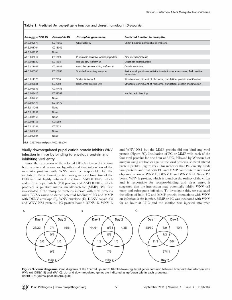
Looking at overlapping DEGs throughout infection with any given
virus, we found considerable variation between timepoints
(Figure 3). During DENV infection, no genes were differentially
expressed at all timepoints, while YFV had seven DURGs and
three DDRGs in common during infection at D1, D2 and D7.
There were slightly more DEGs in common between D1 and D2
for each infection, with 8 DURGs and 31 DDRGs shared on D1
and D2 during infection with DENV (Figure 3).
Author Summary
Dengue (DENV), West Nile (WNV) and Yellow Fever (YFV)viruses are responsible for severe human disease andmortality worldwide. There is no vaccine available fordengue or West Nile virus and no specific antiviral isavailable for any of these viral infections. These viruses aretransmitted to humans through the bite of a mosquitovector. Understanding the effects of viral infection on geneexpression in the mosquito is crucial to the developmentof effective antiviral treatments for mosquitoes and mayenable researchers to interrupt the human-insect infectioncycle. Here we investigate the alterations in geneexpression across the entire Aedes aegypti genome duringinfection with DENV, YFV and WNV over time. We describeseveral genes that share a similar expression profile duringinfection with all three viruses. We also use a WNVmosquito cell, mosquito and mouse model to show thatvirally downregulated genes are inhibitory to infectionwhen overexpressed and that viral regulation of mosquitogenes is tissue-specific. Our results provide an extensiveamount of data highlighting viral gene targets in themosquito during infection. This data may also be used todevelop broad-spectrum anti-flaviviral treatments in mos-quitoes.
Flavivirus Infection Alters Mosquito Transcriptome
PLoS Pathogens | www.plospathogens.org 2 September 2011 | Volume 7 | Issue 9 | e1002189

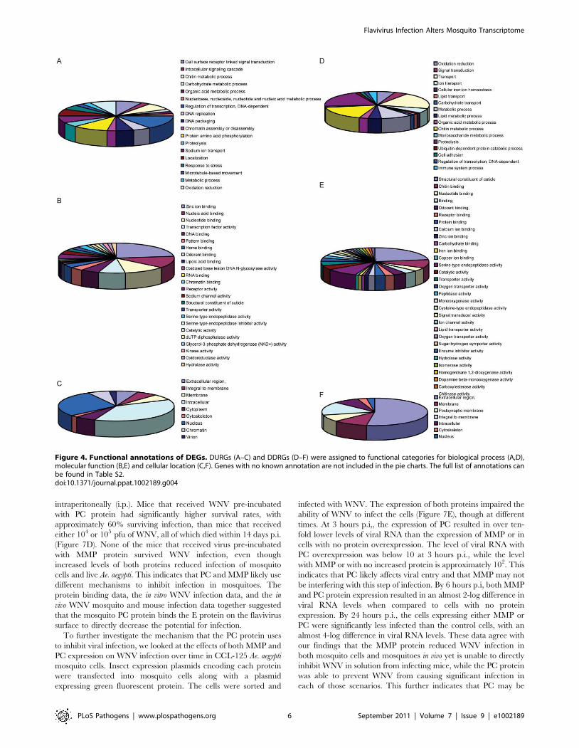
Functional annotations of differentially expressedtranscripts
The 405 genes that were differentially expressed during
infection with one of the flaviviruses were classified into groups
based on biological process (BP), molecular function (MF) and
cellular component (CC) (Figure 4, Tables S2 and S3.) Genes that
did not have any annotation were excluded from analysis. The
largest proportions of DURGs with BP annotation were involved
in DNA-dependent transcription regulation (3%) and protein
amino acid phosphorylation (2%) (Figure 4A). The most abundant
MFs among DURGs were zinc ion binding (9%), nucleic acid
binding (5%), nucleotide binding (4%) and transcription factor
binding (3%) (Figure 4B). The most abundant BP of the DDRGs
were associated with chitin metabolism (12%), transport (7%) and
proteolysis (7%) (Figure 3D) and the MFs were related to structural
constituent of cuticle (23%) and serine-type endopeptidase activity
(10%) (Figure 3E). A similar reduction in chitin-binding proteins
and alteration in transport genes was found during infection of
Aedes with Sindbis virus [25]. In addition, metabolism was
previously found to be altered by DENV infection on day 10
post-infection [10]. Most of the DURGs were found to be
intracellular (9%) and nuclear (7%) (Figure 4C) and most of the
DDRGs were extracellular (10%) (Figure 4F). Functional cluster-
ing of DEGs revealed that the most significant BPs of DURGs
were regulation of transcription (26%) and phosphate metabolic
process (22%) and MF clustered at ion binding (55%) (Figure 5A
and 5B.) The clustering of DURGs placed most of them in
intracellular non-membrane-bound organelles at a surprisingly
high 57%, with 43% at the plasma membrane (Figure 5C). For the
DDRGs, only MF had significant functional clustering, with the
majority involved in peptidase activity (31%), chitin binding (24%)
and ion binding (24%).
Virally-downregulated gene expression is tissue-specificDuring infection of the mosquito, flaviviruses must disseminate
from the midgut (MG) through the body to the salivary gland (SG)
and so likely alter gene expression in different organs at various
times. To determine tissue-specific expression of the identified
DEGs, we infected Ae. aegypti with WNV through blood feeding
and dissected the MG, abdomen (AB) and SG on D1, D2, D7 and
D14 p.i.. We performed qRT-PCR on select DDRGs at each
timepoint to determine levels of gene expression in each tissue
(Figure 6). On D1, all DDRGs tested were significantly
downregulated in the MG, which is expected as this is where
the virus localizes immediately after feeding. Surprisingly,
AAEL011375 was also differentially downregulated in the SG on
D1 and D2 p.i. when the virus is not expected to be present. It is
possible that signalling molecules travel from the infected MG
throughout the mosquito early in infection, affecting gene
expression in other organs. By D7 p.i., many DDRGs are
upregulated in the MG, possibly to compensate for previous
downregulation. At D14 p.i., most DDRGs are downregulated in
the SG, which may be indicative of the virus disseminating to this
organ by this timepoint. An exception to this is AAEL009577,
Figure 1. Genome-wide microarray analysis of the mosquito transcriptome during WNV, DENV and YFV infection. A. Schematic of theexperimental procedure. Ae.aegypti mosquitoes were infected with WNV, DENV or YFV. Microarray analysis was done at 1, 2 and 7 days p.i. for 15,959genes. B. Heatmap for mosquito genes that were $5-fold up-regulated (203 genes) and $10-fold down-regulated (202 genes) during infection withany virus at any timepoint. Red, black and green colors indicate gene expression above, equal to and below the mean, respectively. C,D. Venndiagrams of the $5-fold up- (C) and $10-fold down-regulated (D) genes common and unique to each virus. Timepoints are indicated as D1/D2/D7within each grouping. The complete dataset can be found in Table S1.doi:10.1371/journal.ppat.1002189.g001
Flavivirus Infection Alters Mosquito Transcriptome
PLoS Pathogens | www.plospathogens.org 3 September 2011 | Volume 7 | Issue 9 | e1002189

which is slightly upregulated in the SG and downregulated in the
MG at D14 p.i. This could be due to a precise role the protein
plays in the mosquito during infection that is not related to viral
dissemination. The putative MMP (AAEL003012), found to be
highly downregulated in the whole mosquito during infection with
all three flaviviruses at all timepoints, was significantly under-
expressed in the MG on D1 (12-fold) and then highly upregulated
in the AB by D14 (19.5-fold) p.i. In fact, most of the genes that
were highly downregulated in the whole mosquito during infection
with all three flaviviruses are not significantly downregulated, and
often slightly upregulated, in the AB at most timepoints tested.
These results suggest a complex balance of gene regulation that
occurs in the various organs during flaviviral infection.
Virally-downregulated genes inhibit WNV infection inmosquito cells and live mosquitoes
Six highly DDRGs from different functional groups that shared
similar expression profiles during all three flaviviral infections were
selected for further characterization, testing the notion that these
genes represent a conserved principal in flaviviral infection. We
chose to use WNV to characterize these genes as both mosquito
cells and live mosquitoes are highly susceptible to infection and a
WNV mouse model of infection and disease has been well-
established, with infected mice developing encephalitis that leads
to death [26]. We hypothesized that virally down-regulated genes
may be inhibitory to infection. First, the genes were expressed in
Ae. aegypti cells and the susceptibility of the cells to WNV infection
was examined using immunofluorescence microscopy with
antibodies against WNV envelope protein. A representative image
of WNV-infected cells can be found in Figure S3. The
overexpression of four previously identified virally-DDRG genes,
AAEL001704, AAEL011045, AAEL001022 and AAEL003012,
caused a significant reduction in WNV infection of CCL-125
mosquito cells (266.8+/25.1, 548.75+/27, 247.28+/25.9,
284.05+/26, fold reduction, respectively) compared to GFP alone
(Figure 7A). These genes were also shown to inhibit DENV
infection of mosquito cells (data not shown). Next, the effect of
these proteins on mosquito infection in vivo was investigated. It was
previously reported that whole body transfection (WBT) of DNA
plasmids into mosquitoes results in high expression of target genes
[27]. This method was improved by adding a lipid-based
transfection reagent to the protocol, which enhanced expression
of transfected genes on D3 and D14 post-WBT (p.WBT) (Figure
S4). Ae. aegypti were transfected by intra-thoracic inoculation with
DNA plasmids encoding either GFP, AAEL001704,
AAEL011045, or AAEL003012 and infected with WNV through
blood feeding 6 days p.WBT. At day 10 p.i., the level of WNV in
the mosquitoes was measured using qRT-PCR. The expression of
each of the three genes significantly lowered WNV infection by
approximately one million-fold (Figure 7B). These results strongly
support the hypothesis that these proteins play an important role
during virus infection of the mosquito.
Figure 2. Expression of individual Ae. aegypti DEGs at 4timepoints after WNV or DENV infection. Mosquitoes wereinfected with virus via blood feeding and RNA isolated at 1, 2, 7 and14 days post-infection. qRT-PCR analysis was done to measureexpression of DURGs (A,B) and DDRGs (C,D) after WNV (A,C) or DENV(B,D) infection. Data from 3 separate infections were analyzed intriplicate and plotted on graphs, error bars indicate standard deviation.Fold change in expression was calculated from Ct values, normalized toactin and compared to mock infection.doi:10.1371/journal.ppat.1002189.g002
Flavivirus Infection Alters Mosquito Transcriptome
PLoS Pathogens | www.plospathogens.org 4 September 2011 | Volume 7 | Issue 9 | e1002189
Virally-downregulated pupal cuticle protein inhibits WNVinfection in mice by binding to envelope protein andinhibiting viral entry
Since the expression of the selected DDRGs lowered infection
both in vitro and in vivo, we hypothesized that interaction of the
mosquito proteins with WNV may be responsible for the
inhibition. Recombinant protein was generated from two of the
DDRGs that highly inhibited infection: AAEL011045, which
codes for a pupal cuticle (PC) protein, and AAEL003012, which
produces a putative matrix metalloprotease (MMP). We first
investigated if the mosquito proteins interact with viral proteins
using ELISA assays to detect potential binding of PC and MMP
with DENV envelope (E), WNV envelope (E), DENV capsid (C)
and WNV NS1 proteins. PC protein bound DENV E, WNV E
and WNV NS1 but the MMP protein did not bind any viral
protein (Figure 7C). Incubation of PC or MMP with each of the
four viral proteins for one hour at 37uC, followed by Western blot
analysis using antibodies against the viral proteins, showed altered
protein profiles (Figure S5.) This indicates that PC directly binds
viral proteins and that both PC and MMP contribute to increased
oligomerization of WNV E, DENV E and WNV NS1. Since PC
bound WNV E protein, which is found on the surface of the virion
and is responsible for receptor-binding and virus entry, it
suggested that the interaction may potentially inhibit WNV cell
entry and subsequent infection. To investigate this, we evaluated
the effects of both PC and MMP protein interactions with WNV
on infection in vivo in mice. MMP or PC was incubated with WNV
for an hour at 37uC and the solution was injected into mice
Table 1. Predicted Ae. aegypti gene function and closest homolog in Drosophila.
Ae.aegypti SEQ ID Drosophila ID Drosophila gene name Predicted function in mosquito
AAEL009577 CG17052 Obstructor A Chitin binding, peritrophic membrane
AAEL001704 CG13042
AAEL009750 None
AAEL003012 CG1009 Puromycin-sensitive aminopeptidase Zinc metalloprotease
AAEL001022 CG1803 Regucalcin, isoform D Organism reproduction
AAEL011045 CG13935 cuticular protein 62Bb, isoform A Cuticle structure
AAEL006568 CG16705 Spatzle-Processing enzyme Serine endopeptidase activity, innate immune response, Toll positiveregulation
AAEL011375 CG7996 Snake, isoform A Structural constituent of ribosome, translation, protein modification
AAEL003881 CG2960 Ribosomal protein L40 Structural constituent of ribosome, translation, protein modification
AAEL006536 CG34453
AAEL008415 CG31301 Nucleic acid binding
AAEL009255 None
AAEL002477 CG15479
AAEL014205 None
AAEL012959 None
AAEL004555 None
AAEL001156 CG5280
AAEL013288 CG7523
AAEL008835 None
AAEL009504 None
doi:10.1371/journal.ppat.1002189.t001
Figure 3. Venn diagrams. Venn diagrams of the $5-fold up- and $10-fold down-regulated genes common between timepoints for infection withWNV (A), DENV (B) and YFV (C). Up- and down-regulated genes are indicated as up/down within each grouping.doi:10.1371/journal.ppat.1002189.g003
Flavivirus Infection Alters Mosquito Transcriptome
PLoS Pathogens | www.plospathogens.org 5 September 2011 | Volume 7 | Issue 9 | e1002189
intraperitoneally (i.p.). Mice that received WNV pre-incubated
with PC protein had significantly higher survival rates, with
approximately 60% surviving infection, than mice that received
either 104 or 105 pfu of WNV, all of which died within 14 days p.i.
(Figure 7D). None of the mice that received virus pre-incubated
with MMP protein survived WNV infection, even though
increased levels of both proteins reduced infection of mosquito
cells and live Ae. aegypti. This indicates that PC and MMP likely use
different mechanisms to inhibit infection in mosquitoes. The
protein binding data, the in vitro WNV infection data, and the in
vivo WNV mosquito and mouse infection data together suggested
that the mosquito PC protein binds the E protein on the flavivirus
surface to directly decrease the potential for infection.
To further investigate the mechanism that the PC protein uses
to inhibit viral infection, we looked at the effects of both MMP and
PC expression on WNV infection over time in CCL-125 Ae. aegypti
mosquito cells. Insect expression plasmids encoding each protein
were transfected into mosquito cells along with a plasmid
expressing green fluorescent protein. The cells were sorted and
infected with WNV. The expression of both proteins impaired the
ability of WNV to infect the cells (Figure 7E), though at different
times. At 3 hours p.i,, the expression of PC resulted in over ten-
fold lower levels of viral RNA than the expression of MMP or in
cells with no protein overexpression. The level of viral RNA with
PC overexpression was below 10 at 3 hours p.i., while the level
with MMP or with no increased protein is approximately 102. This
indicates that PC likely affects viral entry and that MMP may not
be interfering with this step of infection. By 6 hours p.i, both MMP
and PC protein expression resulted in an almost 2-log difference in
viral RNA levels when compared to cells with no protein
expression. By 24 hours p.i., the cells expressing either MMP or
PC were significantly less infected than the control cells, with an
almost 4-log difference in viral RNA levels. These data agree with
our findings that the MMP protein reduced WNV infection in
both mosquito cells and mosquitoes in vivo yet is unable to directly
inhibit WNV in solution from infecting mice, while the PC protein
was able to prevent WNV from causing significant infection in
each of those scenarios. This further indicates that PC may be
Figure 4. Functional annotations of DEGs. DURGs (A–C) and DDRGs (D–F) were assigned to functional categories for biological process (A,D),molecular function (B,E) and cellular location (C,F). Genes with no known annotation are not included in the pie charts. The full list of annotations canbe found in Table S2.doi:10.1371/journal.ppat.1002189.g004
Flavivirus Infection Alters Mosquito Transcriptome
PLoS Pathogens | www.plospathogens.org 6 September 2011 | Volume 7 | Issue 9 | e1002189

inhibiting infection from the point of entry by binding to the E
protein while MMP may indirectly inhibit infection at a later
point.
Discussion
Discovery of host factors regulated during viral infection of the
mosquito may identify conserved protein families and pathways
representing both mosquito anti-viral mechanisms as well as
requirements for viral life cycles. Our analysis highlights many
mosquito genes that are important for infection with three globally
(re)emerging flaviviruses, West Nile, dengue and yellow fever. In
our studies, we used Aedes aegypti as an in vivo model of flavivirus
infection. Though the Ae. aegypti mosquito is a major vector for
DENV and YFV transmission in nature, it is generally considered
a secondary vector for the transmission of WNV. The major
vector for WNV transmission is the Culex mosquito, though WNV
has been detected in both Ae. aegypti and Ae. albopictus in nature
[28,29]. In addition, both Aedes and Culex mosquitoes are members
of the Culicinae subfamily of mosquito vectors. Comparative
analysis revealed that the expression profile of 20 significantly
upregulated genes and 15 downregulated genes is quite similar
between the three flaviviruses on D1 of infection, indicating a
potentially conserved transcriptomic signature of flaviviral infec-
tion. Indeed, while Aedes is the major vector for both DENV and
YFV and a secondary vector for WNV, we found a similar overlap
of gene expression profiles between WNV and DENV as we found
for YFV and DENV. This suggests that there are flavivirus-specific
alterations in the mosquito transcriptome regardless of which
flavivirus infects the mosquito as well as which mosquito is the
major vector of the flavivirus used.
One of the genes significantly upregulated during all three
infections was juvenile hormone-inducible protein (AAEL014440),
which has a homolog in Drosophila melanogaster that is thought to
regulate the expression of many other genes [30]. Another gene,
core histone H3 (AAEL003685), was over 4-fold upregulated at all
timepoints during infection with all three viruses. Several viral
proteins target host chromatin and histone proteins to interfere
with host gene expression and nucleosome assembly by various
mechanisms and for diverse purposes [31]. Herpes simplex virus
type 1 (HSV-1) is known to utilize histones for its own genome
during lytic infection [32]. The importance of histone proteins in
flaviviral infection, and infection in the mosquito vector in general,
remains to be investigated. Many genes were found to be
differentially regulated in mosquitoes infected with two of the
three flaviviruses. For example, during infection with both DENV
and WNV, AAEL009750 was over 20-fold downregulated on D1
of infection and significantly lower than mock infection at other
Figure 5. Functional clustering of DEGs. DURGs (A–C) and DDRGs (D) were assigned to functional categories for biological process (A), molecularfunction (B,D) and cellular component (C). Genes with no known annotation are not included in the pie charts. The full list of functional clusteringannotations can be found in Table S3.doi:10.1371/journal.ppat.1002189.g005
Flavivirus Infection Alters Mosquito Transcriptome
PLoS Pathogens | www.plospathogens.org 7 September 2011 | Volume 7 | Issue 9 | e1002189

timepoints but expression was not significantly altered during YFV
infection. This gene codes for a member of the mosquito allergen
proteins, which are known to bind human IgE [33] and could
interfere with viral transmission to humans if produced in
abundance during infection. An interesting point is whether
flaviviral infection is altering gene expression directly or indirectly,
for example through the enrichment of small interfering RNA
molecules. To investigate this, we compared highly downregulated
genes from our DENV infected mosquitoes to the recent
publication by Hess et al regarding small RNA levels during
DENV infection and found several correlations [34]. For example,
AAEL010160 was downregulated 18.8, 59.3 and 7.45-fold on days
1, 2 and 7 of DENV infection in our analysis, respectively, and a
corresponding sense sRNA was enriched with 2.15 log fold-change
on day 2 of DENV infection from the dataset published by Hess et
al. Another gene, AAEL001953, was downregulated 11.84, 19.05
and 3.84-fold on days 1, 2 and 7 during DENV infection in our
analysis, respectively, and a corresponding sense sRNA was
enriched with 3.65 log fold-change on day 2 of DENV infection in
the previous study [34]. This indicates that flaviviruses likely alter
gene expression through both direct and indirect mechanisms
during infection of the mosquito.
The majority of highly altered mosquito transcripts were not
canonical innate immune genes though we were able to correlate
our analysis with previous data on viral infection and mosquito
immunity. Previous studies suggest that depleting PIAS (protein
inhibitor of activated STAT), a negative regulator of the Jak-
STAT pathway, resulted in down-regulation of five antimicrobial
genes (four Cecropin A-like genes and one defensin l-like gene)
that were also downregulated by DENV infection [9]. All five of
these genes were significantly downregulated in our analysis
during infection with all three flaviviruses infections at all three
timepoints. One of these genes, AAEL000611, was highly
downregulated late in infection, with 15-fold, 27-fold and 38-fold
lower expression on D7 of YFV, DENV and WNV, respectively.
Another Cecropin A-like gene, AAEL000627, was also highly
downregulated on D7 of infection, with expression 14-fold, 14-fold
and 41-fold lower during infection with YFV, DENV and WNV,
respectively. This indicates that the mosquito Jak-STAT pathway
is likely involved throughout infection with all three flaviviruses.
This also suggests that YFV, DENV and WNV may have evolved
a conserved mechanism to suppress this antiviral pathway during
infection. The Toll pathway has also been previously implicated in
anti-flaviviral defense by the mosquito [10]. One gene shown to be
involved in the mosquito Toll pathway and downregulated by
DENV, AAEL001929, was 2.5-fold lower on D1 of infection with
YFV and 2.5-fold lower on D2 of DENV infection in our study.
Another Toll gene, AAEL003507, was only significantly down-
regulated during YFV infection, with 2.3-fold and 2.6-fold lower
expression on D1 and D7, respectively. Our analysis also found a
serine protease gene (AAEL006568) to be downregulated during
infection with all three flaviviruses. Previous studies show that
some midgut serine proteases limit DENV-2 infection in Aedes
aegypti [19]. The Drosophila homolog of this gene, Spatzle-
processing enzyme (CG16705), is known to be a positive regulator
of the Toll pathway [35]. This implies that serine proteases may
contribute to the innate immune response to viruses in mosquitoes.
We also demonstrate that virally-regulated gene expression is
tissue-specific. Since flaviviruses must travel from the site of entry
and infection, the midgut, throughout the mosquito body before
reaching the salivary glands, it was likely that the expression of
many genes would be differentially altered in various organs at
different timepoints. Genes which are highly upregulated early in
infection are likely important for flaviviral colonization of the
midgut as well as the start of dissemination out of the midgut.
Alternatively, these genes could represent the innate immune
response of the mosquito to viral replication in the midgut. We saw
several of the flavivirally-down regulated genes in the whole
mosquito also downregulated by WNV infection in the midgut on
D1 of infection, including AAEL011375, which was 17-fold lower
than the mock group. This gene encodes a protein in the trypsin
family, and trypsin silencing has been shown to increase DENV
infection in Aedes [19]. One gene that was highly downregulated in
Figure 6. Alteration in DDRG expression in Ae. aegypti is tissue-specific. Gene expression was analyzed by qRT-PCR in mosquitotissues at select timepoints during WNV infection of Ae. aegyptimosquitoes. The fold change in expression of indicated genes inmidgut (A), salivary gland (B) and abdomen (C), after WNV infectioncompared to mock infection. D1, D2, D7, D14 indicated by represen-tative bars. Data is from 3 separate infection groups and qRT-PCR wasdone in triplicate. Error bars indicate standard deviation.doi:10.1371/journal.ppat.1002189.g006
Flavivirus Infection Alters Mosquito Transcriptome
PLoS Pathogens | www.plospathogens.org 8 September 2011 | Volume 7 | Issue 9 | e1002189

Figure 7. Overexpression of virally down-regulated genes impairs WNV infectivity. A. Overexpression of DDRGs reduces WNV infection ofAe. aegypti mosquito cells. CCL-125 cells were transfected with expression plasmids encoding mosquito DDRGs and infected with WNV 48 hours post-transfection (p.t.). Cells were analyzed for infection by immunofluorescence microscopy 24 hours post-infection (p.i.) using an antibody against WNVenvelope protein. Fold decrease in infection is indicated. Data is pooled from 3 separate experiments, error bars indicate standard deviation. B.Overexpression of DDRGs reduces WNV infection of Ae. aegypti mosquitoes. Mosquitoes were injected with plasmids used in (A) and infected withWNV 6 days p.t. At 10 days p.i., RNA was isolated and qRT-PCR performed to detect WNV. Infection is indicated as ng WNV E/ng actin. p,.001 for all 3genes vs. control, one way ANOVA and Kruskal-Wallis test were used, n = 10. C. ELISA analysis of AAEL011045 (PC protein) binding to WNV E, DENV E,WNV NS1 and DENV capsid. Optical density (O.D.) is shown at 450 nm and normalized to glutathione S-transferase (GST) binding. Data is pooled from3 separate experiments, error bars indicate standard deviation. D. Purified recombinant pupal cuticle protein (PC) (AAEL011045) was incubated withWNV for an hour at 37uC and injected i.p. into c57L/B6 mice. Survival curve was plotted for: 104 WNV alone, 105 WNV alone, 104 WNV+PC, 105
WNV+PC; number refers to viral titer in plaque forming units (pfu). p,.05 using the Logrank test. Results are pooled from 2 separate experiments,total n = 10 for each group. E. Overexpression of PC and MP protein affects WNV infection in mosquito cells. CCL-125 cells were transfected as in (A)and analyzed by qRT-PCR as in (B). Points plotted on line graph are from 3,6,9,12 and 24 h post-infection.doi:10.1371/journal.ppat.1002189.g007
Flavivirus Infection Alters Mosquito Transcriptome
PLoS Pathogens | www.plospathogens.org 9 September 2011 | Volume 7 | Issue 9 | e1002189

the whole mosquito in response to infection with all three
flaviviruses, AAEL003012, encodes the putative matrix metallo-
protease (MMP) protein. This gene was also downregulated in the
midgut, salivary gland and abdomen on D1 of WNV infection,
suggesting that the protein may play a role in controlling the initial
viral infection in the mosquito. Genes downregulated late in
infection are possibly inhibitory to establishment of infection in the
salivary glands or to transmission of virus to a new host. By D14
p.i, many of the identified virally-downregulated genes have lower
expression in the salivary glands, which might be expected as the
virus should be concentrated in this organ by this timepoint.
Interestingly, this is the only organ in which the MMP protein is
still downregulated at D14 p.i., which again indicates that this
protein may be involved in controlling viral replication and/or
infection.
Bioinformatics analysis revealed significant changes in the
expression of genes from diverse cellular processes, including ion
binding, ion transport, metabolic processes and peptidase activity.
In a previous study investigating gene expression in midguts of
WNV-infected Culex mosquitoes on D10 p.i., almost 5% of the
highly upregulated genes were related to ion transport. This
alteration in ion transport molecules was hypothesized to aid in
viral spread through polarized cells by maintaining proper cell
polarity and stable solute transport functions [12]. A study on the
infection of Aedes with Sindbis virus (SINV) found genes involved
in ion transport upregulated D4 p.i. [25]. The same group also saw
a decrease in genes related to chitin binding on D1 of SINV
infection and our analysis revealed a significant reduction in genes
involved with chitin binding and the structural constituent of
cuticle. Metabolism and oxidoreductive processes were previously
found to be major functional groups altered by DENV infection in
Aedes on D10 p.i. [10]. In agreement, our analysis showed that
genes involved with oxidation reduction and zinc ion binding were
highly upregulated and metabolic process and chitin metabolic
process were highly downregulated. We also found that genes
encoding serine protease inhibitors were significantly downregu-
lated by infection with all three flaviviruses. In a study investigating
mammalian genes important in WNV, it was found that silencing
serine peptidase inhibitors significantly increased infection,
indicating that a reduction in these proteins favors viral infection
[36]. This highlights the likely overlap between mosquito and
mammalian flaviviral host factors. We also looked at the cellular
location of the DEGs found during infection. In our analysis, the
majority of virally-upregulated genes produce intracellular and
nuclear proteins and most of the virally-downregulated genes
encoded proteins found in the extracellular region.
Several of the proteins encoded by the most significantly virally-
downregulated genes are shown to be inhibitory to WNV infection
both in mosquito cells and in mosquitoes in vivo. This is direct
evidence that viral infection in the mosquito decreased the
expression of proteins that are likely to impede viral replication
or infection of new cells. Two of these proteins, a pupal cuticle
protein and a matrix metalloprotease, were shown to alter the
protein profiles of WNV E and NS1. In addition, the PC protein
was able to directly bind WNV E protein and inhibit viral
infection in mosquito cells as early as 3 hours post-infection. This
suggests that PC is acting at the step of viral entry, likely by directly
binding the E protein on the virus surface. The MMP protein was
also able to inhibit WNV infection in cells, though at a later
timepoint than PC. When mixed with live WNV, PC protein
enhanced the survival of injected mice, indicating direct action on
the virus to impede infection. These results provide strong
evidence that the virally-downregulated genes identified in this
study likely represent proteins that are inhibitory to flaviviral
infection. To further ensure that our findings are relevant for
WNV infection in nature, we aligned the pupal cuticle protein
from Ae. aegypti with the corresponding Cx. quinquefasciatus protein
and found 92% sequence identity. In addition, we performed a
BLAST of the Ae. aegypti DDRGs that we use in the WNV studies
against the Cx. quinquefasciatus genome and found very high
sequence identity (85–95%). This indicates that the genes
identified as inhibitory to WNV infection in Aedes mosquitoes are
likely also inhibitory to infection in Culex mosquitoes.
This investigation uncovered many previously unknown host
factors differentially regulated by flaviviral infection. This is also
the first study, to our knowledge, to compare infection with three
flaviviruses in the same mosquito at the same timepoints.
Understanding the effects of infection on the mosquito, both
common and unique to individual flaviviruses, will aid in
developing broadly applicable methods to treat and prevent
infection.
Materials and Methods
Cell culture and virus growthThe CCL-125 Aedes aegypti cell line (ATCC, VA) was used for
transfection and infection studies. The cells were grown at 30uCand 5% CO2 in DMEM supplemented with 10% heat-inactivated
fetal bovine serum (Gemini, CA), 1% penicillin-streptomycin and
1% tryptose phosphate broth (Sigma, MO). Flavivirus was grown
in C6/36 Aedes albopictus cell line using the same media. Strains
used were: WNV 2741, DENV-2 New Guinea C, YFV Asibi
strain. Cells were infected at an m.o.i. of 1.0, virus was allowed to
propagate for 6–8 days, supernatant was removed, spun down and
virus stock was stored at 280uC until use.
Mosquito infectionsThe Rockefeller strain of Ae. aegypti mosquitoes were either
infected by intra-thoracic inoculation or blood-feeding, as
indicated in the text and figure legends. For thoracic injections,
virus was used at 6.5 logs per mL and 0.5 mL were injected per
mosquito. For blood feeding, 100 mL of virus was added to 1 mL
serum-inactivated blood from c57L/B6 mice and fed to
mosquitoes for 20 minutes at room temperature using a hemotek
feeder. Mosquitoes were maintained in groups of 10 at 30uC, 80%
humidity. Mosquitoes were supplied raisins as a source of dietary
sugar.
Microarray analysisRNA was isolated from WNV, DENV type 2 or YFV infected
Ae. aegypti mosquitoes on days 1, 2 and 7. RNA was purified using
the Rneasy kit (Qiagen, CA) and hybridized with Nimblegen X4
microarray chips using 81-mer probes designed from 18,000 open
reading frames (ORF) found in the Ae. aegypti genome, with 2
different probes per ORF. The gene expression data was
normalized using quantile normalization [37]. Partek Genomic
Suite v. 6.4 (http://www.partek.com) was used for the statistical
data analysis and ANOVA was applied to identify differentially
expressed genes between each infection versus mock for each
timepoint. False discovery rate was used to adjust the p-value for
multiple testing corrections [38].
Bioinformatics analysisThe functional annotation and clustering of DEGs was
performed using the DAVID Bioinformatics Resource 6.7
[39,40]. Briefly, IDs of DDRGs and DURGs were uploaded
separately to the DAVID web interface and converted to unique
DAVID IDs. The background gene list for the functional
Flavivirus Infection Alters Mosquito Transcriptome
PLoS Pathogens | www.plospathogens.org 10 September 2011 | Volume 7 | Issue 9 | e1002189

clustering consisted of the IDs of all Ae. Aegypti transcripts
represented on the Nimblegen X4 microarray chips. The
functional annotation and clustering were then performed using
the DAVID default parameters.
qRT-PCR analysisRNA was isolated from infected Ae. aegypti mosquitoes on days
1, 2, 7 and 14 and purified using RNeasy kit (Qiagen, CA)
according to manufacturer’s instructions. cDNA was made from
the RNA using a SuperscriptIII kit (Invitrogen, CA). cDNA from
1 mg RNA was used in each quantitative (q)RT-PCR reaction
along with SYBR green chemistry. Fold change in gene expression
was calculated using the CT value differences normalized to actin
expression. Oligos can be found in Table S4.
Immunofluorescence analysisCCL-125 Aedes aegypti cells were infected with WNV at an MOI
of 0.1. 24 hours post-infection, cells were fixed in 4% parafor-
maldehyde for 20 min at RT, washed with PBS(-) and then stained
for infection using an antibody against recombinant WNV E
protein conjugated with TRITC. The antibody was diluted in 1%
BSA at 1/250 and cells were incubated for 20 minutes at RT.
Infection was visualized using fluorescent microscopy.
Protein productionGST-tagged protein was made from two mosquito genes:
AAEL011045, which codes for a pupal cuticle protein, and
AAEL003012, which produces a putative matrix metalloprotease.
Protein was produced in E.coli and batch purified using glutathione
sepharose (GS) (GE, NJ) with centrifugation. Briefly, pelleted
bacteria from 1L culture were lysed and mixed with 2 mL GS
resin with end-over-end mixing for 1 h at RT. The resin was spun
down at 500 rpm, washed with PBS(-) and protein was eluted with
buffer (50 mM Tris-HCl, 10 mM reduced glutathione, pH 8.0).
The GST tags were removed using PreScission Protease enzyme
(GE, NJ).
ELISA analysisBinding between the Ae. aegypti PC or MMP proteins with
flaviviral proteins was investigated using ELISA analysis. Briefly,
5 mg of mosquito or GST control protein was coated onto a 96-
well ELISA plate (Thermo Fisher Sci, MA) and incubated
overnight at 4uC. The plate was blocked with 1% BSA in PBS(-)
and incubated with 1 mg of flaviviral protein (either WNV E,
WNV NS1, DENV E or DENV C) or BSA control for an hour at
RT. The proteins were washed off, secondary-HRP was added for
30 min at RT, washed off and TMB substrate was added for
20 min at RT. Stop solution was added and the O.D. of the wells
read at 450 nm. WNV NS1 protein was a kind gift from Dr.
Michael Diamond (Washington University, MO), WNV and
DENV E were kind gifts from L2 (CT) and recombinant DENV C
was produced in our laboratory.
Western blotsThe Ae. aegypti PC or MMP proteins were incubated with each
of four viral proteins (WNV E, WNV NS1, DENV E, DENV C)
for one hour at 37uC. The solution was run on a 4-12% SDS-
PAGE gel for 1.5 h at 15 milliamps per gel. The proteins were
then transferred to nitrocellulose membrane. The nitrocellulose
was blocked with 5% milk in 1% TBST for 1 h at RT and then
incubated with the appropriate primary antibody overnight at
4uC. The nitrocellulose was washed and then incubated with the
appropriate horseradish peroxidase secondary antibody for 1 h at
RT. The protein blots were incubated with ECL substrates
(Amersham, NJ) for 5 min at RT and then detected on Kodak
film. Antibodies used: anti-WNV envelope (L2, CT), anti-dengue
envelope (L2, CT), anti-WNV NS1 (gift from Dr. Michael
Diamond, Washington University School of Medicine, MO) and
mouse immune serum against recombinant DENV-2 capsid
protein made in our laboratory.
MiceNine week old female C57BL/6 mice were infected with WNV
(with and without mosquito proteins) intraperitoneally (i.p.) at a
dose of 103 plaque forming units (pfu) per mouse. All animal
experimental protocols were approved by the Institutional Animal
Care and Use Committee of Yale University and experiments
were done in a Biosafety Level 3 animal facility according to the
regulations of Yale University.
Transfection of plasmidsAll plasmids were transfected into CCL-125 cells using
Effectene (Qiagen, CA) according to manufacturer’s instructions.
Briefly, for a 10 cm2 plate, 10 mg of DNA was mixed with 500 mL
buffer EC and 32 mL enhancer was added. This was allowed to
incubate for 5 min on the benchtop. Then, 30 mL Effectene
reagent was added and the solution vortexed briefly. After 10 min
incubation, the solution was added to the cells. Expression was
observed 24 h post-transfection and peaked at 48 h.
Accession numbersThe following GENBANK accession numbers were referenced
in the manuscript text: AAEL014440, AAEL004861-RA,
AAEL011045, AAEL003012, AAEL012402, AAEL014108,
AAEL004897, AAEL012770, AAEL000340, AAEL011375,
AAEL009577, AAEL001704, AAEL001022, AAEL003685,
AAEL009750, AAEL010160, AAEL001953, AAEL000611,
AAEL000627, AAEL001929, AAEL003507, AAEL006568,
AAEL011375.
Ethics statementOur study was carried out in strict accordance with the
recommendations Guide for the Care and Use of Laboratory
Animals of the National Institutes of Health. All animal
experimental protocols were approved by the Institutional Animal
Care and Use Committee of Yale University (Protocol Permit
Number: 2008-07941) and experiments were done in a Biosafety
Level 3 animal facility according to the regulations of Yale
University. All efforts were made to minimize suffering.
Supporting Information
Figure S1 Summary of the microarray data. Ae. aegypti
mosquitoes were infected with WNV, DENV or YFV and
microarray analysis was done using RNA isolated on days 1, 2
and 7 post-infection. Mosquito genes that were $5-fold up-
regulated (203 genes) and $10-fold down-regulated (202 genes)
during infection with any virus at any timepoint are designated
differentially expressed genes (DEGs). A. Graph plots the trend of
the DEGs from D0-D8 for all 3 FVs, blue line indicates positive
regulation, red line indicates negative regulation. B. Chart
showing the number of DEGs up- or down-regulated for each
FV at each timepoint.
(PDF)
Figure S2 Detailed heatmaps for DEGs from micro-array analysis. A–G. Detailed heatmaps for Ae. aegypti genes
Flavivirus Infection Alters Mosquito Transcriptome
PLoS Pathogens | www.plospathogens.org 11 September 2011 | Volume 7 | Issue 9 | e1002189

that were $5-fold up-regulated (203 genes) and $10-fold down-
regulated (202 genes) during infection with any virus at any
timepoint. Flavivirus and timepoint are indicated at the bottom of
each heatmap, individual genes are listed on the right.
(PDF)
Figure S3 Immunofluorescence analysis of WNV-infect-ed Ae. aegypti cells. CCL-125 cells were infected with WNV at
an MOI of 0.1 and fixed with 4% paraformaldehyde 24 hours p.i.
Cells were stained with an antibody against the WNV envelope
protein conjugated to TRITC secondary. Scale bar is shown in
lower right corner.
(PDF)
Figure S4 Whole body transfection of Ae. aegyptimosquitoes. Mosquitoes were injected via intra-thoracic inoc-
ulation with insect expression vectors coding for AAEL003012
(MMP) or AAEL011045 (PC). RNA was isolated from mosquitoes
on day 3 and day 14 post-transfection and qRT-PCR analysis was
done to detect levels of expression. Mosquitoes that received the
plasmid coding for the alternate gene were used as controls for
expression of each gene. Each point represents 10 mosquitoes; fold
increase in expression is indicated.
(PDF)
Figure S5 Mosquito MMP and PC change the proteinprofile of WNV proteins. Recombinant MMP protein
(AAEL003012) was incubated with WNV NS1 (A), WNV E (B)
and PC protein (AAEL011045) was incubated with WNV E (C) for
an hour at 37uC. Solutions were run on a 12% SDS-PAGE gel. The
proteins were transferred to nitrocellulose and Western blot analysis
was done using antibodies against the viral proteins. 5 mM ZnCl
was added to one solution of MMP and viral proteins as MMP is a
presumed metalloprotease. Dimers and monomers are indicated.
(PDF)
Table S1 Microarray analysis data. Ae. aegypti mosquitoes
were infected with WNV, DENV or YFV and microarray analysis
was done using RNA isolated on days 1, 2 and 7 post-infection. To
identify fold change in expression, we applied ANOVA to each
infected sample versus mock for each timepoint and applied false
discovery rate (FDR) to adjust the p-value for multiple corrections.
Submitted to NCBI Gene Expression Omnibus.
(XLS)
Table S2 Functional annotations of DEGs. DURGs and
DDRGs were assigned to functional categories for biological
process, molecular function and cellular component.
(XLS)
Table S3 Functional clustering of DEGs. DURGs and
DDRGs were assigned to functional categories for biological
process, molecular function and cellular component. Genes were
analyzed for functional clustering. Enrichment scores are listed for
each cluster.
(XLS)
Table S4 Oligos used for qRT-PCR. qRT-PCR was done
with listed oligos to determine levels of gene expression,
normalized to actin expression.
(XLS)
Acknowledgments
We thank Dr. John F. Anderson and the Connecticut Agricultural
Experiment Station (New Haven, CT) for mosquito rearing, facility use
and experiment insights. We thank individuals at the W.M. Keck Facility
at Yale for the following: Can Bruce for bioinformatic analysis, Aiping Lin
for biostatistic analysis and Irina Tikhonova for microarray analysis. We
thank L2 Diagnostics for providing the anti-WNV antibodies and
recombinant envelope proteins.
Author Contributions
Conceived and designed the experiments: TMC EF JC. Performed the
experiments: TMC JC FMF DLV PW GC. Analyzed the data: TMC JC
SK MNK EF SH. Wrote the paper: TMC EF.
References
1. Mackenzie JS, Gubler DJ, Petersen LR (2004) Emerging flaviviruses: the spread
and resurgence of Japanese encephalitis, West Nile and dengue viruses. Nat Med
10: S98–109.
2. Rappole JH, Derrickson SR, Hubalek Z (2000) Migratory birds and spread of
West Nile virus in the Western Hemisphere. Emerg Infect Dis 6: 319–328.
3. Gubler DJ (2002) Epidemic dengue/dengue hemorrhagic fever as a public
health, social and economic problem in the 21st century. Trends Microbiol 10:
100–103.
4. site PAHOw (2007) Number of reported cases of dengue and dengue
hemorrhagic fever (DHF) in the Americas, by country: figures for 2007.
5. Blair CD, Adelman ZN, Olson KE (2000) Molecular strategies for interrupting
arthropod-borne virus transmission by mosquitoes. Clin Microbiol Rev 13:651–661.
6. Bae HG, Drosten C, Emmerich P, Colebunders R, Hantson P, et al. (2005)
Analysis of two imported cases of yellow fever infection from Ivory Coast andThe Gambia to Germany and Belgium. J Clin Virol 33: 274–280.
7. Tomori O (2004) Yellow fever: the recurring plague. Crit Rev Clin Lab Sci 41:391–427.
8. Monath TP (2006) Yellow fever as an endemic/epidemic disease and prioritiesfor vaccination. Bull Soc Pathol Exot 99: 341–347.
9. Souza-Neto JA, Sim S, Dimopoulos G (2009) An evolutionary conserved
function of the JAK-STAT pathway in anti-dengue defense. Proc Natl AcadSci U S A 106: 17841–17846.
10. Xi Z, Ramirez JL, Dimopoulos G (2008) The Aedes aegypti toll pathwaycontrols dengue virus infection. PLoS Pathog 4: e1000098.
11. Sessions OM, Barrows NJ, Souza-Neto JA, Robinson TJ, Hershey CL, et al.(2009) Discovery of insect and human dengue virus host factors. Nature 458:
1047–1050.
12. Bartholomay LC, Waterhouse RM, Mayhew GF, Campbell CL, Michel K, et al.Pathogenomics of Culex quinquefasciatus and meta-analysis of infection
responses to diverse pathogens. Science 330: 88–90.
13. Cheng G, Cox J, Wang P, Krishnan MN, Dai J, et al. (2010) A C-type lectin
collaborates with a CD45 phosphatase homolog to facilitate West Nile virus
infection of mosquitoes. Cell 142: 714–725.
14. Molina-Cruz A, Gupta L, Richardson J, Bennett K, Black Wt, et al. (2005) Effect
of mosquito midgut trypsin activity on dengue-2 virus infection and
dissemination in Aedes aegypti. Am J Trop Med Hyg 72: 631–637.
15. Baron OL, Ursic-Bedoya RJ, Lowenberger CA, Ocampo CB Differential gene
expression from midguts of refractory and susceptible lines of the mosquito,
Aedes aegypti, infected with Dengue-2 virus. J Insect Sci 10: 41.
16. Smartt CT, Richards SL, Anderson SL, Erickson JS (2009) West Nile virus
infection alters midgut gene expression in Culex pipiens quinquefasciatus Say
(Diptera: Culicidae). Am J Trop Med Hyg 81: 258–263.
17. Bennett KE, Flick D, Fleming KH, Jochim R, Beaty BJ, et al. (2005)
Quantitative trait loci that control dengue-2 virus dissemination in the mosquito
Aedes aegypti. Genetics 170: 185–194.
18. Sim S, Dimopoulos G (2010) Dengue virus inhibits immune responses in Aedes
aegypti cells. PLoS One 5: e10678.
19. Brackney DE, Foy BD, Olson KE (2008) The effects of midgut serine proteases
on dengue virus type 2 infectivity of Aedes aegypti. Am J Trop Med Hyg 79:
267–274.
20. Gould EA, Solomon T (2008) Pathogenic flaviviruses. Lancet 371: 500–509.
21. Nene V, Wortman JR, Lawson D, Haas B, Kodira C, et al. (2007) Genome
sequence of Aedes aegypti, a major arbovirus vector. Science 316: 1718–1723.
22. Vanlandingham DL, McGee CE, Klinger KA, Vessey N, Fredregillo C, et al.
(2007) Relative susceptibilties of South Texas mosquitoes to infection with West
Nile virus. Am J Trop Med Hyg 77: 925–928.
23. CDC (2009) http://www.cdc.gov/ncidod/dvbid/westnile/mosquitoSpecies.
htm.
24. Gaunt MW, Sall AA, de Lamballerie X, Falconar AK, Dzhivanian TI, et al.
(2001) Phylogenetic relationships of flaviviruses correlate with their epidemiol-
ogy, disease association and biogeography. J Gen Virol 82: 1867–1876.
25. Sanders HR, Foy BD, Evans AM, Ross LS, Beaty BJ, et al. (2005) Sindbis virus
induces transport processes and alters expression of innate immunity pathway
genes in the midgut of the disease vector, Aedes aegypti. Insect Biochem Mol
Biol 35: 1293–1307.
Flavivirus Infection Alters Mosquito Transcriptome
PLoS Pathogens | www.plospathogens.org 12 September 2011 | Volume 7 | Issue 9 | e1002189

26. Wang T, Town T, Alexopoulou L, Anderson JF, Fikrig E, et al. (2004) Toll-like
receptor 3 mediates West Nile virus entry into the brain causing lethalencephalitis. Nat Med 10: 1366–1373.
27. Isoe J, Kunz S, Manhart C, Wells MA, Miesfeld RL (2007) Regulated expression
of microinjected DNA in adult Aedes aegypti mosquitoes. Insect Mol Biol 16:83–92.
28. Higgs S, Snow K, Gould EA (2004) The potential for West Nile virus to establishoutside of its natural range: a consideration of potential mosquito vectors in the
United Kingdom. Trans R Soc Trop Med Hyg 98: 82–87.
29. Hubalek Z, Halouzka J (1999) West Nile fever--a reemerging mosquito-borneviral disease in Europe. Emerg Infect Dis 5: 643–650.
30. Dubrovsky EB, Dubrovskaya VA, Bilderback AL, Berger EM (2000) Theisolation of two juvenile hormone-inducible genes in Drosophila melanogaster.
Dev Biol 224: 486–495.31. Wei H, Zhou MM (2010) Viral-encoded enzymes that target host chromatin
functions. Biochim Biophys Acta 1799: 296–301.
32. Kent JR, Zeng PY, Atanasiu D, Gardner J, Fraser NW, et al. (2004) During lyticinfection herpes simplex virus type 1 is associated with histones bearing
modifications that correlate with active transcription. J Virol 78: 10178–10186.33. Peng Z, Xu W, James AA, Lam H, Sun D, et al. (2001) Expression, purification,
characterization and clinical relevance of rAed a 1--a 68-kDa recombinant
mosquito Aedes aegypti salivary allergen. Int Immunol 13: 1445–1452.
34. Hess AM, Prasad AN, Ptitsyn A, Ebel GD, Olson KE, et al. Small RNA
profiling of Dengue virus-mosquito interactions implicates the PIWI RNA
pathway in anti-viral defense. BMC Microbiol 11: 45.
35. Arnot CJ, Gay NJ, Gangloff M;Molecular mechanism that induces activation of
Spatzle, the ligand for the Drosophila Toll receptor. J Biol Chem 285:
19502–19509.
36. Krishnan MN, Ng A, Sukumaran B, Gilfoy FD, Uchil PD, et al. (2008) RNA
interference screen for human genes associated with West Nile virus infection.
Nature 455: 242–245.
37. Bolstad BM, Irizarry RA, Astrand M, Speed TP (2003) A comparison of
normalization methods for high density oligonucleotide array data based on
variance and bias. Bioinformatics 19: 185–193.
38. Xu WW, Carter CJ Parallel multiplicity and error discovery rate (EDR) in
microarray experiments. BMC Bioinformatics 11: 465.
39. Huang da W, Sherman BT, Lempicki RA (2009) Systematic and integrative
analysis of large gene lists using DAVID bioinformatics resources. Nat Protoc 4:
44–57.
40. Dennis G, Jr., Sherman BT, Hosack DA, Yang J, Gao W, et al. (2003) DAVID:
Database for Annotation, Visualization, and Integrated Discovery. Genome Biol
4: P3.
Flavivirus Infection Alters Mosquito Transcriptome
PLoS Pathogens | www.plospathogens.org 13 September 2011 | Volume 7 | Issue 9 | e1002189


![[Lessons learned in the control of Aedes aegypti to address dengue and the emergency of chikungunya in Iquitos, Peru]](https://static.fdokumen.com/doc/165x107/63375c5f4554fe9f0c05c677/lessons-learned-in-the-control-of-aedes-aegypti-to-address-dengue-and-the-emergency.jpg)