Mosquitoes Larvicidal Activity of Ocimum kilimandscharicum ...
Upload
independentCategory
view
0download
0
RESEARCH Open Access
Evidence of co-infection of chikungunya anddensonucleosis viruses in C6/36 cell lines andlaboratory infected Aedes aegypti (L.) mosquitoesAruna Sivaram, Pradip V Barde, Mangesh D Gokhale, Dinesh K Singh, Devendra T Mourya*
Abstract
Background: Densonucleosis viruses are the etiological agents of insect’s disease. We have reported the isolationof densovirus from India and its distribution among the natural populations of Aedes aegypti mosquitoes across thecountry. Since densonucleosis virus persistently infects mosquito populations, and is demonstrated to negativelyaffect multiplication of dengue virus in Aedes albopictus, it would be interesting to study if this virus has a role indetermining the susceptibility of the vector mosquito Ae. aegypti to chikugunya virus.
Methods: Mosquito cell lines and adult Ae. aegypti mosquitoes infected with densovirus were superinfected withChikungunya virus and both the viruses were quantitated by determining their genomic copy number by real timeamplification. Comparison was made between the log of genomic copy numbers of the viruses in the presenceand absence of each other.
Results: The log of copy number of the viruses did not vary due to co-infection. Even though the RNA copynumber of chikungunya virus increased over the period of time, no change was observed in the RNA copynumber between the control and the co-infected group on any given day. Similarly, DNA copy number ofdensovirus also remained unchanged between the control and the co-infected groups.
Conclusion: Chikungunya virus neither stimulates the replication of densovirus nor is its own replicationsuppressed due to co-infection. Ae. aegypti mosquitoes with densovirus infection were as susceptible to infectionby chikungunya virus as the uninfected mosquitoes.
BackgroundDensonucleosis viruses (DNV) are known to persistentlyinfect the mosquito cell lines and mosquito populationsin nature. They belong to the family Parvoviridae andgenus Brevidensovirus. DNV have been isolated fromseveral species of mosquitoes like Culex salinarius [1],Aedes aegypti [2,3], Ae. albopictus [4], Anopheles mini-mus [5] and mosquito cell lines [6]. They are isometric,non-enveloped viruses of about 20 nm in diameter withsingle stranded linear DNA, packaged either as plus or aminus strand [7]. Palindromic sequences that form hairpin structure are found in both the ends of the genome[8,9]. The coding sequences include the genes for nonstructural proteins NS1 and NS2 occupying the 5’ end
of the strand and the genes for structural protein VPoccupying the 3’ end [3,9,10].DNV is pathogenic to mosquitoes, resulting in loss of
mobility, deformity and loss of pigmentation in the mos-quito larvae. A very high rate of mortality (up to 90%)was reported when first instar Ae. aegypti larvae wereexposed to Ae. albopictus DNV [11]. The pathogenicityof the virus is dose dependent, where in at lower dosesof infection with DNV, the larvae survive to form adultsthat carry the infection [4,12]. Virus multiplication hasalso been observed in the ovaries of the infected mos-quitoes, which enable them to transmit the virus verti-cally to the next generation [11,13]. DNV has a role insignificantly reducing the vectorial capacity of the mos-quitoes by reducing mosquito lifespan [13]. DNV hasalso been developed as a transducing vector used tointroduce constructs that interfere with the arboviralinfection in mosquitoes.
* Correspondence: [email protected] Containment Complex, National Institute of Virology, Sus Road,Pashan, Pune 411 021, India
Sivaram et al. Parasites & Vectors 2010, 3:95http://www.parasitesandvectors.com/content/3/1/95
© 2010 Sivaram et al; licensee BioMed Central Ltd. This is an Open Access article distributed under the terms of the Creative CommonsAttribution License (http://creativecommons.org/licenses/by/2.0), which permits unrestricted use, distribution, and reproduction inany medium, provided the original work is properly cited.
Vector competence refers to the intrinsic permissive-ness of an arthropod vector for infection, replicationand transmission of a vertebrate pathogen [14]. Vectorcompetence is governed by intrinsic (genetic) factorsand extrinsic factors [15,16]. The microflora present inthe midgut of mosquito might also have a role in deter-mining their susceptibility to the viruses [17]. SinceDNV has been seen to be persistently infecting mos-quito populations, it would be interesting to study if thisvirus has a role in determining the susceptibility of thevector to other arboviruses. C6/36 mosquito cell cul-tures persistently infected with AalDNV showed mark-edly lower CPE than naive-cell cultures or acutelyAalDNV infected cultures when super-challenged withDENV-2 [18]. Paterson et al [19] reported that persis-tent infection with the low virulence type of DNV didnot prevent severe CPE due to super-challenge with amore virulent type. DNV infected Ae. albopictus mos-quitoes have been reported to show reduced numbers ofDENV-2 when super challenged as compared to thoseuninfected with DNV [20]. Their study also showed thatsuper-challenge with DENV-2 stimulated DNV replica-tion. Recent studies show that mosquito cells canaccommodate balanced, persistent co-infections with aDNV and DENV [21]. When cells dually infected withDENV and DNV were super-challenged with Japaneseencephalitis (JE), cultures were stable without signs ofcytopathology, with 99% cells producing antigens of allthe three viruses [22].Chikungunya fever (CHIK) is a viral disease trans-
mitted by Aedes mosquitoes. CHIKV belongs to thefamily Togaviridae, genus Alphavirus. The disease has asignificant potential to spread globally given the widedistribution of its arthropod vector [23,24]. In IndiaCHIKV re-emerged after nearly 32 years in October2005 in a very high magnitude. During this outbreak ofCHIKV, our laboratory received mosquitoes from whichwe had isolated and characterized Ae. aegypti DNV andstudied its distribution among different Ae. aegyptipopulations across India [3]. Whether the susceptibilityof Ae. aegypti mosquitoes to CHIKV is affected due tocoinfection of CHIKV and DNV is unknown. The pre-sent communication focuses on the effect of co-infec-tion of DNV and CHIKV in the mosquito cell line andin Ae. aegypti mosquitoes.
MethodsVirus stocksApproximately 10,000 fourth instar larvae from the DNVinfected colony of Ae. aegypti maintained at NationalInstitute of Virology (NIV), Pune were triturated in 150ml of Phosphate Buffer Saline (PBS) using a tissue homo-genizer (Fisher Scientific). The resulting slurry was cen-trifuged at 10,000 rpm for 30 min at 4°C to remove the
cell debris. The supernatant was aliquoted and stored at-80°C until used. CHIKV isolate (Strain No: 601573) wasobtained from virus repository of this institute and stockwas prepared in Vero E6 cell lines. These viral stockswere used throughout the study. Both the virus stockswere quantitated by real time PCR.
Cell linesC6/36 cells were grown in Mitsuhashi Maramorosch(MM) media [25], supplemented with 10% fetal bovineserum (FBS) and 1% penicillin - streptomycin. The cellline was maintained at 28°C.
MosquitoesThe Ae. aegypti mosquitoes used for these studies werefrom an insectary maintained at NIV, Pune for the last35 years. The mosquitoes were maintained at 28 ± 2°Cand a relative humidity of 70-80%. The larvae were fedon sterilized yeast tablets and dog biscuits mixed in theproportion of 70 and 30%, respectively. The adults werereared in cages and were fed on 10% glucose. Femalemosquitoes were fed with chicken blood every third dayto obtain eggs.
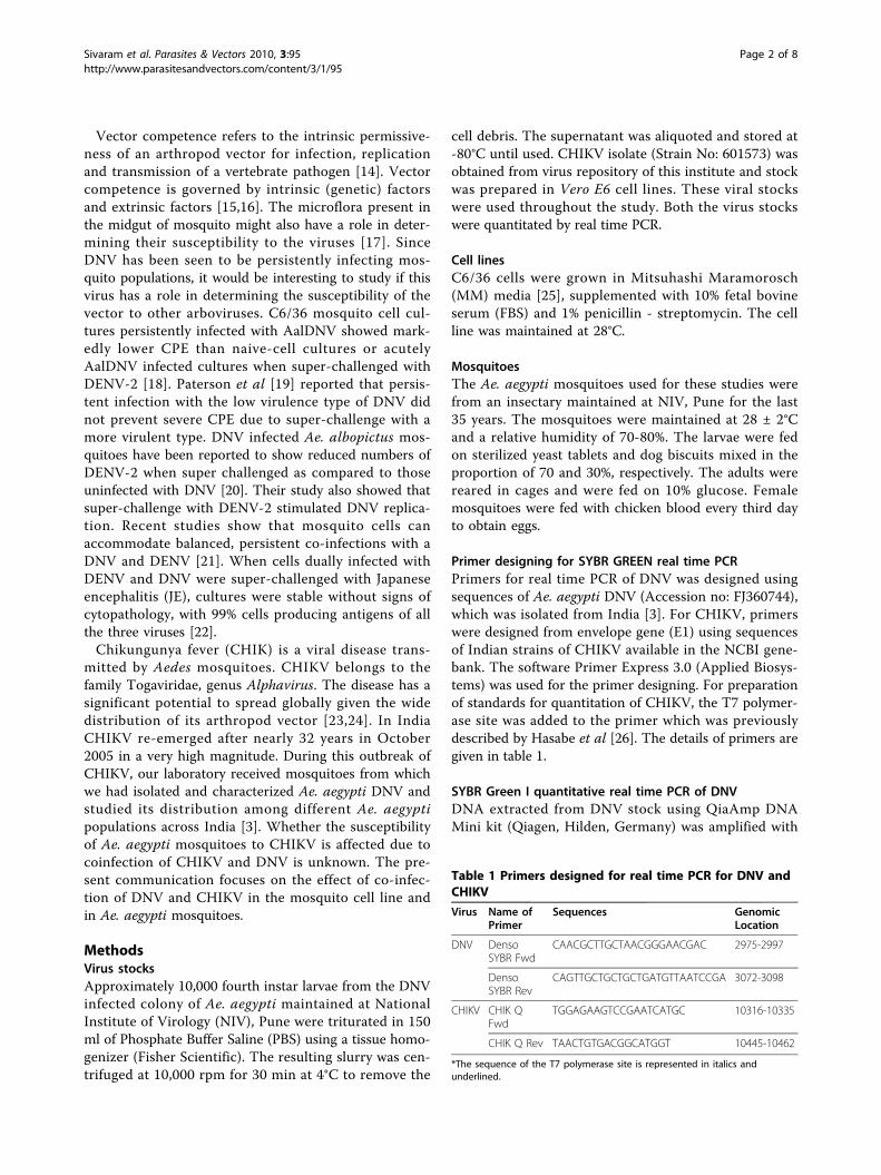
Primer designing for SYBR GREEN real time PCRPrimers for real time PCR of DNV was designed usingsequences of Ae. aegypti DNV (Accession no: FJ360744),which was isolated from India [3]. For CHIKV, primerswere designed from envelope gene (E1) using sequencesof Indian strains of CHIKV available in the NCBI gene-bank. The software Primer Express 3.0 (Applied Biosys-tems) was used for the primer designing. For preparationof standards for quantitation of CHIKV, the T7 polymer-ase site was added to the primer which was previouslydescribed by Hasabe et al [26]. The details of primers aregiven in table 1.
SYBR Green I quantitative real time PCR of DNVDNA extracted from DNV stock using QiaAmp DNAMini kit (Qiagen, Hilden, Germany) was amplified with
Table 1 Primers designed for real time PCR for DNV andCHIKV
Virus Name ofPrimer
Sequences GenomicLocation
DNV DensoSYBR Fwd
CAACGCTTGCTAACGGGAACGAC 2975-2997
DensoSYBR Rev
CAGTTGCTGCTGCTGATGTTAATCCGA 3072-3098
CHIKV CHIK QFwd
TGGAGAAGTCCGAATCATGC 10316-10335
CHIK Q Rev TAACTGTGACGGCATGGT 10445-10462
*The sequence of the T7 polymerase site is represented in italics andunderlined.
Sivaram et al. Parasites & Vectors 2010, 3:95http://www.parasitesandvectors.com/content/3/1/95
Page 2 of 8
DNV specific primers [20]. The amplicon of 1.1 kb wascloned into E. coli cells using TA cloning kit (Invitrogen,Carlsbad, CA) following manufacturer’s protocol. Bluewhite colony screening was used to select the trans-formed cells. Plasmids were extracted from positiveclones using Plasmid Mini kit (Qiagen, Hilden,Germany) and the insert was sequenced on an ABI 3100automated DNA sequencer using Big Dye terminator kit(Applied Biosystems). The concentration of the plasmidwas determined using NanoDrop spectrophotometer(NanoDrop technologies, Inc., USA). The plasmid wasserially diluted 10 fold and used as standard for realtime PCR. The reaction mixture for real time amplifica-tion composed of 12.5 μl of Power SYBR Green PCRMaster Mix (Applied Biosystems, Foster City, CA), 1 μlof each primer (10 pico moles) and 5 μl of templateDNA. The mixture was made up to 25 μl by adding 5.5μl of water. The real time PCR conditions for DNVwere 10 min of initial hot start at 95°C, denaturation at95°C for 15 sec and 1 min of annealing and extension at60°C for 40 cycles. The reactions were carried out inABI 7300 real time PCR system (Applied Bio-systems).The system software provided (SDS) was used to analyzethe results by plotting standard curve.
SYBR Green I quantitative real time RT-PCR of CHIKVRNA was extracted from CHIKV stock using QIAampviral RNA mini kit (Qiagen, Hilden, Germany) and wasreverse transcribed to cDNA using Reverse transcriptionsystem (Promega Corp., Madison, USA) according tothe manufacturer’s protocol. The reverse primerdesigned for real time amplification of CHIKV was usedto synthesize cDNA. The forward primer used toamplify the cDNA contained a site of T7 polymerasewhich could be anchored to the amplicon to enablein vitro transcription. The amplicon of 250 bp was sub-jected to in vitro transcription using T7 RNA polymer-ase by Riboprobe in vitro transcription system (PromegaCorp., Madison, USA) according to manufacturer’sinstructions. The transcript was quantitated using Nano-Drop spectrophotometer. The RNA was reverse tran-scribed to cDNA and serially diluted 10 fold, whichserved as standard. The reaction mix for real timeamplification was prepared similar to that of DNV. Thereal time amplification conditions were 10 min of initialhot start at 95°C, denaturation at 95°C for 15 sec and 30secs of annealing at 50°C and 30 secs of extension at 62°C for 40 cycles. Results were analyzed by plotting stan-dard curve using SDS software.
Determining the effect of co-infection of CHIKV and DNVin C6/36 cell lineC6/36 cells were counted using hemocytometer andwere seeded in 6 well plates at 3000 cells per well.
When the cells were 80% confluent, the media wasremoved and the cells were infected by adding DNV tothe cell sheet. The quantity of DNV used to infect cor-responded to 6.07 × 10⁸ viral particles. The cells wereincubated at 28°C for one hour with intermittent rock-ing of the plate at every 15 min to enable adsorption ofthe virus. After incubation, the inoculum was removed,and the cells were washed with sterile PBS, fed with 2ml of fresh media supplemented with 2% FBS andreturned to the incubator.The cells infected with DNV were incubated for two
days. On day 3 post DNV infection, the cells weresuperinfected with 5.73 × 10⁵ RNA copy number ofCHIKV. All infections and superinfections were done intriplicates. Appropriate controls were used whichinclude cells infected with only CHIKV, only DNV anduninfected cells.Virus was harvested from the infected cells every 24
hr post superinfection for 4 consecutive days byrepeated freeze thaw cycles and centrifugation at 5000rpm for 20 mins at 4°C. The supernatant was dividedinto two batches, one of which was used to quantitateDNV and the other to quantitate CHIKV. Nucleic acidsextracted from these samples were amplified real timeto quantify the viruses using specific primers. Standardswere also amplified along with the samples and the viralgenomic copy numbers were quantitated’ by standardcurve analysis. Each sample was amplified in triplicatesby real time PCR. The copy number of genomes ofDNV and CHIKV in the presence and absence of eachother was compared.
Determining the effect of co-infection of CHIKV and DNVin Ae. aegypti mosquitoesFirst instar Ae. aegypti larvae after one hour of hatching,(n = 3000) were counted and transferred to a beakercontaining 180 ml of water and infected with DNV byadding 20 ml of virus stock to the water in which thelarvae were reared. The quantity of DNV used for infec-tion corresponded to 6.07 × 10⁸ viral particles. After 48hr, the larvae were transferred to a larger pan, fed on amixture of yeast tablet and dog biscuits and were main-tained under normal insectary conditions. Many larvaedied during the course of development. Among thefemales that emerged, 5 individuals were randomlychecked by PCR for DNV infection. The remainingfemales were counted and were infected orally by mem-brane feeding on heparinised blood containing 5.73 ×10⁵ RNA copy number of CHIKV (5 IU/ml of blood-virus mixture) as described by Harada et al [27]. Themosquitoes were allowed to feed for 45 minutes and thefully engorged mosquitoes were segregated and counted.They were maintained under normal insectary condi-tions with 10% glucose as a nutritional supplement.
Sivaram et al. Parasites & Vectors 2010, 3:95http://www.parasitesandvectors.com/content/3/1/95
Page 3 of 8
Appropriate controls were used which include mosqui-toes infected with only CHIKV, only DNV and unin-fected mosquitoes.Twelve mosquitoes were collected and stored in 2
batches of 6 mosquitoes each on alternate day postinfection starting from day 0 up to day 8. DNA wasextracted from one batch to analyse for DNV and RNAfrom the other batch to analyse for CHIKV.Nucleic acid extraction was done from individual mos-
quitoes by triturating the whole mosquito in Nucleasefree water. The nucleic acids were then quantitated byreal time amplification along with the standards usingthe corresponding primers and analyzed by plottingstandard curve. The real time amplification of each sam-ple was performed in triplicates.
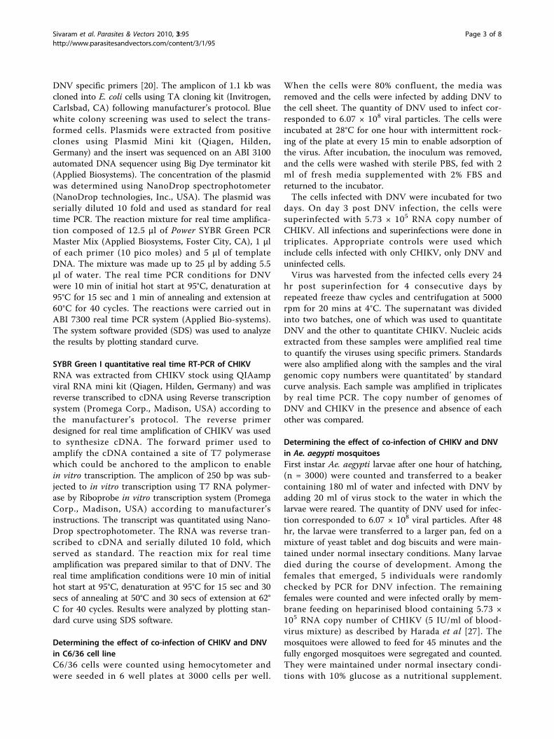
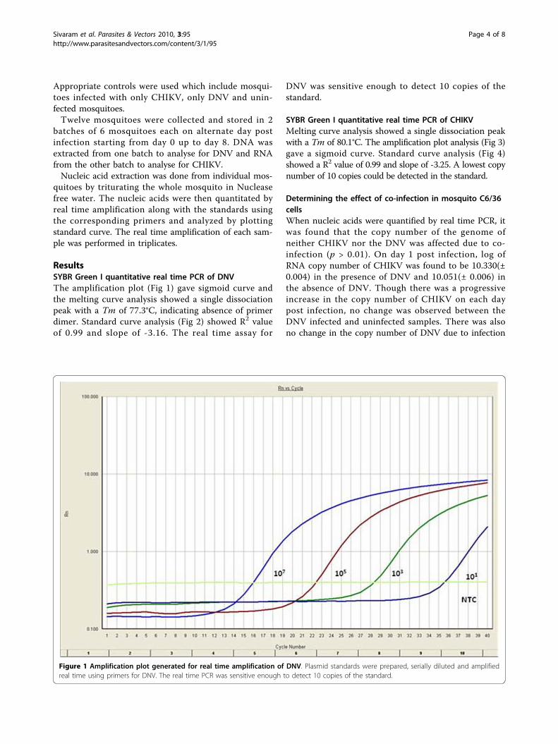
ResultsSYBR Green I quantitative real time PCR of DNVThe amplification plot (Fig 1) gave sigmoid curve andthe melting curve analysis showed a single dissociationpeak with a Tm of 77.3°C, indicating absence of primerdimer. Standard curve analysis (Fig 2) showed R2 valueof 0.99 and slope of -3.16. The real time assay for
DNV was sensitive enough to detect 10 copies of thestandard.
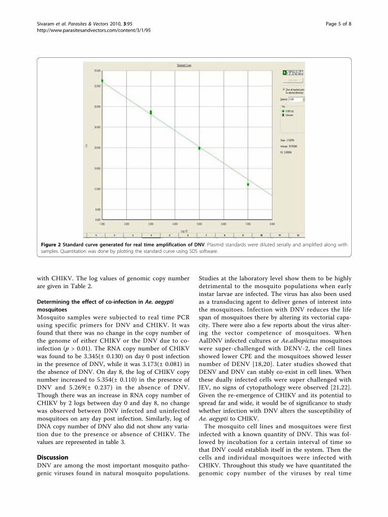
SYBR Green I quantitative real time PCR of CHIKVMelting curve analysis showed a single dissociation peakwith a Tm of 80.1°C. The amplification plot analysis (Fig 3)gave a sigmoid curve. Standard curve analysis (Fig 4)showed a R2 value of 0.99 and slope of -3.25. A lowest copynumber of 10 copies could be detected in the standard.
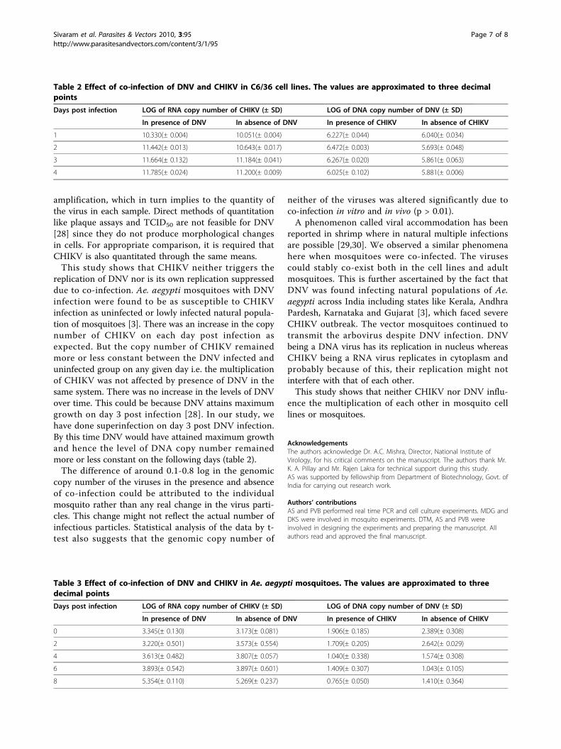
Determining the effect of co-infection in mosquito C6/36cellsWhen nucleic acids were quantified by real time PCR, itwas found that the copy number of the genome ofneither CHIKV nor the DNV was affected due to co-infection (p > 0.01). On day 1 post infection, log ofRNA copy number of CHIKV was found to be 10.330(±0.004) in the presence of DNV and 10.051(± 0.006) inthe absence of DNV. Though there was a progressiveincrease in the copy number of CHIKV on each daypost infection, no change was observed between theDNV infected and uninfected samples. There was alsono change in the copy number of DNV due to infection
Figure 1 Amplification plot generated for real time amplification of DNV. Plasmid standards were prepared, serially diluted and amplifiedreal time using primers for DNV. The real time PCR was sensitive enough to detect 10 copies of the standard.
Sivaram et al. Parasites & Vectors 2010, 3:95http://www.parasitesandvectors.com/content/3/1/95
Page 4 of 8
with CHIKV. The log values of genomic copy numberare given in Table 2.
Determining the effect of co-infection in Ae. aegyptimosquitoesMosquito samples were subjected to real time PCRusing specific primers for DNV and CHIKV. It wasfound that there was no change in the copy number ofthe genome of either CHIKV or the DNV due to co-infection (p > 0.01). The RNA copy number of CHIKVwas found to be 3.345(± 0.130) on day 0 post infectionin the presence of DNV, while it was 3.173(± 0.081) inthe absence of DNV. On day 8, the log of CHIKV copynumber increased to 5.354(± 0.110) in the presence ofDNV and 5.269(± 0.237) in the absence of DNV.Though there was an increase in RNA copy number ofCHIKV by 2 logs between day 0 and day 8, no changewas observed between DNV infected and uninfectedmosquitoes on any day post infection. Similarly, log ofDNA copy number of DNV also did not show any varia-tion due to the presence or absence of CHIKV. Thevalues are represented in table 3.
DiscussionDNV are among the most important mosquito patho-genic viruses found in natural mosquito populations.
Studies at the laboratory level show them to be highlydetrimental to the mosquito populations when earlyinstar larvae are infected. The virus has also been usedas a transducing agent to deliver genes of interest intothe mosquitoes. Infection with DNV reduces the lifespan of mosquitoes there by altering its vectorial capa-city. There were also a few reports about the virus alter-ing the vector competence of mosquitoes. WhenAalDNV infected cultures or Ae.albopictus mosquitoeswere super-challenged with DENV-2, the cell linesshowed lower CPE and the mosquitoes showed lessernumber of DENV [18,20]. Later studies showed thatDENV and DNV can stably co-exist in cell lines. Whenthese dually infected cells were super challenged withJEV, no signs of cytopathology were observed [21,22].Given the re-emergence of CHIKV and its potential tospread far and wide, it would be of significance to studywhether infection with DNV alters the susceptibility ofAe. aegypti to CHIKV.The mosquito cell lines and mosquitoes were first
infected with a known quantity of DNV. This was fol-lowed by incubation for a certain interval of time sothat DNV could establish itself in the system. Then thecells and individual mosquitoes were infected withCHIKV. Throughout this study we have quantitated thegenomic copy number of the viruses by real time
Figure 2 Standard curve generated for real time amplification of DNV. Plasmid standards were diluted serially and amplified along withsamples. Quantitation was done by plotting the standard curve using SDS software.
Sivaram et al. Parasites & Vectors 2010, 3:95http://www.parasitesandvectors.com/content/3/1/95
Page 5 of 8
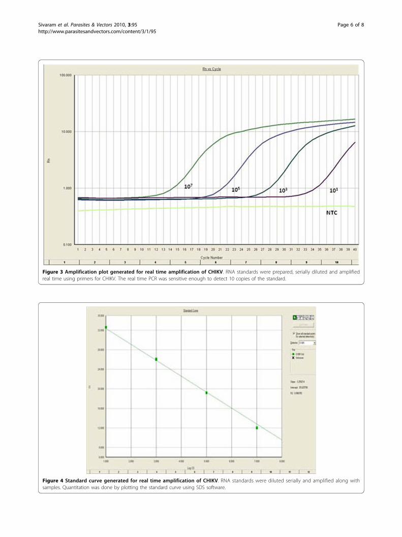
Figure 4 Standard curve generated for real time amplification of CHIKV. RNA standards were diluted serially and amplified along withsamples. Quantitation was done by plotting the standard curve using SDS software.
Figure 3 Amplification plot generated for real time amplification of CHIKV. RNA standards were prepared, serially diluted and amplifiedreal time using primers for CHIKV. The real time PCR was sensitive enough to detect 10 copies of the standard.
Sivaram et al. Parasites & Vectors 2010, 3:95http://www.parasitesandvectors.com/content/3/1/95
Page 6 of 8
amplification, which in turn implies to the quantity ofthe virus in each sample. Direct methods of quantitationlike plaque assays and TCID50 are not feasible for DNV[28] since they do not produce morphological changesin cells. For appropriate comparison, it is required thatCHIKV is also quantitated through the same means.This study shows that CHIKV neither triggers the
replication of DNV nor is its own replication suppresseddue to co-infection. Ae. aegypti mosquitoes with DNVinfection were found to be as susceptible to CHIKVinfection as uninfected or lowly infected natural popula-tion of mosquitoes [3]. There was an increase in the copynumber of CHIKV on each day post infection asexpected. But the copy number of CHIKV remainedmore or less constant between the DNV infected anduninfected group on any given day i.e. the multiplicationof CHIKV was not affected by presence of DNV in thesame system. There was no increase in the levels of DNVover time. This could be because DNV attains maximumgrowth on day 3 post infection [28]. In our study, wehave done superinfection on day 3 post DNV infection.By this time DNV would have attained maximum growthand hence the level of DNA copy number remainedmore or less constant on the following days (table 2).The difference of around 0.1-0.8 log in the genomic
copy number of the viruses in the presence and absenceof co-infection could be attributed to the individualmosquito rather than any real change in the virus parti-cles. This change might not reflect the actual number ofinfectious particles. Statistical analysis of the data by t-test also suggests that the genomic copy number of
neither of the viruses was altered significantly due toco-infection in vitro and in vivo (p > 0.01).A phenomenon called viral accommodation has been
reported in shrimp where in natural multiple infectionsare possible [29,30]. We observed a similar phenomenahere when mosquitoes were co-infected. The virusescould stably co-exist both in the cell lines and adultmosquitoes. This is further ascertained by the fact thatDNV was found infecting natural populations of Ae.aegypti across India including states like Kerala, AndhraPardesh, Karnataka and Gujarat [3], which faced severeCHIKV outbreak. The vector mosquitoes continued totransmit the arbovirus despite DNV infection. DNVbeing a DNA virus has its replication in nucleus whereasCHIKV being a RNA virus replicates in cytoplasm andprobably because of this, their replication might notinterfere with that of each other.This study shows that neither CHIKV nor DNV influ-
ence the multiplication of each other in mosquito celllines or mosquitoes.
AcknowledgementsThe authors acknowledge Dr. A.C. Mishra, Director, National Institute ofVirology, for his critical comments on the manuscript. The authors thank Mr.K. A. Pillay and Mr. Rajen Lakra for technical support during this study.AS was supported by fellowship from Department of Biotechnology, Govt. ofIndia for carrying out research work.
Authors’ contributionsAS and PVB performed real time PCR and cell culture experiments. MDG andDKS were involved in mosquito experiments. DTM, AS and PVB wereinvolved in designing the experiments and preparing the manuscript. Allauthors read and approved the final manuscript.
Table 3 Effect of co-infection of DNV and CHIKV in Ae. aegypti mosquitoes. The values are approximated to threedecimal points
Days post infection LOG of RNA copy number of CHIKV (± SD) LOG of DNA copy number of DNV (± SD)
In presence of DNV In absence of DNV In presence of CHIKV In absence of CHIKV
0 3.345(± 0.130) 3.173(± 0.081) 1.906(± 0.185) 2.389(± 0.308)
2 3.220(± 0.501) 3.573(± 0.554) 1.709(± 0.205) 2.642(± 0.029)
4 3.613(± 0.482) 3.807(± 0.057) 1.040(± 0.338) 1.574(± 0.308)
6 3.893(± 0.542) 3.897(± 0.601) 1.409(± 0.307) 1.043(± 0.105)
8 5.354(± 0.110) 5.269(± 0.237) 0.765(± 0.050) 1.410(± 0.364)
Table 2 Effect of co-infection of DNV and CHIKV in C6/36 cell lines. The values are approximated to three decimalpoints
Days post infection LOG of RNA copy number of CHIKV (± SD) LOG of DNA copy number of DNV (± SD)
In presence of DNV In absence of DNV In presence of CHIKV In absence of CHIKV
1 10.330(± 0.004) 10.051(± 0.004) 6.227(± 0.044) 6.040(± 0.034)
2 11.442(± 0.013) 10.643(± 0.017) 6.472(± 0.003) 5.693(± 0.048)
3 11.664(± 0.132) 11.184(± 0.041) 6.267(± 0.020) 5.861(± 0.063)
4 11.785(± 0.024) 11.200(± 0.009) 6.025(± 0.102) 5.881(± 0.006)
Sivaram et al. Parasites & Vectors 2010, 3:95http://www.parasitesandvectors.com/content/3/1/95
Page 7 of 8
Competing interestsThe authors declare that they have no competing interests.
Received: 13 August 2010 Accepted: 12 October 2010Published: 12 October 2010
References1. Clark TB, Chapman HC: A polyhedrosis in Culex salinarius of Louisiana. J
Invertebr Pathol 1969, 13:312.2. Kittayapong P, Baisley KJ, O’Neill SL: A mosquito densovirus infecting
Aedes aegypti and Aedes albopictus from Thailand. Am J Trop Med Hyg1999, 61:612-617.
3. Sivaram A, Barde PV, Kumar SR, Yadav P, Gokhale MD, Basu A, Mourya DT:Isolation and characterization of densonucleosis virus from Aedesaegypti mosquitoes and its distribution in India. Intervirology 2009,52(1):1-7.
4. Boublik Y, Jousset FX, Bergoin M: Complete nucleotide sequence andgenomic organization of the Aedes albopictus parvovirus (AaPV)pathogenic for Aedes aegypti larvae. Virology 1994, 200:752-763.
5. Rwegoshora RT, Baisley KJ, Kittayapong P: Seasonal and spatial variation innatural densovirus infection in Anopheles minimus S.L. in Thailand.Southeast Asian J Trop Med Publ Health 2000, 31:3-9.
6. Jousset FX, Barreau C, Boublik Y, Cornet M: A parvo-like virus persistentlyinfecting a C6/36 clone of Aedes albopictus mosquito cell line andpathogenic for Aedes aegypti larvae. Virus Res 1993, 29:99-114.
7. Siegl G, Bates RC, Berns KI, Carter BJ, Kelly DC, Kurstak E, Tattersall P:Characteristics and taxonomy of Parvoviridae. Intervirology 1985,23(2):61-73.
8. Barreau C, Jousset FX, Bergoin M: Pathogenicity of the Aedes albopictusparvovirus (AaPV), a denso-like virus, for Ae. aegypti mosquitoes. JInvertebr Pathol 1996, 68:299-309.
9. Chen S, Cheng L, Zhang Q, Lin W, Lu X, Brannan J, Zhou ZH, Zhang J:Genetic, biochemical, and structural characterization of a newdensovirus isolated from a chronically infected Aedes albopictus C6/36cell line. Virology 2004, 318:123-133.
10. Afanasiev BN, Galyov EE, Buchatsky LP, Kozlov YV: Nucleotide sequenceand genomic organization of Aedes densonucleosis virus. Virology 1991,185:323-336.
11. Ledermann JP, Suchman EL, Black WC IV, Carlson JO: Infection andpathogenicity of mosquito densoviruses AeDNV, HeDNV and APeDNV inAedes aegypti mosquitoes (Diptera: Culicidae). J Econ Entomol 2004,97:1827-1835.
12. Buchatsky LP, Bogdanova EN, Kuznetsova MA, Lebedinets NM, Kononko AG,Chabanenko AA, Podrezova LM: Field trial of the viral preparation Virodenon the preimaginal stages of blood-sucking mosquitoes. Meditsinskayaparazitologiya I Parazitare. Bolezni 1987, 4:69-71.
13. Suchman EL, Kononko A, Plake E, Doehling M, Kleler B, Black WC,Buchatsky L, Carlson J: Effects of AeDNV infection on Aedes aegypti (L)lifespan and reproduction. Biol Control 2006, 39:465-473.
14. Black WC, Severson DW: Genetics of vector competence. In Biology ofdisease vectors. Edited by: Marquardt WC, et al. Elsivier Academic Press,California; 2004:167-185.
15. Reeves WC: Factors that influence the probability of epidemics ofWestern equine, St. Louis and California encephalitis in California. VectorViews 1967, 14:13-18.
16. WHO: Vector Ecology. Tech. Rep. Ser 1972, 501:1-38.17. Mourya DT, Pidiyar V, Patole M, Gokhale MD, Shouche Y: Mid gut bacterial
flora of Aedes aegypti affects the susceptibility of mosquitoes to dengueviruses. Dengue Bull 2002, 26:190-194.
18. Burivong P, Pattanakitsakul SN, Thongrungkiat S, Malasit P, Flegel TW:Markedly reduced severity of Dengue virus infection in mosquito cellcultures persistently infected with Aedes albopictus densovirus (AalDNV).Virology 2004, 329:261-269.
19. Paterson A, Robinson E, Suchman E, Afanasiev B, Carlson J: Mosquitodensonucleosis viruses cause dramatically different infection phenotypesin the C6/36 Aedes albopictus cell line. Virology 2005, 337:253-261.
20. Wei W, Shao D, Huang X, Li J, Chen H, Zhang Q, Zhang J: Thepathogenicity of mosquito DNV (C6/36DNV) and its interaction withdengue virus type II in Aedes albopictus. Am J Trop Med Hyg 2006,5:1118-1126.
21. Kanthong N, Khemnu N, Sriurairatana S, Pattanakitsakul S, Malasit P,Flegel TW: Mosquito cells accommodate balanced, persistent co-infections with a densovirus and Dengue virus. Dev Comp Immunol 2008,32:1063-1075.
22. Kanthong N, Khemnu N, Sriurairatana S, Pattanakitsakul S, Malasit P,Flegel TW: Persistent, triple-virus co-infections in mosquito cells. BMCmicrobiology 2010, 10:14.
23. Gratz NG: Emerging and resurging vector-borne diseases. Annu RevEntomol 1999, 44:51-75.
24. Charrel RN, de Lamballerie X, Raoult D: Chikungunya outbreaks-theglobalization of vectorborne diseases. N Engl J Med 2007, 356:769-771.
25. Mitsuhashi J, Maramorosch K: Leafhopper tissue culture: embryonic,nymphal and imaginal tissues from aseptic insects. Contrib BoyceThompson Inst 1964, 22:435-460.
26. Hasebe F, Parquet MC, Pandey BD, Mathenge EGM, Morita K,Balasubramaniam V, Saat Z, Yusop A, Sinniah M, Natkunam S, Igarashi A:Combined detection and genotyping of Chikungunya virus by a specificreverse transcription-polymerase chain reaction. J Med Virol 2002,67:370-374.
27. Harada M, Matsuoka H, Suguri S: A convenient mosquito membranefeeding method. Med Entomol Zool 1996, 47:103-105.
28. Azarkh A, Robinson E, Hirunkanokpun S, Afanasiev B, Kittayapong P,Carlson J, Corsini J: Mosquito Densonucleosis Virus non-structural proteinNS2 is necessary for a productive infection. Virology 2008, 374(1):128-137.
29. Flegel TW: The shrimp response to viral pathogens. In The new wave.Proceedings of the special session on sustainable shrimp aquaculture, WorldAquaculture, Orlando. Edited by: Browdy CL, Jory DE. World AquacultureSociety, Boca Raton; 2001:190-214.
30. Flegel TW, Pasharawipas T: Active viral accommodation: a new conceptfor crustacean response to viral pathogens. In Advances in ShrimpBiotechnology. Edited by: Flegel TW. National Center for GeneticEngineering and Biotechnology, Bangkok; 1998:245-250.
doi:10.1186/1756-3305-3-95Cite this article as: Sivaram et al.: Evidence of co-infection ofchikungunya and densonucleosis viruses in C6/36 cell lines andlaboratory infected Aedes aegypti (L.) mosquitoes. Parasites & Vectors2010 3:95.
Submit your next manuscript to BioMed Centraland take full advantage of:
• Convenient online submission
• Thorough peer review
• No space constraints or color figure charges
• Immediate publication on acceptance
• Inclusion in PubMed, CAS, Scopus and Google Scholar
• Research which is freely available for redistribution
Submit your manuscript at www.biomedcentral.com/submit
Sivaram et al. Parasites & Vectors 2010, 3:95http://www.parasitesandvectors.com/content/3/1/95
Page 8 of 8
Copyright © 2022 FDOKUMEN