058 Effect of Cyclosporine on Left Ventricle Remodeling after Reperfused Myocardial Infarction
6
Effect of Cyclosporine on Left Ventricular Remodeling After Reperfused Myocardial Infarction Nathan Mewton, MD,* Pierre Croisille, MD, PHD,* Gerald Gahide, MD,§ Gilles Rioufol, MD, PHD,*† Eric Bonnefoy, MD, PHD,* Ingrid Sanchez, MD,* Thien Tri Cung, MD,‡§ Catherine Sportouch, MD,‡§ Denis Angoulvant, MD, PHD,*† Gérard Finet, MD, PHD,*† Xavier André-Fouët, MD,*† Geneviève Derumeaux, MD, PHD,*† Christophe Piot, MD, PHD,‡§ Hélène Vernhet, MD,‡§ Didier Revel, MD,* Michel Ovize, MD, PHD*† Lyon and Montpellier, France Objectives This study examined the effect of a single dose of cyclosporine administered at the time of reperfusion on left ventricular (LV) remodeling and function by cardiac magnetic resonance 5 days and 6 months after myocardial infarction. Background In a human study, administration of cyclosporine at the time of acute reperfusion was associated with a smaller infarct size. Methods Twenty-eight patients of the original cyclosporine study had an acute (at 5 days) and a follow-up (at 6 months) cardiac magnetic resonance study to determine LV volumes, mass, ejection fraction, myocardial wall thickness in infarcted and remote noninfarcted myocardium, and infarct size. Results There was a persistent reduction in infarct size at 6 months in the cyclosporine group compared with the control group of patients (29 15 g vs. 38 14 g; p 0.04). There was a significant reduction of LV end-systolic vol- ume (and a trend for LV end-diastolic volume; p 0.07) in the cyclosporine group compared with the control group, both at 5 days and 6 months after infarction. There was no significant difference between the 2 groups in either global LV mass or regional wall thickness of the remote noninfarcted myocardium at 5 days or 6 months. Attenuation of LV dilation and improvement of LV ejection fraction by cyclosporine at 6 months were correlated with infarct size reduction. Conclusions Cyclosporine used at the moment of acute myocardial infarction reperfusion persistently reduces infarct size and does not have a detrimental effect on LV remodeling. These results are preliminary and must be supported by further studies. (Ciclosporin A and Acute Myocardial Infarction; NCT00403728) (J Am Coll Cardiol 2010;55: 1200–5) © 2010 by the American College of Cardiology Foundation Recently, we showed that the administration of cyclosporine at the time of reperfusion in patients undergoing percutaneous coronary intervention for an acute myocardial infarction (AMI) was associated with a reduction in infarct size (1). The rationale of this proof-of-concept study was that cyclosporine is a potent inhibitor of the opening of the mitochondrial permeability transition pore, which plays a crucial role in lethal myocardial reperfusion injury (2–5). Cyclosporine inhibits mitochondrial permeability transition pore opening via its binding to the peptidylprolyl isomerase cyclophilin D located in the mitochondrial matrix (6). See page 1206 However, cyclosporine is not specific for cyclophilin D and also forms a complex with cytosolic cyclophilin A that inhibits the calcium-activated protein phosphatase cal- cineurin. The calcineurin-dependent pathway seems to be crucial for the compensatory myocardial hypertrophy of the remote noninfarcted myocardium during the early post- AMI remodeling stage. Animal studies have found that long-term daily administration of cyclosporine can alter left ventricular (LV) remodeling after AMI and favor the From the *Hospices Civils de Lyon, Université Claude Bernard Lyon, Lyon, France; †Inserm U886, Lyon, France; ‡Inserm U661, Montpellier, France; and the §Hopital Arnaud de Villeneuve, Université de Montpellier I and II, Montpellier, France. Dr. Mewton was supported by a research grant from the French Federation of Cardiology (Fédération Française de Cardiologie). Manuscript received June 24, 2009; revised manuscript received September 16, 2009, accepted October 20, 2009. Journal of the American College of Cardiology Vol. 55, No. 12, 2010 © 2010 by the American College of Cardiology Foundation ISSN 0735-1097/10/$36.00 Published by Elsevier Inc. doi:10.1016/j.jacc.2009.10.052
-
Upload
independent -
Category
Documents
-
view
0 -
download
0
Transcript of 058 Effect of Cyclosporine on Left Ventricle Remodeling after Reperfused Myocardial Infarction

Rtc(
cmc
F†AM(
2
Journal of the American College of Cardiology Vol. 55, No. 12, 2010© 2010 by the American College of Cardiology Foundation ISSN 0735-1097/10/$36.00P
Effect of Cyclosporine on Left VentricularRemodeling After Reperfused Myocardial Infarction
Nathan Mewton, MD,* Pierre Croisille, MD, PHD,* Gerald Gahide, MD,§Gilles Rioufol, MD, PHD,*† Eric Bonnefoy, MD, PHD,* Ingrid Sanchez, MD,*Thien Tri Cung, MD,‡§ Catherine Sportouch, MD,‡§ Denis Angoulvant, MD, PHD,*†Gérard Finet, MD, PHD,*† Xavier André-Fouët, MD,*† Geneviève Derumeaux, MD, PHD,*†Christophe Piot, MD, PHD,‡§ Hélène Vernhet, MD,‡§ Didier Revel, MD,*Michel Ovize, MD, PHD*†
Lyon and Montpellier, France
Objectives This study examined the effect of a single dose of cyclosporine administered at the time of reperfusion on leftventricular (LV) remodeling and function by cardiac magnetic resonance 5 days and 6 months after myocardialinfarction.
Background In a human study, administration of cyclosporine at the time of acute reperfusion was associated with a smallerinfarct size.
Methods Twenty-eight patients of the original cyclosporine study had an acute (at 5 days) and a follow-up (at 6 months)cardiac magnetic resonance study to determine LV volumes, mass, ejection fraction, myocardial wall thicknessin infarcted and remote noninfarcted myocardium, and infarct size.
Results There was a persistent reduction in infarct size at 6 months in the cyclosporine group compared with the controlgroup of patients (29 � 15 g vs. 38 � 14 g; p � 0.04). There was a significant reduction of LV end-systolic vol-ume (and a trend for LV end-diastolic volume; p � 0.07) in the cyclosporine group compared with the controlgroup, both at 5 days and 6 months after infarction. There was no significant difference between the 2 groups ineither global LV mass or regional wall thickness of the remote noninfarcted myocardium at 5 days or 6 months.Attenuation of LV dilation and improvement of LV ejection fraction by cyclosporine at 6 months were correlatedwith infarct size reduction.
Conclusions Cyclosporine used at the moment of acute myocardial infarction reperfusion persistently reduces infarct size anddoes not have a detrimental effect on LV remodeling. These results are preliminary and must be supported byfurther studies. (Ciclosporin A and Acute Myocardial Infarction; NCT00403728) (J Am Coll Cardiol 2010;55:1200–5) © 2010 by the American College of Cardiology Foundation
ublished by Elsevier Inc. doi:10.1016/j.jacc.2009.10.052
Cpc
aiccrAl
ecently, we showed that the administration of cyclosporine athe time of reperfusion in patients undergoing percutaneousoronary intervention for an acute myocardial infarctionAMI) was associated with a reduction in infarct size (1).
The rationale of this proof-of-concept study was thatyclosporine is a potent inhibitor of the opening of theitochondrial permeability transition pore, which plays a
rucial role in lethal myocardial reperfusion injury (2–5).
rom the *Hospices Civils de Lyon, Université Claude Bernard Lyon, Lyon, France;Inserm U886, Lyon, France; ‡Inserm U661, Montpellier, France; and the §Hopitalrnaud de Villeneuve, Université de Montpellier I and II, Montpellier, France. Dr.ewton was supported by a research grant from the French Federation of Cardiology
Fédération Française de Cardiologie).
vManuscript received June 24, 2009; revised manuscript received September 16,
009, accepted October 20, 2009.
yclosporine inhibits mitochondrial permeability transitionore opening via its binding to the peptidylprolyl isomeraseyclophilin D located in the mitochondrial matrix (6).
See page 1206
However, cyclosporine is not specific for cyclophilin Dnd also forms a complex with cytosolic cyclophilin A thatnhibits the calcium-activated protein phosphatase cal-ineurin. The calcineurin-dependent pathway seems to berucial for the compensatory myocardial hypertrophy of theemote noninfarcted myocardium during the early post-MI remodeling stage. Animal studies have found that
ong-term daily administration of cyclosporine can alter left
entricular (LV) remodeling after AMI and favor the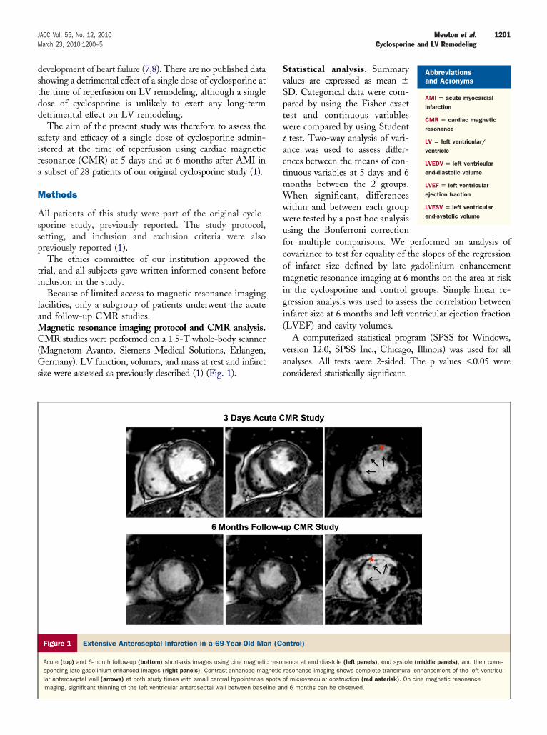
dstdd
sira
M
Assp
ti
faMC(Gs
SvSptwtaetmWwwufcomigi(
vac
1201JACC Vol. 55, No. 12, 2010 Mewton et al.March 23, 2010:1200–5 Cyclosporine and LV Remodeling
evelopment of heart failure (7,8). There are no published datahowing a detrimental effect of a single dose of cyclosporine athe time of reperfusion on LV remodeling, although a singleose of cyclosporine is unlikely to exert any long-termetrimental effect on LV remodeling.The aim of the present study was therefore to assess the
afety and efficacy of a single dose of cyclosporine admin-stered at the time of reperfusion using cardiac magneticesonance (CMR) at 5 days and at 6 months after AMI insubset of 28 patients of our original cyclosporine study (1).
ethods
ll patients of this study were part of the original cyclo-porine study, previously reported. The study protocol,etting, and inclusion and exclusion criteria were alsoreviously reported (1).The ethics committee of our institution approved the
rial, and all subjects gave written informed consent beforenclusion in the study.
Because of limited access to magnetic resonance imagingacilities, only a subgroup of patients underwent the acutend follow-up CMR studies.
agnetic resonance imaging protocol and CMR analysis.MR studies were performed on a 1.5-T whole-body scanner
Magnetom Avanto, Siemens Medical Solutions, Erlangen,ermany). LV function, volumes, and mass at rest and infarct
ize were assessed as previously described (1) (Fig. 1).
Figure 1 Extensive Anteroseptal Infarction in a 69-Year-Old Ma
Acute (top) and 6-month follow-up (bottom) short-axis images using cine magneticsponding late gadolinium-enhanced images (right panels). Contrast-enhanced maglar anteroseptal wall (arrows) at both study times with small central hypointense simaging, significant thinning of the left ventricular anteroseptal wall between basel
tatistical analysis. Summaryalues are expressed as mean �D. Categorical data were com-ared by using the Fisher exactest and continuous variablesere compared by using Studenttest. Two-way analysis of vari-nce was used to assess differ-nces between the means of con-inuous variables at 5 days and 6onths between the 2 groups.hen significant, differences
ithin and between each groupere tested by a post hoc analysissing the Bonferroni correctionor multiple comparisons. We performed an analysis ofovariance to test for equality of the slopes of the regressionf infarct size defined by late gadolinium enhancementagnetic resonance imaging at 6 months on the area at risk
n the cyclosporine and control groups. Simple linear re-ression analysis was used to assess the correlation betweennfarct size at 6 months and left ventricular ejection fractionLVEF) and cavity volumes.
A computerized statistical program (SPSS for Windows,ersion 12.0, SPSS Inc., Chicago, Illinois) was used for allnalyses. All tests were 2-sided. The p values �0.05 wereonsidered statistically significant.
ntrol)
ance at end diastole (left panels), end systole (middle panels), and their corre-resonance imaging shows complete transmural enhancement of the left ventricu-f microvascular obstruction (red asterisk). On cine magnetic resonanced 6 months can be observed.
Abbreviationsand Acronyms
AMI � acute myocardialinfarction
CMR � cardiac magneticresonance
LV � left ventricular/ventricle
LVEDV � left ventricularend-diastolic volume
LVEF � left ventricularejection fraction
LVESV � left ventricularend-systolic volume
n (Co
resonneticpots oine an

R
SuTgCseciCcttbewgs
mttraLdd
vci
cgt
L
a0m
S
V
mL
RC
V
v
1202 Mewton et al. JACC Vol. 55, No. 12, 2010Cyclosporine and LV Remodeling March 23, 2010:1200–5
esults
tudy population. Characteristics of the whole study pop-lation at first admission were previously reported (1).wenty-eight (13 in the control and 15 in the cyclosporineroup) of 58 patients included in the initial study had aMR study done at 5 days and 6 months. In the CMR
ubpopulation of patients, there was no significant differ-nce between the 2 groups for comorbidities, LV andoronary angiography characteristics at admission, or med-cal treatment at 6 months (Table 1).
MR results. INFARCT SIZE. At 5 days, infarct size in theyclosporine group averaged 34 � 14 g versus 48 � 24 g inhe control group (p � 0.03). There was a significant 15%o 20% shrinkage of the infarcted tissue area in the 2 groupsetween the 2 CMR studies. The initial significant differ-nce between the 2 groups was maintained at 6 months,ith the infarct size averaging 29 � 15 g in the cyclosporineroup and 38 � 14 g in the control group (p � 0.04), ashown in Table 2.
The infarct size at 6 months correlated with area at risk aseasured by LV angiography at admission (Fig. 2). Impor-
antly most data points for the cyclosporine group fell belowhe control regression line, confirming that infarct sizeeduction by cyclosporine was independent of the size of therea at risk.V volumes and function at 5 days and 6 months. At 5ays, there were significantly lower left ventricular end-
tudy Population Baseline CharacteristicsTable 1 Study Population Baseline Characteristics
Cyclosporine(n � 15)
Control(n � 13) p Value
Age, yrs 60 � 10 63 � 12 0.45
Sex, M/F 12/3 8/5 0.38
BMI, kg/m2 27 � 4 25 � 4 0.44
Hypertension 4/15 4/13 1.0
Smokers 8/15 7/13 1.0
Dyslipidemia 6/15 4/13 0.41
Diabetes 4/15 3/13 0.67
LV and coronary angiography
Infarct-related artery 0.46
LAD 6 (40%) 7 (54%)
RCA 9 (60%) 6 (46%)
LVEF, % 49 � 9 53 � 11 0.40
ACS, % 41 � 15 39 � 8 0.17
Ischemia time, min 379 � 201 290 � 129 0.31
Treatment at 6 months afterAMI, %
Aspirin 15/15 13/13 1.0
Clopidogrel 15/15 13/13 1.0
ACE inhibitors 13/15 10/13 0.64
Beta-blockers 13/15 11/13 1.0
Statins 14/15 12/13 1.0
alues are expressed as mean � SD, n/N, or n (%).ACE � angiotensin-converting enzyme; ACS � abnormally contracting segments; AMI � acuteyocardial infarction; BMI � body mass index; LAD � left anterior descending coronary artery;
V � left ventricular; LVEF � left ventricular ejection fraction; RCA � right coronary artery.
iastolic volume (LVEDV) and left ventricular end-systolic
olume (LVESV) in the cyclosporine compared with theontrol group (Table 2). There was no significant differencen LVEF between the 2 groups.
At 6 months, we observed a lower LVESV in theyclosporine group compared with the control group. Re-arding the LVEDV, there was a nonsignificant (p � 0.07)rend toward lower value in the cyclosporine group.
In each group, we noticed a nonsignificant decrease inVEDV and LVESV between 5 days and 6 months.There was a significant correlation between infarct size
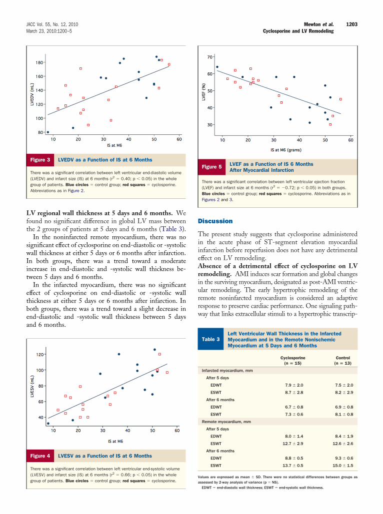
nd both LVEDV (r2 � 0.40; p � 0.05), LVESV (r2 �.66; p � 0.05), and LVEF (r2 � �0.72; p � 0.05) at 6onths in the whole group of patients (Figs. 3 to 5).
Figure 2Infarct Size (CMR 6 Months After AcuteMyocardial Infarction) as a Function of the Sizeof the Area at Risk (LV Angiography at Admission)
There was a significant correlation between the 2 variables in the control group(r2 � 0.84). Data points for the cyclosporine group (red squares) (r2 � 0.67)lie below the regression line for the control group (blue circles). These dataindicate that, for any given area at risk, cyclosporine administration was asso-ciated with a reduction in the resulting infarct size as measured by delayedenhancement cardiac magnetic resonance (CMR). This difference was signifi-cant by analysis of covariance (p � 0.007). ACS � acute coronary syndrome;LV � left ventricular; M6 � 6 months.
esults for Measures ofMR After 5 Days and 6 MonthsTable 2 Results for Measures ofCMR After 5 Days and 6 Months
Cyclosporine(n � 15)
Control(n � 13) p Value
After 5 days
LVEDV, ml 143 � 32 167 � 43 0.05
LVESV, ml 72 � 20 91 � 36 0.04
LVEF, % 50 � 10 47 � 11 0.19
Infarct size, g 34 � 14 48 � 24 0.03
After 6 months
LVEDV, ml 133 � 28 151 � 35 0.07
LVESV, ml 67 � 24 84 � 29 0.05
LVEF, % 51 � 9 46 � 11 0.10
Infarct size, g 29 � 15 38 � 14 0.04
alues are expressed as mean � SD.CMR � cardiac magnetic resonance; LVEDV � left ventricular end-diastolic volume; LVEF � left
entricular ejection fraction; LVESV � left ventricular end-systolic volume.
Lft
swIit
etbea
D
TiieAriurrw
LMM
V
1203JACC Vol. 55, No. 12, 2010 Mewton et al.March 23, 2010:1200–5 Cyclosporine and LV Remodeling
V regional wall thickness at 5 days and 6 months. Weound no significant difference in global LV mass betweenhe 2 groups of patients at 5 days and 6 months (Table 3).
In the noninfarcted remote myocardium, there was noignificant effect of cyclosporine on end-diastolic or -systolicall thickness at either 5 days or 6 months after infarction.
n both groups, there was a trend toward a moderatencrease in end-diastolic and -systolic wall thickness be-ween 5 days and 6 months.
In the infarcted myocardium, there was no significantffect of cyclosporine on end-diastolic or -systolic wallhickness at either 5 days or 6 months after infarction. Inoth groups, there was a trend toward a slight decrease innd-diastolic and -systolic wall thickness between 5 daysnd 6 months.
Figure 3 LVEDV as a Function of IS at 6 Months
There was a significant correlation between left ventricular end-diastolic volume(LVEDV) and infarct size (IS) at 6 months (r2 � 0.40; p � 0.05) in the wholegroup of patients. Blue circles � control group; red squares � cyclosporine.Abbreviations as in Figure 2.
Figure 4 LVESV as a Function of IS at 6 Months
There was a significant correlation between left ventricular end-systolic volume(LVESV) and infarct size (IS) at 6 months (r2 � 0.66; p � 0.05) in the wholegroup of patients. Blue circles � control group; red squares � cyclosporine.
aiscussion
he present study suggests that cyclosporine administeredn the acute phase of ST-segment elevation myocardialnfarction before reperfusion does not have any detrimentalffect on LV remodeling.bsence of a detrimental effect of cyclosporine on LV
emodeling. AMI induces scar formation and global changesn the surviving myocardium, designated as post-AMI ventric-lar remodeling. The early hypertrophic remodeling of theemote noninfarcted myocardium is considered an adaptiveesponse to preserve cardiac performance. One signaling path-ay that links extracellular stimuli to a hypertrophic transcrip-
Figure 5 LVEF as a Function of IS 6 MonthsAfter Myocardial Infarction
There was a significant correlation between left ventricular ejection fraction(LVEF) and infarct size at 6 months (r2 � �0.72; p � 0.05) in both groups.Blue circles � control group; red squares � cyclosporine. Abbreviations as inFigures 2 and 3.
eft Ventricular Wall Thickness in the Infarctedyocardium and in the Remote Nonischemicyocardium at 5 Days and 6 MonthsTable 3
Left Ventricular Wall Thickness in the InfarctedMyocardium and in the Remote NonischemicMyocardium at 5 Days and 6 Months
Cyclosporine(n � 15)
Control(n � 13)
Infarcted myocardium, mm
After 5 days
EDWT 7.9 � 2.0 7.5 � 2.0
ESWT 8.7 � 2.8 8.2 � 2.9
After 6 months
EDWT 6.7 � 0.8 6.9 � 0.8
ESWT 7.3 � 0.6 8.1 � 0.8
Remote myocardium, mm
After 5 days
EDWT 8.0 � 1.4 8.4 � 1.9
ESWT 12.7 � 2.9 12.6 � 2.6
After 6 months
EDWT 8.8 � 0.5 9.3 � 0.6
ESWT 13.7 � 0.5 15.0 � 1.5
alues are expressed as mean � SD. There were no statistical differences between groups as
ssessed by 2-way analysis of variance (p � NS).EDWT � end-diastolic wall thickness; ESWT � end-systolic wall thickness.
tdtCtslphemtita
ooesrc
tatri2utaaeriwawaRiciatroutLm6snw
etmodtnpc
LpCcLtrcot
C
ToLhwTs
RsRu
R
1204 Mewton et al. JACC Vol. 55, No. 12, 2010Cyclosporine and LV Remodeling March 23, 2010:1200–5
ional response of the myocyte uses the Ca2�/calmodulin-ependent phosphatase calcineurin and its downstreamranscriptional effector nuclear factor of activated T cells.yclosporine binds to the mitochondrial cyclophilin D and
hereby inhibits lethal reperfusion injury and reduces infarctize (2,3,9). However, cyclosporine also binds to the cytoso-ic cyclophilin A and inhibits the calcium-activated proteinhosphatase calcineurin; it may therefore influence cardiacypertrophic response and LV remodeling after AMI. Theffects of cyclosporine on immediate and long-term post–yocardial infarction LV remodeling in animal experimen-
al models have been equivocal. In some studies, cyclospor-ne was shown to impede the compensatory hypertrophy ofhe remote noninfarcted myocardium, increase LV dilation,nd decrease the myocardial systolic performance (7,8).
We showed that cyclosporine had no detrimental effectn LV remodeling at 6 months. The regional modificationsf infarcted and noninfarcted myocardium were not differ-nt between cyclosporine and control patients. This tends touggest that a single dose of cyclosporine was enough toeduce infarct size, but not sufficient to modify thealcineurin-mediated hypertrophic response.
This apparent discordance between several experimen-al studies and the present clinical study may be associ-ted with different factors. First, all experimental studieshat found a detrimental effect of cyclosporine usedepeated doses of this drug. In contrast, we used a singlentravenous injection of cyclosporine. Second, we used a.5-mg/kg dose, which was 4 to 10 times lower than thatsed in the previously mentioned studies. Third, theiming of administration of cyclosporine differed greatlymong all studies. Opening of the mitochondrial perme-bility transition pore has been shown to occur in thearly minutes of reperfusion and represents a “point of noeturn” to cell death (10,11). Experimental studies usingschemic post-conditioning have shown that the timeindow to prevent lethal reperfusion injury is very narrow
fter reflow (12,13). This is the reason why cyclosporineas injected before angioplasty of the culprit coronary
rtery in this clinical trial.elationship between LV remodeling and persistent
nfarct size limitation by cyclosporine. At 6 months,yclosporine–treated patients exhibited a 29% reduction ofrreversible myocardial damage, very similar to that observedt 5 days (1). In previous studies, infarct size has been showno be a major prognostic factor, predictive of increased LVemodeling, congestive heart failure, and clinical adverseutcome. In recent studies, when assessed at 4 months andp to 4 years, infarct size in multivariate analysis was one ofhe strongest independent predictors of ejection fraction andV volumes independently of scar localization and trans-urality (14). Other studies assessing LV remodeling at
-month follow-up after AMI with echocardiographyhowed that peak troponin T and peak creatine phosphoki-ase levels, which are directly correlated with infarct size,
ere strong independent predictors of LV functional recov-ry (15,16). Thus, reducing the infarct size is an importantarget for the treatment of patients with ongoing acuteyocardial infarction. Together with a persistent reduction
f infarct size, we found that cyclosporine-treated patientsisplayed a significant attenuation of LVESV with a trendoward a diminution of LVEDV enlargement. The smallumber of patients likely explains the nonsignificant im-rovement of LVEF in the cyclosporine-treated patientsompared with the control patients.
In the whole study population, the LV volumes andVEF modifications followed patterns similar to those ofreviously published data in post-AMI LV remodelingMR studies (14). Furthermore, we found comparable
orrelations between infarct size and LVEF, LVEDV, andVESV with previously published data (17). The fact that
he cyclosporine and the control groups share the sameegression lines indicates that there was no specific effect ofyclosporine on LV remodeling and tells us that thebserved trend toward improvement might be entirely dueo infarct size reduction (Figs. 3 to 5).
onclusions
his study shows that cyclosporine administered at the timef AMI reperfusion does not have a detrimental effect onV remodeling. On the contrary, cyclosporine seems toave a sustained beneficial effect on infarct size reduction,hich might improve the post-infarction remodeling process.hese results are preliminary and must be supported by further
tudies.
eprint requests and correspondence: Prof. Michel Ovize, In-erm U886, Laboratoire de Physiologie Lyon-Nord, 8, avenueockefeller, 69373 Lyon, France. E-mail: [email protected].
EFERENCES
1. Piot C, Croisille P, Staat P, et al. Effect of cyclosporine on reperfusioninjury in acute myocardial infarction. N Engl J Med 2008;359:473–81.
2. Nazareth W, Yafei N, Crompton M. Inhibition of anoxia-inducedinjury in heart myocytes by cyclosporin A. J Mol Cell Cardiol1991;23:1351–4.
3. Duchen MR, McGuinness O, Brown LA, Crompton M. On theinvolvement of a cyclosporin A sensitive mitochondrial pore inmyocardial reperfusion injury. Cardiovasc Res 1993;27:1790–4.
4. Crompton M, Costi A. A heart mitochondrial Ca2(�)-dependentpore of possible relevance to re-perfusion-induced injury. Evidencethat ADP facilitates pore interconversion between the closed and openstates. Biochem J 1990;266:33–9.
5. Di Lisa F, Menabo R, Canton M, Barile M, Bernardi P. Opening ofthe mitochondrial permeability transition pore causes depletion ofmitochondrial and cytosolic NAD� and is a causative event in thedeath of myocytes in postischemic reperfusion of the heart. J BiolChem 2001;276:2571–5.
6. Woodfield K, Ruck A, Brdiczka D, Halestrap AP. Direct demonstra-tion of a specific interaction between cyclophilin-D and the adeninenucleotide translocase confirms their role in the mitochondrial perme-
ability transition. Biochem J 1998;336:287–90.
1
1
1
1
1
1
1
1
K
1205JACC Vol. 55, No. 12, 2010 Mewton et al.March 23, 2010:1200–5 Cyclosporine and LV Remodeling
7. Oie E, Bjornerheim R, Clausen OP, Attramadal H. Cyclosporin Ainhibits cardiac hypertrophy and enhances cardiac dysfunction duringpostinfarction failure in rats. Am J Physiol Heart Circ Physiol2000;278:2115–23.
8. Youn TJ, Piao H, Kwon JS, et al. Effects of the calcineurin dependentsignaling pathway inhibition by cyclosporin A on early and late cardiacremodeling following myocardial infarction. Eur J Heart Fail 2002;4:713–8.
9. Argaud L, Gomez L, Gateau-Roesch O, et al. Trimetazidineinhibits mitochondrial permeability transition pore opening andprevents lethal ischemia-reperfusion injury. J Mol Cell Cardiol2005;39:893–9.
0. Argaud L, Gateau-Roesch O, Chalabreysse L, et al. Preconditioningdelays Ca2�-induced mitochondrial permeability transition. Cardio-vasc Res 2004;61:115–22.
1. Gomez L, Chavanis N, Argaud L, et al. Fas-independent mitochon-drial damage triggers cardiomyocyte death after ischemia-reperfusion.Am J Physiol Heart Circ Physiol 2005;289:H2153–8.
2. Argaud L, Gateau-Roesch O, Raisky O, Loufouat J, Robert D, OvizeM. Postconditioning inhibits mitochondrial permeability transition.
Circulation 2005;111:194–7. m3. Kin H, Zhao ZQ, Sun HY, et al. Postconditioning attenuatesmyocardial ischemia-reperfusion injury by inhibiting events in theearly minutes of reperfusion. Cardiovasc Res 2004;62:74–85.
4. Orn S, Manhenke C, Anand IS, et al. Effect of left ventricular scarsize, location, and transmurality on left ventricular remodeling withhealed myocardial infarction. Am J Cardiol 2007;99:1109 –14.
5. Mollema SA, Liem SS, Suffoletto MS, et al. Left ventricular dyssyn-chrony acutely after myocardial infarction predicts left ventricularremodeling. J Am Coll Cardiol 2007;50:1532–40.
6. Parodi G, Memisha G, Carrabba N, et al. Prevalence, predictors, timecourse, and long-term clinical implications of left ventricular func-tional recovery after mechanical reperfusion for acute myocardialinfarction. Am J Cardiol 2007;100:1718–22.
7. Baks T, van Geuns RJ, Biagini E, et al. Recovery of left ventricularfunction after primary angioplasty for acute myocardial infarction. EurHeart J 2005;26:1070–7.
ey Words: acute myocardial infarction y cyclosporine y remodeling y
agnetic resonance imaging.