Beyond immunosuppression - challenges in the clinical management of lupus nephritis

Journal of Controlled Release 156 (2011) 406–412
Contents lists available at ScienceDirect
Journal of Controlled Release
j ourna l homepage: www.e lsev ie r.com/ locate / jconre l
NANOMEDICIN
E
Cyclosporine A-loaded and stem cell-seeded electrospun nanofibers for cell-basedtherapy and local immunosuppression
Vladimir Holan a,b,⁎, Milada Chudickova a,b, Peter Trosan a,b, Eliska Svobodova a,b, Magdalena Krulova a,b,Sarka Kubinova c, Eva Sykova c, Jakub Sirc d, Jiri Michalek d, Martina Juklickova e,Marcela Munzarova e, Alena Zajicova a
a Institute of Molecular Genetics, Academy of Sciences of the Czech Republic, 142 20 Prague, Czech Republicb Faculty of Science, Charles University, 128 44 Prague, Czech Repubicc Institute of Experimental Medicine, Academy of Sciences of the Czech Republic, 142 20 Prague, Czech Republicd Institute of Macromolecular Chemistry, Academy of Sciences of the Czech Republic, 162 06 Prague, Czech Republice Elmarco, 460 07 Liberec, Czech Republic
⁎ Corresponding author at: Institute of Molecular Gethe Czech Republic, Videnska 1083, 142 20 Prague 4241063226; fax: +420 224310955.
E-mail address: [email protected] (V. Holan).
0168-3659/$ – see front matter © 2011 Elsevier B.V. Adoi:10.1016/j.jconrel.2011.07.022
a b s t r a c t
a r t i c l e i n f oArticle history:Received 26 April 2011Accepted 15 July 2011Available online 23 July 2011
Keywords:Poly(L-lactic acid)NanofibersCyclosporine AInflammationImmunosuppression
Cyclosporine A (CsA), a potent immunosuppressive drug with low water solubility, was dissolved in poly(L-lactic acid) (PLA) solution, and nanofibers were fabricated from this mixture by electrospinningtechnology. The addition of CsA into the PLA solution and the conditions of the electrospinning process didnot influence the structure of the nanofibers nor affect the pharmacological activity of CsA. Study of the CsArelease behavior in culture medium showed a release for at least 96 h. After the topical application of CsA-loaded nanofibers on skin allografts in vivo, the release was significantly slower and about 35% of the drugwas still retained in the nanofibers on day 8. The addition of CsA-loaded nanofibers into cultures of mousespleen cells stimulated with Concanavalin A selectively inhibited T cell functions; the activity of stimulatedmacrophages or the growth of non-T-cell populations was not suppressed in the presence of CsA-loadednanofibers. The covering of skin allografts with CsA-loaded nanofibers significantly attenuated the localproduction of the proinflammatory cytokines IL-2, IFN-γ and IL-17. These results suggest that CsA-loadedelectrospun nanofibers can serve as effective drug carriers for the local/topical suppression of aninflammatory reaction and simultaneously could be used as scaffolds for cell-based therapy.
netics, Academy of Sciences of, Czech Republic. Tel.: +420
ll rights reserved.
© 2011 Elsevier B.V. All rights reserved.
1. Introduction
Nanofibers prepared by the electrospinning technology have turnedout to be convenient and promising carriers for targeted drug deliveryor as scaffolds for cell transfer in regenerative medicine. So far, a widerange of various molecules have been incorporated into the polymersused for nanofiber formation or bound on the surface of the nanofibers[1–4]. The release of therapeutic molecules from nanofibers dependson the type of polymer as well as the molecular weight andhydrophobicity/hydrophilicity of the drug. To modulate the kineticsof drug release from nanofibers, different forms of polymers orcomposite nanofiber structures, including nanofibers, nanoparticulesor self-assembling proteins, have been tested [5–8]. These materialshave proven to be suitable carriers for a number of molecules, includingantibiotics, vitamins, growth factors and chemotherapeutics [9].
In addition, nanofibers have been successfully used as scaffolds forcell-based therapy in regenerative medicine. It has been shown thatvarious cell types can be grown on nanofibers which represent anoptimal matrix [10–13]. If allogeneic cells are used for cell-basedtherapy, local immunosuppression, in addition to a systemic suppres-sion of immunity, would be useful for protecting the foreign cells fromimmunological rejection. For this purpose, the development ofbiocompatible nanofibers that could serve as a carrier of the cells andthat could simultaneously release a selective immunosuppressive drugwould be of the utmost priority.
To approach this task, we prepared and tested nanofibers thatwere fabricated by the original needleless electrospinning technol-ogy from a biocompatible polymer, poly(L-lactic acid) (PLA).Nanofibers prepared from PLA can be seeded with different celltypes and can serve as carriers for cell-based therapy. We haverecently shown that cell-seeded nanofibers can be succesfully used asa scaffold for stem cell transfer to treat ocular surface injuries andlimbal stem cell deficiencies [13]. Here we demonstrate that PLAnanofibers can be simultaneously loaded with the immunosuppres-sive drug Cyclosporine A (CsA), a widely used selective inhibitor ofcalcineurin and T-cell functions. So far, CsA has been incorporated

407V. Holan et al. / Journal of Controlled Release 156 (2011) 406–412
NANOMEDICIN
E
into various types of nanoparticules, nanostructures or micells toprolong its therapeutic efficacy after oral administration or oculardelivery [14–17]. We show here that CsA can be incorporated intoPLA nanofibers during the non-jet electrospinning process. Thesenanofibers with encapsulated CsA can be used for the localsuppression of harmful T-cell-mediated immune reactions and,after seeding with cells, could simultaneously serve as scaffolds forcell-based therapy.
2. Materials and methods
2.1. Mice
Mice of both sexes of the inbred strains BALB/c and C57BL/6Sn(B6) were used in the experiments at the age of 7–10 weeks. Theanimals were obtained from the breeding unit of the Institute ofMolecular Genetics, Prague. The use of animals was approved by thelocal Animal Ethics Committee of the Institute of Molecular Genetics.
2.2. Materials and nanofiber preparation
The PLA polymer was purchased from Nature Works LLC(Minnetonka, MN). This material was dissolved in chloroform at 7weight percent (henceforth abbreviated as wt%), and two othersolvents, 1,2-dichlorethane (29 wt.%) and ethyl acetate (10 wt.%)(both purchased from PENTA, Prague, Czech Republic), were added tothis solution. The mixture was stirred until a homogeneous polymersolution was obtained. CsA (TEVA Czech Industries, Opava, CzechRepublic) was dissolved in the prepared polymer solution to selectedconcentrations (1, 2.5, 5 and 10 wt%) and the solution stirred until thedrug was dissolved. The modified needleless NanospiderTM technol-ogy [18], in which polymeric jets are spontaneously formed fromliquid surfaces on a rotating spinning electrode, was used for thepreparation of the nanofibers. This Nanospider technology flexiblyenables the formation of fibers tens of nanometers to tens ofmicrometers in diameter and the preparation of nanofiber matriceswith masses per unit area ranging from 1 to 100 g per m2. During thisstudy, nanofiber materials with a mass per unit area of 10 g/m2 andcontaining, if not indicated otherwise, 10 wt.% CsA were used. Thesenanofibers had a diameter ranging from 290 to 539 nm and contained1 μg of CsA/mm2. The morphology of CsA-free and CsA-loaded PLAnanofibers and their nanofibrous architecture were analyzed usingscanning electron microscopy (SEM).
2.3. T-cell proliferation assay and assessment of immunosuppression invitro
Single cell suspensions of spleen cells from BALB/c mice wereprepared in RPMI 1640 medium (Sigma, St. Louis, MO) containing10% fetal calf serum (FCS, Sigma), antibiotics (100 U/ml of penicillin,100 μg/ml of streptomycin), 10 mM HEPES buffer and 5×10⁻⁵ M2-mercapoethanol (hereafter referred to as complete RPMI 1640medium). The cells (0.5×10⁶/ml)were cultured in a volume of 200 μlof complete RPMI 1640 medium in 96-well tissue culture plates(Nunc, Roskilde, Netherlands) unstimulated or stimulated with1.0 μg/ml of Concanavalin A (Con A, Sigma). To determine cellproliferation,
3H-thymidine (1 μCi/well, Nuclear Research Institute,
Rez, Czech Republic) was added to each well for the last 6 h of a 72-hincubation period. The cells were harvested using an Automash 2000cell harvestor (Dynex, Chantilly, VA) and the radioactivity incorpo-rated in the cells was measured using a Tri-Carb 2900TR scintilationcounter (Packard, Meridien, CT).
To characterize the immunosuppressive potential of CsA-loadednanofibers, nanofiber samples of variable size (ranging from 1 mm2 to25 mm2) and with different concentrations of CsA (0, 1, 2.5, 5 or10 wt.%) were added into wells with Con A-activated spleen cells. Free
CsA at concentrations ranging from 1 pg/ml to 100 μg/ml was addedto cultures of Con A-stimulated cells as a positive control.
2.4. Determination of the kinetics of CsA release from nanofibers in vitro
Samples of nanofibers (size 5×5 mm, mass per unit area 10 g/m2,10 wt.% CsA) were soaked in wells with 250 μl of RPMI 1640 mediumwith 10% FCS at laboratory temperature. The nanofibers wererepeatedly transferred after 0.5, 1, 2, 4, 8, 12, 24, 48, 72 and 96 hinto new wells containing fresh medium. The supernatants from theindividual wells after each transfer were harvested, stored at −20 °Cand tested for the inhibition of Con A-induced T-cell proliferation andIL-2 production.
2.5. Proliferation of nonlymphoid cells
Limbal stem cells (LSCs) were obtained by enzyme digestion oflimbal tissue from BALB/c mice as we have described [19]. Mousemesenchymal stem cells (MSCs) were prepared from bone marrowisolated from the femurs and tibias of BALB/c mice as described [20].The mouse embryonal fibroblast cell line 3T3 was obtained from theAmerican Cell Culture Collection. LSCs,MSCs or 3T3 cellswere culturedat a concentration of 25×103/ml in a volume of 200 μl of completeRPMI 1640medium in 96-well tissue culture plates (Nunc) alone or inthe presence of CsA-free or CsA-loaded nanofibers (sample size3×3 mm, mass per unit area 10 g/m2, 10 wt.% CsA). The proliferationof the cells was determined by
3H-thymidine added to the cultures for
the last 6 h of a 48-h incubation period.To determine the growth of LSCs, MSCs and 3T3 cells on plastic
and nanofibers, nanofiber scaffolds were cut into squares (approx-imately 1.5×1.5 cm) and fixed into CellCrownTM24 inserts (Scaffdex,Tampere, Finland). The inserts with nanofibers were transferred into24-well tissue culture plates (Corning Co., Corning, NY). One hundredthousand cells in a volume of 700 μl of complete RPMI 1640 mediumwere transferred into each well. The plates were incubated for 24 h,and cell viability andmetabolic activity were determined by theWSTassay [13]. The assay is based on the ability of living cells to cleavetetrazolium salts by mitochondrial dehydrogenases into watersoluble formazan, which is then measured by spectrophotometry.In brief, WST-1 reagent (Roche, Mannheim, Germany) (10 μl/100 μlof the medium) was added to each well, and the plates wereincubated for another 4 h to form formazan. The absorbance wasmeasured using a Sunrise Remote ELISA Reader (Gröding, Austria) at450 nm.
2.6. Production and detection of cytokines and nitric oxide (NO)
Spleen cells from BALB/c mice (0.5×10⁶/ml) were stimulated withCon A (1.0 μg/ml) in a volume of 800 μl of culture medium in 48-welltissue culture plates (Corning) in the absence or presence of nanofibersamples (size 3×3 mm, mass per unit area 10 g/m2, 10 wt.% CsA). Theconcentrations of IL-2, IFN-γ and IL-17 in the supernatants wereassessed by an enzyme-linked immunosorbent assay (ELISA) usingcapture and detection anti-cytokine antibodies purchased fromPharMingen (San Diego, CA) (IL-2 and IFN-γ detection) or R & DSystems (Minneapolis, MN) (IL-17 detection) and following theinstructions of the manufacturers.
For the production of IL-1β, IL-6 and NO, mouse peritonealmacrophages (1×10⁶ cells/ml) were stimulated with lipopolysaccha-ride (LPS, 1.0 μg/ml, Difco Laboratories, Detroit, MI) and IFN-γ(5 ng/ml) in the absence or presence of nanofibers. The concentra-tions of IL-1β and IL-6 in the supernatants were measured by ELISAusing capture and detection antibodies purchased from R & D Systems(for detection of IL-1β) or from PharMingen (for IL-6 detection). Thelevels of NO in the supernatants were detected after 48 h using theGriess reaction [21].
A
B
C
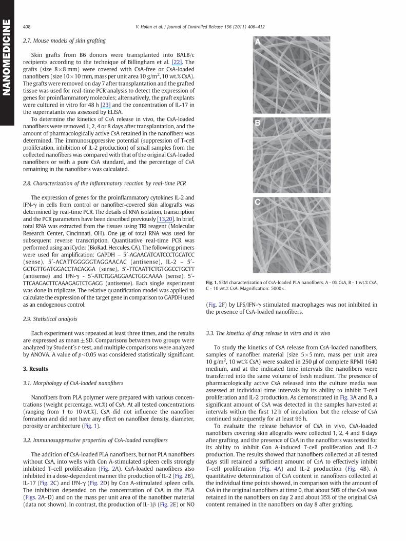
Fig. 1. SEM characterization of CsA-loaded PLA nanofibers. A - 0% CsA, B - 1 wt.% CsA,C - 10 wt.% CsA. Magnification: 5000×.
408 V. Holan et al. / Journal of Controlled Release 156 (2011) 406–412
NANOMEDICIN
E
2.7. Mouse models of skin grafting
Skin grafts from B6 donors were transplanted into BALB/crecipients according to the technique of Billingham et al. [22]. Thegrafts (size 8×8 mm) were covered with CsA-free or CsA-loadednanofibers (size 10×10 mm,mass per unit area 10 g/m2, 10 wt.% CsA).The graftswere removed on day 7 after transplantation and the graftedtissue was used for real-time PCR analysis to detect the expression ofgenes for proinflammatorymolecules; alternatively, the graft explantswere cultured in vitro for 48 h [23] and the concentration of IL-17 inthe supernatants was assessed by ELISA.
To determine the kinetics of CsA release in vivo, the CsA-loadednanofiberswere removed 1, 2, 4 or 8 days after transplantation, and theamount of pharmacologically active CsA retained in the nanofibers wasdetermined. The immunosuppressive potential (suppression of T-cellproliferation, inhibition of IL-2 production) of small samples from thecollected nanofibers was comparedwith that of the original CsA-loadednanofibers or with a pure CsA standard, and the percentage of CsAremaining in the nanofibers was calculated.
2.8. Characterization of the inflammatory reaction by real-time PCR
The expression of genes for the proinflammatory cytokines IL-2 andIFN-γ in cells from control or nanofiber-covered skin allografts wasdetermined by real-time PCR. The details of RNA isolation, transcriptionand the PCR parameters have been described previously [13,20]. In brief,total RNA was extracted from the tissues using TRI reagent (MolecularResearch Center, Cincinnati, OH). One μg of total RNA was used forsubsequent reverse transcription. Quantitative real-time PCR wasperformedusing an iCycler (BioRad,Hercules, CA). The followingprimerswere used for amplification: GAPDH – 5´-AGAACATCATCCCTGCATCC(sense), 5´-ACATTGGGGGTAGGAACAC (antisense), IL-2 – 5´-GCTGTTGATGGACCTACAGGA (sense), 5´-TTCAATTCTGTGGCCTGCTT(antisense) and IFN-γ - 5´-ATCTGGAGGAACTGGCAAAA (sense), 5´-TTCAAGACTTCAAAGAGTCTGAGG (antisense). Each single experimentwas done in triplicate. The relative quantification model was applied tocalculate the expression of the target gene in comparison to GAPDHusedas an endogenous control.
2.9. Statistical analysis
Each experiment was repeated at least three times, and the resultsare expressed as mean±SD. Comparisons between two groups wereanalyzed by Student´s t-test, andmultiple comparisons were analyzedby ANOVA. A value of pb0.05 was considered statistically significant.
3. Results
3.1. Morphology of CsA-loaded nanofibers
Nanofibers from PLA polymer were prepared with various concen-trations (weight percentage, wt.%) of CsA. At all tested concentrations(ranging from 1 to 10 wt.%), CsA did not influence the nanofiberformation and did not have any effect on nanofiber density, diameter,porosity or architecture (Fig. 1).
3.2. Immunosuppressive properties of CsA-loaded nanofibers
The addition of CsA-loaded PLA nanofibers, but not PLA nanofiberswithout CsA, into wells with Con A-stimulated spleen cells stronglyinhibited T-cell proliferation (Fig. 2A). CsA-loaded nanofibers alsoinhibited in a dose-dependentmanner the production of IL-2 (Fig. 2B),IL-17 (Fig. 2C) and IFN-γ (Fig. 2D) by Con A-stimulated spleen cells.The inhibition depended on the concentration of CsA in the PLA(Figs. 2A–D) and on the mass per unit area of the nanofiber material(data not shown). In contrast, the production of IL-1β (Fig. 2E) or ΝΟ
(Fig. 2F) by LPS/IFN-γ stimulated macrophages was not inhibited inthe presence of CsA-loaded nanofibers.
3.3. The kinetics of drug release in vitro and in vivo
To study the kinetics of CsA release from CsA-loaded nanofibers,samples of nanofiber material (size 5×5 mm, mass per unit area10 g/m2, 10 wt.% CsA) were soaked in 250 μl of complete RPMI 1640medium, and at the indicated time intervals the nanofibers weretransferred into the same volume of fresh medium. The presence ofpharmacologically active CsA released into the culture media wasassessed at individual time intervals by its ability to inhibit T-cellproliferation and IL-2 production. As demonstrated in Fig. 3A and B, asignificant amount of CsA was detected in the samples harvested atintervals within the first 12 h of incubation, but the release of CsAcontinued subsequently for at least 96 h.
To evaluate the release behavior of CsA in vivo, CsA-loadednanofibers covering skin allografts were collected 1, 2, 4 and 8 daysafter grafting, and the presence of CsA in the nanofibers was tested forits ability to inhibit Con A-induced T-cell proliferation and IL-2production. The results showed that nanofibers collected at all testeddays still retained a sufficient amount of CsA to effectively inhibitT-cell proliferation (Fig. 4A) and IL-2 production (Fig. 4B). Aquantitative determination of CsA content in nanofibers collected atthe individual time points showed, in comparison with the amount ofCsA in the original nanofibers at time 0, that about 50% of the CsA wasretained in the nanofibers on day 2 and about 35% of the original CsAcontent remained in the nanofibers on day 8 after grafting.

A
0
20
40
60
80
100
120
140
- + 0 1 2.5 5 10
Cpm
x 1
0-3
B
0100200300400500600700800900
- + 0 1 2.5 5 10
C
0
100
200
300
400
500
600
- + 0 1 2.5 5 10
D
0
200
400
600
800
1000
1200
1400
- + 0 1 2.5 5 10
E
0
50
100
150
200
250
- + 0 1 2.5 5 10
IL-1
2 (p
g/m
l)IL
-2 (
pg/m
l)
IL-1
7 (p
g/m
l)
F
0
5
10
15
20
25
30
- + 0 1 2.5 5 10CsA (wt %)CsA (wt %) CsA (wt %)
CsA (wt %) CsA (wt %)CsA (wt %)
Fig. 2. Selective inhibition of T-cell proliferation and cytokine production by CsA-loaded PLA nanofibers. Mouse spleen cells were cultured unstimulated (−), stimulated with Con A(+) or stimulatedwith Con A in the presence of nanofibermaterial (size 2×2 mm,mass per unit area 10 g/m2) with the indicated content (wt.%) of CsA. Cell proliferation (A) and theproduction of IL-2 (B), IL-17 (C) and IFN-γ (D) were determined. The production of IL-1β (E) and ΝΟ (F) by macrophages stimulated with LPS/IFN-γ in the presence of nanofiberswith the indicated content of CsA was also assessed.
409V. Holan et al. / Journal of Controlled Release 156 (2011) 406–412
NANOMEDICIN
E
3.4. The growth of cells on CsA-loaded PLA nanofibers
Three types of cells, LSCs, MSCs and 3T3 fibroblasts, were culturedfor 72 h in plastic tissue culture plates in the presence of CsA-loaded
A
0
20
40
60
80
100
120
140
- + 0.5 1 2 4 6 8 12 24 48 72 96
Time (hours)
B
0
100
200
300
400
500
600
- + 0.5 1 2 4 6 8 12 24 48 72 96
Time (hours)
IL-2
(pg
/ml)
Cpm
x 1
0-3
Fig. 3. The kinetics of CsA release from nanofibers in vitro. CsA-loaded nanofibers weresoaked in culture medium and transferred at the indicated time intervals into freshmedium. Samples of medium after each transfer were harvested and tested for theirability to inhibit Con A-induced T-cell proliferation (A) and IL-2 production (B).
PLA nanofibers and cell proliferation was determined according to3H-thymidine incorporation. It was observed that the cells grew inthe presence of CsA-loaded nanofibers comparably as in the presence
Days after grafting
A
0
20
40
60
80
100
120
140
160
- + 0 1 2 4 8
B
0
100
200
300
400
500
600
700
800
- + 0 1 2 4 8
Days after grafting
IL-2
(pg
/ml)
Cpm
x 1
0-3
Fig. 4. Thekinetics of CsA release invivo.Nanofibers (10 wt.%CsA) coveringa skin allograftwere harvested on the day of grafting (day 0) and 1, 2, 4 and 8 days after transplantation.Small pieces (2×2 mm) of these nanofibers were added to cultures of Con A-stimulatedspleen cells, and cell proliferation (A) and IL-2 production (B) were determined.

A
0
1
2
3
4
N B6 0 10
CsA (wt%)
Rel
ativ
e ge
ne e
xpre
ssio
n
B
20
40
60
80
lativ
e ge
ne e
xpre
ssio
n
**
*
410 V. Holan et al. / Journal of Controlled Release 156 (2011) 406–412
NANOMEDICIN
E
of nanofibers prepared from PLA without CsA or in wells withoutnanofiber samples (data not shown). In addition, the cells wereseeded on plastic 24-well tissue culture plates, on PLA nanofiberswithout CsA or on CsA-loaded nanofibers, and cell viability andmetabolic activity were determined after a 24-hour incubationperiod by the WST assay. Fig. 5 shows that all cell types grew onCsA-containing nanofibers comparably as on nanofibers without CsAor on tissue culture plastic.
3.5. Suppression of a local inflammatory reaction in vivo
The rejection of a skin allograft induces a local inflammatory reactionthat can be detected by the expression of genes for the proinflammatorycytokines IL-2, IL-17 and IFN-γ (Fig. 6). Covering the sides of skinallografts with CsA-loaded nanofibers significantly inhibited theexpression of the genes for IL-2 (Fig. 6A) and IFN-γ (Fig. 6B) andattenuated the production of IL-17 in cultured explants of skin allograftsthat had been covered by CsA-loaded nanofibers (Fig. 6C).
4. Discussion
The transplantation of organs or the therapeutic transfer ofallogeneic cells requires extensive systemic immunosuppression to
A
0
0,5
1
1,5
2
2,5
3
P 0 10
CsA (wt%)
CsA (wt%)
CsA (wt%)
O.D
. (nm
)
B
0
0,5
1
1,5
2
2,5
3
P 0 10
O.D
. (nm
)
C
0
0,2
0,4
0,6
0,8
1
1,2
P 0 10
O.D
. (nm
)
Fig. 5. The growth of LSCs, MSCs and 3T3 fibroblasts on CsA-loaded nanofibers. MouseLSCs (A), MSCs (B) or 3T3 fibroblasts (C) were grown on plastic (tissue culture plates, P)or on PLA nanofibers with the indicated content (wt.%) of CsA, and cell proliferation wasdetermined after a 24-h incubation period.
0N B6 0 10
CsA (wt%)
Re
C
0
100
200
300
400
N B6 0 10
CsA (wt%)
***IL-1
7 (p
g/m
l)
Fig. 6. Suppression of local inflammatory reactions by CsA-loaded nanofibers. Theexpression of genes for IL-2 (A) or IFN-γ (B) was detected in normal B6 skin (N), inuntreated skin allografts (B6) or in allografts that were covered with nanofiberswithout CsA (0) or with 10 wt% CsA (10). The comparative Ct method was used todetermine the extent of target gene expression normalized to an internal GAPDHcontrol. In addition, explants of normal skin or of skin allografts were removed on day 7after transplantation and were cultured for 48 h. The presence of IL-17 in thesupernatants was determined by ELISA (C). Each bar represents the mean±SD from 5to 6 determinations. Values with asterisks are significantly different (*pb0.05,**pb0.01, ***pb0.001) from the positive control (allografts without nanofibers).
inhibit the deleterious immune response to antigens on the graftedcells. Similarly, immunosuppression is also the first choice of treat-ment to manage harmful inflammation. The systemic administrationof immunosuppressive drugs is regularly associated with side effectsthat prevent the use of effective doses of the drugs. To avoid thisproblem, the local delivery of the drugs offers a promising solution.Searching for an optimal carrier for targeted drug delivery, nanotech-nologies provide a perspective tool. So far, various nanoparticules,nanofibers, nanocomposites and other nanomaterials have beentested [4–8,14–17]. Here, we show that CsA, a potent inhibitor ofT-cell functions, can be efficiently encapsulated into PLA nanofibersand used for the local suppression of T-cell reactivity and theinhibition of inflammatory reactions.
CsA represents one of the most widely used immunosuppressivedrugs, but its use is limited by its side effects, such as nephrotoxicity,

411V. Holan et al. / Journal of Controlled Release 156 (2011) 406–412
NANOMEDICIN
E
gingivitis and hirsutism, regularly observed at higher drug doses. Atlower concentrations, CsA selectively inhibits T-cell functionswithout any apparent effect on other cell populations. Therefore,we dissolved CsA into a PLA solution and prepared nanofiberscaffolds with different concentrations (1–10 wt.%) of CsA. Wecalculated that 1 mm2 of CsA-loaded nanofiber material with10 wt.% CsA contains 1 μg of CsA. Although CsA has extremely lowaqueous solubility (6.6 μg/ml) [24], it has been shown thatconcentrations of the drug as low as 50–200 ng/g of tissue aresufficient to suppress T-cell reactivity and inflammation [25]. Theaddition of CsA-loaded nanofibers into cultures of spleen cellsstimulated with Con A inhibited T-cell proliferation and suppressedthe production of T-cell cytokines. The production of IL-1β, IL-6 or NOby activated macrophages and the growth of LSCs, MSCs or 3T3fibroblasts were not inhibited in the presence of CsA-loadednanofibers. These results indicate that the incorporation of CsA intothe PLA solution and the electrospinning of this mixture did notimpair the immunosuppressive properties of CsA. Studies of thekinetics of CsA release in aqueous solutions revealed that themajority of CsA was released from the nanofibers into the culturemedium within the first few hours, but a significant amount of CsAwas retained in the nanofibers and gradually released for more than96 h. The sustained release of CsA for a few days is different from thepattern of release of water-soluble small molecular weight sub-stances which are rapidly released from nanofibers within a fewminutes or hours [6,26]. The delayed release of CsA from thenanofibers may be associated with the very low solubility of CsA inaqueous solutions. Although attempts have been made to controldrug release behavior [8], the release of a significant amount of thedrug initially, coupledwith a sustained release for a prolonged periodof time, may be of great interest, especially in situations in which aharmful inflammatory reaction must be rapidly suppressed and thenkept at an acceptable level. Moreover, the drug release profile in vivomay be quite different from that observed in aqueous solutions invitro. Indeed, we observed that CsA-loaded nanofibers retain asubstantial amount of the drug for more than 8 days when they areplaced on skin allografts in vivo.
Although the potential use of drug-loaded nanofibers has beenextensively discussed, there are only a few studies showing theeffectiveness of drug-loaded nanofibers in biological systems in vitro[7,27,28] and in vivo [29,30]. Our data presented here show for thefirst time that CsA-loaded nanofibers fabricated from PLA polymereffectively and selectively inhibit the proliferation of activated T cellsand suppress the production of T-cell cytokines in vitro. Moreover,using an experimental model of skin transplantation in mice, weshowed that covering skin allograft with CsA-loaded nanofiberssignificantly inhibited the local production of the proinflammatorycytokines IL-2, IL-17 and IFN-γ.
In addition to their ability to serve as drug carriers, CsA-loadednanofibers could be used as scaffolds for the growth and transfer of non-T-cell populations.We showed that LSCs,MSCs and3T3fibroblasts growon CsA-loaded nanofibers comparably as on nanofibers prepared from aCsA-free PLA. Since nanofibers prepared from PLA polymer havecomparable mechanical properties as nanofibers prepared from poly-amid,whichwe recently described and used for LSC andMSC transfer totreat ocular surface injuries [13], CsA-loaded and simultaneously cell-seeded nanofibers appear to be potential scaffolds for cell transfer inregenerative medicine.
In conclusion, we showed that the immunosuppressive drug CsAcan be incorporated directly into electrospun nanofibers without anyloss of its pharmacological activity. Such CsA-loaded nanofibers can beused as drug carriers for the local suppression of inflammatoryreactions and as cell scaffolds for tissue repair and regeneration. Theseobservations suggest that CsA-loaded PLA nanofibers may beextremely useful for simultaneous cell-based therapy and the localsuppression of T-cell-mediated immune reactions.
Acknowledgments
This work was supported by grant KAN200520804 from the GrantAgency of the Academy of Sciences of the Czech Republic, grantsP304/11/0653, P301/11/1568 and 310/08/H077 from the Grant Agencyof the CzechRepublic, projects 1 M0506 andMSM0021620858 from theMinistry of Education of the Czech Republic and project AVOZ50520514from theAcademyof Sciences of theCzechRepublic. The authorsdeclareno conflict of interest.
References
[1] J. Zeng, X. Xu, X. Chen, Q. Liang, X. Bian, L. Yang, X. Jing, Biodegradable electrospunfibers for drug delivery, J. Control. Release 92 (2003) 227–231.
[2] X. Xu, X. Chen, P. Ma, X. Wang, X. Jing, The release behaviour of doxorubicinhydrochloride from medicated fibers prepared by emulsion-electrospinning, Eur.J. Pharm. Biopharm. 70 (2008) 165–170.
[3] C. Xie, X. Luo, Y. Yang, W. Cui, J. Zou, S. Zhou, Release modulation and cytotoxicityof hydroxycamptothecin-loaded electrospun fibers with 2-hydroxy-propyl-beta-cyclodextrin inoculations, Int. J. Pharm. 391 (2010) 55–64.
[4] S.G. Kumbar, L.-S. Nair, S. Bhattacharyya, C.T. Laurencin, Polymeric nanofibers asnovel carriers for the delivery of therapeutic molecules, J. Nanosci. Nanotechnol. 6(2006) 2591–2607.
[5] C. Chen, G. Lv, C. Pan, M. Song, C. Wu, D. Guo, X. Wang, B. Chen, Z. Gu, Poly(lactidacid) (PLA) based nanocomposites—a novel way of drug-releasing, Biomed.Mater. 2 (2007) L1–L4.
[6] Y. Wang, B. Wang, W. Qiao, T. Yin, A novel controlled release drug delivery systemfor multiple drugs based on electrospun nanofibers containing nanoparticules,J. Pharm. Sci. 99 (2010) 4805–4811.
[7] M. Arlt, D. Haase, S. Hampel, S. Oswald, A. Bachmatiuk, R. Klingrer, R. Schulze, M.Ritschel, A. Leonhardt, S. Fuessel, B. Buchler, K. Kraemer, M.P. Wirth, Delivery ofcarboplatin by carbon-based nanocontainers mediates increased cancer celldeath, Nanotechnology 21 (2010) 335101.
[8] T. Okuda, K. Tominaga, S. Kidoak, Time-programmed dual release formulation bymultilayered drug-loaded nanofiber meshes, J. Control. Release 143 (2010)258–264.
[9] S.Y. Chew, Y. Wen, Y. Dzenis, K.W. Leong, The role of electrospinning in theemerging field of nanomedicine, Curr. Pharm. Des. 12 (2006) 4751–4770.
[10] J. Xie, S.M. Willerth, X. Li, M.R. Macewan, A. Rader, S.E. Sakiyama-Elbert, Y. Xioa,The differentiation of embryonic stem cells seeded on electrospun nanofibers intoneural lineages, Biomaterials 30 (2009) 354–362.
[11] X. Xin, M. Hussain, J.J. Mao, Continuing differentiation of human mesenchymalstem cells and induced chondrogenic and osteogenic lineages in electrospun PLGAnanofiber scaffold, Biomaterials 28 (2007) 316–325.
[12] S.M. Hashemi, M. Soleimani, S.S. Zargarian, V. Haddadi-Asi, N. Ahmadbeigi, S.Soudi, Y. Gheisari, A. Hajarizadeh, V. Mohammedi, In vitro differentiation ofhuman cord blood-derived unrestricted stem cells into hepatocyte-like cells onpoly(ypsilon-caprolactone) nanofiber scaffolds, Cells Tissues Organs 190 (2009)135–149.
[13] A. Zajicova, K. Pokorna, A. Lencova, M. Krulova, E. Svobodova, S. Kubinova, E.Sykova, M. Pradny, J. Michalek, J. Svobodova, M. Munzarova, V. Holan, Treatmentof ocular surface injuries by limbal and mesenchymal stem cells growing onnanofiber scaffolds, Cell Transplant 19 (2010) 1281–1290.
[14] S.Q. Wang, J.D. Dai, Z. Chen, T. Zhang, G.M. Xia, T. Nagai, Q. Zhang, Bioavailabilityand pharmacokinetics of cyclosporine A-loaded pH-sensitive nanoparticles fororal administration, J. Control. Release 97 (2004) 421–429.
[15] H.M. Aliabadi, D.R. Brocks, P. Mahdipoor, A. Lavasanifar, A novel use of an in vitromethod to predict the in vivo stability of block copolymer based nano-container, J.Control. Release 122 (2007) 63–70.
[16] J. Azzi, L. Teng, R. Moore, R. Tong, N. El Haddad, T. Akivoshi, B. Mfarrej, S. Yang, M.Jurewicz, T. Ichimura, N. Lindeman, J. Cheng, R. Abdi, Polylactide-cyclosporin Ananoparticles for targeted immunosuppression, FASEB J. 24 (2010) 3927–3936.
[17] P. Aksungur, M. Demirbilek, E.B. Denkbas, J. Vandervoort, A. Ludwig, N. Unlü,Development and characterization of Cyclosporine A loaded nanoparticles forocular drug delivery: cellular toxicity, uptake, and kinetic studies, J. Control.Release 151 (2011) 286–294.
[18] O. Jirsak, F. Sanetrnik, D. Lukas, K. Kotek, L. Martinova, J. Chaloupek, inventors; U.S.patent No. WO205024101.2005.
[19] M. Krulova, K. Pokorna, A. Lencova, A. Zajicova, J. Fric, M. Filipec, J.V. Forrester, V.Holan, A rapid separation of two distinct populations of corneal epithelial cellswith limbal stem cell characteristics in the mouse, Invest. Ophthalmol. Vis. Sci. 49(2008) 3903–3908.
[20] V. Holan, K. Pokorna, J. Procházkova, M. Krulova, A. Zajicova, Immunoregulatoryproperties of mouse limbal stem cells, J. Immunol. 184 (2010) 2124–2129.
[21] L.C. Green, D.A. Wagner, J. Glogowski, P.L. Skipper, J.S. Wishnok, S.R. Tannenbaum,Analysis of nitrate, nitrite, and [15N]nitrate in biological fluids, Anal. Biochem. 126(1982) 131–138.
[22] R.E. Billingham, L. Brent, P.B.Medawar, Quantitative studieson tissue transplantationimmunity. I. The survival times of skin homografts exchanged between members ofdifferent inbred strains of mice, Proc. R. Soc. Lond. (Biol.) 143 (1954) 43–58.
[23] M. Krulova, A. Zajicova, J. Fric, V. Holan, Alloantigen-induced, T-cell-dependentproduction of nitric oxide by macrophages infiltrating skin allografts in mice,Transpl. Int. 15 (2002) 108–116.

412 V. Holan et al. / Journal of Controlled Release 156 (2011) 406–412
NANOMEDICIN
E
[24] S.D. Mithani, V. Bakatselou, C.N. TenHoor, J.B. Dressman, Estimation of theincrease in solubility of drugs as a function of bile salt concentration, Pharm. Res.13 (1996) 163–167.
[25] R.I. Kaswan, Intraocular penetration of topically applied cyclosporine, Transpl.Proc. 20 (1988) 650–655.
[26] E.R. Kanawy, G.L. Bowling, K. Mansfield, J. Layman, D.G. Simpson, E.H. Sanders, G.E.Wnek, Release of tetracycline hydrochloride from electrospun poly(ethylene-co-vinylacetate), poly(lactid acid), and a blend, J. Control. Release 81 (2002)57–64.
[27] J.G. Merrell, S.W. McLaughlin, L. Tie, C.T. Laurencin, A.F. Chen, L.S. Nair, Curcuminloaded poly(ε-caprolactone) nanofibers: diabetic wound dressing with antioxi-
dant and anti-inflammatory properties, Clin. Exp. Pharmacol. Physiol. 36 (2009)1149–1156.
[28] F. Wang, Z. Li, K. Tamama, C.K. Sen, J. Guan, Fabrication and characterization ofprosurvival growth factor releasing, anisotropic scaffolds for enhancedmesenchymalstemcell survival/growthandorientation, Biomacromolecules 10 (2009) 2609–2618.
[29] S.Y. Chew, R. Mi, K.W. Leong, Aligned protein-polymer composite fibers enhancenerve regeneration: a potential tissue-engineering platform, Adv. Func. Mater. 17(2007) 1288–1296.
[30] I.C. Liao, K.W. Leong, Efficacy of engineered FVII-producing skeletal muscleenhanced by growth factor-releasing co-axial electrospun fibers, Biomaterials 32(2011) 1669–1677.
Copyright © 2022 FDOKUMEN