Activation of NF B by Double-Stranded RNA (dsRNA) in the Absence of Protein Kinase R and RNase L...
12
MOLECULAR AND CELLULAR BIOLOGY, 0270-7306/01/$04.0010 DOI: 10.1128/MCB.21.1.61–72.2001 Jan. 2001, p. 61–72 Vol. 21, No. 1 Copyright © 2001, American Society for Microbiology. All Rights Reserved. Activation of NF-kB by Double-Stranded RNA (dsRNA) in the Absence of Protein Kinase R and RNase L Demonstrates the Existence of Two Separate dsRNA-Triggered Antiviral Programs MIHAIL S. IORDANOV, 1 JOHN WONG, 1 JOHN C. BELL, 2,3 AND BRUCE E. MAGUN 1 * Department of Cell and Developmental Biology, Oregon Health Sciences University, Portland, Oregon 97201, 1 and Ottawa Regional Cancer Center Research Laboratories, Ottawa, Ontario K1H 8L6, 2 and Department of Biochemistry, University of Ottawa, Ottawa, Ontario K1H 8M5, 3 Canada Received 27 June 2000/Returned for modification 8 August 2000/Accepted 22 September 2000 Double-stranded RNA (dsRNA) of viral origin triggers two programs of the innate immunity in virus- infected cells. One is intended to decrease the rate of host cell protein synthesis and thus to prevent viral replication. This program is mediated by protein kinase R (PKR) and by RNase L and contributes, eventually, to the self-elimination of the infected cell via apoptosis. The second program is responsible for the production of antiviral (type I) interferons and other alarmone cytokines and serves the purpose of preparing naive cells for the viral invasion. This second program requires the survival of the infected cell and depends on the expression of antiapoptotic genes through the activation of the NF-kB transcription factor. The second program therefore relies on ongoing transcription and translation. It has been proposed that PKR plays an essential role in the activation of NF-kB by dsRNA. Here we present evidence that the dsRNA-induced NF-kB activity and the expression of beta interferon and inflammatory cytokines do not require either PKR or RNase L. Our results indicate, therefore, that the two dsRNA-activated programs are separate and can function independently of each other. At the cellular level, the innate immune response to viruses relies on the execution of two apparently conflicting cellular programs: cell suicide (apoptosis) and survival. The first pro- gram is probably most efficient for viral infections that are initiated by a small number of infected cells at a local site of virus entry. In such case, it seems beneficial (for the organism) for this first population of infected cells to undergo a rapid process of self-elimination through apoptosis, thus preventing further infection. That this first line of antiviral defense is widely used is evident from the multitude of antiapoptotic strategies employed by viruses. Viral genomes encode a grow- ing number of apoptosis-inhibiting proteins, such as the ade- novirus E1B protein (45, 72), the baculovirus p35 protein (5, 44, 58), the cowpox virus CrmA protein (17, 63), the poxvirus and gammaherpesvirus v-FLIP proteins (66), and others (for a review, see reference 62). Genetic evidence from mice (48) (see below) demonstrates that inhibition of apoptosis by the virus is critical for the virulence of encephalomyocarditis virus (EMCV), a picornavirus that is lethal to infected mice. A common viral intermediate that is recognized by specific cellular sensory systems to trigger apoptosis is viral double- stranded RNA (dsRNA). The best-characterized effect of dsRNA on cells is the inhibition of protein synthesis in host cells. The cellular dsRNA-detecting systems that are responsi- ble for the translational inhibition in response to viral infection are the dsRNA-activated protein kinase (PKR) and the cou- pled 2-5 oligoadenylate synthase/RNase L system. PKR (39) is a dormant enzyme directly activated by binding of dsRNA (for recent reviews, see references 28 and 73). A major substrate of PKR is the a-subunit of the eukaryotic translation initiation factor 2 (eIF-2a) (22, 35). Phosphorylation of eIF-2a greatly reduces the rate of initiation of translation (50). The 2-5 oli- goadenylate synthase/RNase L system is composed of a family of dsRNA-dependent enzymes known as 29-59 oligoadenylate synthetases (OAS) (7, 24, 43) and the dormant cytosolic RNase L (82). Upon dsRNA binding, OAS produce unusual second messengers, short 29-59-linked oligoadenylates (2-5A), which, in turn, specifically bind to and activate RNase L (41). Acti- vated RNase L cleaves diverse RNA substrates, including 18S and 28S rRNA, thus inhibiting cellular protein synthesis (26, 51, 52, 74). Fibroblasts from mice nullizygous for both PKR and RNase L alleles are unable to inhibit protein synthesis when challenged with dsRNA (26), thus demonstrating that these two enzymes are both required and sufficient for the translational inhibition caused by dsRNA. Recently, both PKR (2, 3, 15, 20, 34, 53, 77) and RNase L (7–9, 13, 83, 85) have been found to mediate dsRNA-induced apoptosis. The mech- anisms of involvement of PKR and RNase L in the dsRNA- triggered apoptosis are, however, poorly understood. Consid- ering the role of both PKR and RNase L in inhibiting protein synthesis, one obvious candidate for a death-inducing signal is the impaired process of translation. A sustained inhibition of protein synthesis is sufficient to trigger apoptosis in cells in a way that is independent of the particular means of achieving translational inhibition (27). However, other (more direct) * Corresponding author. Mailing address: Department of Cell and Developmental Biology, Oregon Health Sciences University, Portland, OR 97201. Phone: (503) 494-7811. Fax: (503) 494-4253. E-mail: [email protected]. 61
Transcript of Activation of NF B by Double-Stranded RNA (dsRNA) in the Absence of Protein Kinase R and RNase L...
MOLECULAR AND CELLULAR BIOLOGY,0270-7306/01/$04.0010 DOI: 10.1128/MCB.21.1.61–72.2001
Jan. 2001, p. 61–72 Vol. 21, No. 1
Copyright © 2001, American Society for Microbiology. All Rights Reserved.
Activation of NF-kB by Double-Stranded RNA (dsRNA) in theAbsence of Protein Kinase R and RNase L Demonstrates the
Existence of Two Separate dsRNA-TriggeredAntiviral Programs
MIHAIL S. IORDANOV,1 JOHN WONG,1 JOHN C. BELL,2,3 AND BRUCE E. MAGUN1*
Department of Cell and Developmental Biology, Oregon Health Sciences University, Portland, Oregon 97201,1 andOttawa Regional Cancer Center Research Laboratories, Ottawa, Ontario K1H 8L6,2 and Department
of Biochemistry, University of Ottawa, Ottawa, Ontario K1H 8M5,3 Canada
Received 27 June 2000/Returned for modification 8 August 2000/Accepted 22 September 2000
Double-stranded RNA (dsRNA) of viral origin triggers two programs of the innate immunity in virus-infected cells. One is intended to decrease the rate of host cell protein synthesis and thus to prevent viralreplication. This program is mediated by protein kinase R (PKR) and by RNase L and contributes, eventually,to the self-elimination of the infected cell via apoptosis. The second program is responsible for the productionof antiviral (type I) interferons and other alarmone cytokines and serves the purpose of preparing naive cellsfor the viral invasion. This second program requires the survival of the infected cell and depends on theexpression of antiapoptotic genes through the activation of the NF-kB transcription factor. The secondprogram therefore relies on ongoing transcription and translation. It has been proposed that PKR plays anessential role in the activation of NF-kB by dsRNA. Here we present evidence that the dsRNA-induced NF-kBactivity and the expression of beta interferon and inflammatory cytokines do not require either PKR or RNaseL. Our results indicate, therefore, that the two dsRNA-activated programs are separate and can functionindependently of each other.
At the cellular level, the innate immune response to virusesrelies on the execution of two apparently conflicting cellularprograms: cell suicide (apoptosis) and survival. The first pro-gram is probably most efficient for viral infections that areinitiated by a small number of infected cells at a local site ofvirus entry. In such case, it seems beneficial (for the organism)for this first population of infected cells to undergo a rapidprocess of self-elimination through apoptosis, thus preventingfurther infection. That this first line of antiviral defense iswidely used is evident from the multitude of antiapoptoticstrategies employed by viruses. Viral genomes encode a grow-ing number of apoptosis-inhibiting proteins, such as the ade-novirus E1B protein (45, 72), the baculovirus p35 protein (5,44, 58), the cowpox virus CrmA protein (17, 63), the poxvirusand gammaherpesvirus v-FLIP proteins (66), and others (for areview, see reference 62). Genetic evidence from mice (48)(see below) demonstrates that inhibition of apoptosis by thevirus is critical for the virulence of encephalomyocarditis virus(EMCV), a picornavirus that is lethal to infected mice.
A common viral intermediate that is recognized by specificcellular sensory systems to trigger apoptosis is viral double-stranded RNA (dsRNA). The best-characterized effect ofdsRNA on cells is the inhibition of protein synthesis in hostcells. The cellular dsRNA-detecting systems that are responsi-ble for the translational inhibition in response to viral infection
are the dsRNA-activated protein kinase (PKR) and the cou-pled 2-5 oligoadenylate synthase/RNase L system. PKR (39) isa dormant enzyme directly activated by binding of dsRNA (forrecent reviews, see references 28 and 73). A major substrate ofPKR is the a-subunit of the eukaryotic translation initiationfactor 2 (eIF-2a) (22, 35). Phosphorylation of eIF-2a greatlyreduces the rate of initiation of translation (50). The 2-5 oli-goadenylate synthase/RNase L system is composed of a familyof dsRNA-dependent enzymes known as 29-59 oligoadenylatesynthetases (OAS) (7, 24, 43) and the dormant cytosolic RNaseL (82). Upon dsRNA binding, OAS produce unusual secondmessengers, short 29-59-linked oligoadenylates (2-5A), which,in turn, specifically bind to and activate RNase L (41). Acti-vated RNase L cleaves diverse RNA substrates, including 18Sand 28S rRNA, thus inhibiting cellular protein synthesis (26,51, 52, 74). Fibroblasts from mice nullizygous for both PKRand RNase L alleles are unable to inhibit protein synthesiswhen challenged with dsRNA (26), thus demonstrating thatthese two enzymes are both required and sufficient for thetranslational inhibition caused by dsRNA. Recently, both PKR(2, 3, 15, 20, 34, 53, 77) and RNase L (7–9, 13, 83, 85) havebeen found to mediate dsRNA-induced apoptosis. The mech-anisms of involvement of PKR and RNase L in the dsRNA-triggered apoptosis are, however, poorly understood. Consid-ering the role of both PKR and RNase L in inhibiting proteinsynthesis, one obvious candidate for a death-inducing signal isthe impaired process of translation. A sustained inhibition ofprotein synthesis is sufficient to trigger apoptosis in cells in away that is independent of the particular means of achievingtranslational inhibition (27). However, other (more direct)
* Corresponding author. Mailing address: Department of Cell andDevelopmental Biology, Oregon Health Sciences University, Portland,OR 97201. Phone: (503) 494-7811. Fax: (503) 494-4253. E-mail:[email protected].
61
mechanisms of dsRNA-induced cell death, which are indepen-dent of the state of cellular translation, are very likely to exist.
The second dsRNA-initiated program for antiviral defenseinvolves the production and secretion by the infected cells ofalarmone cytokines, the best-studied examples of which are thealpha, beta, and omega interferons, [for reviews, see references16 and 54]). The importance of these interferons for conferringviral resistance to naive cells has been demonstrated by thestrong sensitivity to viral infections of mice lacking the com-mon subunit of the alpha, beta, and omega interferon receptor(40). It is thought that these interferons exert their pleiotropicantiviral actions by preparing cells to interfere with multiple,virus-specific steps of the viral replication cycle, including viralentry, uncoating, transcription, RNA stability, maturation, as-sembly, and release (for a review, see reference 54). Interfer-ons are also important for the ability of adaptive immunity totake over the innate immune response in combating the virus.For instance, mice lacking the interferon alpha, beta, andomega receptor are unable to mount a cytotoxic T-lymphocyteresponse to infection with lymphocytic choriomeningitis virus(40).
A crucial step in the virus-induced beta interferon produc-tion appears to be its transcriptional upregulation by viraldsRNA (for a review, see reference 37). The highly specifictranscriptional induction of the beta interferon gene by viruseshas been best studied for the human beta interferon genepromoter/enhancer region. This region contains a set of regu-latory elements called positive regulatory domains (PRDI toPRDIV). PRDII, PRDI-III, and PRDIV bind the transcriptionfactors NF-kB, IRF-1 (or IRF-3), and ATF-2/c-Jun, respec-tively (for a review, see reference 37). Importantly, NF-kBappears to be absolutely required for the virus-induced activa-tion of the human beta interferon promoter (18, 64, 65).
For the second (interferon-dependent) program of innateantiviral immune response to be successful, the proapoptoticresponse of the infected cells must be suppressed, at least forthe time required to complete the production and secretion ofalarmone cytokines. Interestingly, genetic evidence stronglysuggests that NF-kB not only plays an important role in theproduction of beta interferon but also is essential in suppress-ing virus-induced apoptosis. Mice engineered to lack the p50/NF-kB1 subunit of NF-kB (see below) are resistant to infec-tion with EMCV (48, 49). This surprising result (which seemsto contradict the important role of NF-kB in combating viralinfections) is explained by the discovery that EMCV-infectedcells from p50/NF-kB1-nullizygous mice (as well as from miceengineered to lack the other common subunit of NF-kB, p65/RelA [see below]) undergo very rapid apoptosis before thevirus could reproduce (48). These results demonstrate the ap-optosis-suppressing function of NF-kB in EMCV infection.The antiapoptotic role of NF-kB is thought to result from theNF-kB-dependent transcriptional activation of several apopto-sis-inhibiting genes, such as the genes encoding the inhibitor-of-apoptosis proteins (IAPs; for a review, see reference 32)IAP-1, IAP-2, and X-linked IAP (X-IAP1) (12, 55, 70), Bcl-XL
(10, 33, 68), and A1/Bfl1 (33, 57, 69, 86).How is NF-kB activated in general and by viruses and
dsRNA in particular? NF-kB is a collective name for a groupof homo- and heterodimeric transcriptional regulators (activa-tors or repressors) consisting in vertebrates, of the polypeptide
products of the p50/p105(nfkb1), p52/p100(nfkb2), c-rel, relA,and relB genes (for reviews, see references 21 and 42). Inmammalian cells, the most common NF-kB complex is thep50/NF-kB1–p65/RelA heterodimer, and it is this combinationthat is most commonly referred to as NF-kB proper (42). Anessential role in the regulation of NF-kB is played by a familyof inhibitory proteins, collectively termed IkBs (the family en-compasses IkB-a, IkB-b, IkB-ε, and Bcl-3 [for a review, seereference 31]). In nonstimulated cells, IkBs sequester the p50/NF-kB1–p65/RelA heterodimer in the cytoplasm, thus pre-venting it from localizing in the nucleus and stimulating thetranscription of NF-kB-dependent genes (for a review, seereference 31). With the notable exception of UV radiation, apotent NF-kB activator, most stimuli that activate NF-kB (in-cluding viruses and dsRNA) cause the phosphorylation ofserine residues 32 and 36 in IkB-a (and of the correspondingserine residues in IkB-b). The phosphorylation of IkB causesits rapid polyubiquitinylation and degradation by the 26S pro-teasome, thus releasing NF-kB and allowing it to translocate tothe nucleus (most extensively reviewed in references 30, 31,and 80). A multicomponent IkB kinase (IKK) complex hasbeen purified, molecularly cloned, and found to consist of twohomologous catalytic subunits (IKK1/a and IKK2/b) (14, 38,81) and a noncatalytic subunit (IKKg, also known as NEMO)(75). Genetic inactivation of IKK in mice demonstrated thatIKK2/b and IKKg (but not IKK1/a) are required for the IkBphosphorylation and subsequent NF-kB activation in responseto most agents (25, 36, 46, 59, 61). Due to the lack of suitabletargeted gene inactivation models, however, the modes of up-stream regulation of IKK activity are currently completely un-known, even though several kinases have been proposed to actupstream of IKKs. For instance, in striking contrast to allexperimental evidence concerning the role of PKR in trigger-ing the protein synthesis-inhibiting and proapoptotic programof antiviral innate immunity, this kinase has been proposed tobe a major mediator of virus- and dsRNA-induced activationof NF-kB. Using mouse embryonic fibroblasts (MEF) and 3T3-like fibroblast cell lines from one of the two published PKRgenetic knockouts (76), several groups found these cells to bedeficient in dsRNA-induced NF-kB activation, thus postulat-ing an important role for this kinase in activating NF-kB inresponse to viruses (11, 19, 79).
We considered that this postulated role of PKR in dsRNA-induced NF-kB activation ultimately imposes the paradoxicalsituation that the same dsRNA-sensing molecule (PKR) wouldtrigger the execution of the two opposing antiviral programs inthe same cell: the program that attempts to eliminate theinfected cell through translational inhibition and apoptosis andthe program that attempts to suppress apoptosis throughNF-kB activation. To resolve this paradox, we have employeda panel of primary MEF or 3T3-like cell lines from two inde-pendent successful attempts to inactivate the PKR gene inmice (1, 76), from the RNase L-deficient mice (83), and frommice with a double deficiency in both the PKR and the RNaseL genes (26, 84). Our study demonstrates that neither PKR norRNase L is required for the activation of NF-kB by dsRNA orEMCV. Furthermore, we found that the ability of dsRNA tostimulate the expression of beta interferon and of the inflam-matory cytokines interleukin-6 (IL-6) and tumor necrosis fac-tor alpha (TNF-a) was also independent of the presence of
62 IORDANOV ET AL. MOL. CELL. BIOL.
PKR. Thus, the “translational inhibition/pro-apoptotic pro-gram” and the “biosynthetic/anti-apoptotic program,” eachtriggered by viral dsRNA, appear to be mechanistically sepa-rate and to function independently of one another.
MATERIALS AND METHODS
Chemicals. Lipofectin reagent was from Gibco BRL/Life Technologies. pI-pCwas from Midland Certified Reagent Co. and was stored at 220°C as a 10-mg/mlstock solution in double-distilled deionized water. The proteasome inhibitorsbenzyloxycarbonylleucyl-leucyl-leucine aldehyde (MG 132) and benzyloxycar-bonyliso-leucyl-glutamyl(OtBu)-alanyl-leucine aldehyde (proteasome inhibitorI) were purchased both from Alexis Biochemicals and from Calbiochem, andthere was no detectable difference in their activities. Recombinant humanTNF-a was from R&D Systems. All radiochemicals were from DuPont NENResearch Products.
Cells. All cells were maintained in Dulbecco’s modified Eagle’s medium(DMEM) supplemented with 10% calf serum (HyClone, Logan, Ut.). pkr1/1
(EX12) and pkr0/0(EX12) MEF have been described previously (1) and werereferred to there as pkr1/1 and pkr0/0 cells. pkr1/1(EX213) and pkr0/0(EX213)MEF were described previously (76) and were also referred to there as pkr1/1
and pkr0/0 cells. The rnasel1/1/pkr1/1, rnasel2/2/pkr1/1, and rnasel2/2/pkr2/2
3T3-like fibroblasts were described previously (84) and are referred to here bythe same names as in reference 26.
Lipofectin-mediated delivery of pI-pC. The procedure for treatment of cellswith pI-pC was the same as described in reference 26. For each milliliter of finalvolume of Lipofectin mixture, an initial concentrated mixture (containing Lipo-fectin and pI-pC) was prepared in one-quarter of the final volume (250 ml). Tothis end, 10 ml of Lipofectin (1 mg/ml) was added to serum- and antibiotic-freeDMEM and mixed, and the desired amount of pI-pC was added (in a volume of250 ml). This mixture was left for 10 min at room temperature. Finally, theremaining three-quarters of the final volume (750 ml) was added. Before theapplication of the Lipofectin–pI-pC mixtures, the cells were washed once withserum-free DMEM.
Preparation of cell lysates for immunoblot analysis. To avoid any possibledephosphorylation or proteolytic degradation of the proteins of interest, the cellswere typically harvested by aspirating the cell culture medium, scraping the cellsdirectly on the tissue culture plate in 23 sodium dodecyl sulfate-polyacrylamidegel electrophoresis (SDS-PAGE) sample-loading buffer, and subjecting them toheat denaturation at 95°C for 5 min. Cell lysates were stored at 270°C.
Antibodies and immunoblot analyses. The antibodies against IkB-b (C-20),phospho-(serine-32)–IkB-a (B-9), PKR (M-515 and D-20), and p65/RelA (C-20)and the blocking peptide solutions used in the experiment in Fig. 3 (p65/RelAC-20 peptide and MEKK1 C-22 peptide) were from Santa Cruz Biotechnologies.For the antibody blocking, 1 volume of the anti-p65/RelA antibody was prein-cubated with 5 volumes of the respective blocking peptide solution for 6 h atroom temperature. The antibody against IkB-a (13996E) was from Pharmingen.The antibodies against the dually phosphorylated forms of JNK and p38a mito-gen-activated protein (MAP) kinase were from New England BioLabs. Theantibody against the phosphorylated form of eIF-2a was from Research Genet-ics. The electrophoretic separation of proteins in SDS-PAGE and electrotransferto a polyvinylidene difluoride membrane (Millipore) were performed using stan-dard procedures. Immunoprobing with specific antibodies and enhanced chemi-luminescence detection (DuPont NEN Research Products) were performed asspecified by the respective manufacturers.
Immunocytochemical staining of p65/RelA. Cells were grown on Thermanoxcoverslips (Nunc). After the appropriate treatments, the cells were fixed in cold(220°C) methanol for 5 min, dried, and stored at 220°C. Blocking was per-formed with 1.5% normal goat serum in PBS for 1 h followed by incubation withthe primary antibody (anti-p65/RelA [C-20 from Santa Cruz] at a 1:800 dilutionin PBS with 1.5% serum) for 1 h. After the cells were washed in PBS, incubationwith secondary antibody was performed with biotinylated anti-rabbit immuno-globulin G (1:500 dilution in PBS with 1.5% serum) for 1 h, followed again bywashing. Endogenous peroxidase activity was quenched with 2% hydrogen per-oxide in PBS for 30 min. After being washed, the cells were incubated withVectaStain Elite ABC reagent (Vector Laboratories) for 1 h. Finally, the cellswere washed, incubated with diaminobenzidine (Sigma) until the desired stainintensity developed, and rinsed with water. Photographs were taken using aCoolSnap digital camera mounted on a Zeiss microscope.
EMSA. Nuclear extracts were prepared and used in electrophoretic mobilityshift assays as described in references 4 and 56. Briefly, cells were collected byscraping in ice-cold PBS, sedimented, and resuspended in 100 ml of lysis buffer
(10 mM HEPES [pH 7.9], 1 mM EDTA, 60 mM KCl, 0.5% Nonidet P-40[NP-40], 1 mM dithiothreitol [DTT], protease inhibitor cocktail [Roche Molec-ular Biochemicals]). After 5 min on ice, nuclei were sedimented at 1,200 3 g for5 min. The supernatant was used as the cytoplasmic extract. The nuclei werewashed with lysis buffer without NP-40 and suspended in 100 ml of nuclear buffer(250 mM Tris-HCl [pH 7.8], 60 mM KCl, 1 mM DTT, protease inhibitor cock-tail). Nuclei were lysed by three cycles of freezing and thawing in liquid nitrogenand ice. The nuclear extracts were cleared by centrifugation at 13,000 3 g for 15min. EMSAs were done as described in reference 4: the binding reaction wasperformed in a volume of 20 ml with 5 mg of nuclear protein in a buffer containing12 mM HEPES (pH 7.8), 62.5 mM Tris-HCl (pH 7.8), 60 mM KCl, 0.6 mMEDTA, 12% glycerol, 5 mM DTT, 2 mg of bovine serum albumin, and 1 mg ofpoly(dI-dC). 32P-radiolabeled consensus double-stranded NF-kB-binding oligo-nucleotide (59-AGT TGA GGG GAC TTT CCC AGG C-39) or the correspond-ing oligonucleotide with a single-base point mutation (59-AGT TGA GGC GACTTT CCC AGG C-39) from Santa Cruz Biotechnologies were used as probes.For the competition experiments, a consensus double-stranded p53-binding oli-gonucleotide (59-TAC AGA ACA TGT CTA AGC ATG CTG GGG-39) fromSanta Cruz Biotechnologies was used as a nonspecific competitor.
RNA isolation and Northern blot detection of mRNA. Total cellular-RNA wasisolated using TRIzol reagent (GIBCO BRL) as specified by the manufacturer.The multiprobe detection of beta interferon, IL-6, and TNF-a was performedusing a RiboQuant RNase protection assay kit with a mCK-3b multiprobe tem-plate (Pharmingen) as specified by the manufacturer. A 10-mg portion of totalRNA was used.
Determination of IL-6 production. The production of IL-6 was determinedquantitatively using the Quantikine M mouse IL-6 enzyme-linked immunosor-bent assay (R&D Systems) as specified by the manufacturer and as describedpreviously (26).
RESULTS
EMCV infection causes the proteolytic degradation of IkB-bin both pkr1/1 and pkr0/0 MEF. To investigate the possible roleof PKR in virus-induced activation of NF-kB, we employedprimary fibroblasts derived from wild-type (pkr1/1) or pkr0/0
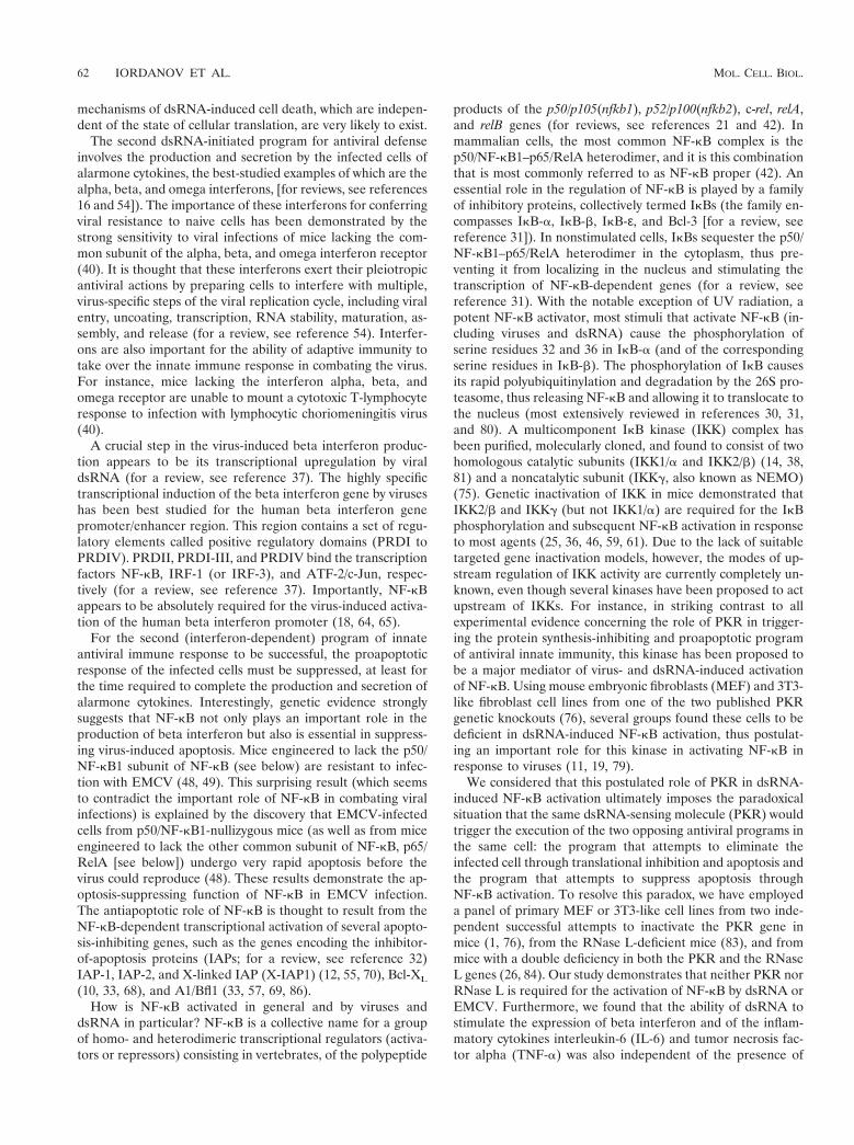
mouse embryos established in the laboratory of one of us (1).Since the inactivation of PKR alleles in these mice wasachieved through a homologous recombination event involvingexon 12 of the PKR gene, these MEF are referred to hereafteras pkr1/1(EX12) and pkr0/0(EX12), respectively. Later, whenMEF from a different PKR knockout (inactivating the PKRgene exons 2 and 3) (76) are used (see below), these cell will bereferred to as pkr1/1(EX213) and pkr0/0(EX213), respec-tively. At 2 h after infection with EMCV, there was a detect-able decrease in the steady-state levels of IkB-b both in thepkr1/1(EX12) and in the pkr0/0(EX12) MEF (Fig. 1A, lanes 3and 7). Four hours after the infection, IkB-b levels in both thepkr1/1(EX12) and the pkr0/0(EX12) MEF were reduced to aminor fraction of those in the control cells (compare lanes 2and 6 with lanes 4 and 8). The absence of PKR in the pkr0/0
(EX12) MEF was confirmed using two independent PKR an-tisera (compare lanes 1 to 4 with lanes 5 to 8). At 4 h after theinfection with EMCV, the levels of PKR in the wild-type MEFwere reduced (lane 4), probably reflecting the overall inhibi-tion of protein synthesis and the subsequent turnover of PKRprotein in these cells.
dsRNA triggers the phosphorylation, polyubiquitinylation,and proteosome-mediated degradation of IkBs in both pkr1/1
(EX12) and pkr0/0(EX12) MEF. The degradation of IkB-b inEMCV-infected MEF was paralleled by the phosphorylationof the stress-activated protein kinases (SAPK) p38a MAP ki-nase (Fig. 1A, lanes 3, 4, 7, and 8), JNK1, and JNK2 (notshown). Previously, we reported that SAPK are potently acti-vated by dsRNA (26). We set out, therefore, to investigate thespecific role of dsRNA in virus-induced activation of NF-kB,
VOL. 21, 2001 PKR-INDEPENDENT ANTIVIRAL RESPONSE 63
independent of viral proteins that, in many cases, also modu-late NF-kB activity. To achieve this goal, we used pI-pC, asynthetic dsRNA, which was delivered into cells via lipofection(see Materials and Methods). First, we set out to confirm thatthe deletion of exon 12 of PKR in the pkr0/0(EX12) MEFresulted in a complete abrogation of PKR activity. Treatmentof pkr1/1(EX12) MEF with pI-pC (hereafter referred to asdsRNA) caused the phosphorylation of eIF-2a at serine-51(Fig. 1B, lane 3). Identically treated pkr0/0(EX12) MEF failedto display the phosphorylation of eIF-2a at serine-51 (lane 6).In contrast to these results and in agreement with our previousfindings (26), the p38a MAP kinase was phosphorylated inresponse to dsRNA both in the PKR-containing and in thePKR-deficient cells (lanes 3 and 6). We concluded, therefore,
that the deletion of exon 12 of PKR in the pkr0/0(EX12) MEFhas resulted indeed in a complete abrogation of PKR activity.
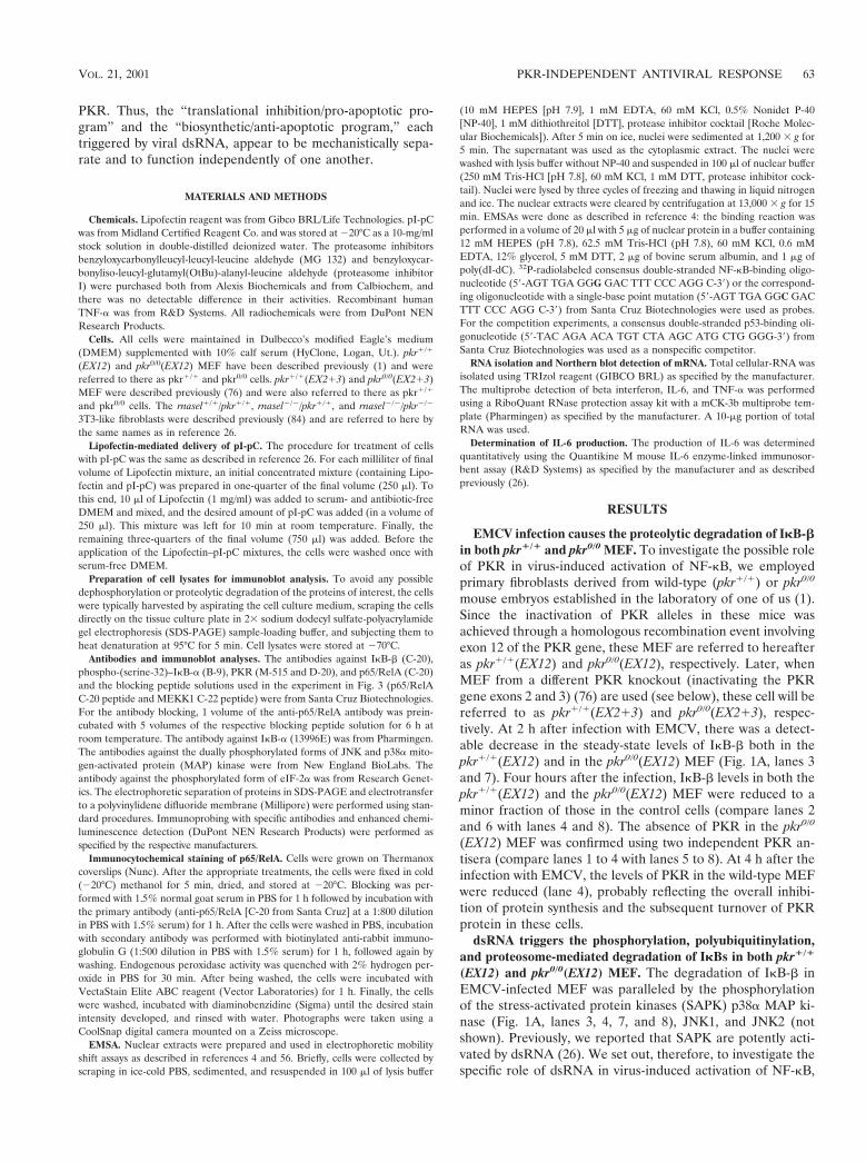
We next addressed the dsRNA-induced signaling to NF-kB.Treatment of either pkr1/1(EX12) or pkr0/0(EX12) MEF withpI-pC for 1 h resulted in a detectable increase in serine-32phosphorylation of IkB-a as measured by immunoblot analysisusing an antibody recognizing specifically only the serine-32-phosphorylated form of IkB-a (Fig. 2A, upper panels, lanes 2and 4). Consistent with the role of IkB-a phosphorylation in itsdegradation by the ubiquitin-proteosome system (for a review,see reference 31), the levels of IkB-a were significantly re-duced in dsRNA-treated cells (lower panels, lanes 2 and 4). To
FIG. 1. (A) EMCV-induced degradation of IkB-b. pkr1/1(EX12)and pkr0/0(EX12) MEF (;2 3 106 cells) were infected, where indi-cated, with EMCV (100 PFU per cell) in 2 ml of serum-free mediumfor 1 h, after which time the excess virus was removed by extensivewashing of the cells with serum-free medium. The cells were furtherincubated in serum-free medium. At 2 or 4 h after the removal of theextracellular virus, the mock-infected or infected cells were harvestedand the cell lysates were processed for the immunoblot detection ofIkB-b (top panel) as described in Materials and Methods. The mem-branes were stripped and reprobed consecutively with an anti-phos-phorylated p38 MAP kinase antibody (second panel from top) andwith the M-515 (third panel from top) and D-20 (bottom panel) PKRantisera. A nonspecific band recognized by the D-20 antibody is indi-cated by an asterisk. (B) Lack of PKR activity in the pkr0/0(EX12)MEF. pkr1/1(EX12) and pkr0/0(EX12) MEF were left untreated (Co)or were treated with Lipofectin (LF) alone or with pI-pC (10 mg/ml) inthe presence of Lipofectin (LF 1 dsRNA). At 3 h after the treatments,the phosphorylation states of eIF-2a and p38a MAP kinase wereassessed in immunoblot analyses.
FIG. 2. dsRNA-triggered phosphorylation, polyubiquitinylation,and proteosome-mediated degradation of IkBs in both pkr1/1(EX12)and pkr0/0(EX12) MEF. (A) pkr1/1(EX12) and pkr0/0(EX12) MEFwere treated with Lipofectin alone (lanes Control) or with pI-pC (10mg/ml) in the presence of Lipofectin (lanes dsRNA). Note that thesame procedure for Lipofectin treatment (with or without pI-pC) ap-plies to all experiment presented in Fig. 2 to 9 and is described inMaterials and Methods. At 1 h after the treatment, the cells wereharvested and processed for immunoblot analysis of IkBa using eithera phospho-(Ser32)-IkBa-specific antibody (upper panels) or an IkBa-specific antibody (lower panels). Treatment of cells with Lipofectindoes not affect IkBb or any other NFkB-related signaling pathwayinvestigated in this work (data not shown). (B) pkr1/1(EX12) andpkr0/0(EX12) MEF were treated as in panel A, except that, whereindicated, the cells were pretreated for 25 min with benzyloxycarbon-yliso-leucyl-glutamyl(OtBu)-alanyl-leucine aldehyde (IEAL), benzy-loxycarbonyl-leucyl-leucyl-leucine aldehyde (LLL), or the respectivesolvents for each of them, methanol (MeOH) or dimethyl sulfoxide(DMSO). IkBb steady-state levels were monitored in an immunoblotanalysis.
64 IORDANOV ET AL. MOL. CELL. BIOL.
investigate whether IkB-b is also degraded by the ubiquitin-proteosome system in response to dsRNA, we employed twopeptide proteosome inhibitors, benzyloxycarbonylleucyl-leucyl-leucine aldehyde (labeled LLL in Fig. 2B) and benzyl-oxycarbonyliso-leucyl-glutamyl(OtBu)-alanyl-leucine aldehyde(labeled IEAL in Fig. 2B). Treatment of either pkr1/1(EX12)or pkr0/0(EX12) MEF with dsRNA for 1 h led to a detectablereduction in the steady-state levels of IkB-b in both the pkr1/1
(EX12) and the pkr0/0(EX12) MEF (Fig. 2B, narrow panels,lane 6). Pretreatment of the cells with either of the two pro-teosome inhibitors prevented the dsRNA-induced degradationof IkB-b (narrow panels, lanes 9 and 10). Furthermore, in thepresence of both dsRNA and the proteasome inhibitors, thecells accumulated multiple anti-IkB-b-immunoreactive bandswith reduced mobility in SDS-PAGE (wide panels, lanes 9 and10; note that the wide panels represent a longer film exposureof the same immunoblots presented in the narrow panels). Theappearance of these novel forms of anti-IkB-b immunoreac-tivity with reduced mobility is consistent with unimpaired levelsof dsRNA-induced polyubiquitinylation of IkB-b but a blockedpolyubiquitinylation-induced degradation of IkB-b by the pro-teasome. Importantly, identical effects on the IkBs of theseproteasome inhibitors were observed in cells stimulated withTNF-a, a well-established inducer of proteasome-mediateddegradation of IkBs (reference 67 and data not shown). Noneof the dsRNA-induced effects on IkBs (phosphorylation,polyubiquitinylation, and proteasome-dependent degradation)appeared to require the presence of PKR. Neither single-stranded RNA (pI or pC), nor dsDNA (p[d(IC)]) had anyeffect on IkB-b or IkB-a (data not shown), indicating that theeffects observed using pI-pC represent a bona fide dsRNAresponse. Despite the limited ability to compare the behaviorof IkB-b and IkB-a in the same assay, the results shown in Fig.2 favor the conclusion that both IkBs are addressed by dsRNA-induced signal transduction pathways in a similar manner.
dsRNA-induced IkB degradation coincides with transloca-tion of NF-kB to the nucleus, independent of the presence orthe absence of PKR. To investigate and determine conclusivelyif PKR may be required for a functional activation of NF-kBdownstream of IkB phosphorylation and degradation, we em-ployed pkr1/1(EX12) and pkr0/0(EX12) MEF and pkr1/1
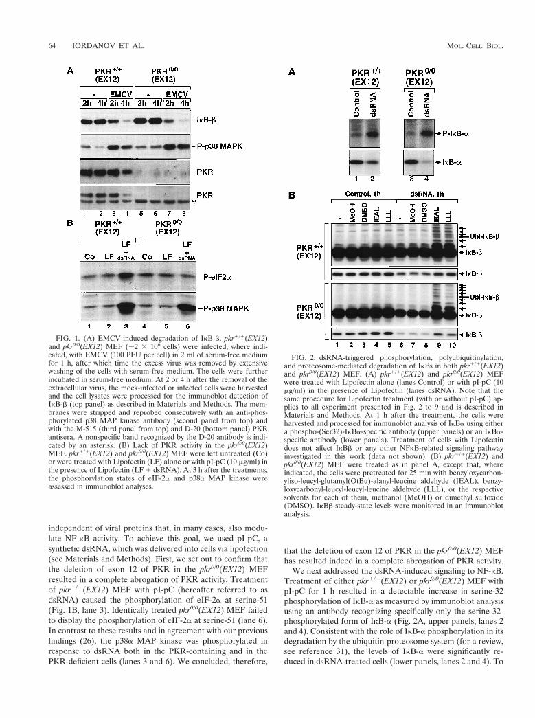
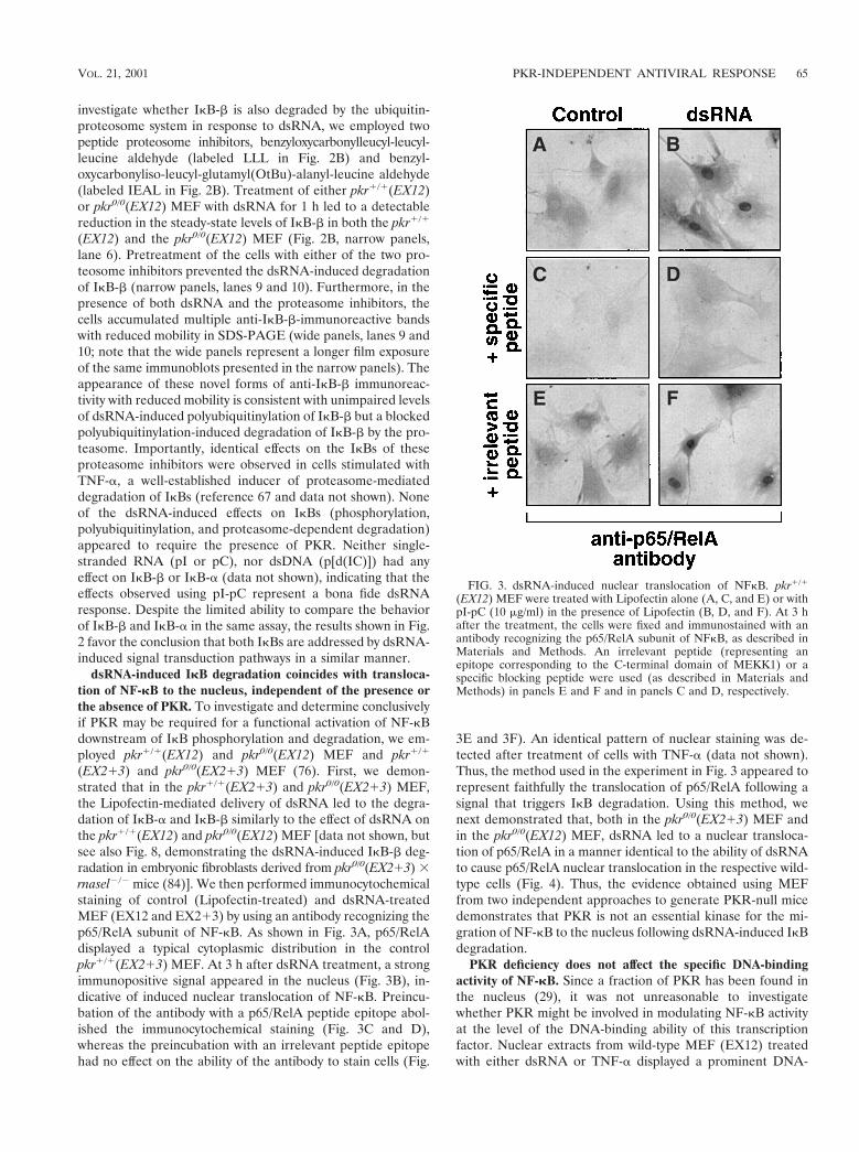
(EX213) and pkr0/0(EX213) MEF (76). First, we demon-strated that in the pkr1/1(EX213) and pkr0/0(EX213) MEF,the Lipofectin-mediated delivery of dsRNA led to the degra-dation of IkB-a and IkB-b similarly to the effect of dsRNA onthe pkr1/1(EX12) and pkr0/0(EX12) MEF [data not shown, butsee also Fig. 8, demonstrating the dsRNA-induced IkB-b deg-radation in embryonic fibroblasts derived from pkr0/0(EX213) 3rnasel2/2 mice (84)]. We then performed immunocytochemicalstaining of control (Lipofectin-treated) and dsRNA-treatedMEF (EX12 and EX213) by using an antibody recognizing thep65/RelA subunit of NF-kB. As shown in Fig. 3A, p65/RelAdisplayed a typical cytoplasmic distribution in the controlpkr1/1(EX213) MEF. At 3 h after dsRNA treatment, a strongimmunopositive signal appeared in the nucleus (Fig. 3B), in-dicative of induced nuclear translocation of NF-kB. Preincu-bation of the antibody with a p65/RelA peptide epitope abol-ished the immunocytochemical staining (Fig. 3C and D),whereas the preincubation with an irrelevant peptide epitopehad no effect on the ability of the antibody to stain cells (Fig.
3E and 3F). An identical pattern of nuclear staining was de-tected after treatment of cells with TNF-a (data not shown).Thus, the method used in the experiment in Fig. 3 appeared torepresent faithfully the translocation of p65/RelA following asignal that triggers IkB degradation. Using this method, wenext demonstrated that, both in the pkr0/0(EX213) MEF andin the pkr0/0(EX12) MEF, dsRNA led to a nuclear transloca-tion of p65/RelA in a manner identical to the ability of dsRNAto cause p65/RelA nuclear translocation in the respective wild-type cells (Fig. 4). Thus, the evidence obtained using MEFfrom two independent approaches to generate PKR-null micedemonstrates that PKR is not an essential kinase for the mi-gration of NF-kB to the nucleus following dsRNA-induced IkBdegradation.
PKR deficiency does not affect the specific DNA-bindingactivity of NF-kB. Since a fraction of PKR has been found inthe nucleus (29), it was not unreasonable to investigatewhether PKR might be involved in modulating NF-kB activityat the level of the DNA-binding ability of this transcriptionfactor. Nuclear extracts from wild-type MEF (EX12) treatedwith either dsRNA or TNF-a displayed a prominent DNA-
FIG. 3. dsRNA-induced nuclear translocation of NFkB. pkr1/1
(EX12) MEF were treated with Lipofectin alone (A, C, and E) or withpI-pC (10 mg/ml) in the presence of Lipofectin (B, D, and F). At 3 hafter the treatment, the cells were fixed and immunostained with anantibody recognizing the p65/RelA subunit of NFkB, as described inMaterials and Methods. An irrelevant peptide (representing anepitope corresponding to the C-terminal domain of MEKK1) or aspecific blocking peptide were used (as described in Materials andMethods) in panels E and F and in panels C and D, respectively.
VOL. 21, 2001 PKR-INDEPENDENT ANTIVIRAL RESPONSE 65
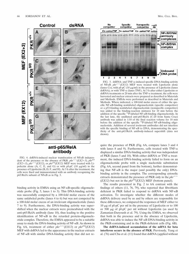
binding activity in EMSA using an NF-kB-specific oligonucle-otide probe (Fig. 5, lanes 1 to 3). This DNA-binding activitywas successfully competed by a 100-fold molar excess of thesame unlabeled probe (lanes 4 to 6) but was not competed bya 100-fold molar excess of an irrelevant oligonucleotide (lanes7 to 9). Furthermore, the DNA-binding activity was super-shifted when the nuclear extracts were preincubated with theanti-p65/RelA antibody (lane 10), thus leading to the positiveidentification of NF-kB in the retarded protein-oligonucle-otide complex. Therefore, the EMSA appeared to be a suitableassay to study the DNA-binding activity of NF-kB. As shown inFig. 6A, treatment of either pkr1/1(EX12) or pkr0/0(EX12)MEF with dsRNA led to the appearance in the nuclear extractsof NF-kB with similar DNA-binding activity that did not re-
quire the presence of PKR (Fig. 6A, compare lanes 3 and 4with lanes 8 and 9). Furthermore, cells treated with TNF-adisplayed a similar DNA-binding activity that was independentof PKR (lanes 5 and 10). With either dsRNA or TNF-a treat-ment, the induced DNA-binding activity failed to form on anoligonucleotide probe with a single nucleotide substitution(Fig. 6A, second panel from the bottom), further demonstrat-ing that NF-kB is the major (and possibly the only) DNA-binding activity in the complex. The corresponding cytosolicextracts demonstrated the presence of PKR only in the pkr1/1
(EX12) but not in the pkr0/0(EX12) MEF (bottom panel).The results presented in Fig. 2 to 6A contrast with the
findings of others (11, 76, 79), who reported that fibroblastsdeficient in PKR failed to respond to dsRNA with NF-kBactivation. To investigate whether the different modes ofdsRNA delivery used by us and by others are responsible forthese differences, we compared the responses of MEF either to10 mg of pI-pC per ml in the presence of Lipofectin or to 100or 500 mg of pI-pC per ml without Lipofectin (as used byZamanian-Daryoush et al. 79). Using the EMSA, we observedthat both in the presence and in the absence of Lipofectin,dsRNA was able to induce the NF-kB DNA-binding activity inthe PKR-containing and in the PKR-deficient cells (Fig. 6B).
The dsRNA-induced accumulation of the mRNA for betainterferon occurs in the absence of PKR. Previously, Yang etal. (76) and Chu et al. (11) found that dsRNA (applied without
FIG. 4. dsRNA-induced nuclear translocation of NFkB indepen-dent of the presence or the absence of PKR. pkr1/1(EX213), pkr0/0
(EX213), pkr1/1(EX12), or pkr0/0(EX12) MEF were treated with Li-pofectin alone (A, C, E, and G) or with pI-pC (10 mg/ml) in thepresence of Lipofectin (B, D, F, and H). At 3 h after the treatment, thecells were fixed and immunostained with an antibody recognizing thep65/RelA subunit of NFkB as in Fig. 3.
FIG. 5. dsRNA- and TNF-a-induced specific DNA-binding activityof NFkB. pkr1/1(EX12) MEF were treated with Lipofectin alone(lanes Co), with pI-pC (10 mg/ml) in the presence of Lipofectin (lanesdsRNA), or with TNF-a (lanes TNF). At 3 h after either Lipofectin ordsRNA treatments or 20 min after the TNF-a treatment, the cells wereharvested and nuclear extracts were prepared as described in Materialsand Methods. EMSAs were performed as described in Materials andMethods. Where indicated, a 100-fold molar excess of either the spe-cific NF-kB-binding nonlabeled oligonucleotide (specific competitor)or a p53-binding nonlabeled oligonucleotide (nonspecific competitor)was added to the binding-reaction mixtures for 10 min before theaddition of the specific 32P-labeled NF-kB-binding oligonucleotide. Inthe last lane, the undiluted anti-p65/RelA (C-20 from Santa Cruz)antibody was added in 1/10 of the final reaction volume for 10 minbefore the addition of the specific 32P-labeled NF-kB-binding oligo-nucleotide. Addition of several irrelevant antibodies did not interferewith the specific binding of NF-kB to DNA, demonstrating the spec-ificity of the anti-p65/RelA antibody-induced supershift (data notshown).
66 IORDANOV ET AL. MOL. CELL. BIOL.
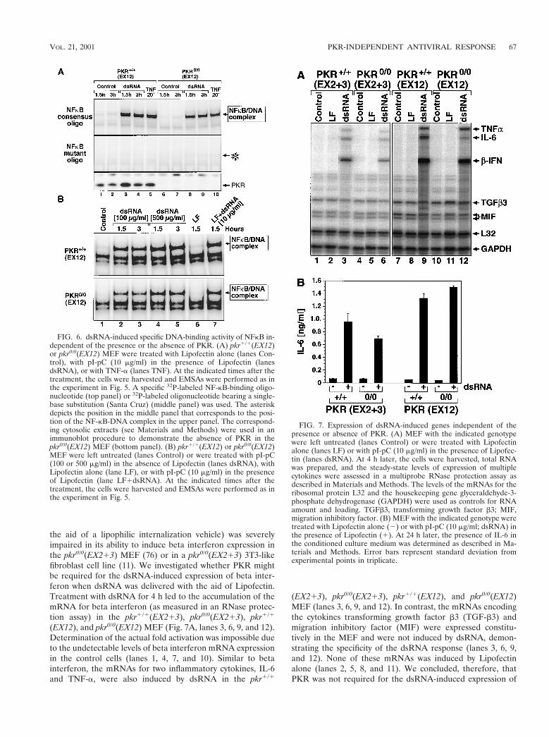
the aid of a lipophilic internalization vehicle) was severelyimpaired in its ability to induce beta interferon expression inthe pkr0/0(EX213) MEF (76) or in a pkr0/0(EX213) 3T3-likefibroblast cell line (11). We investigated whether PKR mightbe required for the dsRNA-induced expression of beta inter-feron when dsRNA was delivered with the aid of Lipofectin.Treatment with dsRNA for 4 h led to the accumulation of themRNA for beta interferon (as measured in an RNase protec-tion assay) in the pkr1/1(EX213), pkr0/0(EX213), pkr1/1
(EX12), and pkr0/0(EX12) MEF (Fig. 7A, lanes 3, 6, 9, and 12).Determination of the actual fold activation was impossible dueto the undetectable levels of beta interferon mRNA expressionin the control cells (lanes 1, 4, 7, and 10). Similar to betainterferon, the mRNAs for two inflammatory cytokines, IL-6and TNF-a, were also induced by dsRNA in the pkr1/1
(EX213), pkr0/0(EX213), pkr1/1(EX12), and pkr0/0(EX12)MEF (lanes 3, 6, 9, and 12). In contrast, the mRNAs encodingthe cytokines transforming growth factor b3 (TGF-b3) andmigration inhibitory factor (MIF) were expressed constitu-tively in the MEF and were not induced by dsRNA, demon-strating the specificity of the dsRNA response (lanes 3, 6, 9,and 12). None of these mRNAs was induced by Lipofectinalone (lanes 2, 5, 8, and 11). We concluded, therefore, thatPKR was not required for the dsRNA-induced expression of
FIG. 6. dsRNA-induced specific DNA-binding activity of NFkB in-dependent of the presence or the absence of PKR. (A) pkr1/1(EX12)or pkr0/0(EX12) MEF were treated with Lipofectin alone (lanes Con-trol), with pI-pC (10 mg/ml) in the presence of Lipofectin (lanesdsRNA), or with TNF-a (lanes TNF). At the indicated times after thetreatment, the cells were harvested and EMSAs were performed as inthe experiment in Fig. 5. A specific 32P-labeled NF-kB-binding oligo-nucleotide (top panel) or 32P-labeled oligonucleotide bearing a single-base substitution (Santa Cruz) (middle panel) was used. The asteriskdepicts the position in the middle panel that corresponds to the posi-tion of the NF-kB-DNA complex in the upper panel. The correspond-ing cytosolic extracts (see Materials and Methods) were used in animmunoblot procedure to demonstrate the absence of PKR in thepkr0/0(EX12) MEF (bottom panel). (B) pkr1/1(EX12) or pkr0/0(EX12)MEF were left untreated (lanes Control) or were treated with pI-pC(100 or 500 mg/ml) in the absence of Lipofectin (lanes dsRNA), withLipofectin alone (lane LF), or with pI-pC (10 mg/ml) in the presenceof Lipofectin (lane LF1dsRNA). At the indicated times after thetreatment, the cells were harvested and EMSAs were performed as inthe experiment in Fig. 5.
FIG. 7. Expression of dsRNA-induced genes independent of thepresence or absence of PKR. (A) MEF with the indicated genotypewere left untreated (lanes Control) or were treated with Lipofectinalone (lanes LF) or with pI-pC (10 mg/ml) in the presence of Lipofec-tin (lanes dsRNA). At 4 h later, the cells were harvested, total RNAwas prepared, and the steady-state levels of expression of multiplecytokines were assessed in a multiprobe RNase protection assay asdescribed in Materials and Methods. The levels of the mRNAs for theribosomal protein L32 and the housekeeping gene glyceraldehyde-3-phosphate dehydrogenase (GAPDH) were used as controls for RNAamount and loading. TGFb3, transforming growth factor b3; MIF,migration inhibitory factor. (B) MEF with the indicated genotype weretreated with Lipofectin alone (2) or with pI-pC (10 mg/ml; dsRNA) inthe presence of Lipofectin (1). At 24 h later, the presence of IL-6 inthe conditioned culture medium was determined as described in Ma-terials and Methods. Error bars represent standard deviation fromexperimental points in triplicate.
VOL. 21, 2001 PKR-INDEPENDENT ANTIVIRAL RESPONSE 67
beta interferon mRNA or of IL-6 and TNF-a mRNAs. PKRwas also not required for the dsRNA-induced accumulation ofthe mRNA for the NF-kB-dependent antiapoptotic gene iap-2(data not shown).
PKR contributes, in mouse fibroblasts, about 50% of theoverall inhibition of translation in response to dsRNA (theother 50% being contributed by the 2-5 oligoadenylate syn-thase/RNase L system) (26). It was therefore reasonable tospeculate that activated PKR may negatively affect gene ex-pression via its inhibitory action on protein synthesis. To ad-dress this question, we employed a highly sensitive enzyme-linked immunosorbent assay for the detection of IL-6 anddetermined whether the presence or absence of PKR affectsthe expression and/or secretion of this cytokine. As shown inFig. 7B, the levels of IL-6 in the culture medium were markedlyelevated 24 h after the treatment with dsRNA in the pkr1/1
(EX213), pkr0/0(EX213), pkr1/1(EX12), and pkr0/0(EX12)MEF. We were unable, therefore, to discern a clear pattern ofPKR involvement in the production of IL-6 in response todsRNA.
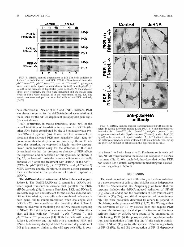
The dsRNA-induced activation of NF-kB does not requireRNase L. The OAS/2-5A/RNase L system is a dsRNA-acti-vated signal transduction cascade that parallels the PKR/eIF-2a cascade (54). In mouse fibroblasts, PKR and RNase Lare solely required and sufficient for the dsRNA-induced inhi-bition of translation, since cells with a combined deficiency inboth genes fail to inhibit translation when challenged withdsRNA (26). We considered the possibility that RNase Lmight be involved in mediating the dsRNA-induced signalingto NF-kB. To test this hypothesis, we employed 3T3-like fibro-blast cell lines with pkr1/1/rnasel1/1, pkr1/1/rnasel2/2, andpkr2/2/rnasel2/2 genotypes (84). Both the cells with a singleRNase L deficiency and the cells with a combined PKR andRNase L deficiency displayed dsRNA-induced degradation ofIkB-b in a manner similar to the wild-type cells (Fig. 8, com-
pare lanes 1 to 3 with lanes 4 to 6). Furthermore, in each cellline, NF-kB translocated to the nucleus in response to dsRNAtreatment (Fig. 9). We concluded, therefore, that neither PKRnor RNase L is a critical component in mediating the dsRNA-induced signaling to NF-kB.
DISCUSSION
The most important result of this study is the demonstrationof a novel response of cells to viral dsRNA that is independentof the dsRNA-activated PKR. Surprisingly, we found that thisresponse includes the dsRNA-induced activation of NF-kB(Fig. 2 to 6, 8, and 9) and the production of beta (“fibroblast”)interferon (Fig. 7A), two critical components of innate immu-nity that were previously described by others to depend, infibroblasts, on the presence of PKR (11, 76, 79). We argue thatthe activation of NF-kB by dsRNA does not require PKRbecause the following critical steps of activation of this tran-scription factor by dsRNA were found to be unimpaired incells lacking PKR: (i) the phosphorylation, polyubiquitinyla-tion, and degradation of IkB (Fig. 2); (ii) the nuclear translo-cation of NF-kB (Fig. 4); (iii) the specific DNA-binding activityof NF-kB (Fig. 6); and (iv) the induction of NF-kB-dependent
FIG. 8. dsRNA-induced degradation of IkB-b in cells deficient inRNase L or both RNase L and PKR. 3T3-like fibroblast cell lines withpkr1/1/rnasel1/1, pkr1/1/rnasel2/2, and pkr2/2/rnasel2/2 genotypeswere treated with Lipofectin alone (lanes Control) or with pI-pC (10mg/ml) in the presence of Lipofectin (lanes dsRNA). At the indicatedtimes after treatment, the cells were harvested and the steady-statelevels of IkB-b were assessed as in the experiment in Fig. 1A. Themembranes were stripped and reprobed with an anti-PKR antibody(D-20).
FIG. 9. dsRNA-induced nuclear translocation of NFkB in cells de-ficient in RNase L or both RNase L and PKR. 3T3-like fibroblast celllines with pkr1/1/rnasel1/1, pkr1/1/rnasel2/2, and pkr2/2/rnasel2/2 ge-notypes were treated with Lipofectin alone (Control) or with pI-pC (10mg/ml) in the presence of Lipofectin (dsRNA). At 3 h after treatment,the cells were fixed and immunostained with an antibody recognizingthe p65/RelA subunit of NFkB as in the experiment in Fig. 3.
68 IORDANOV ET AL. MOL. CELL. BIOL.
genes, such as the genes for beta interferon, IL-6, TNF-a, andIAP-2 (Fig. 7 and data not shown) (for an extended list ofestablished NF-kB-dependent genes, see reference 42 and ref-erences therein).
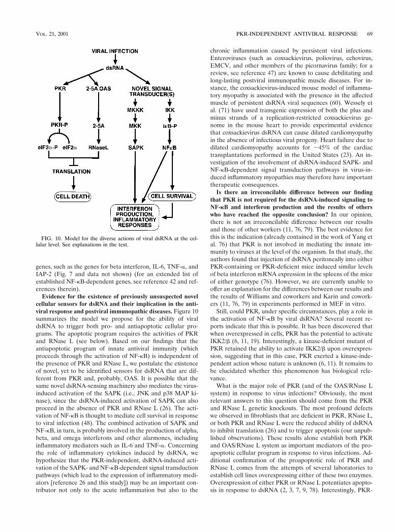
Evidence for the existence of previously unsuspected novelcellular sensors for dsRNA and their implication in the anti-viral response and postviral immunopathic diseases. Figure 10summarizes the model we propose for the ability of viraldsRNA to trigger both pro- and antiapoptotic cellular pro-grams. The apoptotic program requires the activities of PKRand RNase L (see below). Based on our findings that theantiapoptotic program of innate antiviral immunity (whichproceeds through the activation of NF-kB) is independent ofthe presence of PKR and RNase L, we postulate the existenceof novel, yet to be identified sensors for dsRNA that are dif-ferent from PKR and, probably, OAS. It is possible that thesame novel dsRNA-sensing machinery also mediates the virus-induced activation of the SAPK (i.e., JNK and p38 MAP ki-nase), since the dsRNA-induced activation of SAPK can alsoproceed in the absence of PKR and RNase L (26). The acti-vation of NF-kB is thought to mediate cell survival in responseto viral infection (48). The combined activation of SAPK andNF-kB, in turn, is probably involved in the production of alpha,beta, and omega interferons and other alarmones, includinginflammatory mediators such as IL-6 and TNF-a. Concerningthe role of inflammatory cytokines induced by dsRNA, wehypothesize that the PKR-independent, dsRNA-induced acti-vation of the SAPK- and NF-kB-dependent signal transductionpathways (which lead to the expression of inflammatory medi-ators [reference 26 and this study]) may be an important con-tributor not only to the acute inflammation but also to the
chronic inflammation caused by persistent viral infections.Enteroviruses (such as coxsackievirus, poliovirus, echovirus,EMCV, and other members of the picornavirus family; for areview, see reference 47) are known to cause debilitating andlong-lasting postviral immunopathic muscle diseases. For in-stance, the coxsackievirus-induced mouse model of inflamma-tory myopathy is associated with the presence in the affectedmuscle of persistent dsRNA viral sequences (60). Wessely etal. (71) have used transgenic expression of both the plus andminus strands of a replication-restricted coxsackievirus ge-nome in the mouse heart to provide experimental evidencethat coxsackievirus dsRNA can cause dilated cardiomyopathyin the absence of infectious viral progeny. Heart failure due todilated cardiomyopathy accounts for ;45% of the cardiactransplantations performed in the United States (23). An in-vestigation of the involvement of dsRNA-induced SAPK- andNF-kB-dependent signal transduction pathways in virus-in-duced inflammatory myopathies may therefore have importanttherapeutic consequences.
Is there an irreconcilable difference between our findingthat PKR is not required for the dsRNA-induced signaling toNF-kB and interferon production and the results of otherswho have reached the opposite conclusion? In our opinion,there is not an irreconcilable difference between our resultsand those of other workers (11, 76, 79). The best evidence forthis is the indication (already contained in the work of Yang etal. 76) that PKR is not involved in mediating the innate im-munity to viruses at the level of the organism. In that study, theauthors found that injection of dsRNA peritoneally into eitherPKR-containing or PKR-deficient mice induced similar levelsof beta interferon mRNA expression in the spleens of the miceof either genotype (76). However, we are currently unable tooffer an explanation for the differences between our results andthe results of Williams and coworkers and Karin and cowork-ers (11, 76, 79) in experiments performed in MEF in vitro.
Still, could PKR, under specific circumstances, play a role inthe activation of NF-kB by viral dsRNA? Several recent re-ports indicate that this is possible. It has been discovered thatwhen overexpressed in cells, PKR has the potential to activateIKK2/b (6, 11, 19). Interestingly, a kinase-deficient mutant ofPKR retained the ability to activate IKK2/b upon overexpres-sion, suggesting that in this case, PKR exerted a kinase-inde-pendent action whose nature is unknown (6, 11). It remains tobe elucidated whether this phenomenon has biological rele-vance.
What is the major role of PKR (and of the OAS/RNase Lsystem) in response to virus infections? Obviously, the mostrelevant answers to this question should come from the PKRand RNase L genetic knockouts. The most profound defectswe observed in fibroblasts that are deficient in PKR, RNase L,or both PKR and RNase L were the reduced ability of dsRNAto inhibit translation (26) and to trigger apoptosis (our unpub-lished observations). These results alone establish both PKRand OAS/RNase L system as important mediators of the pro-apoptotic cellular program in response to virus infections. Ad-ditional confirmation of the proapoptotic role of PKR andRNase L comes from the attempts of several laboratories toestablish cell lines overexpressing either of these two enzymes.Overexpression of either PKR or RNase L potentiates apopto-sis in response to dsRNA (2, 3, 7, 9, 78). Interestingly, PKR-
FIG. 10. Model for the diverse actions of viral dsRNA at the cel-lular level. See explanations in the text.
VOL. 21, 2001 PKR-INDEPENDENT ANTIVIRAL RESPONSE 69
overexpressing cells are also more sensitive to the cytotoxiceffects of TNF-a (78), an effect probably resulting from anincreased sensitivity to apoptotic stimuli in the face of com-promised sustainability of the process of protein synthesis. Isthe PKR-mediated inhibition of protein synthesis per se a maincause of PKR-induced apoptosis? Some experimental evidencesuggests that this may be the case. For instance, apoptosisinduced by an overexpression of PKR could be counteractedby the concomitant overexpression of a nonphosphorylatableform of eIF-2a (20, 53). Furthermore, expression of a mutantform of eIF-2a that mimics phosphorylated eIF-2a was foundto induce apoptosis by itself (53).
Finally, although the evidence provided in this study doesnot support a role for PKR in mediating the expression ofNF-kB-dependent genes, our conclusions should not be inter-preted as an attempt to rule out the possible participation ofPKR in signal transduction to the nucleus. Further studies,especially those using powerful DNA array technologies, arelikely to provide a conclusive answer in the near future. Fur-thermore, we have recently reported that the PKR- and RNaseL-mediated inhibition of protein synthesis (but not PKR andRNase L per se) plays a critical role in the ability of dsRNA totrigger the activation of JNK (26). This establishes the inter-esting possibility that dsRNA-activated JNK may play a role inmediating dsRNA-induced apoptosis. This possibility is cur-rently being investigated.
ACKNOWLEDGMENTS
We thank Olga Ryabinina, Thanh-Hoai Dinh, and Paul Spitz forexcellent technical assistance. We thank Bryan Williams for thepkr1/1(EX213) and pkr0/0(EX213) MEF and Robert Silverman forthe rnasel1/1Ppkr1/1, rnasel2/2Ppkr1/1, and rnasel2/2Ppkr2/2 3T3-like fibroblasts and for the EMCV.
This work was supported by U.S. Public Health Service grants CA-39360 and ES-08456 to B.E.M. and by an N. L. Tartar Research FundFellowship to M.S.I. J.C.B. is supported by the National Cancer Insti-tute of Canada and the Canadian Cancer Society.
REFERENCES
1. Abraham, N., D. F. Stojdl, P. I. Duncan, N. Methot, T. Ishii, M. Dube, B. C.Vanderhyden, H. L. Atkins, D. A. Gray, M. W. McBurney, A. E. Koromilas,E. G. Brown, N. Sonenberg, and J. C. Bell. 1999. Characterization of trans-genic mice with targeted disruption of the catalytic domain of the double-stranded RNA-dependent protein kinase, PKR. J. Biol. Chem. 274:5953–5962.
2. Balachandran, S., C. N. Kim, W. C. Yeh, T. W. Mak, K. Bhalla, and G. N.Barber. 1998. Activation of the dsRNA-dependent protein kinase, PKR,induces apoptosis through FADD-mediated death signaling. EMBO J. 17:6888–6902.
3. Balachandran, S., P. C. Roberts, T. Kipperman, K. N. Bhalla, R. W. Com-pans, D. R. Archer, and G. N. Barber. 2000. Alpha/beta interferons poten-tiate virus-induced apoptosis through activation of the FADD/caspase-8death signaling pathway. J. Virol. 74:1513–1523.
4. Bender, K., M. Gottlicher, S. Whiteside, H. J. Rahmsdorf, and P. Herrlich.1998. Sequential DNA damage-independent and -dependent activation ofNF-kappaB by UV. EMBO J. 17:5170–5181.
5. Birnbaum, M. J., R. J. Clem, and L. K. Miller. 1994. An apoptosis-inhibitinggene from a nuclear polyhedrosis virus encoding a polypeptide with Cys/Hissequence motifs. J. Virol. 68:2521–2528.
6. Bonnet, M. C., R. Weil, E. Dam, A. G. Hovanessian, and E. F. Meurs. 2000.PKR stimulates NF-kB irrespective of its kinase function by interacting withthe IkB kinase complex. Mol. Cell. Biol. 20:4532–4542.
7. Castelli, J., K. A. Wood, and R. J. Youle. 1998. The 2-5A system in viralinfection and apoptosis. Biomed. Pharmacother. 52:386–390.
8. Castelli, J. C., B. A. Hassel, A. Maran, J. Paranjape, J. A. Hewitt, X. L. Li,Y. T. Hsu, R. H. Silverman, and R. J. Youle. 1998. The role of 29-59 oligoad-enylate-activated ribonuclease L in apoptosis. Cell Death Differ. 5:313–320.
9. Castelli, J. C., B. A. Hassel, K. A. Wood, X. L. Li, K. Amemiya, M. C.Dalakas, P. F. Torrence, and R. J. Youle. 1997. A study of the interferon
antiviral mechanism: apoptosis activation by the 2-5A system. J. Exp. Med.186:967–972.
10. Chen, C., L. C. Edelstein, and C. Gelinas. 2000. The Rel/NF-kB familydirectly activates expression of the apoptosis inhibitor Bcl-x(L). Mol. Cell.Biol. 20:2687–2695.
11. Chu, W. M., D. Ostertag, Z. W. Li, L. Chang, Y. Chen, Y. Hu, B. Williams,J. Perrault, and M. Karin. 1999. JNK2 and IKKbeta are required for acti-vating the innate response to viral infection. Immunity 11:721–731.
12. Chu, Z. L., T. A. McKinsey, L. Liu, J. J. Gentry, M. H. Malim, and D. W.Ballard. 1997. Suppression of tumor necrosis factor-induced cell death byinhibitor of apoptosis c-IAP2 is under NF-kappaB control. Proc. Natl. Acad.Sci. USA 94:10057–10062.
13. Diaz-Guerra, M., C. Rivas, and M. Esteban. 1997. Activation of the IFN-inducible enzyme RNase L causes apoptosis of animal cells. Virology 236:354–363.
14. DiDonato, J. A., M. Hayakawa, D. M. Rothwarf, E. Zandi, and M. Karin.1997. A cytokine-responsive IkappaB kinase that activates the transcriptionfactor NF-kappaB. Nature 388:548–554.
15. Donze, O., J. Dostie, and N. Sonenberg. 1999. Regulatable expression of theinterferon-induced double-stranded RNA dependent protein kinase PKRinduces apoptosis and fas receptor expression. Virology 256:322–329.
16. Foster, G. R., and N. B. Finter. 1998. Are all type I human interferonsequivalent? J. Viral Hepatol. 5:143–152.
17. Gagliardini, V., P. A. Fernandez, R. K. Lee, H. C. Drexler, R. J. Rotello,M. C. Fishman, and J. Yuan. 1994. Prevention of vertebrate neuronal deathby the crmA gene. Science 263:826–828.
18. Garoufalis, E., I. Kwan, R. Lin, A. Mustafa, N. Pepin, A. Roulston, J.Lacoste, and J. Hiscott. 1994. Viral induction of the human beta interferonpromoter: modulation of transcription by NF-kappa B/rel proteins and in-terferon regulatory factors. J. Virol. 68:4707–4715.
19. Gil, J., J. Alcami, and M. Esteban. 2000. Activation of NF-kappa B by thedsRNA-dependent protein kinase, PKR involves the I kappa B kinase com-plex. Oncogene 19:1369–1378.
20. Gil, J., J. Alcami, and M. Esteban. 1999. Induction of apoptosis by double-stranded-RNA-dependent protein kinase (PKR) involves the alpha subunitof eukaryotic translation initiation factor 2 and NF-kB. Mol. Cell. Biol.19:4653–4663.
21. Gilmore, T. D. 1999. The Rel/NF-kappaB signal transduction pathway: in-troduction. Oncogene 18:6842–6844.
22. Gross, M., W. M. Knish, and A. Kwan. 1981. Rabbit reticulocyte double-stranded RNA-activated protein kinase and the hemin-controlled transla-tional repressor phosphorylate the same Mr 1500 peptide of eukaryoticinitiation factor 2 alpha. FEBS Lett. 125:223–226.
23. Hosenpud, J. D., R. J. Novick, L. E. Bennett, B. M. Keck, B. Fiol, and O. P.Daily. 1996. The Registry of the International Society for Heart and LungTransplantation: thirteenth official report—1996. J. Heart Lung Transplant.15:655–674.
24. Hovanessian, A. G. 1991. Interferon-induced and double-stranded RNA-activated enzymes: a specific protein kinase and 29,59-oligoadenylate syn-thetases. J. Interferon Res. 11:199–205.
25. Hu, Y., V. Baud, M. Delhase, P. Zhang, T. Deerinck, M. Ellisman, R.Johnson, and M. Karin. 1999. Abnormal morphogenesis but intact IKKactivation in mice lacking the IKKalpha subunit of IkappaB kinase. Science284:316–320.
26. Iordanov, M. S., J. M. Paranjape, A. Zhou, J. Wong, B. R. Williams, E. F.Meurs, R. H. Silverman, and B. E. Magun. 2000. Activation of p38 mitogen-activated protein kinase and c-Jun NH2-terminal kinase by double-strandedRNA and encephalomyocarditis virus: involvement of RNase L, proteinkinase R, and alternative pathways. Mol. Cell. Biol. 20:617–627.
27. Iordanov, M. S., O. P. Ryabinina, J. Wong, T. H. Dinh, D. L. Newton, S. M.Rybak, and B. E. Magun. 2000. Molecular determinants of apoptosis inducedby the cytotoxic ribonuclease onconase: evidence for cytotoxic mechanismsdifferent from inhibition of protein synthesis. Cancer Res. 60:1983–1994.
28. Jagus, R., B. Joshi, and G. N. Barber. 1999. PKR, apoptosis and cancer. Int.J. Biochem. Cell Biol. 31:123–138.
29. Jeffrey, I. W., S. Kadereit, E. F. Meurs, T. Metzger, M. Bachmann, M.Schwemmle, A. G. Hovanessian, and M. J. Clemens. 1995. Nuclear localiza-tion of the interferon-inducible protein kinase PKR in human cells andtransfected mouse cells. Exp. Cell Res. 218:17–27.
30. Karin, M. 1999. The beginning of the end: IkappaB kinase (IKK) andNF-kappaB activation. J. Biol. Chem. 274:27339–27342.
31. Karin, M. 1999. How NF-kappaB is activated: the role of the IkappaB kinase(IKK) complex. Oncogene 18:6867–6874.
32. LaCasse, E. C., S. Baird, R. G. Korneluk, and A. E. MacKenzie. 1998. Theinhibitors of apoptosis (IAPs) and their emerging role in cancer. Oncogene17:3247–3259.
33. Lee, H. H., H. Dadgostar, Q. Cheng, J. Shu, and G. Cheng. 1999. NF-kappaB-mediated up-regulation of Bcl-x and Bfl-1/A1 is required for CD40survival signaling in B lymphocytes. Proc. Natl. Acad. Sci. USA 96:9136–9141.
34. Lee, S. B., D. Rodriguez, J. R. Rodriguez, and M. Esteban. 1997. Theapoptosis pathway triggered by the interferon-induced protein kinase PKR
70 IORDANOV ET AL. MOL. CELL. BIOL.
requires the third basic domain, initiates upstream of Bcl-2, and involvesICE-like proteases. Virology 231:81–88.
35. Levin, D. H., R. Petryshyn, and I. M. London. 1981. Characterization ofpurified double-stranded RNA-activated eIF-2 alpha kinase from rabbitreticulocytes. J. Biol. Chem. 256:7638–7641.
36. Li, Z. W., W. Chu, Y. Hu, M. Delhase, T. Deerinck, M. Ellisman, R. Johnson,and M. Karin. 1999. The IKKbeta subunit of IkappaB kinase (IKK) isessential for nuclear factor kappaB activation and prevention of apoptosis. J.Exp. Med. 189:1839–1845.
37. Maniatis, T., J. V. Falvo, T. H. Kim, T. K. Kim, C. H. Lin, B. S. Parekh, andM. G. Wathelet. 1998. Structure and function of the interferon-beta enhan-ceosome. Cold Spring Harbor Symp. Quant. Biol. 63:609–620.
38. Mercurio, F., H. Zhu, B. W. Murray, A. Shevchenko, B. L. Bennett, J. Li,D. B. Young, M. Barbosa, M. Mann, A. Manning, and A. Rao. 1997. IKK-1and IKK-2: cytokine-activated IkappaB kinases essential for NF-kappaBactivation. Science 278:860–866.
39. Meurs, E., K. Chong, J. Galabru, N. S. Thomas, I. M. Kerr, B. R. Williams,and A. G. Hovanessian. 1990. Molecular cloning and characterization of thehuman double-stranded RNA-activated protein kinase induced by inter-feron. Cell 62:379–390.
40. Muller, U., U. Steinhoff, L. F. Reis, S. Hemmi, J. Pavlovic, R. M. Zinkerna-gel, and M. Aguet. 1994. Functional role of type I and type II interferons inantiviral defense. Science 264:1918–1921.
41. Nilsen, T. W., D. L. Wood, and C. Baglioni. 1981. 29,59-Oligo(A)-activatedendoribonuclease. Tissue distribution and characterization with a bindingassay. J. Biol. Chem. 256:10751–10754.
42. Pahl, H. L. 1999. Activators and target genes of Rel/NF-kappaB transcrip-tion factors. Oncogene 18:6853–6866.
43. Player, M. R., and P. F. Torrence. 1998. The 2-5A system: modulation ofviral and cellular processes through acceleration of RNA degradation. Phar-macol. Ther. 78:55–113.
44. Rabizadeh, S., D. J. LaCount, P. D. Friesen, and D. E. Bredesen. 1993.Expression of the baculovirus p35 gene inhibits mammalian neural celldeath. J. Neurochem. 61:2318–2321.
45. Rao, L., M. Debbas, P. Sabbatini, D. Hockenbery, S. Korsmeyer, and E.White. 1992. The adenovirus E1A proteins induce apoptosis, which is inhib-ited by the E1B 19-kDa and Bcl-2 proteins. Proc. Natl. Acad. Sci. USA89:7742–7746.
46. Rudolph, D., W. C. Yeh, A. Wakeham, B. Rudolph, D. Nallainathan, J.Potter, A. J. Elia, and T. W. Mak. 2000. Severe liver degeneration and lackof NF-kappaB activation in NEMO/IKKgamma-deficient mice. Genes Dev.14:854–862.
47. Rueckert, R. 1996. Picornaviridae: the viruses and their replication. p. 477–522. In B. Fields, D. Knipe, and P. Howley (ed.), Virology, 3rd ed. Lippin-cott-Raven, Philadelphia, Pa.
48. Schwarz, E. M., C. Badorff, T. S. Hiura, R. Wessely, A. Badorff, I. M. Verma,and K. U. Knowlton. 1998. NF-kB-mediated inhibition of apoptosis is re-quired for encephalomyocarditis virus virulence: a mechanism of resistancein p50 knockout mice. J. Virol. 72:5654–5660.
49. Sha, W. C., H. C. Liou, E. I. Tuomanen, and D. Baltimore. 1995. Targeteddisruption of the p50 subunit of NF-kappa B leads to multifocal defects inimmune responses. Cell 80:321–330.
50. Siekierka, J., V. Manne, and S. Ochoa. 1984. Mechanism of translationalcontrol by partial phosphorylation of the alpha subunit of eukaryotic initia-tion factor 2. Proc. Natl. Acad. Sci. USA 81:352–356.
51. Silverman, R. H., P. J. Cayley, D. H. Wreschner, M. Knight, C. S. Gilbert,R. E. Brown, and I. M. Kerr. 1981. 2-5A(pppA29p59A29p59A) in interferon-treated encephalomyocarditis virus-infected mouse L-cells. Tex. Rep. Biol.Med. 41:479–486.
52. Silverman, R. H., J. J. Skehel, T. C. James, D. H. Wreschner, and I. M. Kerr.1983. rRNA cleavage as an index of ppp(A29p)nA activity in interferon-treated encephalomyocarditis virus-infected cells. J. Virol. 46:1051–1055.
53. Srivastava, S. P., K. U. Kumar, and R. J. Kaufman. 1998. Phosphorylationof eukaryotic translation initiation factor 2 mediates apoptosis in response toactivation of the double-stranded RNA-dependent protein kinase. J. Biol.Chem. 273:2416–2423.
54. Stark, G. R., I. M. Kerr, B. R. Williams, R. H. Silverman, and R. D.Schreiber. 1998. How cells respond to interferons. Annu. Rev. Biochem.67:227–264.
55. Stehlik, C., R. de Martin, I. Kumabashiri, J. A. Schmid, B. R. Binder, andJ. Lipp. 1998. Nuclear factor (NF)-kappaB-regulated X-chromosome-linkediap gene expression protects endothelial cells from tumor necrosis factoralpha-induced apoptosis. J. Exp. Med. 188:211–216.
56. Stein, B., H. J. Rahmsdorf, A. Steffen, M. Litfin, and P. Herrlich. 1989.UV-induced DNA damage is an intermediate step in UV-induced expressionof human immunodeficiency virus type 1, collagenase, c-fos, and metallo-thionein. Mol. Cell. Biol. 9:5169–5181.
57. Stroka, D. M., A. Z. Badrichani, F. H. Bach, and C. Ferran. 1999. Overex-pression of A1, an NF-kappaB-inducible anti-apoptotic bcl gene, inhibitsendothelial cell activation. Blood 93:3803–3810.
58. Sugimoto, A., P. D. Friesen, and J. H. Rothman. 1994. Baculovirus p35prevents developmentally programmed cell death and rescues a ced-9 mu-
tant in the nematode Caenorhabditis elegans. EMBO J. 13:2023–2028.59. Takeda, K., O. Takeuchi, T. Tsujimura, S. Itami, O. Adachi, T. Kawai, H.
Sanjo, K. Yoshikawa, N. Terada, and S. Akira. 1999. Limb and skin abnor-malities in mice lacking IKKalpha. Science 284:313–316.
60. Tam, P. E., and R. P. Messner. 1999. Molecular mechanisms of coxsackievi-rus persistence in chronic inflammatory myopathy: viral RNA persiststhrough formation of a double-stranded complex without associated genomicmutations or evolution. J. Virol. 73:10113–10121.
61. Tanaka, M., M. E. Fuentes, K. Yamaguchi, M. H. Durnin, S. A. Dalrymple,K. L. Hardy, and D. V. Goeddel. 1999. Embryonic lethality, liver degenera-tion, and impaired NF-kappa B activation in IKK-beta-deficient mice. Im-munity 10:421–429.
62. Teodoro, J. G., and P. E. Branton. 1997. Regulation of apoptosis by viralgene products. J. Virol. 71:1739–1746.
63. Tewari, M., and V. M. Dixit. 1995. Fas- and tumor necrosis factor-inducedapoptosis is inhibited by the poxvirus crmA gene product. J. Biol. Chem.270:3255–3260.
64. Thanos, D., and T. Maniatis. 1992. The high mobility group protein HMGI(Y) is required for NF-kappa B-dependent virus induction of the humanIFN-beta gene. Cell 71:777–789.
65. Thanos, D., and T. Maniatis. 1995. Identification of the rel family membersrequired for virus induction of the human beta interferon gene. Mol. Cell.Biol. 15:152–164.
66. Thome, M., P. Schneider, K. Hofmann, H. Fickenscher, E. Meinl, F. Neipel,C. Mattmann, K. Burns, J. L. Bodmer, M. Schroter, C. Scaffidi, P. H.Krammer, M. E. Peter, and J. Tschopp. 1997. Viral FLICE-inhibitory pro-teins (FLIPs) prevent apoptosis induced by death receptors. Nature 386:517–521.
67. Traenckner, E. B., H. L. Pahl, T. Henkel, K. N. Schmidt, S. Wilk, and P. A.Baeuerle. 1995. Phosphorylation of human I kappa B-alpha on serines 32 and36 controls I kappa B-alpha proteolysis and NF-kappa B activation in re-sponse to diverse stimuli. EMBO J. 14:2876–2883.
68. Tsukahara, T., M. Kannagi, T. Ohashi, H. Kato, M. Arai, G. Nunez, Y.Iwanaga, N. Yamamoto, K. Ohtani, M. Nakamura, and M. Fujii. 1999.Induction of Bcl-x(L) expression by human T-cell leukemia virus type 1 Taxthrough NF-kappaB in apoptosis-resistant T-cell transfectants with Tax.J. Virol. 73:7981–7987.
69. Wang, C. Y., D. C. Guttridge, M. W. Mayo, and A. S. Baldwin, Jr. 1999.NF-kappaB induces expression of the Bcl-2 homologue A1/Bfl-1 to prefer-entially suppress chemotherapy-induced apoptosis. Mol. Cell. Biol. 19:5923–5929.
70. Wang, C. Y., M. W. Mayo, R. G. Korneluk, D. V. Goeddel, and A. S. Baldwin,Jr. 1998. NF-kappaB antiapoptosis: induction of TRAF1 and TRAF2 andc-IAP1 and c-IAP2 to suppress caspase-8 activation. Science 281:1680–1683.
71. Wessely, R., K. Klingel, L. F. Santana, N. Dalton, M. Hongo, W. JonathanLederer, R. Kandolf, and K. U. Knowlton. 1998. Transgenic expression ofreplication-restricted enteroviral genomes in heart muscle induces defectiveexcitation-contraction coupling and dilated cardiomyopathy. J. Clin. Investig.102:1444–1453.
72. White, E., P. Sabbatini, M. Debbas, W. S. Wold, D. I. Kusher, and L. R.Gooding. 1992. The 19-kilodalton adenovirus E1B transforming protein in-hibits programmed cell death and prevents cytolysis by tumor necrosis factoralpha. Mol. Cell. Biol. 12:2570–2580.
73. Williams, B. R. 1999. PKR: a sentinel kinase for cellular stress. Oncogene18:6112–6120.
74. Wreschner, D. H., T. C. James, R. H. Silverman, and I. M. Kerr. 1981.Ribosomal RNA cleavage, nuclease activation and 2-5A(ppp(A29p)nA) ininterferon-treated cells. Nucleic Acids Res. 9:1571–1581.
75. Yamaoka, S., G. Courtois, C. Bessia, S. T. Whiteside, R. Weil, F. Agou, H. E.Kirk, R. J. Kay, and A. Israel. 1998. Complementation cloning of NEMO, acomponent of the IkappaB kinase complex essential for NF-kappaB activa-tion. Cell 93:1231–1240.
76. Yang, Y. L., L. F. Reis, J. Pavlovic, A. Aguzzi, R. Schafer, A. Kumar, B. R.Williams, M. Aguet, and C. Weissmann. 1995. Deficient signaling in micedevoid of double-stranded RNA-dependent protein kinase. EMBO J. 14:6095–6106.
77. Yeung, M. C., D. L. Chang, R. E. Camantigue, and A. S. Lau. 1999. Inhibitoryrole of the host apoptogenic gene PKR in the establishment of persistentinfection by encephalomyocarditis virus in U937 cells. Proc. Natl. Acad. Sci.USA 96:11860–11860.
78. Yeung, M. C., J. Liu, and A. S. Lau. 1996. An essential role for the inter-feron-inducible, double-stranded RNA-activated protein kinase PKR in thetumor necrosis factor-induced apoptosis in U937 cells. Proc. Natl. Acad. Sci.USA 93:12451–12455.
79. Zamanian-Daryoush, M., T. H. Mogensen, J. A. DiDonato, and B. R. Wil-liams. 2000. NF-kB activation by double-stranded-RNA-activated proteinkinase (PKR) is mediated through NF-kB-inducing kinase and IkB kinase.Mol. Cell. Biol. 20:1278–1290.
80. Zandi, E., and M. Karin. 1999. Bridging the gap: composition, regulation,and physiological function of the IkB kinase complex. Mol. Cell. Biol. 19:4547–4551.
VOL. 21, 2001 PKR-INDEPENDENT ANTIVIRAL RESPONSE 71

81. Zandi, E., D. M. Rothwarf, M. Delhase, M. Hayakawa, and M. Karin. 1997.The IkappaB kinase complex (IKK) contains two kinase subunits, IKKalphaand IKKbeta, necessary for IkappaB phosphorylation and NF-kappaB acti-vation. Cell 91:243–252.
82. Zhou, A., B. A. Hassel, and R. H. Silverman. 1993. Expression cloning of2-5A-dependent RNAase: a uniquely regulated mediator of interferon ac-tion. Cell 72:753–765.
83. Zhou, A., J. Paranjape, T. L. Brown, H. Nie, S. Naik, B. Dong, A. Chang, B.Trapp, R. Fairchild, C. Colmenares, and R. H. Silverman. 1997. Interferonaction and apoptosis are defective in mice devoid of 29,59-oligoadenylate-
dependent RNase L. EMBO J. 16:6355–6363.84. Zhou, A., J. M. Paranjape, S. D. Der, B. R. Williams, and R. H. Silverman.
1999. Interferon action in triply deficient mice reveals the existence of al-ternative antiviral pathways. Virology 258:435–440.
85. Zhou, A., J. M. Paranjape, B. A. Hassel, H. Nie, S. Shah, B. Galinski, andR. H. Silverman. 1998. Impact of RNase L overexpression on viral andcellular growth and death. J. Interferon Cytokine Res. 18:953–961.
86. Zong, W. X., L. C. Edelstein, C. Chen, J. Bash, and C. Gelinas. 1999. Theprosurvival Bcl-2 homolog Bfl-1/A1 is a direct transcriptional target of NF-kappaB that blocks TNFalpha-induced apoptosis. Genes Dev. 13:382–387.
72 IORDANOV ET AL. MOL. CELL. BIOL.