The molecular mechanism of dsRNA processing by a bacterial ...
14
Published online 27 March 2019 Nucleic Acids Research, 2019, Vol. 47, No. 9 4707–4720 doi: 10.1093/nar/gkz208 The molecular mechanism of dsRNA processing by a bacterial Dicer Lan Jin 1 , He Song 1 , Joseph E. Tropea 1 , Danielle Needle 1 , David S. Waugh 1 , Shuo Gu 2,* and Xinhua Ji 1,* 1 Macromolecular Crystallography Laboratory, National Cancer Institute, Frederick, MD 21702, USA and 2 RNA Biology Laboratory, National Cancer Institute, Frederick, MD 21702, USA Received October 10, 2018; Revised March 01, 2019; Editorial Decision March 05, 2019; Accepted March 17, 2019 ABSTRACT Members of the ribonuclease (RNase) III family regu- late gene expression by processing dsRNAs. It was previously shown that Escherichia coli (Ec) RNase III recognizes dsRNA with little sequence specificity and the cleavage products are mainly 11 nucleotides (nt) long. It was also shown that the mutation of a glutamate (EcE38) to an alanine promotes genera- tion of siRNA-like products typically 22 nt long. To fully characterize substrate specificity and product size of RNase IIIs, we performed in vitro cleavage of dsRNAs by Ec and Aquifex aeolicus (Aa) enzymes and delineated their products by next-generation se- quencing. Surprisingly, we found that both enzymes cleave dsRNA at preferred sites, among which a gua- nine nucleotide was enriched at a specific position (+3G). Based on sequence and structure analyses, we conclude that RNase IIIs recognize +3G via a conserved glutamine (EcQ165/AaQ161) side chain. Abolishing this interaction by mutating the glutamine to an alanine eliminates the observed +3G prefer- ence. Furthermore, we identified a second gluta- mate (EcE65/AaE64), which, when mutated to ala- nine, also enhances the production of siRNA-like products. Based on these findings, we created a bac- terial Dicer that is ideally suited for producing hetero- geneous siRNA cocktails to be used in gene silenc- ing studies. INTRODUCTION Found in all kingdoms of life, the ribonuclease III (RNase III) family endoribonucleases regulate gene expression by specifically processing double-stranded (ds) RNAs (1,2). The founding member of the family, Escherichia coli RNase III, was discovered in 1968 (3). Bacterial RNase III plays a major role in ribosomal RNA (rRNA) processing (4). It is also involved in post-transcriptional gene expression con- trol (5) and defense against viral infection (6,7). Knocking out or mutating RNase III in E. coli (Ec) or Bacillus subtilis induces global gene expression changes (8,9). The produc- tion of a set of Staphylococcus aureus small RNAs is depen- dent on RNase III (10,11). Other representative members of the RNase III family in- clude yeast Rnt1p, human Drosha and human Dicer. Like bacterial RNase III, yeast Rnt1p also functions in the pro- cessing of rRNA (12). In addition, it is involved in the pro- cessing of small non-coding RNA (13) and the degradation of messenger RNA (mRNA) (14). The two human RNase III enzymes, Drosha and Dicer, are more specialized. Dicer plays a role in the production of small interfering RNA (siRNA). Along with Drosha, it is also involved in the bio- genesis of microRNA (miRNA), a master gene regulator in eukaryotic cells (15–20). The RNase III nuclease domain (RIIID) dimerizes, which creates a catalytic valley that ac- commodates a dsRNA substrate (21,22). Bacterial RNase III and yeast Rnt1p have a single RIIID; these enzymes function as homodimers (23,24). Human Dicer and Drosha have two RIIIDs in the same molecule; these enzymes func- tion as monomers (19,20). Both RNase III and Dicer can digest long dsRNA substrates, for which two distinct mech- anisms have been previously described. One mechanism fea- tures an end-in strategy, in which the enzyme recognizes the termini of a long dsRNA, cleaves both RNA strands and removes a small RNA duplex upon product release. Both enzymes can adopt this end-in mechanism and suc- cessively remove small RNA duplexes from the dsRNA ter- mini (5,20,24). Whereas RNase III typically measures 11 nucleotides (nt) from the termini for cleavage (Figure 1A), Dicer typically measures 22 nt (Figure 1B). A second mech- anism features an inside-out scheme, in which multiple RI- IID dimers bind consecutively to a long dsRNA and cleave the substrate simultaneously, producing siRNA-like small RNA duplexes, the length of which is the distance between consecutive active centers. This inside-out mechanism has been observed for EcRNase III (Figure 1A) under special * To whom correspondence should be addressed. Tel: +1 301 846 5035; Fax: +1 301 846 6073; Email: [email protected] Correspondence may also be addressed to Shuo Gu. Tel: +1 301 846 5447; Email: [email protected] Published by Oxford University Press on behalf of Nucleic Acids Research 2019. This work is written by (a) US Government employee(s) and is in the public domain in the US. Downloaded from https://academic.oup.com/nar/article/47/9/4707/5420528 by guest on 17 April 2022
-
Upload
khangminh22 -
Category
Documents
-
view
2 -
download
0
Transcript of The molecular mechanism of dsRNA processing by a bacterial ...

Published online 27 March 2019 Nucleic Acids Research, 2019, Vol. 47, No. 9 4707–4720doi: 10.1093/nar/gkz208
The molecular mechanism of dsRNA processing by abacterial DicerLan Jin1, He Song1, Joseph E. Tropea1, Danielle Needle1, David S. Waugh 1, Shuo Gu 2,*
and Xinhua Ji 1,*
1Macromolecular Crystallography Laboratory, National Cancer Institute, Frederick, MD 21702, USA and 2RNABiology Laboratory, National Cancer Institute, Frederick, MD 21702, USA
Received October 10, 2018; Revised March 01, 2019; Editorial Decision March 05, 2019; Accepted March 17, 2019
ABSTRACT
Members of the ribonuclease (RNase) III family regu-late gene expression by processing dsRNAs. It waspreviously shown that Escherichia coli (Ec) RNaseIII recognizes dsRNA with little sequence specificityand the cleavage products are mainly 11 nucleotides(nt) long. It was also shown that the mutation of aglutamate (EcE38) to an alanine promotes genera-tion of siRNA-like products typically 22 nt long. Tofully characterize substrate specificity and productsize of RNase IIIs, we performed in vitro cleavage ofdsRNAs by Ec and Aquifex aeolicus (Aa) enzymesand delineated their products by next-generation se-quencing. Surprisingly, we found that both enzymescleave dsRNA at preferred sites, among which a gua-nine nucleotide was enriched at a specific position(+3G). Based on sequence and structure analyses,we conclude that RNase IIIs recognize +3G via aconserved glutamine (EcQ165/AaQ161) side chain.Abolishing this interaction by mutating the glutamineto an alanine eliminates the observed +3G prefer-ence. Furthermore, we identified a second gluta-mate (EcE65/AaE64), which, when mutated to ala-nine, also enhances the production of siRNA-likeproducts. Based on these findings, we created a bac-terial Dicer that is ideally suited for producing hetero-geneous siRNA cocktails to be used in gene silenc-ing studies.
INTRODUCTION
Found in all kingdoms of life, the ribonuclease III (RNaseIII) family endoribonucleases regulate gene expression byspecifically processing double-stranded (ds) RNAs (1,2).The founding member of the family, Escherichia coli RNaseIII, was discovered in 1968 (3). Bacterial RNase III plays amajor role in ribosomal RNA (rRNA) processing (4). It is
also involved in post-transcriptional gene expression con-trol (5) and defense against viral infection (6,7). Knockingout or mutating RNase III in E. coli (Ec) or Bacillus subtilisinduces global gene expression changes (8,9). The produc-tion of a set of Staphylococcus aureus small RNAs is depen-dent on RNase III (10,11).
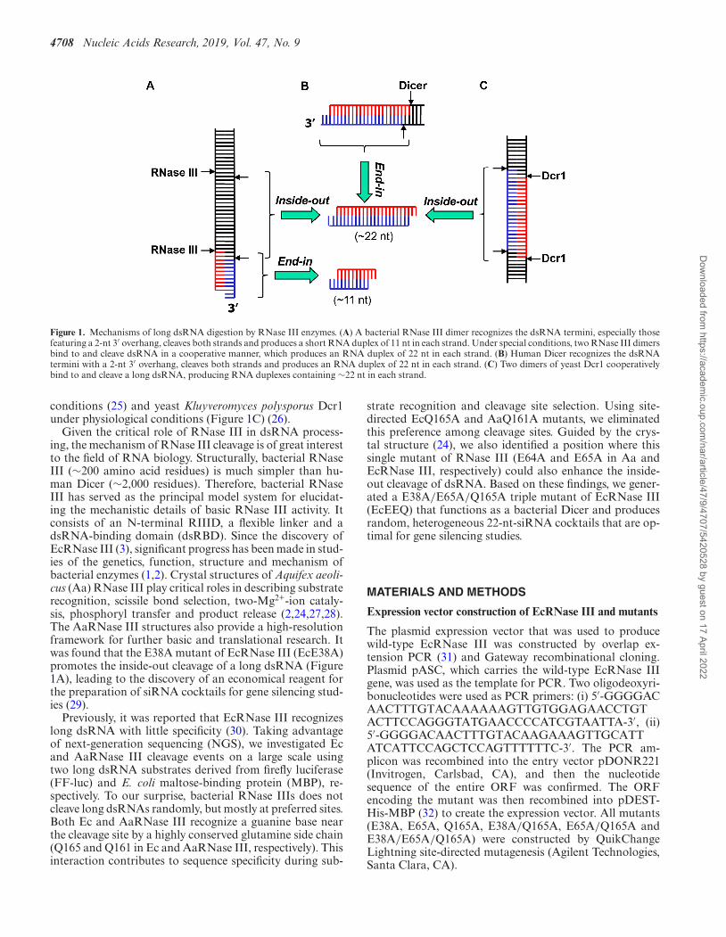
Other representative members of the RNase III family in-clude yeast Rnt1p, human Drosha and human Dicer. Likebacterial RNase III, yeast Rnt1p also functions in the pro-cessing of rRNA (12). In addition, it is involved in the pro-cessing of small non-coding RNA (13) and the degradationof messenger RNA (mRNA) (14). The two human RNaseIII enzymes, Drosha and Dicer, are more specialized. Dicerplays a role in the production of small interfering RNA(siRNA). Along with Drosha, it is also involved in the bio-genesis of microRNA (miRNA), a master gene regulator ineukaryotic cells (15–20). The RNase III nuclease domain(RIIID) dimerizes, which creates a catalytic valley that ac-commodates a dsRNA substrate (21,22). Bacterial RNaseIII and yeast Rnt1p have a single RIIID; these enzymesfunction as homodimers (23,24). Human Dicer and Droshahave two RIIIDs in the same molecule; these enzymes func-tion as monomers (19,20). Both RNase III and Dicer candigest long dsRNA substrates, for which two distinct mech-anisms have been previously described. One mechanism fea-tures an end-in strategy, in which the enzyme recognizesthe termini of a long dsRNA, cleaves both RNA strandsand removes a small RNA duplex upon product release.Both enzymes can adopt this end-in mechanism and suc-cessively remove small RNA duplexes from the dsRNA ter-mini (5,20,24). Whereas RNase III typically measures 11nucleotides (nt) from the termini for cleavage (Figure 1A),Dicer typically measures 22 nt (Figure 1B). A second mech-anism features an inside-out scheme, in which multiple RI-IID dimers bind consecutively to a long dsRNA and cleavethe substrate simultaneously, producing siRNA-like smallRNA duplexes, the length of which is the distance betweenconsecutive active centers. This inside-out mechanism hasbeen observed for EcRNase III (Figure 1A) under special
*To whom correspondence should be addressed. Tel: +1 301 846 5035; Fax: +1 301 846 6073; Email: [email protected] may also be addressed to Shuo Gu. Tel: +1 301 846 5447; Email: [email protected]
Published by Oxford University Press on behalf of Nucleic Acids Research 2019.This work is written by (a) US Government employee(s) and is in the public domain in the US.
Dow
nloaded from https://academ
ic.oup.com/nar/article/47/9/4707/5420528 by guest on 17 April 2022
4708 Nucleic Acids Research, 2019, Vol. 47, No. 9
Figure 1. Mechanisms of long dsRNA digestion by RNase III enzymes. (A) A bacterial RNase III dimer recognizes the dsRNA termini, especially thosefeaturing a 2-nt 3′ overhang, cleaves both strands and produces a short RNA duplex of 11 nt in each strand. Under special conditions, two RNase III dimersbind to and cleave dsRNA in a cooperative manner, which produces an RNA duplex of 22 nt in each strand. (B) Human Dicer recognizes the dsRNAtermini with a 2-nt 3′ overhang, cleaves both strands and produces an RNA duplex of 22 nt in each strand. (C) Two dimers of yeast Dcr1 cooperativelybind to and cleave a long dsRNA, producing RNA duplexes containing ∼22 nt in each strand.
conditions (25) and yeast Kluyveromyces polysporus Dcr1under physiological conditions (Figure 1C) (26).
Given the critical role of RNase III in dsRNA process-ing, the mechanism of RNase III cleavage is of great interestto the field of RNA biology. Structurally, bacterial RNaseIII (∼200 amino acid residues) is much simpler than hu-man Dicer (∼2,000 residues). Therefore, bacterial RNaseIII has served as the principal model system for elucidat-ing the mechanistic details of basic RNase III activity. Itconsists of an N-terminal RIIID, a flexible linker and adsRNA-binding domain (dsRBD). Since the discovery ofEcRNase III (3), significant progress has been made in stud-ies of the genetics, function, structure and mechanism ofbacterial enzymes (1,2). Crystal structures of Aquifex aeoli-cus (Aa) RNase III play critical roles in describing substraterecognition, scissile bond selection, two-Mg2+-ion cataly-sis, phosphoryl transfer and product release (2,24,27,28).The AaRNase III structures also provide a high-resolutionframework for further basic and translational research. Itwas found that the E38A mutant of EcRNase III (EcE38A)promotes the inside-out cleavage of a long dsRNA (Figure1A), leading to the discovery of an economical reagent forthe preparation of siRNA cocktails for gene silencing stud-ies (29).
Previously, it was reported that EcRNase III recognizeslong dsRNA with little specificity (30). Taking advantageof next-generation sequencing (NGS), we investigated Ecand AaRNase III cleavage events on a large scale usingtwo long dsRNA substrates derived from firefly luciferase(FF-luc) and E. coli maltose-binding protein (MBP), re-spectively. To our surprise, bacterial RNase IIIs does notcleave long dsRNAs randomly, but mostly at preferred sites.Both Ec and AaRNase III recognize a guanine base nearthe cleavage site by a highly conserved glutamine side chain(Q165 and Q161 in Ec and AaRNase III, respectively). Thisinteraction contributes to sequence specificity during sub-
strate recognition and cleavage site selection. Using site-directed EcQ165A and AaQ161A mutants, we eliminatedthis preference among cleavage sites. Guided by the crys-tal structure (24), we also identified a position where thissingle mutant of RNase III (E64A and E65A in Aa andEcRNase III, respectively) could also enhance the inside-out cleavage of dsRNA. Based on these findings, we gener-ated a E38A/E65A/Q165A triple mutant of EcRNase III(EcEEQ) that functions as a bacterial Dicer and producesrandom, heterogeneous 22-nt-siRNA cocktails that are op-timal for gene silencing studies.
MATERIALS AND METHODS
Expression vector construction of EcRNase III and mutants
The plasmid expression vector that was used to producewild-type EcRNase III was constructed by overlap ex-tension PCR (31) and Gateway recombinational cloning.Plasmid pASC, which carries the wild-type EcRNase IIIgene, was used as the template for PCR. Two oligodeoxyri-bonucleotides were used as PCR primers: (i) 5′-GGGGACAACTTTGTACAAAAAAGTTGTGGAGAACCTGTACTTCCAGGGTATGAACCCCATCGTAATTA-3′, (ii)5′-GGGGACAACTTTGTACAAGAAAGTTGCATTATCATTCCAGCTCCAGTTTTTTC-3′. The PCR am-plicon was recombined into the entry vector pDONR221(Invitrogen, Carlsbad, CA), and then the nucleotidesequence of the entire ORF was confirmed. The ORFencoding the mutant was then recombined into pDEST-His-MBP (32) to create the expression vector. All mutants(E38A, E65A, Q165A, E38A/Q165A, E65A/Q165A andE38A/E65A/Q165A) were constructed by QuikChangeLightning site-directed mutagenesis (Agilent Technologies,Santa Clara, CA).
Dow
nloaded from https://academ
ic.oup.com/nar/article/47/9/4707/5420528 by guest on 17 April 2022
Nucleic Acids Research, 2019, Vol. 47, No. 9 4709
Expression and purification of EcRNase III proteins
The His6-MBP tagged wild-type EcRNase III and its mu-tants were expressed in E. coli BL21(DE3) CodonPlus-RILcells (ThermoFisher Scientific, Waltham, MA). The cellswere cultivated in Luria-Bertani (LB) broth containing 100�g ml−1 ampicillin, 30 �g ml−1 chloramphenicol and 0.2%glucose to mid-log phase at 37◦C, induced by the addi-tion of isopropyl �-D-1-thiogalactopyranoside (IPTG) toa final concentration of 1 mM, and shaken overnight at18◦C. Harvested cultures were lysed by sonication and, fol-lowing removal of the insoluble cell debris by centrifuga-tion, the supernatant was applied to a HisTrap FF column(GE Healthcare Life Sciences, Pittsburgh, PA). The His6-MBP tag was removed by incubating overnight with 0.5 mgml−1 S219V TEV protease (33) at 4◦C and passing througha second HisTrap FF column. Proteins were further puri-fied with a HiLoad (26/60) Superdex 200 gel filtration col-umn (GE Healthcare Life Sciences) and the quality wasanalyzed by sodium dodecyl sulfate-polyacrylamide elec-trophoresis (SDS-PAGE) and electrospray mass spectrom-etry (LC-ESMS). The final products were concentrated to10 mg ml−1 and stored at −80◦C.
Expression vector construction of AaRNase III and mutants
Protein expression vectors were generated by Gateway™ re-combinational cloning as previously described (34). Briefly,entry clones carried the wild-type RNase III gene or its mu-tants in the pDONR201 (Invitrogen, Carlsbad, CA) back-bone. All mutations were verified by DNA sequencing usingprimers PE367 (5′-TCG CGT TAA CGC TAG CAT GGATCT C-3′) and PE240 (5′-GTA ACA TCA GAG ATT TTGAGA CAC-3′). Entry clones were recombined by GatewayLR reaction with pET-DEST42 (Invitrogen, Carlsbad, CA)to produce expression vectors.
An entry clone encoding the E110Q mutant of AaRNaseIII was constructed for experiments related to this work byoverlap extension PCR (31) using four primers: PE1426,5′-GGG GAC AAG TTT GTA CAA AAA AGC AGGCTT TAA GAA GGA GAT ATA CAT ATG AAA ATGTTG GAG CAA CTT G-3′; PE1427, 5′-CTG CCC AAAGAG CTT GAA ATA CGT CTC CTA-3′; PE1428, 5′-TAG GAG ACG TAT TTC AAG CTC TTT GGG CAG-3′;PE1429, 5′-GGG GAC CAC TTT GTA CAA GAA AGCTGG GTT ATT ATT CTG ATT CCT CCA GTA ATTT-3′. First, two separate PCRs with primer pairs, PE1426-PE1427 and PE1428-PE1429, introduced an E110Q muta-tion and removed the C-terminal hexahistidine tag from anantecedent E110K mutant vector. Then, the products fromthe first two PCRs were used in a third reaction with primersPE1426 and PE1429 to generate full-length product for re-combination into pDONR201 via the Gateway BP reac-tion, which produced entry vector pBA1665. The resultantE110Q entry vector was reverted to the wild-type by site-directed mutagenesis using primers PE2679 (5′-GGA GACGTA TTT GAA GCT CTT TGG GCA GCG G-3′) andPE2680 (5′-CCG CTG CCC AAA GAG CTT CAA ATACGT CTC C-3′) to generate entry clone pBA2518, whichwas subsequently recombined into pET-DEST42 to pro-duce pBA2520, the expression vector for untagged, wild-type AaRNase III.
The E37A mutant was produced by site-directed muta-genesis of the wild-type entry clone pBA2518 with primersPE2677 (5′-CTC AAA AAA AGA ACA CTA CGC AACTCT TGA GTT CCT CGG C-3′) and PE2678 (5′-GCCGAG GAA CTC AAG AGT TGC GTA GTG TTC TTTTTT TGA G-3′) to produce entry clone pBA2519, whichwas recombined with pET-DEST42 to generate the expres-sion vector pBA2521.
The wild-type (pBA2518) and E37A mutant (pBA2519)entry clones were used as templates in QuikChangeLightning site-directed mutagenesis reactions with primersA.a161-A-F (5′-ATA CTT CAG GAG ATC ACT GCAAAA CGA TGG AAG GAA AGA-3′) and A.a161-A-R(5′-TCT TTC CTT CCA TCG TTT TGC AGT GAT CTCCTG AAG TAT-3′) to produce an entry clone encoding theQ161A mutation alone (pDN2749) and one encoding bothE37A and Q161A (pDN2750). pDN2749 and pDN2750were recombined into pET-DEST42 to generate the Q161A(pDN2755) and E37A/Q161A (pDN2754) expression vec-tor.
Single, double and triple mutants were created by addingthe E64A mutation via QuikChange Lightning to the wild-type (pBA2518), Q161A (pDN2749) and E37A/Q161A(pDN2750) entry vectors using primers PE3025 (5′-AGGGAG ATA AAA AGC CTG CCC TTT TGT TGG GGGAA-3′) and PE3026 (5′-TTC CCC CAA CAA AAG GGCAGG CTT TTT ATC TCC CT-3′). Following sequenceverification, entry clones pDN2954 (E64A), pDN2955(E64A/Q161A) and pDN3061 (E37A/E64A/Q161A) wererecombined with pET-DEST42 to generate expression vec-tors pDN2961, pDN2962 and pDN3063, respectively.
Expression and purification of AaRNase III proteins
The AaRNase III protein was overproduced in E. coliand purified as described (28). All expression vectors weretransformed into E. coli strain BL21(DE3)-CodonPlus-RIL (ThermoFisher Scientific). Cells were grown to mid-logphase at 37◦C in LB broth containing 100 �g ml−1 ampi-cillin, 30 �g ml−1 chloramphenicol and 0.2% glucose. Over-production of the recombinant protein was induced withisopropyl �-D-1-thiogalactopyranoside (IPTG) at a finalconcentration of 1 mM for 4 h at 30◦C. The cells were pel-leted by centrifugation and stored at −80◦C.
All procedures were performed at 4–8◦C unless otherwisestated. E. coli cell paste from 6 L of culture, expressing AaR-Nase III, was suspended in ice-cold 50 mM sodium phos-phate (pH 8), 25 mM NaCl buffer (buffer A) containing1 mM benzamidine and Complete™ EDTA-free proteaseinhibitor cocktail (Roche Diagnostics Corporation, Indi-anapolis, IN), and lysed with an APV-1000 homogenizer(SPX Corporation, Charlotte, NC) at 10,000 psi. The ho-mogenate was centrifuged at 30,000 × g for 30 min and thesupernatant was heat-treated at 80◦C for 20 min. Followinga 30-min incubation on ice, insoluble material was removedby centrifugation and the supernatant was filtered through a0.2-�m polyethersulphone membrane. The sample was ap-plied to a HiPrep 16/10 SP FF column (GE Healthcare LifeSciences) equilibrated in buffer A. The column was washedto baseline with buffer A and eluted with a linear gradientof NaCl to 1 M. Fractions containing recombinant protein
Dow
nloaded from https://academ
ic.oup.com/nar/article/47/9/4707/5420528 by guest on 17 April 2022

4710 Nucleic Acids Research, 2019, Vol. 47, No. 9
were pooled and concentrated using an Ultracel® 10 kDaultrafiltration disc (EMD Millipore Corporation, Billerica,MA). The concentrate was fractionated on a HiLoad 26/60Superdex 75 pg column (GE Healthcare Life Sciences) equi-librated in 25 mM Tris–HCl (pH 7.2), 600 mM NaCl buffer.Peak fractions containing AaRNase III were pooled and di-luted with 50 mM Tris–HCl (pH 7.5) buffer to reduce theNaCl concentration to 200 mM. The sample was appliedto a 15 ml AGPoly(I)·Poly(C)™ Type 6 column (AmershamBiosciences/GE Healthcare Life Sciences) equilibrated in50 mM Tris–HCl (pH 7.5), 200 mM NaCl buffer. The col-umn was washed to baseline with equilibration buffer andeluted with a linear gradient of NaCl to 1 M. Fractionscontaining recombinant protein were pooled, concentratedand subjected to a second round of size exclusion chro-matography as described above. The final product was di-luted with 25 mM Tris–HCl (pH 7.2) buffer to reduce theNaCl concentration to 300 mM and concentrated using anUltracel® 10 kDa ultrafiltration disc. Concentrations weredetermined spectrophotometrically using molar extinctioncoefficients derived from the ExPASy ProtParam tool (35).Aliquots were flash-frozen in liquid nitrogen and stored at−80◦C. Purity was judged to be >95% by sodium dodecylsulfate-polyacrylamide gel electrophoresis. The molecularweights were confirmed by LC-ESMS.
Cleavage assays and electrophoresis
As templates for in vitro transcription, pshicheck2 plasmidfor Firefly luciferase and gateway pDest-HisMBP plasmidfor MBP were PCRed by (i) 5′-TAATACGACTCACTATAGGGAGAATGGCCGATGCTAAGAACATT-3′, (ii)5′-AATTAACCCTCACTAAAGGGAGATTACACGGCGATCTTGCCGCC-3′, (iii) 5′-AATTAACCCTCACTAAAGGGAGATTACACGGCGATCTTGCCGCC-3′, (iv)5′-AATTAACCCTCACTAAAGGGAGATTTTTTGTACAAACTTGTGA-3′. The PCR amplicon from (i) and(ii), (iii) and (iv) was purified, respectively. In vitro transcrip-tion was carried out by using Maxi script (ThermoFisherScientific) as per the manufacturer’s instructions.
RNase III cleavage reactions were done using 4 mg ofpurified His-tagged protein and 500 ng of either FF-lucor MBP dsRNA as the substrate in New England BiolabsBuffer 2 [10 mM Tris–HCl (pH 7.9), 10 mM MgCl2, 50 mMNaCl and 1 mM dithiothreitol] and incubated for 30 min at37◦C (EcRNase III) or 30 min at 60◦C (AaRNase III). Forprotein titration reactions, 4 mg of purified His-tagged pro-tein was used for the highest amount and serially diluted.Reactions were stopped by the addition of DNA loadingdye [20 mM EDTA (pH 8.0), 1.25% SDS and 10% glycerol]and incubated for 10 min at 65◦C before loading onto a 20%polyacrylamide/TBE gel (Thermo-Fisher Scientific). Afterelectrophoresis, gels were stained with EtBr and imaged us-ing a ChemiDoc Touch imaging system (Bio-Rad, Hurcules,CA).
Library preparation and next generation sequencing
Prior to library preparation, RNAs from the cleavage as-say were extracted and quantified using a Qubit 2.0 Flu-orometer (ThermoFisher Scientific). Total RNA was also
analyzed with an Agilent Bioanalyzer 2100 (Agilent Tech-nologies) to ensure appropriate RNA quality. Sequencing li-braries were prepared using an Illumina® TruSeq® RNAsample preparation kit according to the manufacturer’s in-structions. The 3′ and 5′ RNA adapters were ligated tothe cleavage products sequentially followed by reverse tran-scription and PCR amplification. The cDNA constructswere recovered from TBE gel electrophoresis and furtherpurified by ethanol precipitation. The cDNA libraries werepooled and sequenced using an Illumina MiSeq ReagentKit v3. Sequencing resulted in approximately 500,000 readsper sample. For each library, we used 100 ng of RNA as astarting material.
Programming and analysis of next-generation sequencingdata
We removed the 3′ adaptor sequences by customized scripts.Reads without the 3′ adaptor sequences or shorter than 6 ntwere discarded. Using Bowtie (version 1.1.1) (36) with thecommand ‘bowtie -v 0 –best –sam,’ we aligned the result-ing reads to the coding sequence of either FF-luc or MBPwithout allowing mismatch. The resulting SAM files wereconverted to BAM files, sorted, indexed by SAMtools (37)and eventually visualized with IGV (38). Length distribu-tion analysis of mapped reads and cleavage sites analysiswere carried out subsequently by customized scripts. Theconsensuses motif of preferred cleavage sites or mappablereads was identified by Weblogo (39).
RESULTS
Bacterial RNase III cleaves long dsRNA at preferred sites
To investigate how bacterial RNase III cleaves dsRNAs, wepurified Ec and AaRNase III proteins and incubated eachwith the dsRNA substrate corresponding to the FF-luc cod-ing sequence (Figure 2A). Consistent with a previous report(29), both enzymes cleaved the 1676-base pair (bp) FF-lucdsRNA into small RNA fragments, ranging from 10 to 15nt in length (Figure 2B).
To further characterize these cleavage events, we con-verted the library of digestion products into DNA, clonedthe fragments and then subjected them to NGS. Four in-dependent cleavage and subsequent NGS experiments wereperformed with EcRNase III and three were performedwith AaRNase III, resulting in 1 026 111 and 509 780 reads,respectively. About 90% of these reads (869,360 and 486,711for Ec and AaRNase III, respectively) could be mappedwithout mismatch to either the sense or antisense sequenceof the FF-luc dsRNA substrate. These small RNA readsare in sufficient abundance to provide an extensive coverageover the relatively small (1676-bp) dsRNA substrate (∼300–500 fold), demonstrating the robustness of this approach foranalyzing in vitro cleavage products of RNase III.
Although the cleavage products cover the entire substratesequence, the distribution of their abundance is far fromeven. Whereas most of the small RNAs were detected fewerthan 100 times, certain reads were found over 10,000 times.Mapping all of these reads back to the substrate, we iden-tified several ‘hot spots’ along the FF-luc sequence, each
Dow
nloaded from https://academ
ic.oup.com/nar/article/47/9/4707/5420528 by guest on 17 April 2022

Nucleic Acids Research, 2019, Vol. 47, No. 9 4711
A B
C
D
Figure 2. Bacterial RNase III cleaves long dsRNA at preferred sites. (A) Schematic representation of the in vitro cleavage experiment. (B) dsRNA of FF-lucsequence was cleaved by either Aquifex aeolicus or Escherichia coli RNase III. The cleavage products were separated on 20% polyacrylamide non-denaturing(native) gels and detected by staining. Synthetic single-stranded RNAs with certain length were used as markers. After next-generation sequencing, readsof cleavage products were mapped back to (C) FF-luc or (D) MBP, respectively. Coverage plots are presented.
of which consists of one or several highly abundant read(s)(Figure 2C). Of note, this coverage distribution pattern ishighly consistent among replicates but slightly different be-tween EcRNase III- and AaRNase III-treated samples, in-dicating that the observed ‘hot spots’ are not a trivial resultof sequencing bias.
Similar results were obtained when we replaced thedsRNA substrate with the coding sequence of MBP (Fig-ure 2D), demonstrating that the bacterial RNase III doesnot cleave dsRNA randomly but rather at preferred sites.More importantly, these results suggest that the underlyingmechanism for the formation of these hot spots might in-volve features that are intrinsic to the long dsRNA sequenceper se.
RNase III recognizes +3G in selecting cleavage sites
Given that the substrates used in our assay are dsRNAswithout internal structure, we reason that the preferredcleavage sites might contain conserved sequence motifs thatare recognized by bacterial RNase III. To test this directly,we first sought to identify hotspots of AaRNase III cleav-age sites, which can be defined from either the 5′ or 3′ endof the cleavage products. Based on a total of 973,422 countsof ends (2 × 486,711 reads), 1,674 cleavage sits were identi-fied along the 1,676-bp substrate, indicating that RNase IIIcleavage was extensive and happened at nearly every pos-sible position. Nonetheless, the frequency varied dramat-ically. While many cleavage sites were supported by onlyfew reads, several cleavage sites generated over 10,000 cor-
Dow
nloaded from https://academ
ic.oup.com/nar/article/47/9/4707/5420528 by guest on 17 April 2022

4712 Nucleic Acids Research, 2019, Vol. 47, No. 9
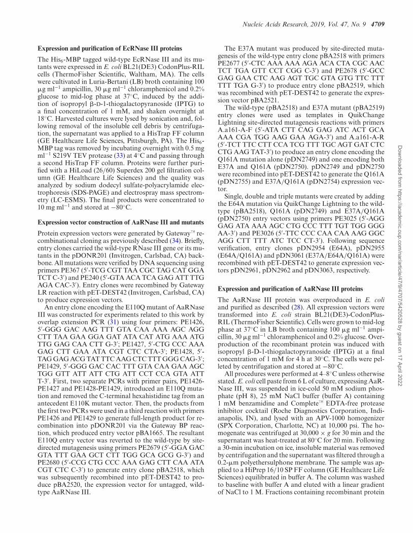
responding reads (Figure 3A). Using five times the averageread as a threshold, we identified 141 highly preferred cleav-age sites (Figure 3A). Overlapping nucleotide sequencesaround these cleavage sites of FF-luc dsRNA revealed thatnucleotide G is enriched in the +3 position (+3G) and C isenriched to a lesser extent in the -6 position (-6C), relativeto the position of cleavage (Figure 3B). The same analysisof MBP dsRNA cleavage site hotspots (102 unique cleav-age site sequences) also generated the +3G/-6C motif (Fig-ure 3B), demonstrating the intrinsic nature of this identifiedmotif.
Consistent with the identified +3G/-6C motif, furtheranalysis revealed that around 30% of all preferred cleavagesites on FF-luc and MBP contain both the +3G and -6C,which is significantly higher than the percentage expectedby chance (1/16 or 6.25%, Figure 3C). However, this stillleaves more than half of the preferred cleavage sites unex-plained. Since RNase III cleaves dsRNA as a dimer and thecleavage sites on the sense and antisense strands of dsRNAare 2 bp apart, the -6C on the sense strand is equivalent tothe +3G on the antisense strand (Figure 3D). This led us tohypothesize that RNase III only recognizes the +3G. Theenrichment of -6C is an indirect result of the other RNaseIII subunit recognizing the +3G on the antisense strand(Figure 3D). In such a scenario, a cleavage site containingeither a +3G or a -6C can be recognized by the RNase IIIdimer. In support of this idea, we found that over 75% ofthose preferred cleavage sites on FF-luc and MBP have ei-ther +3G or -6C and >25% of these have neither (Figure3C).
Parallel analyses of EcRNase III cleavage products iden-tified 120 and 78 preferred cleavage sites on FF-luc andMBP dsRNA, respectively. Consistent with the results ob-tained with AaRNase III, a guanine nucleotide is enrichedin the +3 position (+3G) (Supplementary Figure S1A) andover 70% of those preferred cleavage sites contain either a+3G or a -6C (Supplementary Figure S1B). These resultsstrongly suggest that the preference of +3G in cleavage siteselection is a conserved mechanism for bacterial RNase IIIs.Interestingly, a SS (S stands for G or C) motif is also en-riched at the -1/-2 position among the preferred cleavagesites of EcRNase III (Supplementary Figure S1A), indicat-ing that EcRNase III might recognize these two nucleotidesin addition to the +3G during target site selection.
Taken together, these results indicate that preferredcleavage sites (hot spots) are a result of sequence-specificenzyme–substrate interaction. The sites that can be recog-nized by either subunit of the RNase III dimer are cleavedat a higher frequency.
A conserved glutamine side chain in dsRBD recognizes the+3G near the cleavage site
Based on the crystal structure of AaRNase III in complexwith dsRNA, the first �-helix of the dsRBD is an importantdeterminant for substrate selection. Residues T154, Q157,E158 and Q161 form hydrogen bonds with 2′-hydroxylgroups for specific recognition of RNA substrates (24). Inaddition, the Q161 side chain, which is conserved in bac-terial RNase IIIs (Supplementary Figure S2A), forms twobase-specific hydrogen bonds with the +3G nucleotide, in-
dicating that Q161 is responsible for sequence-specific sub-strate recognition (Figure 4A). Unlike +3G, the -6C basedoes not form any hydrogen bonds with the protein (24).
To test the functional role of Q161 directly, we created theQ161A mutant of AaRNase III (AaQ161A) and performedthe cleavage and subsequent NGS analysis. As expected, theconsensus sequences surrounding cleavage sites are com-pletely abolished for the sequences of both FF-luc and MBP(Figure 4B). In addition, the percentage of cleavage sitescontaining +3G and/or -6C dropped to a level close to thatexpected by chance (Figure 4C). Together, these results in-dicate that the +3G preference observed in wild-type AaR-Nase III cleavage products is not due to a bias introducedduring cloning or sequencing. Instead, it is caused by thespecific interaction between residue Q161 of AaRNase IIIand dsRNA substrate.
While a wealth of structural information is available forAaRNase III (1,2), 3D structure of EcRNase III remainsto be determined. Nonetheless, given that the Q161 sidechain is conserved in bacterial RNase IIIs (Supplemen-tary Figure S2A), we hypothesize that the correspondingresidue Q165 is responsible for the +3G preference in EcR-Nase III cleavage site selection. Indeed, mutating Q165 toan alanine (Q165A) completely obliterated the +3G prefer-ence among preferred cleavage sites (Supplementary FigureS3). Together, these results demonstrate that the specific in-teraction between the conserved glutamine side chain anddsRNA substrate contributes to the sequence preference of+3G during cleavage site selection.
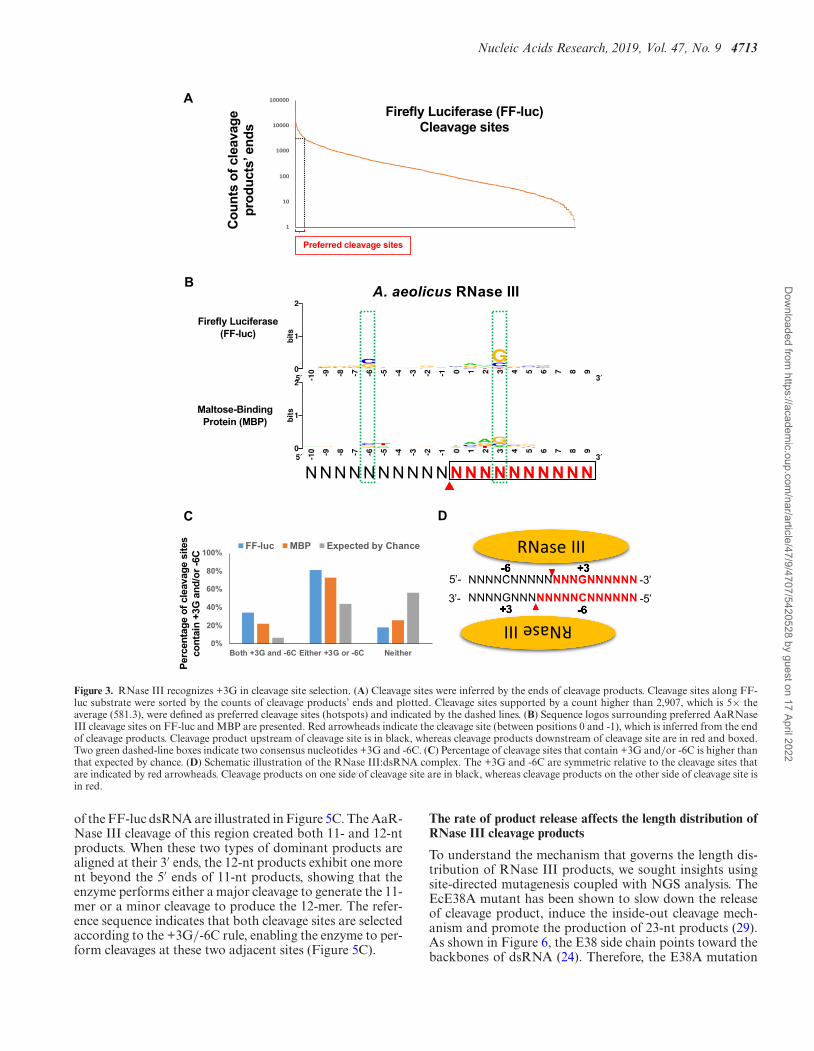
Bacterial RNase III cleaves dsRNA into small RNA duplexeswith distinct patterns of length distribution
Bacterial RNase III cleaves both natural and syntheticdsRNA into small duplex products ranging from 10 to 18bp in length (5,40). Consistently, our NGS results show thatthe Ec and AaRNase IIIs cleave dsRNA into small RNAduplexes within the range of 6–17 and 6–19 bp, respectively(Figure 5A). The dominant length of EcRNase III prod-ucts is 12 nt in a single strand, followed by 9, 11 and 13nt, whereas that of AaRNase III are mainly 11 nt. Interest-ingly, the pattern of product length distribution is species(E. coli or A. aeolicus) dependent but sequence (FF-luc orMBP) independent. Whereas patterns derived from differ-ent sequences by the same RNase III are similar, patternsfrom the same sequence by different RNase IIIs are distinct(Figure 5A), which appears to be determined by certain in-trinsic features of RNase III structures. Of note, the cleavagereactions were performed at different temperatures: 37◦Cfor EcRNase III and 60◦C for AaRNase III because AaR-Nase III is inactive at 37◦C (Supplementary Figure S4). Al-though unlikely, we cannot exclude the possibility that thedistinctions in cleavage pattern between EcRNase III andAaRNase III are partially due to the difference in cleavagereaction temperature.
The NGS analysis provides detailed insights into themechanism of cleavage site selection by bacterial RNase III.Mapping cleavage products with various lengths to the sub-strates yields distinct landscapes of hot spots (Figure 5B),indicating that products with variable lengths were gener-ated by distinctive cleavage events. Details at one ‘hot spot’
Dow
nloaded from https://academ
ic.oup.com/nar/article/47/9/4707/5420528 by guest on 17 April 2022
Nucleic Acids Research, 2019, Vol. 47, No. 9 4713
A
B
C D
Figure 3. RNase III recognizes +3G in cleavage site selection. (A) Cleavage sites were inferred by the ends of cleavage products. Cleavage sites along FF-luc substrate were sorted by the counts of cleavage products’ ends and plotted. Cleavage sites supported by a count higher than 2,907, which is 5× theaverage (581.3), were defined as preferred cleavage sites (hotspots) and indicated by the dashed lines. (B) Sequence logos surrounding preferred AaRNaseIII cleavage sites on FF-luc and MBP are presented. Red arrowheads indicate the cleavage site (between positions 0 and -1), which is inferred from the endof cleavage products. Cleavage product upstream of cleavage site is in black, whereas cleavage products downstream of cleavage site are in red and boxed.Two green dashed-line boxes indicate two consensus nucleotides +3G and -6C. (C) Percentage of cleavage sites that contain +3G and/or -6C is higher thanthat expected by chance. (D) Schematic illustration of the RNase III:dsRNA complex. The +3G and -6C are symmetric relative to the cleavage sites thatare indicated by red arrowheads. Cleavage products on one side of cleavage site are in black, whereas cleavage products on the other side of cleavage site isin red.
of the FF-luc dsRNA are illustrated in Figure 5C. The AaR-Nase III cleavage of this region created both 11- and 12-ntproducts. When these two types of dominant products arealigned at their 3′ ends, the 12-nt products exhibit one morent beyond the 5′ ends of 11-nt products, showing that theenzyme performs either a major cleavage to generate the 11-mer or a minor cleavage to produce the 12-mer. The refer-ence sequence indicates that both cleavage sites are selectedaccording to the +3G/-6C rule, enabling the enzyme to per-form cleavages at these two adjacent sites (Figure 5C).
The rate of product release affects the length distribution ofRNase III cleavage products
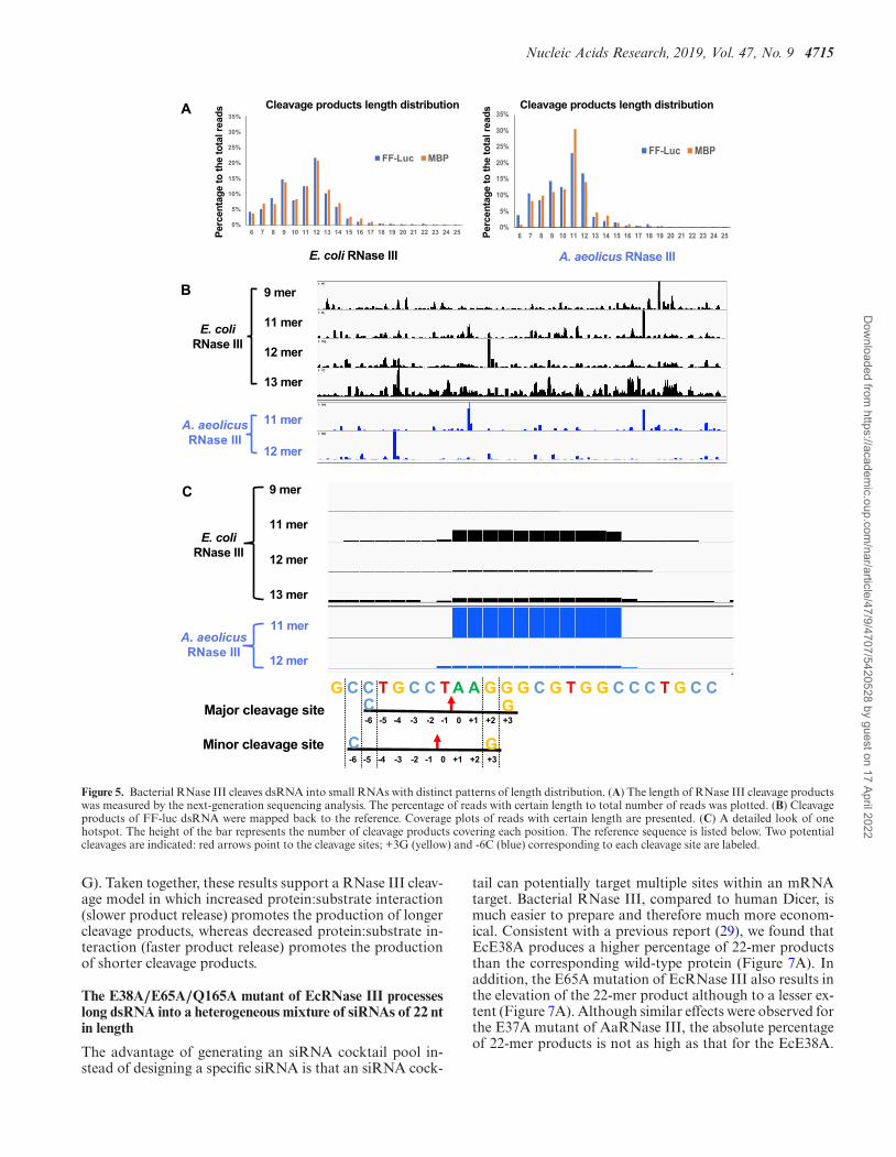
To understand the mechanism that governs the length dis-tribution of RNase III products, we sought insights usingsite-directed mutagenesis coupled with NGS analysis. TheEcE38A mutant has been shown to slow down the releaseof cleavage product, induce the inside-out cleavage mech-anism and promote the production of 23-nt products (29).As shown in Figure 6, the E38 side chain points toward thebackbones of dsRNA (24). Therefore, the E38A mutation
Dow
nloaded from https://academ
ic.oup.com/nar/article/47/9/4707/5420528 by guest on 17 April 2022

4714 Nucleic Acids Research, 2019, Vol. 47, No. 9
A
B
C
Figure 4. Residue Q161 of AaRNase III recognizes the +3G near the cleavage site. (A) On the left: schematic illustration of the crystal structure of AaRNaseIII in complex with dsRNA (PDB entry: 2EZ6); On the right: residue Q161 recognizes the +3G by forming two base-specific hydrogen bonds and onehydrogen bond with the 2′-hydroxyl group. (B) The consensus sequence of cleavage site is abolished when residue 161 was mutated from Q to A. Sequencelogos were created as illustrated in Figure 3. (C) Percentage of highly preferable cleavage sites of the AaRNase III Q161A mutant, with or without +3Gand/or -6C, is similar to that expected by chance.
removes a repulsive protein–RNA interaction and therebystabilizes the protein:dsRNA complex. Similarly, the E65side chain also points toward the dsRNA backbone (Fig-ure 6A). We predict that the E65A mutation should alsostabilize the protein:dsRNA complex and promote the pro-duction of longer RNA products. Unlike E38 and E65, sidechain Q165 forms three hydrogen bonds to the +3G (Fig-ure 4A). The Q165A mutation, while eliminating sequencepreference (Supplementary Figure S3), should destabilizethe protein:dsRNA complex and thereby promote the pro-duction of shorter RNA products. Our working hypothe-sis is that the balance between the stabilization and destabi-lization effects on the protein:dsRNA complex dictates theproduct length distribution.
We expressed and purified the EcE38A, EcE65A andEcQ165A mutant proteins. Using the FF-luc and MBPdsRNAs as substrates, the cleavage products were se-quenced and analyzed. As expected, the percentage of 22-mer product produced by either EcE38A or EcE65A is sig-nificantly increased when compared to the wild-type (Fig-ure 6B and C), whereas the percentage of 22-mer productby EcQ165A is either slightly increased for the MBP or de-creased for the FF-luc dsRNA (Figure 6D). Interestingly,the relative abundances of all cleavage products changed,with a turning point around the 12-nt product. Further-more, the degree of changes is approximately proportionalto the product lengths. The corresponding mutants of theAaRNase III produced similar results (Figure 6E, F and
Dow
nloaded from https://academ
ic.oup.com/nar/article/47/9/4707/5420528 by guest on 17 April 2022
Nucleic Acids Research, 2019, Vol. 47, No. 9 4715
A
B
C
Figure 5. Bacterial RNase III cleaves dsRNA into small RNAs with distinct patterns of length distribution. (A) The length of RNase III cleavage productswas measured by the next-generation sequencing analysis. The percentage of reads with certain length to total number of reads was plotted. (B) Cleavageproducts of FF-luc dsRNA were mapped back to the reference. Coverage plots of reads with certain length are presented. (C) A detailed look of onehotspot. The height of the bar represents the number of cleavage products covering each position. The reference sequence is listed below. Two potentialcleavages are indicated: red arrows point to the cleavage sites; +3G (yellow) and -6C (blue) corresponding to each cleavage site are labeled.
G). Taken together, these results support a RNase III cleav-age model in which increased protein:substrate interaction(slower product release) promotes the production of longercleavage products, whereas decreased protein:substrate in-teraction (faster product release) promotes the productionof shorter cleavage products.
The E38A/E65A/Q165A mutant of EcRNase III processeslong dsRNA into a heterogeneous mixture of siRNAs of 22 ntin length
The advantage of generating an siRNA cocktail pool in-stead of designing a specific siRNA is that an siRNA cock-
tail can potentially target multiple sites within an mRNAtarget. Bacterial RNase III, compared to human Dicer, ismuch easier to prepare and therefore much more econom-ical. Consistent with a previous report (29), we found thatEcE38A produces a higher percentage of 22-mer productsthan the corresponding wild-type protein (Figure 7A). Inaddition, the E65A mutation of EcRNase III also results inthe elevation of the 22-mer product although to a lesser ex-tent (Figure 7A). Although similar effects were observed forthe E37A mutant of AaRNase III, the absolute percentageof 22-mer products is not as high as that for the EcE38A.
Dow
nloaded from https://academ
ic.oup.com/nar/article/47/9/4707/5420528 by guest on 17 April 2022
4716 Nucleic Acids Research, 2019, Vol. 47, No. 9
B E
FC
D G
A
Figure 6. Length distribution of cleavage products is affected by the rate of product release. (A) On the left: cartoon illustration of the crystal structure ofthe AaRNase III dimer in complex with dsRNA (PDB entry 2EZ6); on the right: zoom-in window shows relative positioning of side chains E37, E64 andQ161, of which the counterparts in EcRNase III are E38, E65 and Q165, respectively. (B–G) FF-luc and MBP dsRNAs were cleaved by various RNaseIII mutants. The length of products was compared to that of the wild-type. Fold changes in log scale are plotted against product lengths for EcE38A (B),EcE65A (C), EcQ165A (D), AaE37A (E), AaE64A (F) and AaQ161A (G).
Hence, we focused on the E. coli enzyme for further opti-mization of siRNA production.
To eliminate the substrate specificity governed by the+3G/-6C rule, we introduced the Q165A mutation intoEcE38A and EcE65A, resulting in two double mutants,EcE38A/Q165A and EcE65A/Q165A, respectively. Somedecrease in the production of 22-mer may happen be-
cause the Q165A mutation weakens the protein:dsRNAinteraction. As expected, the two double mutants exhib-ited elevated production of the 22-mer products but notas much as EcE38A (Figure 7B). To maximize the pro-duction of 22-mer products, we generated a triple mutant,EcE38A/E65A/Q165A (EcEEQ). Using the FF-luc andMBP dsRNAs as substrates, we carried out in vitro cleav-
Dow
nloaded from https://academ
ic.oup.com/nar/article/47/9/4707/5420528 by guest on 17 April 2022

Nucleic Acids Research, 2019, Vol. 47, No. 9 4717
A
B
C
Figure 7. Engineered EcRNase III cleaves in vitro transcribed dsRNA into a heterogeneous mixture of siRNAs with a narrow size distribution centeredat 22 nt. (A) The percentage of 22-nt products to the total number of reads was plotted for EcRNase III and its single mutants. (B) The percentage of22-nt products to the total number of reads was plotted for EcRNase III and its double and triple mutants. (C) No consensus sequence of 22-nt cleavageproducts were detected in the triple mutant (EEQ) of EcRNase III.
age and NGS analysis. The results show that the EcEEQmutant protein produces more 22-nt RNA products thanthe two single mutants (Figure 7A), the two double mu-tants and the wild-type enzyme (Figure 7B). In addition,although EcEEQ only abolished the +3G preference dur-ing cleavage site selection, there are in general no consensussequences among the 22-mer products from the triple mu-tant (Figure 7C).
Furthermore, 87% of 22-mer reads mapping to onestrand of FF-Luc dsRNA can pair with a read mapping tothe other strand of FF-luc dsRNA, in a manner that bothends have a 2-nt 3′ overhang (Supplementary Figure S5).
Similar result (89%) was observed for the 22-mer cleavageproducts of MBP dsRNA (Supplementary Figure S5B). Ofnote, these percentages are the upper-bound estimates sincethe number of reads mapped to each strand of a duplex isoften unmatched. Finally, we analyzed the cleavage prod-ucts of EcEEQ at variable time points. Similar to wild-typeEcRNase III, cleavage products were detectable 10 min af-ter reaction initiation, indicating that the activity of EcEEQis comparable to that of wild-type EcRNase III (Supple-mentary Figure S6). Consistent with a previous study (29),the 22-mer products generated by the inside-out mechanismwere apparently protected by EcEEQ from being further
Dow
nloaded from https://academ
ic.oup.com/nar/article/47/9/4707/5420528 by guest on 17 April 2022
4718 Nucleic Acids Research, 2019, Vol. 47, No. 9
processed and accumulated over time (Supplementary Fig-ure S6).
Together, these results demonstrate that the new reagentEcEEQ cleaves long dsRNAs into a mixture of random, het-erogeneous 22-nt products that are ideal for targeting mul-tiple sites on a target mRNA in gene silencing studies.
DISCUSSION
It was previously shown that EcRNase III recognizesdsRNA with little specificity and no specific features are re-quired for cleavage (30), which is in line with the notion thatbacterial RNase III recognizes substrate structure ratherthan sequence (1). Here, we provide compelling evidencedemonstrating that bacterial RNase III does recognize acertain sequence feature around the cleavage site throughits dsRBD. Interestingly, a recent publication indicates thatB. subtilis Mini-III, an RNase III enzyme without dsRBD,can also recognize a specific sequence motif near the cleav-age site (41). Together, these results suggest that the mode ofRNase III–substrate interaction is more complicated thanpreviously believed. Besides structure, the sequence of RNAsubstrates also contributes to cleavage specificity and effi-cacy.
By demonstrating that both AaRNase III and EcR-Nase III recognize the +3G via the conserved glutamineside chain, we provided one of the first structural in-sights into the understanding of how bacterial RNase IIIachieves sequence specificity during cleavage site determi-nation (1,2,42,43). The A-form dsRNA is characterized bya deep and narrow major groove, where access to the basesis hindered, and a wide and shallow minor groove, wherethe edges of the bases are readily accessible. Both func-tional and structural information suggest that the dsRBDperforms a direct readout of RNA sequence in the minorgroove (44). Although the dsRBD of AaRNase III con-tacts the minor groove extensively, only three base-specifichydrogen bonds are formed between RNA and two highlyconserved glutamine side chains, Q157 and Q161, in thefirst �-helix of its dsRBD, known as RNA-binding motif1 (RBM1) (24). The side chain Q157 carboxamide groupforms one hydrogen bond with either a U base (24) or an Abase (27) in the -5 position, indicating that the UA (Supple-mentary Figure S2B) and AU (Supplementary Figure S2C)bp are functionally equivalent. It has been shown that a CGor GC bp substitution strongly inhibits catalysis and thatthe Q157A mutation results in a defect in substrate bind-ing (45). In contrast, the Q161 carboxamide group specif-ically recognizes the +3G base with two hydrogen bonds(Figure 4A), of which the critical role in cleavage site selec-tion is revealed by our in vitro cleavage experiments and theNGS analysis of cleavage products. The cleavage productsare analyzed by gel electrophoresis and the cleavage sitesare mapped by primer extension. Despite many successes,these approaches are low-throughput in nature. The num-ber of cleavage events that can be investigated in each studyis rather limited. Recent advances in sequencing technologyare making it possible to monitor many cleavage events inparallel. In this study, using NGS with dsRNAs of FF-Lucand MBP as substrates, we characterized more than 3,000cleavage events. Each of them was backed up by multiple (up
to several thousand) small RNAs detected by NGS. In ad-dition, the results are highly consistent among multiple re-peats. Together with two recent reports (41,43,46), our studydemonstrates that NGS is a powerful approach in studyingRNase III cleavage.
Both EcRNase III and AaRNase III recognize the +3G.Interestingly, the former, but not the latter, has an addi-tional preference of SS (S = G or C) at the -1 and -2 po-sitions (Supplementary Figure S1A). This difference wouldexplain, at least in part, the distinct patterns of preferredcleavage sites (Figure 5B). While EcRNase III’s Q165 isresponsible for recognizing the +3G during cleavage, theidentity of residues that interact with the -1S/-2S remainselusive. Future high-resolution structures of the complexformed by EcRNase III and its substrate should give addi-tional insights into the underlying mechanisms of the -1S/-2S preference. Finally, our result is highly consistent with arecent study where the transcriptome-wide cleavage sites ofEcRNase III were mapped in vivo (43). The enrichments ofboth +3G and -1S/-2S were observed among sequences sur-rounding the cleavage sites on endogenous substrates, indi-cating that our conclusion can apply to EcRNase III cleav-ages under physiological conditions.
It is intriguing to ask why RNase III has evolved to recog-nize specific sequences near the cleavage sites. It is likely thatjust the dsRNA structure is not sufficient to define cleav-age sites in cellular targets. The +3G as well as -1S/-2Srecognitions may function as additional determinants forthe enzyme to perform cleavage only at desired sites. In fact,it has been recently shown that a set of Dicer-like RNaseIII enzymes in Paramecium cleave dsRNA in a sequence-specific manner to enable precise targeting of transposon-derived IESs, playing important roles in the development ofParamecium (46). RNase III mutants whose sequence pref-erence was abolished will serve as a valuable resource to fur-ther interrogate the biological function of RNase III. Giventhat the interaction between dsRBD and the +3G is ob-served in both EcRNase III and AaRNase III, we speculatethat the same mechanism may be conserved in eukaryoticcounterparts as well, in which it might play important func-tional roles. Future studies should provide more insightsinto the mechanism of cleavage site selection by eukaryoticRNase III enzymes.
We demonstrate that bacterial RNase III cleaves longdsRNA into a set of small RNAs with distinct patterns oflength distribution (Figure 5A). A likely scenario is thatRNase III places its cleavage site approximately N nt awayfrom the end of dsRNA substrate. Whereas the ideal valueof N is 11 as defined by high-resolution structures (24,27),the range of N is ∼6–19 that is dictated by the recognition of+3G (this study). Therefore, it is the combination of struc-tural requirement and sequence recognition that makes the11- and 12-mer the dominant species of cleavage products ofwild-type RNase III. It was reported that the EcE38A pro-motes the inside-out cleavage of a long dsRNA by reducingsubstrate release after cleavage (29). Here, we provide ad-ditional evidence supporting this model by demonstratingthat other mutants that potentially alter substrate releaserate also promote or inhibit the inside-out mode of cleav-age.
Dow
nloaded from https://academ
ic.oup.com/nar/article/47/9/4707/5420528 by guest on 17 April 2022
Nucleic Acids Research, 2019, Vol. 47, No. 9 4719
Finally, our study has important implications for RNAinterference (RNAi) technology, specifically for the prepa-ration of heterogeneous siRNA cocktails. To eliminate thesequence specificity and promote the production efficiency,we have developed the EcEEQ mutant protein. In addi-tion to the 22-nt siRNAs, engineered RNase III also gen-erates 21- and 23-nt duplexes but in much smaller amounts.The size distribution of the siRNA cocktail pool gener-ated by EcRNase III mutants (Supplementary Figure S2)is narrow and centered at 22 nt (Supplementary Figure S7),mimicking the typical size distribution of good miRNA li-braries previously characterized by large-scale profiling ofmiRNAs (47). Therefore, bacterial RNase III is an excel-lent reagent to produce siRNA cocktails and EcEEQ is themost efficient because it not only produces more siRNAsthan any other mutant (Supplementary Figure S7), but alsoproduces random, heterogeneous siRNAs that are ideal forgene silencing studies. It has been two decades since thediscovery of RNAi (48), which was recognized by the No-bel Prize in Physiology or Medicine in 2006. As a Nobel-winning technique, RNAi got its first-ever drug approval(patisiran) by the Food and Drug Administration in Au-gust 2018 (49). This landmark drug shows the promise ofRNAi. Our EcEEQ triple mutant serves as an efficient andeconomical reagent for the discovery of more siRNA drugsthat mute disease-causing genes like the Huntington’s.
DATA AVAILABILITY
The Small RNA-Seq datasets generated in this study areavailable on NCBI GEO under the accession numberGSE120052 (ylsrecyglzifxwt - reviewer token).
SUPPLEMENTARY DATA
Supplementary Data are available at NAR Online.
ACKNOWLEDGEMENTS
We thank Donald Court and Alexander Wlodawer for read-ing the manuscript and discussions, Brian Austin for tech-nical assistance, and the Biophysics Resource in the Struc-tural Biophysics Laboratory, Center for Cancer Research,National Cancer Institute for use of the LC/ESMS instru-ment. The content of this publication does not necessarilyreflect the views or policies of the Department of Health andHuman Services, nor does mention of trade names, com-mercial products or organizations imply endorsement bythe US Government.
FUNDING
Intramural Research Program of the NIH, Center for Can-cer Research, National Cancer Institute. Funding for openaccess charge: National Institutes of Health, Intramural Re-search Program.Conflict of interest statement. None declared.
REFERENCES1. Nicholson,A.W. (2014) Ribonuclease III mechanisms of
double-stranded RNA cleavage. Wiley Interdiscip. Rev. RNA, 5,31–48.
2. Court,D.L., Gan,J., Liang,Y.-H., Shaw,G.X., Tropea,J.E.,Costantino,N., Waugh,D.S. and Ji,X. (2013) RNase III: Genetics andFunction; Structure and mechanism. Annu. Rev. Genet., 47, 405–431.
3. Robertson,H.D., Webster,R.E. and Zinder,N.D. (1968) Purificationand properties of ribonuclease III from Escherichiacoli. J. Biol.Chem., 243, 82–91.
4. Watson,N. and Apirion,D. (1985) Molecular cloning of the gene forthe RNA-processing enzyme RNase III of Escherichiacoli. Proc. Natl.Acad. Sci. U.S.A., 82, 849–853.
5. Court,D.L. (1993) In: Belasco,JG and Brawerman,G (eds). Control ofMessenger RNA Stability. Academic Press, NY, pp. 71–116.
6. Langenberg,W.G., Zhang,L., Court,D.L., Giunchedi,L. and Mitra,A.(1997) Transgenic tobacco plants expressing the bacterial rnc generesist virus infection. Mol. Breed., 3, 391–399.
7. Deltcheva,E., Chylinski,K., Sharma,C.M., Gonzales,K., Chao,Y.,Pirzada,Z.A., Eckert,M.R., Vogel,J. and Charpentier,E. (2011)CRISPR RNA maturation by trans-encoded small RNA and hostfactor RNase III. Nature, 471, 602–607.
8. Stead,M.B., Marshburn,S., Mohanty,B.K., Mitra,J., Pena Castillo,L.,Ray,D., van Bakel,H., Hughes,T.R. and Kushner,S.R. (2011)Analysis of Escherichiacoli RNase E and RNase III activity invivousing tiling microarrays. Nucleic Acids Res., 39, 3188–3203.
9. Durand,S., Gilet,L., Bessieres,P., Nicolas,P. and Condon,C. (2012)Three essential ribonucleases-RNase Y, J1, and III-control theabundance of a majority of Bacillus subtilis mRNAs. PLoS Genet., 8,e1002520.
10. Lasa,I., Toledo-Arana,A., Dobin,A., Villanueva,M., de losMozos,I.R., Vergara-Irigaray,M., Segura,V., Fagegaltier,D.,Penades,J.R., Valle,J. et al. (2011) Genome-wide antisensetranscription drives mRNA processing in bacteria. Proc. Natl. Acad.Sci. U.S.A., 108, 20172–20177.
11. Lioliou,E., Sharma,C.M., Caldelari,I., Helfer,A.C., Fechter,P.,Vandenesch,F., Vogel,J. and Romby,P. (2012) Global regulatoryfunctions of the Staphylococcus aureus endoribonuclease III in geneexpression. PLoS Genet., 8, e1002782.
12. Abou Elela,S., Igel,H. and Ares,M. Jr (1996) RNase III cleaveseukaryotic preribosomal RNA at a U3 snoRNP-dependent site. Cell,85, 115–124.
13. Ghazal,G., Ge,D., Gervais-Bird,J., Gagnon,J. and Abou Elela,S.(2005) Genome-wide prediction and analysis of yeast RNaseIII-dependent snoRNA processing signals. Mol. Cell Biol., 25,2981–2994.
14. Gagnon,J., Lavoie,M., Catala,M., Malenfant,F. and Elela,S.A. (2015)Transcriptome wide annotation of eukaryotic RNase III reactivityand degradation signals. PLoS Genet., 11, e1005000.
15. Lee,R.C., Feinbaum,R.L. and Ambros,V. (1993) The C. elegansheterochronic gene lin-4 encodes small RNAs with antisensecomplementarity to lin-14. Cell, 75, 843–854.
16. Carthew,R.W. (2001) Gene silencing by double-stranded RNA. Curr.Opin. Cell Biol., 13, 244–248.
17. Bernstein,E., Caudy,A.A., Hammond,S.M. and Hannon,G.J. (2001)Role for a bidentate ribonuclease in the initiation step of RNAinterference. Nature, 409, 363–366.
18. Filippov,V., Solovyev,V., Filippova,M. and Gill,S.S. (2000) A noveltype of RNase III family proteins in eukaryotes. Gene, 245, 213–221.
19. Kwon,S.C., Nguyen,T.A., Choi,Y.G., Jo,M.H., Hohng,S., Kim,V.N.and Woo,J.S. (2016) Structure of human DROSHA. Cell, 164, 81–90.
20. Liu,Z., Wang,J., Cheng,H., Ke,X., Sun,L., Zhang,Q.C. andWang,H.W. (2018) Cryo-EM structure of human dicer and itscomplexes with a Pre-miRNA substrate. Cell, 173, 1191–1203.
21. MacRae,I.J., Li,F., Zhou,K., Cande,W.Z. and Doudna,J.A. (2006)Structure of Dicer and mechanistic implications for RNAi. ColdSpring Harb. Symp. Quant. Biol., 71, 73–80.
22. Blaszczyk,J., Tropea,J.E., Bubunenko,M., Routzahn,K.M.,Waugh,D.S., Court,D.L. and Ji,X. (2001) Crystallographic andmodeling studies of RNase III suggest a mechanism fordouble-stranded RNA cleavage. Structure, 9, 1225–1236.
23. Liang,Y.H., Lavoie,M., Comeau,M.A., Abou Elela,S. and Ji,X.(2014) Structure of a eukaryotic RNase III postcleavage complexreveals a double-ruler mechanism for substrate selection. Mol. Cell,54, 431–444.
24. Gan,J., Tropea,J.E., Austin,B.P., Court,D.L., Waugh,D.S. and Ji,X.(2006) Structural insight into the mechanism of double-strandedRNA processing by ribonuclease III. Cell, 124, 355–366.
Dow
nloaded from https://academ
ic.oup.com/nar/article/47/9/4707/5420528 by guest on 17 April 2022
4720 Nucleic Acids Research, 2019, Vol. 47, No. 9
25. Yang,D., Buchholz,F., Huang,Z., Goga,A., Chen,C.Y., Brodsky,F.M.and Bishop,J.M. (2002) Short RNA duplexes produced by hydrolysiswith Escherichiacoli RNase III mediate effective RNA interference inmammalian cells. Proc. Natl. Acad. Sci. U.S.A., 99, 9942–9947.
26. Weinberg,D.E., Nakanishi,K., Patel,D.J. and Bartel,D.P. (2011) Theinside-out mechanism of Dicers from budding yeasts. Cell, 146,262–276.
27. Gan,J., Shaw,G., Tropea,J.E., Waugh,D.S., Court,D.L. and Ji,X.(2008) A stepwise model for double-stranded RNA processing byribonuclease III. Mol. Microbiol., 67, 143–154.
28. Gan,J., Tropea,J.E., Austin,B.P., Court,D.L., Waugh,D.S. and Ji,X.(2005) Intermediate states of ribonuclease III in complex withdouble-stranded RNA. Structure , 13, 1435–1442.
29. Xiao,J., Feehery,C.E., Tzertzinis,G. and Maina,C.V. (2009) E. coliRNase III(E38A) generates discrete-sized products from longdsRNA. RNA, 15, 984–991.
30. Lamontagne,B. and Elela,S.A. (2004) Evaluation of the RNAdeterminants for bacterial and yeast RNase III binding and cleavage.J. Biol. Chem., 279, 2231–2241.
31. Ho,S.N., Hunt,H.D., Horton,R.M., Pullen,J.K. and Pease,L.R.(1989) Site-directed mutagenesis by overlap extension using thepolymerase chain reaction. Gene, 77, 51–59.
32. Nallamsetty,S., Austin,B.P., Penrose,K.J. and Waugh,D.S. (2005)Gateway vectors for the production of combinatorially-taggedHis6-MBP fusion proteins in the cytoplasm and periplasm ofEscherichia coli. Protein Sci., 14, 2964–2971.
33. Kapust,R.B., Tozser,J., Fox,J.D., Anderson,D.E., Cherry,S.,Copeland,T.D. and Waugh,D.S. (2001) Tobacco etch virus protease:mechanism of autolysis and rational design of stable mutants withwild-type catalytic proficiency. Protein. Eng., 14, 993–1000.
34. Blaszczyk,J., Gan,J., Tropea,J.E., Court,D.L., Waugh,D.S. and Ji,X.(2004) Noncatalytic assembly of ribonuclease III withdouble-stranded RNA. Structure, 12, 457–466.
35. Gasteiger,E., Gattiker,A., Hoogland,C., Ivanyi,I., Appel,R.D. andBairoch,A. (2003) ExPASy: The proteomics server for in-depthprotein knowledge and analysis. Nucleic Acids Res., 31, 3784–3788.
36. Langmead,B., Trapnell,C., Pop,M. and Salzberg,S.L. (2009) Ultrafastand memory-efficient alignment of short DNA sequences to thehuman genome. Genome Biol., 10, R25.
37. Li,H., Handsaker,B., Wysoker,A., Fennell,T., Ruan,J., Homer,N.,Marth,G., Abecasis,G., Durbin,R. and Genome Project Data
Processing, S. (2009) The sequence Alignment/Map format andSAMtools. Bioinformatics, 25, 2078–2079.
38. Robinson,J.T., Thorvaldsdottir,H., Winckler,W., Guttman,M.,Lander,E.S., Getz,G. and Mesirov,J.P. (2011) Integrative genomicsviewer. Nat. Biotechnol., 29, 24–26.
39. Crooks,G.E., Hon,G., Chandonia,J.M. and Brenner,S.E. (2004)WebLogo: a sequence logo generator. Genome Res., 14, 1188–1190.
40. Robertson,H.D. and Dunn,J.J. (1975) Ribonucleic acid processingactivity of Escherichiacoli ribonuclease III. J. Biol. Chem., 250,3050–3056.
41. Glow,D., Pianka,D., Sulej,A.A., Kozlowski,L.P., Czarnecka,J.,Chojnowski,G., Skowronek,K.J. and Bujnicki,J.M. (2015)Sequence-specific cleavage of dsRNA by Mini-III RNase. NucleicAcids Res., 43, 2864–2873.
42. Lamontagne,B. and Abou Elela,S. (2004) Evaluation of the RNAdeterminants for bacterial and yeast RNase III binding and cleavage.J. Biol. Chem., 279, 2231–2241.
43. Altuvia,Y., Bar,A., Reiss,N., Karavani,E., Argaman,L. andMargalit,H. (2018) In vivo cleavage rules and target repertoire ofRNase III in Escherichia coli. Nucleic Acids Res., 46, 10380–10394.
44. Masliah,G., Barraud,P. and Allain,F.H. (2013) RNA recognition bydouble-stranded RNA binding domains: a matter of shape andsequence. Cell Mol. Life Sci., 70, 1875–1895.
45. Shi,Z., Nicholson,R.H., Jaggi,R. and Nicholson,A.W. (2011)Characterization of Aquifex aeolicus ribonuclease III and thereactivity epitopes of its pre-ribosomal RNA substrates. NucleicAcids Res., 39, 2756–2768.
46. Hoehener,C., Hug,I. and Nowacki,M. (2018) Dicer-like enzymes withsequence cleavage preferences. Cell, 173, 234–247.
47. Chu,A., Robertson,G., Brooks,D., Mungall,A.J., Birol,I., Coope,R.,Ma,Y., Jones,S. and Marra,M.A. (2016) Large-scale profiling ofmicroRNAs for The Cancer Genome Atlas. Nucleic Acids Res., 44, e3.
48. Fire,A., Xu,S., Montgomery,M.K., Kostas,S.A., Driver,S.E. andMello,C.C. (1998) Potent and specific genetic interference bydouble-stranded RNA in Caenorhabditiselegans. Nature, 391,806–811.
49. Adams,D., Gonzalez-Duarte,A., O’Riordan,W.D., Yang,C.C.,Ueda,M., Kristen,A.V., Tournev,I., Schmidt,H.H., Coelho,T.,Berk,J.L. et al. (2018) Patisiran, an RNAi therapeutic, for hereditarytransthyretin amyloidosis. N. Engl. J. Med., 379, 11–21.
Dow
nloaded from https://academ
ic.oup.com/nar/article/47/9/4707/5420528 by guest on 17 April 2022