Expansion of family Reoviridae to include nine-segmented dsRNA viruses: Isolation and...
12
Expansion of family Reoviridae to include nine-segmented dsRNA viruses: Isolation and characterization of a new virus designated aedes pseudoscutellaris reovirus assigned to a proposed genus (Dinovernavirus) i Houssam Attoui a, * , Fauziah Mohd Jaafar a , Mourad Belhouchet a , Philippe Biagini a , Jean-Franc ¸ois Cantaloube a , Philippe de Micco a , Xavier de Lamballerie a,b a Unite ´ des Virus Emergents EA3292, Laboratoire de Virologie Mole ´culaire, Tropicale et Transfusionnelle, Faculte ´ de Me ´decine de Marseille, 27 Boulevard Jean Moulin, 13005 Marseille cedex 5, France b Maladies virales e ´mergentes et syste `mes d’information UR 034. Institut de Recherche pour le De ´veloppement, Marseille, France Received 13 June 2005; returned to author for revision 1 August 2005; accepted 21 August 2005 Available online 19 September 2005 Abstract Family Reoviridae is known, by definition, to contain dsRNA viruses with 10 –12 genome segments. We report here the characterization of the first member of this family with a nine-segmented genome. This virus was isolated from Aedes pseudoscutellaris mosquito cells and designated aedes pseudoscutellaris reovirus (APRV). Virions are single-shelled with turrets but are non-occluded by contrast to cypoviruses. APRVreplicates in various mosquito cell lines, but not in mice or mammalian cells. Complete sequence analysis showed that APRV is phylogenetically related to cypoviruses, fijiviruses and oryzaviruses. The maximum amino acid identities with cypoviruses, oryzaviruses or fijiviruses in the polymerase, are compatible with values observed between these genera and lower than values within a given genus. This suggests that APRV should be classified within a new genus that we designated Dinovernavirus (sigla from D: Double-stranded, i: insect, nove: nine from the latin ‘‘novem’’, rna: RNA, virus) in family Reoviridae. D 2005 Elsevier Inc. All rights reserved. Keywords: ds-RNA viruses; Reoviridae; Single-shelled turreted virus; Nine-segmented dsRNA genome; Aedes pseudoscutellaris reovirus; Dinovernavirus Introduction Since the original characterization of an orthoreovirus by Sabin in 1959 (Sabin, 1959), numerous other viruses have been identified with genomes composed of multiple (10, 11 or 12) segments of linear double-stranded RNA (dsRNA). These viruses have been isolated from a wide range of host species, including mammals, birds, reptiles, fish, crustaceans, marine protists, insects, ticks, arachnids, plants and fungi. They currently include a total of 75 virus species, which are classified as members of 12 different genera within the family Reoviridae, with a further ¨30 tentative or Funassigned_ species reported to date (Joklik, 1983; Mertens et al., 2005; Brussaard et al., 2004). The genera Coltivirus , Orbivirus and Seadornavirus , include arboviruses that infect animals (Mertens et al., 2005; Attoui et al., 2005a, 2005b). Many of the plant reoviruses (Fijivirus , Oryzavirus and Phytoreovirus ) are also transmitted between their hosts by vector insects (such as leafhoppers) and are therefore plant arboviruses. However, viruses belonging to the other genera (Aquareovirus , Cypovirus , Idnoreovirus , Mycoreovirus , Orthoreovirus and Rotavirus ) are thought to be transmitted horizontally or vertically between individual host organisms, often by an oral/fecal route. Reoviruses are usually regarded as non-enveloped, although some can acquire a transient membrane-envelope during virion 0042-6822/$ - see front matter D 2005 Elsevier Inc. All rights reserved. doi:10.1016/j.virol.2005.08.028 Virology 343 (2005) 212 – 223 www.elsevier.com/locate/yviro i Accession numbers: DQ08276 – DQ08284. * Corresponding author. Fax: +33 4 91 32 44 95. E-mail addresses: [email protected], [email protected] (H. Attoui).
-
Upload
independent -
Category
Documents
-
view
1 -
download
0
Transcript of Expansion of family Reoviridae to include nine-segmented dsRNA viruses: Isolation and...

lsevier.com/locate/yviro
Virology 343 (200
Expansion of family Reoviridae to include nine-segmented dsRNA viruses:
Isolation and characterization of a new virus designated aedes
pseudoscutellaris reovirus assigned to a
proposed genus (Dinovernavirus)i
Houssam Attoui a,*, Fauziah Mohd Jaafar a, Mourad Belhouchet a, Philippe Biagini a,
Jean-Francois Cantaloube a, Philippe de Micco a, Xavier de Lamballerie a,b
a Unite des Virus Emergents EA3292, Laboratoire de Virologie Moleculaire, Tropicale et Transfusionnelle, Faculte de Medecine de Marseille,
27 Boulevard Jean Moulin, 13005 Marseille cedex 5, Franceb Maladies virales emergentes et systemes d’information UR 034. Institut de Recherche pour le Developpement, Marseille, France
Received 13 June 2005; returned to author for revision 1 August 2005; accepted 21 August 2005
Available online 19 September 2005
Abstract
Family Reoviridae is known, by definition, to contain dsRNAviruses with 10–12 genome segments. We report here the characterization of the
first member of this family with a nine-segmented genome. This virus was isolated from Aedes pseudoscutellaris mosquito cells and designated
aedes pseudoscutellaris reovirus (APRV). Virions are single-shelled with turrets but are non-occluded by contrast to cypoviruses. APRV replicates
in various mosquito cell lines, but not in mice or mammalian cells. Complete sequence analysis showed that APRV is phylogenetically related to
cypoviruses, fijiviruses and oryzaviruses. The maximum amino acid identities with cypoviruses, oryzaviruses or fijiviruses in the polymerase, are
compatible with values observed between these genera and lower than values within a given genus. This suggests that APRV should be classified
within a new genus that we designated Dinovernavirus (sigla from D: Double-stranded, i: insect, nove: nine from the latin ‘‘novem’’, rna: RNA,
virus) in family Reoviridae.
D 2005 Elsevier Inc. All rights reserved.
Keywords: ds-RNA viruses; Reoviridae; Single-shelled turreted virus; Nine-segmented dsRNA genome; Aedes pseudoscutellaris reovirus; Dinovernavirus
Introduction
Since the original characterization of an orthoreovirus by
Sabin in 1959 (Sabin, 1959), numerous other viruses have
been identified with genomes composed of multiple (10, 11
or 12) segments of linear double-stranded RNA (dsRNA).
These viruses have been isolated from a wide range of host
species, including mammals, birds, reptiles, fish, crustaceans,
marine protists, insects, ticks, arachnids, plants and fungi.
They currently include a total of 75 virus species, which are
0042-6822/$ - see front matter D 2005 Elsevier Inc. All rights reserved.
doi:10.1016/j.virol.2005.08.028
i Accession numbers: DQ08276–DQ08284.
* Corresponding author. Fax: +33 4 91 32 44 95.
E-mail addresses: [email protected],
[email protected] (H. Attoui).
classified as members of 12 different genera within the
family Reoviridae, with a further ¨30 tentative or
Funassigned_ species reported to date (Joklik, 1983; Mertens
et al., 2005; Brussaard et al., 2004). The genera Coltivirus,
Orbivirus and Seadornavirus, include arboviruses that infect
animals (Mertens et al., 2005; Attoui et al., 2005a, 2005b).
Many of the plant reoviruses (Fijivirus, Oryzavirus and
Phytoreovirus) are also transmitted between their hosts by
vector insects (such as leafhoppers) and are therefore plant
arboviruses. However, viruses belonging to the other genera
(Aquareovirus , Cypovirus , Idnoreovirus , Mycoreovirus ,
Orthoreovirus and Rotavirus) are thought to be transmitted
horizontally or vertically between individual host organisms,
often by an oral/fecal route.
Reoviruses are usually regarded as non-enveloped, although
some can acquire a transient membrane-envelope during virion
5) 212 – 223
www.e
H. Attoui et al. / Virology 343 (2005) 212–223 213
morphogenesis or cell exit (Murphy et al., 1968; Martin et al.,
1998; Mertens et al., 2005; Owens et al., 2004; Mohd Jaafar et
al., 2005a, 2005b). Reovirus particles have icosahedral
symmetry, with a diameter of approximately 60 to 85 nm
and can be subdivided into two groups, the Fturreted_, and Fnon-turreted_ viruses. Core particles of the non-turreted viruses havea protein-bilayer structure, with an inner Fsubcore_ layer that haspseudo T = 2 symmetry (also described as T = 1), surrounded by
a core-surface layer with T = 13 symmetry. This gives the cores
a smooth or bristly surface appearance and a relatively uniform
diameter (e.g. rotaviruses or orbiviruses—Grimes et al., 1998;
Baker et al., 1999; Mertens et al., 2005). In contrast, cores of the
turreted viruses have 12 icosahedrally arranged projections
(called turrets or spikes), situated on the core surface, one at
each of the 5-fold axes (e.g. orthoreoviruses or cypoviruses)
(Baker et al., 1999; Hill et al., 1999; Nibert and Schiff, 2001). In
contrast to most other reoviruses, the cypoviruses lack an outer-
capsid layer, having only a single-shelled, turreted structure.
Reoviruses have previously been isolated from tick cells
derived from wild caught ticks. These include St. Croix River
virus (SCRV, an orbivirus), which was isolated from IDE2 tick
cells, established from eggs of wild caught Ixodes scapulris tick
(Attoui et al., 2001). We report the isolation of a novel single-
shelled dsRNA virus from a continuous mosquito cell line
established in 1974 (AP61 cells from Aedes pseudoscutellaris—
Varma et al., 1974). The virus is identified as aedes pseudoscu-
tellaris reovirus (APRV). The virus, which persistently infects
AP61 cells but shows no visible cytopathic effect. Electron
microscopy of purified APRV-1 showed a well defined, turreted
and single-shelled capsid. The only single-shelled turreted
reoviruses that have previously been identified belong to the
genus Cypovirus, although these normally have a 10-segmented
dsRNA genome. However, unlike APRV-1, which is a non-
occluded virus, the cypoviruses can usually become occluded
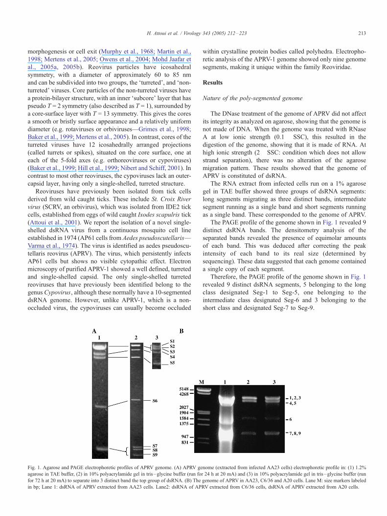
Fig. 1. Agarose and PAGE electrophoretic profiles of APRV genome. (A) APRV ge
agarose in TAE buffer, (2) in 10% polyacrylamide gel in tris–glycine buffer (run fo
for 72 h at 20 mA) to separate into 3 distinct band the top group of dsRNA. (B) The
in bp; Lane 1: dsRNA of APRV extracted from AA23 cells. Lane2: dsRNA of AP
within crystalline protein bodies called polyhedra. Electropho-
retic analysis of the APRV-1 genome showed only nine genome
segments, making it unique within the family Reoviridae.
Results
Nature of the poly-segmented genome
The DNase treatment of the genome of APRV did not affect
its integrity as analyzed on agarose, showing that the genome is
not made of DNA. When the genome was treated with RNase
A at low ionic strength (0.1� SSC), this resulted in the
digestion of the genome, showing that it is made of RNA. At
high ionic strength (2� SSC: condition which does not allow
strand separation), there was no alteration of the agarose
migration pattern. These results showed that the genome of
APRV is constituted of dsRNA.
The RNA extract from infected cells run on a 1% agarose
gel in TAE buffer showed three groups of dsRNA segments:
long segments migrating as three distinct bands, intermediate
segment running as a single band and short segments running
as a single band. These corresponded to the genome of APRV.
The PAGE profile of the genome shown in Fig. 1 revealed 9
distinct dsRNA bands. The densitometry analysis of the
separated bands revealed the presence of equimolar amounts
of each band. This was deduced after correcting the peak
intensity of each band to its real size (determined by
sequencing). These data suggested that each genome contained
a single copy of each segment.
Therefore, the PAGE profile of the genome shown in Fig. 1
revealed 9 distinct dsRNA segments, 5 belonging to the long
class designated Seg-1 to Seg-5, one belonging to the
intermediate class designated Seg-6 and 3 belonging to the
short class and designated Seg-7 to Seg-9.
nome (extracted from infected AA23 cells) electrophoretic profile in: (1) 1.2%
r 24 h at 20 mA) and (3) in 10% polyacrylamide gel in tris–glycine buffer (run
genome of APRV in AA23, C6/36 and A20 cells. Lane M: size markers labeled
RV extracted from C6/36 cells, dsRNA of APRV extracted from A20 cells.

Fig. 2. Electron micrographs of purified APRV particles. (A) Electron
micrograph showing 2 purified APRV particles (right) stained with potassium
phosphotungstate and one core particle of the non-turreted Banna virus (on the
left for the purpose of comparison). (B) Electron micrograph of APRV showing
the turrets extending above the particle surface. The scale bar represents 50 nm
H. Attoui et al. / Virology 343 (2005) 212–223214
In order to rule out the presence of any 10th segment (which
might be in a truncated form), gels were overloaded with
genome extract; however, this failed to identify any band other
than the 9 bands constituting the genome.
Virus replication in mosquito cells
The viral genome was visible in RNA extracts (Fig. 1)
AA23, C6/36 cells and A20 cells that were infected with AP61
lysate.
The extracts of AE and Aw-albus cells incubated with AP61
lysate (or with virus purified from infected AA23) showed no
visible poly-segmented dsRNA profiles. These extracts were
also negative by RT-PCR which showed that these cell lines do
not support the replication of aedes pseudoscutellaris reovirus.
The extract of AP61 cells showed no visible poly-
segmented dsRNA profiles but were positive by RT-PCR
analysis suggesting that these cells contained low amounts of
the virus genome (see below).
Virus presence in AP61 cells from various sources
All three AP61 cell lines were found to contain the aedes
pseudoscutellaris reovirus. PCR using aedes pseudoscutellaris
reovirus segments 6 and 7 specific primers produced amplicons
with the expected sizes. The products were sequenced and their
sequence was found to be 100% identical. Amplicons were
only detectable using a nested PCR assay. The number of viral
genome copies per cell was calculated using FTaqman_quantitative PCR methodology as between 1 and 5.
Effect of 2-aminopurine on virus replication in AP61 cells
The cells treated with 2-aminopurine (2-AM) showed an
increase in the number of viral particles. The quantification by
Taqman has shown that 60–80 APRV genomes could be
estimated per AP61 cell. This represented an increase of over
10-folds in the initial number of viral genome copies estimated
by the same methodology.
Virus replication in mammalian cells and in mice
Extracts of mammalian cells from the three passages in
mice, failed to generate any cDNA products, using the primers
specific to APRV segments 6 and 7. The RNA extracted from
blood of the injected mouse also remained negative for APRV
(by RT-PCR) from day 0 to day 12 post-injection.
Electron microscopy
Purified APRV particles analyzed by TEM showed a
morphology typical of the cores of turreted reoviruses (Fig.
2). In particular, the morphology of APRV was similar to that
observed for cypoviruses (Mertens et al., 2005; Hill et al.,
1999). The mean diameter of the particle is approximately 49–
50 nm with a central part that is 36–37 nm. Turrets were
visible projecting from the particle surface.
.
Physicochemical properties of aedes pseudoscutellaris
reovirus particles
The virion infectivity was not affected by treatment with 1%
deoxycholate but was totally destroyed after 0.1% SDS
treatment. Repeated treatment with Freon 113 or Vertrel XF
did not affect the infectivity.
Freezing the virus at �80 -C or at �20 -C destroyed
infectivity, even in the presence of 50% FBS. However, the
virus is stable at 4 -C for at least 5 months and at room
temperature for up to 3 weeks. Heating to 50–60 -C for 30 min
also abolished infectivity.
Infectivity was retained at pH values between 6 and 8.
Between pH 4 and 5 or between pH 9 and 10, the infectivity is
reduced by a factor of 10�1. The virion morphology was
considerably distorted at pH lower than 5 and virions were
completely disrupted at pH lower than 3.5.
Sequence analysis and comparison
Segments 1 to 9 of APRV were analyzed and their
sequences deposited in GenBank under accession numbers
(DQ08276–DQ08284). The positive strands of all nine
segments of the APRV genome have conserved sequences in
the 5V and 3V non-coding regions (NCR) (5V-AGUUA/UAA
A/
C
A/C———
U/GUUnnn
C/Unn
A/UAGU-3V, where n = any nucle-
otide; Table 1). Comparisons of these conserved termini with
those of member-viruses of the genera Cypovirus, Oryzavirus
and Fijivirus (Table 1) showed that only the first 3 nucleotides

Table 1
Lengths of dsRNA segments 1 to 9, encoded putative proteins, conserved sequences at the 5V and 3V non-coding regions of aedes pseudoscutellaris reovirus
(A) Mass*: calculated theoretical molecular mass. (B) At the 5V end, the first three nucleotides (AGU) are conserved between cypoviruses (CPV), fijiviruses (NLRV:
Nilaparvata lugens reovirus, MRCV: Mal de Rio Cuarto virus, FDV: Fiji disease virus, RBSDV: Rice black streak dwarf virus) and APRV (boxed). At the 3V end,notice that there is no conservation between APRVand the other genera. However, the last two nucleotides in oryzaviruses (GC) seem to be conserved with those of
certain cypoviruses.
H. Attoui et al. / Virology 343 (2005) 212–223 215
in the 5VNCR (AGU) are conserved between APRV, cypoviruses
and NLRV (Nilaparvata lugens reovirus, Fijivirus). The 3Vtermini of APRV differs from those of the cypoviruses, NLRV
and RRSV (Rice ragged stunt virus, Oryzavirus).
The first and last nucleotides of the APRV genome
segments are complementary (A and U), by contrast to those
of cypoviruses and fijiviruses which are not. The mean G + C
content of the APRV genome is 34.4%, compared to 34.8% for
NLRV, 44.7% for RRSV and 43% for the cypoviruses.
Sequence comparison to other viruses, particularly those of
family Reoviridae, has shown significant aa identities (in all
proteins encoded by the genome), with other reoviruses.
Members of the genera Cypovirus, Oryzavirus and Fiji-
virus, which are also turreted, showed the highest aa identities
(19–31%—partial sequences: Fig. 3 and Table 2). However,
weaker aa identities were also detected with other proteins
belonging to members of the genera of other turreted viruses
such as Mycoreovirus, Coltivirus and Orthoreovirus as shown
in Table 2. Interestingly, the protein encoded by segment
8 (VP8) of APRV showed a partial match (aa: 131–260, 27%
identity) to a non-turreted virus protein namely the NSP3 of
rotavirus A (RvA). NSP3 of RvA has been found to be
involved in translational regulation and host cell shut-off
through binding to eIF4G1 factor (Deo et al., 2002; Groft and
Burley, 2002).
It is interesting to note that the VP1 (1189 aa long) of APRV
showed a significant match (22% aa identity, with a probability
of 2e� 6) over a sequence between aa 756 and aa 1173 (a 418
aa-long fragment) to a minor structural protein (accession
number AAC60740) of a ssDNA parvo-like virus isolated from
the silkworm (Bando et al., 1995). Observation of significant
similarities between dsRNA viruses and DNA viruses is rather
unusual. The VP3 of APRV also showed some similarity to the
histidine kinase of Heliothis zea virus (accession number
AAN04301: aa 256–544, identity 18%).
Therefore, putative functions of VP1 to VP9 of APRV were
proposed based on the identified similarities, as shown in Table
2. These comparisons, particularly with cypoviruses, fijiviruses
and oryzaviruses, seem to suggest that there is no equivalent to
polyhedrin gene present in cypoviruses and to confirm the
status of APRV as an authentic nine-segmented virus that is
distinct from the cypoviruses.
Phylogenetic analysis
The virus RNA-dependent RNA polymerase (RdRp) of
member viruses of family Reoviridae is considered as the
only target gene which allows relevant phylogenetic analysis
within this family (Attoui et al., 2002). It has been
suggested as a general rule that a value lower than 30%
aa identity among RdRps distinguishes genera (Attoui et al.,
2002). However, two exceptions to this rule have been
discussed: the case of aquareoviruses and orthoreoviruses
which clearly have a common ancestor with an aa identity
of 42% and that of rotavirus B which is only 22% identical
to other rotaviruses.

Fig. 3. Correspondence between aedes pseudoscutellaris reovirus (APRV) and cypovirus or oryzavirus or fijivirus. Pol: RNA-dependent RNA polymerase.
H.Atto
uiet
al./Viro
logy343(2005)212–223
216

Table 2
Amino acid identities between APRV proteins and other Reoviridae proteins
APRV/function
by similarity
CPV1 (position)
[%]
RRSV (position)
[%]
NLRV (position)
[%]
CpMYRV-1 (position)
[%]
CTFV (position)
[%]
MRV3 (position)
[%]
VP1/possible
cell attachment
VP3 (768–1157)
[23]
P1 (963–1208)
[22]
j1 (44–231)
[25]
VP2a/RdRp VP2 (23–1206)
[26]
P4 (68–1030)
[23]
VP1a (599–624)
[22]
VP1a (62–877)
[20]
VP3/T2 VP1 (6–1302)
[21]
P3 (398–645)
[21]
VP5 (339–698)
[21]
VP4/non-structural NSP5 (795–874)
[28]
P5 (727–789)
[31]
VP5/possible
capping enzyme
VP4 (38–1045)
[22]
P2 (589–1102)
[19]
VP6/possible NTPase VP6 (1–555)
[22]
NS7 (399–591)
[23]
VP7 (399–591)
[23]
VP6 (413–446)
[43]
VP10 (345–491)
[25]
VP7/non-structural VP7 (17–85)
[27]
VP8/possible
translational regulation
VP9/Viral inclusion
bodies
VP9 (169–218)
[31]
The AA identities calculated between putative aedes pseudoscutellaris reovirus proteins and other Reoviridae members are given between brackets. Values between
parenthesis are the positions of aa. CPV1: Cypovirus type 1 (Cypovirus); RRSV: Rice ragged stunt virus (Oryzavirus); NLRV: Nilaparvata lugens reovirus
(Fijivirus); RvA: Rotavirus A (Rotavirus); CpMYRV-1: Mycoreovirus 1 (Mycoreovirus); CTFV: Colorado tick fever virus (Coltivirus); MRV3: Mammalian
orthoreovirus serotype 3 (Orthoreovirus).a Virus RdRp.
H. Attoui et al. / Virology 343 (2005) 212–223 217
The sequence comparison clearly showed that aedes pseudos-
cutellaris reovirus is related to cypoviruses, fijiviruses and
oryzaviruses. AA identity in the RdRp gene is 26, 23 and 22%,
respectively (Table 2). Therefore, the newly identified aedes
pseudoscutellaris reovirus belongs to a new separate genus.
However, RdRp gene analysis suggests a common ancestry
between these 4 groups of viruses. Sequence alignment showed
that the most conserved region among the polymerases lies
within the core domain of the enzyme (located at similar
positions in RdRps of different reoviruses) between aa 634 and
761 of APRV. This region and the whole of the polymerase
sequence were used in phylogenetic comparisons (Figs. 4A and
B, respectively). Evolutionary relationship between aedes
pseudoscutellaris reovirus, cypoviruses, fijiviruses and oryza-
viruses was further confirmed by homology depicted in other
genes (see Fig. 4 and Table 2).
Discussion
Insect and tick cell lines have been used in routine isolation of
arboviruses. Many insect or tick cells have been found to contain
endogenous viruses, infecting the cell line in a persistent manner.
Examples are the Aedes albopictus C6/36 cells (Jousset et al.,
1993) containing a densovirus (ssDNA), the A. pseudoscutel-
laris AP61 cells containing also a densovirus (Gorziglia et al.,
1980) and the Ixodes scapularis IDE2 cells containing an
orbivirus (Attoui et al., 2001) designated St. Croix river virus
(dsRNA). Moreover, C6/36 cells and other mosquito cells were
found to contain a genome of a flavivirus under a DNA form,
integrated in the cell chromosomes (Crochu et al., 2004).
We have identified a dsRNAvirus that infects persistently the
A. pseudoscutellaris AP61 cell line. A. pseudoscutellaris
mosquitoes are known to be confined to the south pacific region
(Marks, 1954). AP61 cells were obtained from 3 independent
sources and the presence of the virus was confirmed in each
culture. This virus was designated aedes pseudoscutellaris
reovirus. The negative staining electronmicroscopy showed that
aedes pseudoscutellaris reovirus is single-shelled with a
morphology similar to that of cypoviruses. However, by contrast
to cypoviruses, aedes pseudoscutellaris reovirus is non-occlud-
ed. The characterization of the dsRNA genome has shown that
aedes pseudoscutellaris reovirus genome is composed of 9
dsRNA segments with a total length 23,355 bp.
We shall discuss hereunder the taxonomic status of aedes
pseudoscutellaris reovirus. Despite the fact that its dsRNA
genome is made of 9 segments (while Reoviridae genomes by
definition contain 10–12 segments), APRV is clearly a
member of family Reoviridae. First, its polymerase is
significantly related to those of cypoviruses, oryzaviruses
and fijiviruses, and homology can also be detected in various
other genes. Second, the morphology of the virus particle is
similar to that of turreted cores of members of the family and
to cypoviruses. Third, other general features including the size
of the virion, the length of the genome and the physico-
chemical properties are similar to those of members of this
family.
To date, insect reoviruses have been classified into two
genera: (i) the occluded viruses belong to genus Cypovirus,
and are characterized by missing capsid protein layers
(Mertens et al., 2005; (ii) the non-occluded viruses have
been recently classified within genus Idnoreovirus and
include 10-segmented viruses from Drosophila melanogaster,
Ceratitis capitata, Hyposoter exiguae and Diadromus pul-
chellus (Mertens et al., 2005). However, aedes pseudoscu-

Fig. 4. Neighbor-joining tree built with the available sequences of RdRps of representative members of family Reoviridae. The accession numbers and abbreviations are those used in Table 2. (A) Tree built with partial
sequences of the polymerases (aa 634 and 761 relative to APRV polymerase). (B) Tree built with full-length sequences of the polymerases.
H.Atto
uiet
al./Viro
logy343(2005)212–223
218

H. Attoui et al. / Virology 343 (2005) 212–223 219
tellaris reovirus cannot be convincingly assigned to either of
these two genera. The purified aedes pseudoscutellaris reovirus
particle morphology is highly similar to that of cypoviruses;
however, APRV particles are found naturally non-occluded by
contrast to cypoviruses. In addition, the genetic relationship
between aedes pseudoscutellaris reovirus and cypoviruses is
that existing between distinct genera and comparable to the
genetic relationship between aedes pseudoscutellaris reovirus
and oryzaviruses or aedes pseudoscutellaris reovirus and
fijiviruses. Aedes pseudoscutellaris reovirus possesses con-
served terminal regions in all 9 segments. The 3VNCR does not
show conservation with that of cypoviruses while the first 3
nucleotides (AGU) in the 5V NCR are conserved among aedes
pseudoscutellaris reovirus and cypoviruses. However, they are
also conserved between aedes pseudoscutellaris reovirus and
fijiviruses (www.iah.bbsrc.ac.uk/dsRNA_virus_proteins/CPV-
RNA-Termin.htm). The G + C content (34.4%) of aedes
pseudoscutellaris reovirus genome is comparable to that of
fijiviruses (34.8%) and lower than those of oryzaviruses
(44.7%) and cypoviruses (43%). Accordingly, genetic anal-
ysis does not provide evidence that aedes pseudoscutellaris
reovirus belongs to genus Cypovirus. Regarding the relation-
ship to idnoreoviruses, these viruses have a different
morphology (triple layered particles) and genetic analysis
shows only 8% aa identity with the RdRp gene of
Diadromus pulchellus reovirus (DPRV, Idnoreovirus, Renault
et al., 2005). Altogether, these arguments exclude the
belonging of aedes pseudoscutellaris reovirus to genus
Idnoreovirus.
Our phylogenetic analysis including representative mem-
bers of the different genera of family Reoviridae does not
suggest that aedes pseudoscutellaris reovirus could be
classified as a member of any of these genera. Accordingly,
data provided in this work make aedes pseudoscutellaris
reovirus the first representative of a new taxonomic group of
dsRNA viruses, suggesting that it should be recognized as
the type species within a distinct new genus of the family
Reoviridae. This genus was designated Dinovernavirus (sigla
from D: Double-stranded, i: insect, nove: nine from the latin
‘‘novem’’ for the 9 segments, rna: RNA, virus). It is the first
genus within family Reoviridae with a reported representative
member having a nine-segmented dsRNA genome, therefore
expanding the definition of the family to viruses with 9–12
dsRNA genome segments. A proposal to recognize the new
genus of nine-segmented viruses within family Reoviridae
was made to the ICTV meeting in San Francisco, July 2005.
It received initial support and was passed for public
consultation.
The discovery of this new genus and its relation to cypo-
viruses, oryzaviruses and fijiviruses, following the reported
relationship between orthoreoviruses and aquareoviruses
(Attoui et al., 2002) or between coltiviruses and mycoviruses
(Suzuki et al., 2004) provides increasing evidence that the
evolutionary origin of the different genera within family
Reoviridae is not polyphyletic. This might represent a starting
point for a possible reconstruction of the evolutionary history of
this family.
Materials and methods
Cell cultures
AA23 (A. albopictus) cells were grown in a mixture of 1
part of Mitsuhashi/Maramorosh insect medium and 1 part of
Schinder’s insect medium supplemented with 10% fetal
bovine serum (FBS) at 28 -C. C6/36 (A. albopictus) cells,
A20 and AE cell lines (both from Aedes aegypti), AP61 (A.
pseudoscutellaris) cells and Aw-albus (Aedes w-albus) were
grown in Leibovitz’s L-15 medium supplemented with 10%
FBS at 28 -C.The AP61 cells were obtained from 3 different sources (Dr.
J.-P. Durand: IMTSSA virology unit, Marseilles, France; Pr.
E.A. Gould: Institute for ecology and hydrology, Oxford, UK;
Pr. R. B. Tesh: center for tropical diseases at UTMB, Texas).
The mammalian cells L929 (mouse fibroblast), BHK-21
(hamster kidney), Vero (monkey kidney), BGM (monkey
kidney), Hep-2 (human cervix cancer) and MRC-5 (human
lung fibroblasts) were grown in Eagle’s Minimum Essential
Medium (EMEM) supplemented with 10% FBS at 37 -C under
CO2.
Virus propagation, purification and electron microscopy
AP61 cells (25 cm2 flask) were grown to confluence and
pelleted at 2000 g. The cells were lysed in deionized water (18
MV) and the lysate was buffered with serum-free L-15
medium. 200 Al of the lysate suspension was incubated for 1
h at 28 -C with a monolayer of AA23 cells (25 cm2 flask) and 7
ml of complete L-15 was subsequently added. The culture was
incubated at 28 -C for 5 days. 200 Al of the supernatant of thisculture was used to re-infect fresh AA23 cultures and 3
passages in AA23 cells were performed. APRV was then
plaque purified from the supernatant of the 3rd passage in
AA23 cells overlaid with seaplaque agarose and plaques were
identified by staining with Trypan blue. The plaque purification
process was repeated 3 times.
The virus was further propagated in 175 cm2 culture
flasks, and infected cells were harvested at day 7 post-
infection (pi). The cells and supernatant were centrifuged
at 3000 � g and the supernatant was recovered. Virus
from supernatant was concentrated by centrifugal ultrafil-
tration using PES ultrafiltration units with 5000 MWCO
(Vivasciences).
Cells were lysed by treatment with 18 MV deionized water
and mixed with the virus from supernatant. The suspension
was made 100 mM Tris–HCl pH 7.5. The mixture was
treated with an equal volume of Vertrel XF, centrifuged at
2000 � g and the supernatant was recovered, layered on top
of a discontinuous Optiprep gradient (10, 20, 30 40 and 50%
optiprep in 100 mM Tris–HCl pH 7.5) and centrifuged for 2
h at 21,0000 � g in an SW41 rotor (Mohd Jaafar et al.,
2005b). The light blue virus band was recovered at the
interface of the 40 and 55% layers.
The virus was adsorbed to formvar/carbon coated grids,
stained with 2% potassium tungstate for 30 s and dried prior to

H. Attoui et al. / Virology 343 (2005) 212–223220
being examined by electron microscopy on a Philips Morgagni
280 transmission electron microscope (TEM).
Assay of aedes pseudoscutellaris reovirus replication in
various mosquito and mammalian cells
The virus was inoculated into insect cell lines, namely C6/
36, A20, AE and Aw-albus. It was also inoculated into the
mammalian cells L929, BHK-21, Vero, BGM, Hep-2 and
MRC-5. For this purpose, 100 Al of the supernatant of APRV-infected AA23 culture was added to the cell monolayers (25
cm2) and incubated at 28 -C for the mosquito cell lines and
at 37 -C (or 33 -C) for the mammalian cell lines for 1 h. The
cells were washed twice with PBS and the culture medium
was added. At day 5 pi, the cells were lysed with deionized
water, mixed with the supernatant and used to re-infect new
cells. This was repeated for 3 passages. In each passage, the
RNA was extracted from cells and supernatant at day 5 pi
(see below) and processed for agarose gel electrophoresis
and/or RT-PCR using specific APRV primers as described
below.
Isolation and purification of nucleic acids
APRV dsRNA was extracted from infected cells using a
commercially available guanidinium isothiocyanate based
procedure (RNA NOW reagent: Biogentex, TX, USA). Briefly,
cells from culture flasks were scraped off and pelleted by
centrifugation at 800 � g at 4 -C for 10 min. The pellet was
dissolved in 500 Al of ‘‘lysis reagent: 6 M guanidinium
isothiocyanate’’ then mixed with 500 Al of the extraction
reagent. Two hundred microliters of chloroform was added and
the mixture was shaken for 1 min, kept for 10 min on ice, then
centrifuged at 12,000 � g for 10 min at 4 -C. The supernatantwas recovered, mixed with 900 Al of 100% isopropanol and
incubated overnight at �20 -C. The RNA was pelleted by
centrifugation at 18,000 � g, for 10 min at 4 -C, washed with
75% ethanol, dried and dissolved in water. The dsRNA was
further purified by precipitating high molecular weight ssRNAs
in 2 M LiCl, as described elsewhere (Attoui et al., 2000).
Nature of the poly-segmented genome
The extracted viral genome was treated with nucleases to
verify the nature of the nucleic acids. Briefly, 2 Ag of the
genome extract was incubated with 100 units of RNase-free
DNase I (Invitrogen) for 1 h at 37 -C to verify if the genome
contained any DNA. Similarly, 2 Ag of the genome was
incubated with 5 Ag/ml of RNase A (Roche) in 2� SSC and in
0.1� SSC medium (1� SSC: 0.015 M sodium citrate and 0.15
M NaCl).
The RNA extract from infected cells was run on a 1%
agarose gel in TAE buffer or 10% polyacrylamide gel in tris–
glycine buffer as described by Laemmli (1970).
The bands separated by PAGE were assayed by densitom-
etry using the BioDocAnalyze software (Biometra) in order to
assay the number of copies per separated band.
RT-PCR of the RNA extract from the tested cell lines
The dsRNA was copied into cDNA using random hexanu-
cleotide primers as previously described (Attoui et al., 2000).
Briefly, the dsRNAwas denatured in 15% DSMO by heating at
100 -C for 1 min and incubated immediately on ice. The reverse
transcription was realized using the Superscript III reverse
transcriptase (Invitrogen) at 42 -C.The resulting cDNA was PCR amplified using primers
designed from segments 6 and 7. The sequences and positions
of these primers are shown in Table 3.
Assay of aedes pseudoscutellaris reovirus replication in mice
Ten-week-old mice were inoculated intraperitoneally with
200 pfu of APRV. Blood was recovered from the caudal vein at
days 0, 3, 6, 9 and 12. The blood samples were frozen at �20
-C. The frozen blood was extracted using the RNA NOW
reagent (Biogentex) as described by the manufacturer and
processed for RT-PCR using specific APRV primers as
described below.
Physicochemical properties of aedes pseudoscutellaris
reovirus particles
The virion infectivity was assayed following treatment with
detergents including 1% sodium deoxycholate as described
earlier (Borden et al., 1971) and 0.1% SDS. Treatment with
Freon 113 or its ozone-friendly replacement 2,3-dihydrodeca-
fluoropentane known as Vertrel XF (Dupont) was also assayed.
The effect of temperature (over a range from �80 -C to + 60
-C) and of pH (pH 3 to pH 10) on the infectivity was assayed.
Quantification of the virus in AP61 cells
The virus titer in AP61 cells was assayed by RT-PCR
Taqman methodology, using the ABI PRISM 7000 Sensor
detection system (ABI). The primers and probes were designed
from the sequence of segment 2. The forward primer was 5V-TCATCACACGCAAATGTTGACTT-3V (position: 2010–
2032), the reverse primer was 5V-GCTATTGCTATACTT-TTCCCGAATTC-3V (position: 2093–2068) and the probe
was 5V-ACTAATAGAAACTGCAGCAACTACATTCAC-
TAA-3V (position: 2034–2066).Cells were counted using Neubauer hemocytometer, lysed
with deionized water and treated with 5 Ag/ml of RNAse A at
37 -C for 30 min to remove ssRNA. Virus RNA was extracted
using RNA NOW as described above, denatured at 100 -C for
4 min and reverse transcribed using random hexaprimers and
Superscript III enzyme. As a standard for the determination of
the copy number, segment 2 was purified on gel and quantified
by UV spectrophotometry.
Effect of 2-aminopurine on virus replication in AP61 cells
2-Aminopurine (2-AM) is known to inhibit the dsRNA-
inducible protein kinase (PKR) in mammalian cells, therefore

Table 3
First round and nested PCR primers designed from the sequences of segments 6 and 7 of APRV
Primer name Specificity Sequence (5VY3V) Position Orientation Amplicon size in bp
Api6S1 Segment 6 GAAGCACATACCATCAGAGATAGC 452–475 Sense 485
Api6R1 Segment 6 GGTCATCAAATACACTGGCAACGC 936–913 Anti-sense
Api6S2 Segment 6 AAGTCGTGTTGATTAATAAGACAGG 619–643 Sense 250
Api6R2 Segment 6 TTCATTACTGGTACGCCATGCACG 868–845 Anti-sense
Api7S1 Segment 7 CCGGAGATACCAGTCTACCAGAC 312–334 Sense 406
Api7R1 Segment 7 TCTCCATATTCCGAGTCTCAACG 717–694 Anti-sense
Api7S2 Segment 7 ATCTAGTACGTCAGCCACCTATGG 384–407 Sense 250
Api7R2 Segment 7 TCCTCGTTTGCGTCGACATATTCC 633–610 Anti-sense
H. Attoui et al. / Virology 343 (2005) 212–223 221
enhancing replication of dsRNA viruses (Strong et al., 1998).
Insect cells, such as D. melanogaster, were found to contain a
homologue to PKR identified as PERK (PKR-like ER kinase)
Table 4
Sequences used in RdRps phylogenetic analysis of aedes pseudoscutellaris reovirus
Species Isolate
Genus Seadornavirus (12 segments)
Banna virus Ch
Kadipiro virus Java-7075
Genus Coltivirus (12 segments)
Colorado tick fever virus Florio
Eyach virus Fr578
Genus Orthoreovirus (10 segments)
Mammalian orthoreovirus Lang strain
Jones strain
Dearing strain
Genus Orbivirus (10 segments)
African horse sickness virus Serotype 9
Bluetongue virus Serotype 2
Serotype 10
Serotype 11
Serotype 13
Serotype 17
Palyam virus Chuzan
St. Croix river virus SCRV
Genus Rotavirus (11 segments)
Rotavirus A Bovine strain U
Simian strain S
Rotavirus B Human/murine
Rotavirus C Porcine Cowde
Genus Aquareovirus (11 segments)
Golden shiner reovirus GSRV
Grass Carp reovirus GCRV-873
Chum salmon reovirus CSRV
Striped bass reovirus SBRV
Genus Fijivirus (10 segments)
Nilaparvata lugens reovirus Izumo strain
Genus Phytoreovirus (10 segments)
Rice dwarf virus Isolate China
Isolate H
Isolate A
Genus Oryzavirus (10 segments)
Rice ragged stunt virus Thai strain
Genus Cypovirus (10 segments)
Bombyx mori cytoplasmic polyhedrosis virus 1 Strain I
Dendrlymus punctatus cytoplasmic polyhedrosis 1 DsCPV-1
Lymantria dispar cytoplasmic polyhedrosis 14 LdCPV-114
Genus Mycoreovirus (11 or 12 segments)
Rosellinia anti-rot virus W370
Cryphonectria parasitica reovirus 9B21
which specifically phoshorylates eIF2a factor (Pomar et al.,
2003). Subconfluent monolayers of AP61 cells were incubat-
ed in the presence of L-15 culture medium with or without 5
: the abbreviations listed are those used in Fig. 4
Abbreviation Accession number
BAV-Ch AF168005
KDV-Ja7075 AF133429
CTFV-Fl AF134529
EYAV-Fr578 AF282467
MRV-1 M24734
MRV-2 M31057
MRV-3 M31058
AHSV-9 U94887
BTV-2 L20508
BTV-10 X12819
BTV-11 L20445
BTV-13 L20446
BTV-17 L20447
CHUV Baa76549
SCRV AF133431
K BoRV-A/UK X55444
A11 SiRV-A/SA11 AF015955
strain IDIR Hu/MuRV-B/IDIR M97203
n strain PoRV-C/Co M74216
GSRV AF403399
GCRV AF260511
CSRV AF418295
SBRV AF450318
NLRV-Iz D49693
RDV-Ch U73201
RDV-H D10222
RDV-A D90198
RRSV-Th U66714
BmCPV-1 AF323782
DsCPV-1 AAN46860
LdCPV-114 AAK73087
RaRV AB102674
CPRV AY277888
H. Attoui et al. / Virology 343 (2005) 212–223222
mM 2-AM. The RNA was extracted at 24, 48 and 72 h post-
addition of 2-AM and analyzed by gel electrophoresis, RT-
PCR and quantitative Taqman RT-PCR.
Cloning of the dsRNA segments
The genome segments of APRV were copied into cDNA,
cloned and sequenced as previously described (Attoui et al.,
2000). Briefly, a defined 3V-amino blocked oligodeoxyribonu-
cleotide was ligated to both of the 3V ends of the dsRNA
segments, using T4 RNA ligase, followed by reverse transcrip-
tion and PCR amplification using a complementary primer.
PCR amplicons were analyzed by agarose gel electrophoresis,
ligated into the PGEM-T cloning vector (Promega) and
transfected into competent XL-blue E. coli. Insert sequences
were determined using M13 universal primers, the D-Rhoda-
mine DNA sequencing kit and an ABI prism 377 sequence
analyzer (Perkin Elmer).
Sequence analysis
All sequence alignments were performed using the Clustal W
software program (Thompson et al., 1994). Phylogenetic ana-
lyses were carried out with the software program MEGA3
(Kumar et al., 2004) using the p-distance determination algo-
rithm. Sequence relatedness is reported as percentage identity.
Analysis of APRV sequence data was performed with a
local BLAST program implemented in the DNATools package
(version 5.2.018, S.W. Rasmussen, Valby data center, Den-
mark), using a database built from all available sequences of
virus-members of family Reoviridae.
The RNA polymerase sequence of APRV was compared
with the sequences of RNA-dependent RNA polymerases of
representative strains of viruses representing the genera of the
family Reoviridae. GenBank accession numbers are provided
in Table 4.
Acknowledgments
The authors wish to thank Nicolas Aldrovandi for the exce-
llent assistance in electron microscopy. This study was sup-
ported by EU Grant ‘‘Reo ID’’ number QLK2-2000-00143. The
‘‘Unite des Virus Emergents’’ is an associated research unit of the
Institut de Recherche pour le Developpement (IRD). This study
was supported in part by the IRD, EFS Alpes-Mediterranee and
EU project VIZIER.
References
Attoui, H., Billoir, F., Cantaloube, J.F., Biagini, P., de Micco, P., de
Lamballerie, X., 2000. Strategies for the sequence determination of viral
dsRNA genomes. J. Virol. Methods 89, 147–158.
Attoui, H., Stirling, J.M., Munderloh, U.G., Billoir, F., Brookes, S.M.,
Burroughs, J.N., de Micco, P., Mertens, P.P.C., de Lamballerie, X., 2001.
Complete sequence characterisation of the genome of the St. Croix River
virus, a new orbivirus isolated from Ixodes scapularis cells. J. Gen. Virol.
82, 795–804.
Attoui, H., Fang, Q., Mohd Jaafar, F., Cantaloube, J.F., Biagini, P., de Micco, P.,
de Lamballerie, X., 2002. Common evolutionary origin of aquareoviruses
and orthoreoviruses revealed by genome characterization of Golden shiner
reovirus, Grass carp reovirus, Striped bass reovirus and golden ide reovirus
(genus Aquareovirus, family Reoviridae). J. Gen. Virol. 83, 1941–1951.
Attoui, H., de Lamballerie, X., Mertens, P.P.C., 2005a. Coltivirus, Reoviridae.
In: Fauquet, C.M., Mayo, M.A., Maniloff, J., Desselberger, U., Ball, L.A.
(Eds.), Virus Taxonomy. Eighth Report of the International Committee on
Taxonomy of Viruses. Elsevier/Academic Press, London, pp. 497–503.
Attoui, H., Mohd Jaafar, F., de Lamballerie, X., Mertens, P.P.C., 2005b.
Seadornavirus, Reoviridae. In: Fauquet, C.M.,. Mayo, M.A, Maniloff, J.,
Desselberger, U., Ball, L.A. (Eds.), Virus Taxonomy. Eighth Report of the
International Committee on Taxonomy of Viruses. Elsevier/Academic
Press, London, pp. 503–510.
Baker, T.S., Olson, N.H., Fuller, S.D., 1999. Adding the third dimension to
virus life cycles: three-dimensional reconstruction of icosahedral viruses
from cryo-electron micrographs. Microbiol. Mol. Biol. Rev. 63, 862–922.
Bando, H., Hayakawa, T., Asano, S., Sahara, K., Nakagaki, M., Iizuka, T.,
1995. Analysis of the genetic information of a DNA segment of a new virus
from silkworm. Arch. Virol. 140, 1147–1155.
Borden, E.C., Shope, R.E., Murphy, F.A., 1971. Physicochemical and
morphological relationships of some arthropode-borne viruses to blue-
tongue virus—A new taxonomic group. Physicochemical and serological
studies. J. Gen. Virol. 13, 261–271.
Brussaard, C.P.D., Noordeloos, A.A.M., Sandaa, R.A., Heldal, M., Bratbak, G.,
2004. Discovery of a dsRNA virus infecting the marine photosynthetic
protist Micromonas pusilla. Virology 319, 280–291.
Crochu, S., Cook, S., Attoui, H., Charrel, R.N., De Chesse, R., Belhouchet, M.,
Lemasson, J.J., de Micco, P., de Lamballerie, X., 2004. Sequences of
flavivirus-related RNA viruses persist in DNA form integrated in the
genome of Aedes spp. mosquitoes. J. Gen. Virol. 85, 1971–1980.
Deo, R.C., Groft, C.M., Rajashankar, K.R., Burley, S.K., 2002. Recognition of
the rotavirus mRNA 3V consensus by an asymmetric NSP3 homodimer. Cell
106, 71–81.
Groft, C.M., Burley, S.K., 2002. Recognition of eIF4G by rotavirus NSP3
reveals a basis for mRNA circularization. Mol. Cell 9, 1273–1283.
Gorziglia, M., Botero, L., Gil, F., Esparza, J., 1980. Preliminary characteriza-
tion of virus-like particles in a mosquito (Aedes pseudoscutellaris) cell line
(Mos. 61). Intervirology 13, 232–240.
Grimes, J.M., Burroughs, J.N., Gouet, P., Diprose, J.M., Malby, R., Zientara, S.,
Mertens, P.P.C., Stuart, D.I., 1998. The atomic structure of bluetongue virus
core. Nature 359, 470–478.
Hill, C.L., Booth, T.F., Prasad, B.V., Grimes, J.M., Mertens, P.P.C., Sutton,
G.C., Stuart, D.I., 1999. The structure of a cypovirus and the functional
organization of dsRNA viruses. Nat. Struct. Biol. 6, 565–568.
Joklik, W.K., 1983. The Reoviridae. Plenum Press, New York.
Jousset, F.X., Barreau, C., Boublik, Y., Cornet, M.A., 1993. Parvo-like virus
persistently infecting a C6/36 clone of Aedes albopictus mosquito cell line
and pathogenic for Aedes aegypti larvae. Virus Res. 29, 99–114.
Kumar, S., Tamura, K., Nei, M., 2004. MEGA3: integrated software for
molecular evolutionary genetics analysis and sequence alignment. Brief.
Bioinform. 5, 150–163.
Laemmli, U.K., 1970. Cleavage of structural proteins during the assembly of
the head of bacteriophage T4. Nature 227, 680–685.
Marks, E.N., 1954. A review of the Aedes scutellaris subgroup with a study of
variation in Aedes pseudoscutellaris (Theobald) (Diptera: Culicidae). Bull.
Br. Mus. (Nat. Hist.), B 3, 347–414.
Martin, L.A., Meyer, A.J., O’Hara, R.S., Fu, H., Knowles, N.J., Mertens, P.P.C.,
1998. Phylogenetic analysis of African horse sickness virus genome
segment 10: sequence variation, virulence characteristics and cell exit.
Arch. Virol., Suppl. 14, 281–293.
Mertens, P.P.C., Attoui, H., Duncan, R., Dermody, T.S., 2005. Reoviridae. In:
Fauquet, C.M., Mayo, M.A., Maniloff, J., Desselberger, U., Ball, L.A.
(Eds.), Virus Taxonomy. Eighth Report of the International Committee on
Taxonomy of Viruses. Elsevier/Academic Press, London, pp. 447–454.
Mohd Jaafar, F., Attoui, H., Bahar, M.W., Siebold, C., Sutton, G., Mertens,
P.P.C., de Micco, P., Stuart, D.I., de Lamballerie, X., 2005a. The structure
and function of the outer coat protein VP9 of Banna virus. Structure 13,
17–29.

H. Attoui et al. / Virology 343 (2005) 212–223 223
Mohd Jaafar, F., Attoui, H., Mertens, P., de Micco, P., de Lamballerie, X., 2005b.
Structural organisation of a human encephalitic isolate of Banna virus (genus
Seadornavirus, family Reoviridae). J. Gen. Virol. 86, 1141–1146.
Murphy, F.A., Coleman, P.H., Harrison, A.K., Gary Jr., G.W., 1968. Colorado
tick fever virus: an electron microscopic study. Virology 35, 28–40.
Nibert, M.L., Schiff, L.A., 2001. Reoviruses and their replication. In: Knipe,
D.M., Howley, P.M., Griffin, D.E., Lamb, R.A., Martin, M.A., Roizman,
B., Straus, S.E. (Eds.), Fields Virology, 4th edR Lippincott Williams and
Wilkins, Philadelphia, PA, pp. 1679–1728.
Owens, R.J., Limn, C., Roy, P., 2004. Role of an arbovirus nonstructural
protein in cellular pathogenesis and virus release. J. Virol. 78, 6649–6656.
Pomar, N., Berlanga, J.J., Campuzano, S., Hernandez, G., Elias, M., de Haro,
C., 2003. Functional characterization of Drosophila melanogaster PERK
eukaryotic initiation factor 2alpha (eIF2alpha) kinase. Eur. J. Biochem. 270,
293–306.
Renault, S., Stasiak, K., Federici, B., Bigot, Y., 2005. Commensal and
mutualistic relationships of reoviruses with their parasitoid wasp hosts.
J. Insect. Physiol. 51, 137–148.
Sabin, A.B., 1959. Reoviruses: a new group of respiratory and enteric
viruses formerly classified as ECHO 10 is described. Science 130,
1387–1389.
Strong, J.E., Coffey, M.C., Tang, D., Sabinin, P., Lee, P.W., 1998. The
molecular basis of viral oncolysis: usurpation of the Ras signaling pathway
by reovirus. EMBO J. 17, 3351–3362.
Suzuki, N., Supyani, S., Maruyama, K., Hillman, B.I., 2004. Complete genome
sequence of Mycoreovirus-1/Cp9B21, a member of a novel genus within
the family Reoviridae, isolated from the chestnut blight fungus Cryphonec-
tria parasitica. J. Gen. Virol. 85, 3437–3448.
Thompson, J.D., Higgins, D.G., Gibson, T.J., 1994. CLUSTAL W: improving
the sensitivity of progressive multiple sequence alignment through
sequence weighting, position-specific gap penalties and weight matrix
choice. Nucleic Acids Res. 22, 4673–4680.
Varma, M.G., Pudney, M., Leake, C.J., 1974. Cell lines from larvae of Aedes
(Stegomyia) malayensis Colless and Aedes (S) pseudoscutellearis (Theo-
bald) and their infection with some arboviruses. Trans. R. Soc. Trop. Med.
Hyg. 68, 374–382.