Characterization of three members of the Arabidopsis carotenoid cleavage dioxygenase family...
12
Characterization of three members of the Arabidopsis carotenoid cleavage dioxygenase family demonstrates the divergent roles of this multifunctional enzyme family Michele E. Auldridge 1,† , Anna Block 1 , Jonathan T. Vogel 1 , Carole Dabney-Smith 1 , Isabelle Mila 2 , Mondher Bouzayen 2 , Maria Magallanes-Lundback 3 , Dean DellaPenna 3 , Donald R. McCarty 1 and and Harry J. Klee 1,* 1 Plant Molecular and Cellular Biology Program, PO Box 110690, University of Florida, Gainesville, FL 32611, USA, 2 UMR990 INRA/INP-ENSA Toulouse, Ge ´ nomique et Biotechnologie des Fruits, Chemin de Borde Rouge, 31326 Castanet- Tolosan, France, and 3 Department of Biochemistry and Molecular Biology, Michigan State University, East Lansing, MI 48824, USA Received 27 May 2005; revised 3 November 2005; accepted 22 November 2005. * For correspondence (fax: þ1 352 846 2063; e-mail [email protected]fl.edu). † Present address: The Jack H. Skirball Center for Chemical Biology and Proteomics, The Salk Institute, 10010 N. Torrey Pines Road, La Jolla, CA 92037, USA. Summary Arabidopsis thaliana has nine genes that constitute a family of putative carotenoid cleavage dioxygenases (CCDs). While five members of the family are believed to be involved in synthesis of the phytohormone abscisic acid, the functions of the other four enzymes are less clear. Recently two of the enzymes, CCD7/MAX3 and CCD8/MAX4, have been implicated in synthesis of a novel apocarotenoid hormone that controls lateral shoot growth. Here, we report on the molecular and genetic interactions between CCD1, CCD7/MAX3 and CCD8/ MAX4. CCD1 distinguishes itself from other reported CCDs as being the only member not targeted to the plastid. Unlike ccd7/max3 and ccd8/max4, both characterized as having highly branched phenotypes, ccd1 loss-of-function mutants are indistinguishable from wild-type plants. Thus, even though CCD1 has similar enzymatic activity to CCD7/MAX3, it does not have a role in synthesis of the lateral shoot growth inhibitor. Rather, it may have a role in synthesis of apocarotenoid flavor and aroma volatiles, especially in maturing seeds where loss of function leads to significantly higher carotenoid levels. Keywords: branching, hormones, seeds. Introduction Apocarotenoids are a class of terpenoid compounds gener- ated by oxidative cleavage of carotenoids. They perform essential biological functions in plants, animals and photo- synthetic bacteria. Examples of biologically active apoca- rotenoids include the phytohormone abscisic acid (ABA) and retinol (vitamin A), an essential component of the visual cycle in animals. Economically, apocarotenoids are valued as colorants and spices. Examples are bixin, an apocarote- noid used as a colorant in foods and cosmetics, and saffron, a spice extracted from the styles of crocus flowers (Winter- halter and Rouseff, 2002). The biosynthesis of apocarote- noids begins with cleavage of a carotenoid molecule at one of its double bonds. In plants, this cleavage can occur non- enzymatically but the predominant mechanism is enzymat- ically via the activity of a family of carotenoid cleavage dioxygenases (Booker et al., 2004; Bouvier et al., 2003; Iuchi et al., 2001; Schwartz et al., 1997, 2001; Sorefan et al., 2003; Tan et al., 1997, 2003). The first gene encoding a carotenoid cleavage dioxyge- nase was isolated from the maize (Zea mays) ABA-deficient viviparous mutant, vp14. VP14 catalyzes the first step in ABA biosynthesis, the cleavage of either of two 9-cis-epoxyca- rotenoids, violoxanthin or neoxanthin at the 11,12 double bond. Because of its preferred substrate, VP14 is called a 9- cis-epoxycarotenoid dioxygenase (NCED) (Schwartz et al., 1997). In the Arabidopsis (A. thaliana) genome, nine putative carotenoid cleavage dioxygenases have been identified based on sequence homology to VP14 (Table 1). Four of the Arabidopsis dioxygenases (NCED2, NCED3, NCED6 and NCED9) have the same activity as VP14 and are designated 982 ª 2006 The Authors Journal compilation ª 2006 Blackwell Publishing Ltd The Plant Journal (2006) 45, 982–993 doi: 10.1111/j.1365-313X.2006.02666.x
-
Upload
independent -
Category
Documents
-
view
0 -
download
0
Transcript of Characterization of three members of the Arabidopsis carotenoid cleavage dioxygenase family...
Characterization of three members of the Arabidopsiscarotenoid cleavage dioxygenase family demonstrates thedivergent roles of this multifunctional enzyme family
Michele E. Auldridge1,†, Anna Block1, Jonathan T. Vogel1, Carole Dabney-Smith1, Isabelle Mila2, Mondher Bouzayen2, Maria
Magallanes-Lundback3, Dean DellaPenna3, Donald R. McCarty1 and and Harry J. Klee1,*
1Plant Molecular and Cellular Biology Program, PO Box 110690, University of Florida, Gainesville, FL 32611, USA,2UMR990 INRA/INP-ENSA Toulouse, Genomique et Biotechnologie des Fruits, Chemin de Borde Rouge, 31326 Castanet-
Tolosan, France, and3Department of Biochemistry and Molecular Biology, Michigan State University, East Lansing, MI 48824, USA
Received 27 May 2005; revised 3 November 2005; accepted 22 November 2005.*For correspondence (fax: þ1 352 846 2063; e-mail [email protected]).†Present address: The Jack H. Skirball Center for Chemical Biology and Proteomics, The Salk Institute, 10010 N. Torrey Pines Road, La Jolla, CA 92037, USA.
Summary
Arabidopsis thaliana has nine genes that constitute a family of putative carotenoid cleavage dioxygenases
(CCDs). While five members of the family are believed to be involved in synthesis of the phytohormone abscisic
acid, the functions of the other four enzymes are less clear. Recently two of the enzymes, CCD7/MAX3 and
CCD8/MAX4, have been implicated in synthesis of a novel apocarotenoid hormone that controls lateral shoot
growth. Here, we report on the molecular and genetic interactions between CCD1, CCD7/MAX3 and CCD8/
MAX4. CCD1 distinguishes itself from other reported CCDs as being the only member not targeted to the
plastid. Unlike ccd7/max3 and ccd8/max4, both characterized as having highly branched phenotypes, ccd1
loss-of-function mutants are indistinguishable from wild-type plants. Thus, even though CCD1 has similar
enzymatic activity to CCD7/MAX3, it does not have a role in synthesis of the lateral shoot growth inhibitor.
Rather, it may have a role in synthesis of apocarotenoid flavor and aroma volatiles, especially in maturing
seeds where loss of function leads to significantly higher carotenoid levels.
Keywords: branching, hormones, seeds.
Introduction
Apocarotenoids are a class of terpenoid compounds gener-
ated by oxidative cleavage of carotenoids. They perform
essential biological functions in plants, animals and photo-
synthetic bacteria. Examples of biologically active apoca-
rotenoids include the phytohormone abscisic acid (ABA) and
retinol (vitamin A), an essential component of the visual
cycle in animals. Economically, apocarotenoids are valued
as colorants and spices. Examples are bixin, an apocarote-
noid used as a colorant in foods and cosmetics, and saffron,
a spice extracted from the styles of crocus flowers (Winter-
halter and Rouseff, 2002). The biosynthesis of apocarote-
noids begins with cleavage of a carotenoid molecule at one
of its double bonds. In plants, this cleavage can occur non-
enzymatically but the predominant mechanism is enzymat-
ically via the activity of a family of carotenoid cleavage
dioxygenases (Booker et al., 2004; Bouvier et al., 2003; Iuchi
et al., 2001; Schwartz et al., 1997, 2001; Sorefan et al., 2003;
Tan et al., 1997, 2003).
The first gene encoding a carotenoid cleavage dioxyge-
nase was isolated from the maize (Zea mays) ABA-deficient
viviparous mutant, vp14. VP14 catalyzes the first step in ABA
biosynthesis, the cleavage of either of two 9-cis-epoxyca-
rotenoids, violoxanthin or neoxanthin at the 11,12 double
bond. Because of its preferred substrate, VP14 is called a 9-
cis-epoxycarotenoid dioxygenase (NCED) (Schwartz et al.,
1997). In the Arabidopsis (A. thaliana) genome, nine putative
carotenoid cleavage dioxygenases have been identified
based on sequence homology to VP14 (Table 1). Four of
the Arabidopsis dioxygenases (NCED2, NCED3, NCED6 and
NCED9) have the same activity as VP14 and are designated
982 ª 2006 The AuthorsJournal compilation ª 2006 Blackwell Publishing Ltd
The Plant Journal (2006) 45, 982–993 doi: 10.1111/j.1365-313X.2006.02666.x

NCEDs. NCED5 has high homology to VP14, though its
activity has not been determined (Iuchi et al., 2001; Tan
et al., 2003). The remaining four proteins diverge from the
NCEDs and have been given the generic designation caro-
tenoid cleavage dioxygenases (CCDs). These include CCD1,
CCD4, CCD7 and CCD8.
CCD1 (Schwartz et al., 2001) and CCD7 (Booker et al.,
2004; Schwartz et al., 2004) have previously been shown to
have activity on multiple carotenoid substrates. They are
specific in their site of cleavage, both having activity at the
9,10 double bond of their substrates. Although CCD1 cleaves
symmetrically at both the 9,10 and 9¢,10¢ bonds, CCD7 has
been reported to cleave only asymmetrically. Thus, CCD1
cleaves b-carotene to produce two C13 products (both b-ionone) and one central C14 dialdehyde (Schwartz et al.,
2001), while CCD7 produces one b-ionone and the C27 10¢-apo-b-carotenal (Schwartz et al., 2004). AtCCD8 has been
shown to further catabolize the C27 apocarotenoid derived
from CCD7 cleavage of b-carotene (Schwartz et al., 2004).
This result suggests that CCD7 and CCD8 may act sequen-
tially in a metabolic pathway. Consistent with this interpret-
ation, loss-of-function mutants of either CCD7 or CCD8 have
a developmentally altered phenotype. The ccd7 and ccd8
mutants are allelic to max3 and max4, respectively. These
mutants, named for more axillary growth, exhibit prolifer-
ation of both leaves and inflorescences emerging from
typically dormant axillary buds (Booker et al., 2004; Sorefan
et al., 2003). Thus, the available data are consistent with a
model in which they act sequentially to produce an inhibitor
of lateral shoot growth. Because the apocarotenoid gener-
ated by CCD7/MAX3 and CCD8/MAX4 action is graft trans-
missible (Booker et al., 2004; Sorefan et al., 2003), it
probably represents a novel class of apocarotenoid phyto-
hormone.
Results
Phenotypes of loss-of-function CCD mutants
In order to address the relative roles of CCD1, CCD7 and
CCD8 in the growth and development of Arabidopsis plants,
we identified loss-of-function mutations in each gene
(Figure 1). For CCD7/MAX3 we used either a previously
described ethylmethane sulfonate (EMS)-induced allele,
max3-9 (Booker et al., 2004), or a T-DNA knockout obtained
from the Salk population,max3-11. This line has an insertion
in the fifth intron of CCD7 and effectively removes the sixth
(and final) exon. Loss-of-function mutants for CCD8 were
isolated from the Wisconsin Knockout Facility (Krysan et al.,
1999) and the SAIL populations (Sessions et al., 2002) and
designated max4-5 and max4-6 to conform to the already
established nomenclature of mutations at the CCD8/MAX4
locus. The T-DNA insertions in the fifth and sixth exons are
present inmax4-5 andmax4-6, respectively. A single loss-of-
function mutant for CCD1 was obtained from the SAIL pop-
ulation of insertional mutants. This line contains a T-DNA
within the sixth intron and was designated ccd1-1. Because
only a single insertional mutant was available for CCD1, the
level of CCD1 transcript was validated by real-time TaqMan
reverse transcriptase (RT)-PCR. The mutant had no detect-
able CCD1 transcript.
Both max4-5 and max4-6 exhibited a significant decrease
in petiole length and were highly branched. The axillary
buds, which are typically delayed in growth in wild-type
plants, grew out to produce leaves and inflorescences. The
max4-5 and max4-6 plants had smaller rosette diameters
Table 1 Comparison of the carotenoid cleavage dioxygenases(CCD) and NCED gene structures and identities to VP14
Familymember Gene ID Intron no.
% Identity toVP14
AtCCD1 At3g63520 13 37AtNCED2 At4g18350 0 64AtNCED3 At3g14440 0 67AtCCD4 At4g19170 0 41AtNCED5 At1g30100 0 66AtNCED6 At3g24220 0 57AtCCD7 At2g44990 5 21AtCCD8 At4g32810 5 26AtNCED9 At1g78390 0 67
Figure 1. Carotenoid cleavage dioxygenases
(CCD) mutant alleles.
Exons are represented by thick lines, introns by
thin lines and insertions by inverted triangles.
The start (ATG) and stop points for translation
are indicated. Insertion events were identified
from the Syngenta (SAIL), the Salk Institute
(SIGNAL) and the University of Wisconsin (UW)
T-DNA insertion collections. All of the insertion
points were verified by sequencing of the
junction fragments. The max3-11 and max4-6
insertions are tandem T-DNA repeats. The
EMS-induced max3-9 allele was previously
reported in Booker et al. (2004).
Arabidopsis CCD family 983
ª 2006 The AuthorsJournal compilation ª 2006 Blackwell Publishing Ltd, The Plant Journal, (2006), 45, 982–993
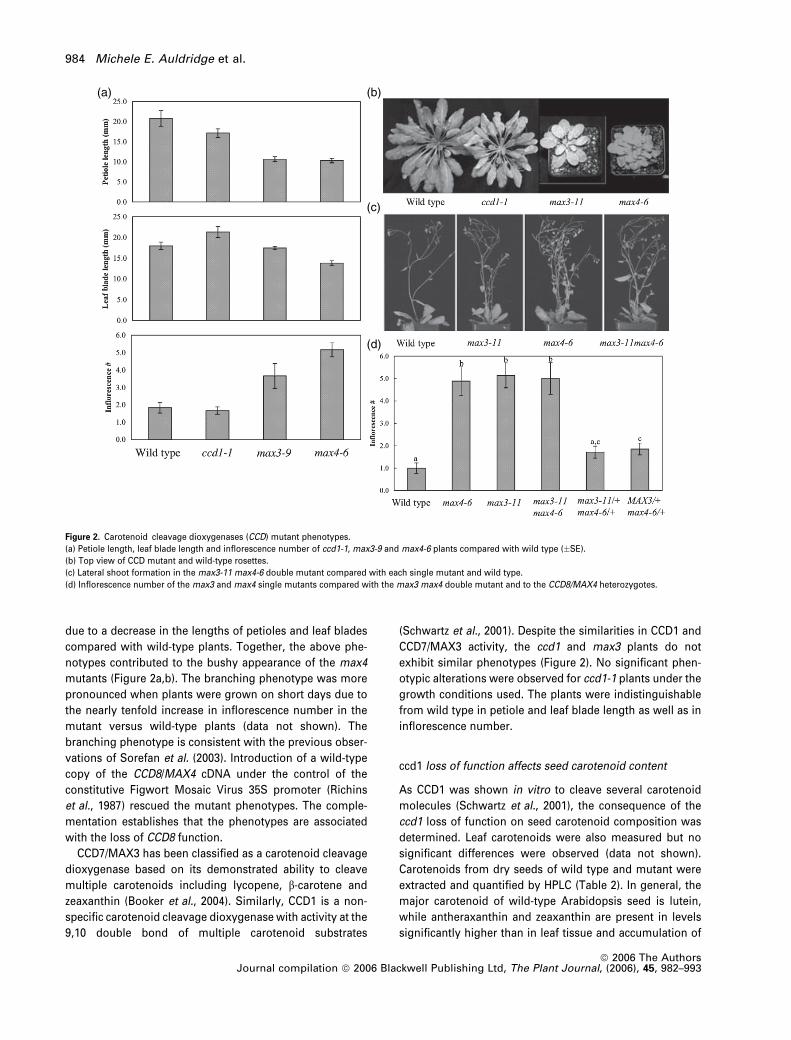
due to a decrease in the lengths of petioles and leaf blades
compared with wild-type plants. Together, the above phe-
notypes contributed to the bushy appearance of the max4
mutants (Figure 2a,b). The branching phenotype was more
pronounced when plants were grown on short days due to
the nearly tenfold increase in inflorescence number in the
mutant versus wild-type plants (data not shown). The
branching phenotype is consistent with the previous obser-
vations of Sorefan et al. (2003). Introduction of a wild-type
copy of the CCD8/MAX4 cDNA under the control of the
constitutive Figwort Mosaic Virus 35S promoter (Richins
et al., 1987) rescued the mutant phenotypes. The comple-
mentation establishes that the phenotypes are associated
with the loss of CCD8 function.
CCD7/MAX3 has been classified as a carotenoid cleavage
dioxygenase based on its demonstrated ability to cleave
multiple carotenoids including lycopene, b-carotene and
zeaxanthin (Booker et al., 2004). Similarly, CCD1 is a non-
specific carotenoid cleavage dioxygenase with activity at the
9,10 double bond of multiple carotenoid substrates
(Schwartz et al., 2001). Despite the similarities in CCD1 and
CCD7/MAX3 activity, the ccd1 and max3 plants do not
exhibit similar phenotypes (Figure 2). No significant phen-
otypic alterations were observed for ccd1-1 plants under the
growth conditions used. The plants were indistinguishable
from wild type in petiole and leaf blade length as well as in
inflorescence number.
ccd1 loss of function affects seed carotenoid content
As CCD1 was shown in vitro to cleave several carotenoid
molecules (Schwartz et al., 2001), the consequence of the
ccd1 loss of function on seed carotenoid composition was
determined. Leaf carotenoids were also measured but no
significant differences were observed (data not shown).
Carotenoids from dry seeds of wild type and mutant were
extracted and quantified by HPLC (Table 2). In general, the
major carotenoid of wild-type Arabidopsis seed is lutein,
while antheraxanthin and zeaxanthin are present in levels
significantly higher than in leaf tissue and accumulation of
(a) (b)
(c)
(d)
Figure 2. Carotenoid cleavage dioxygenases (CCD) mutant phenotypes.
(a) Petiole length, leaf blade length and inflorescence number of ccd1-1, max3-9 and max4-6 plants compared with wild type (�SE).
(b) Top view of CCD mutant and wild-type rosettes.
(c) Lateral shoot formation in the max3-11 max4-6 double mutant compared with each single mutant and wild type.
(d) Inflorescence number of the max3 and max4 single mutants compared with the max3 max4 double mutant and to the CCD8/MAX4 heterozygotes.
984 Michele E. Auldridge et al.
ª 2006 The AuthorsJournal compilation ª 2006 Blackwell Publishing Ltd, The Plant Journal, (2006), 45, 982–993
b-carotene is extremely low. ccd1-1 seed had a 37% increase
in total carotenoids relative to wild type, due mainly to a
greater than twofold and fourfold increase in violaxanthin
and neoxanthin, respectively. This is reflected in a near
doubling of the absolute levels of b-carotene-derived xan-
thophylls in ccd1-1. The lutein level in ccd1-1 was increased
to the greatest extent in absolute terms comparedwith other
carotenoids. To validate that the increased carotenoid con-
tent was due to the loss of CCD1 function, the ccd1-1mutant
was complemented with a 35S-CCD1 cDNA construct. We
identified three independent lines with transgene expres-
sion at or above the level of the endogenous gene. In each
case, the transgene significantly reduced seed carotenoid
content relative to the ccd1-1 parent (P < 0.01; data not
shown). These data indicate that CCD1 metabolizes
carotenoids in seeds and its loss leads to higher seed
carotenoid content.
Double-mutant analysis
The identical phenotypes of the max3 and max4 mutants,
together with the in vitro CCD7/MAX3 and CCD8/MAX4 en-
zyme data (Schwartz et al., 2004) support a model in which
CCD7/MAX3 and CCD8/MAX4 act in a single pathway to
synthesize an inhibitor of bud outgrowth in Arabidopsis. It is
also possible that CCD7/MAX3 and CCD8/MAX4 act in
independent pathways both of which contribute to the pro-
duction of branch-inhibiting compounds. If the latter were
true, then a double max3 max4 mutant may be predicted to
have an additive phenotype compared with either single
mutant. Therefore, a cross between max3-11 and max4-6
was made. The max3-11 max4-6 double mutant was phe-
notypically indistinguishable from either single mutant
(Figure 2c,d) indicating an interaction consistent with both
genes functioning in the same pathway. Interestingly, both
classes of plants genotyped as heterozygous for CCD8/
MAX4 (max3/þ,max4/þ andMAX3/MAX3,max4/þ) showed
a slight increase in inflorescence number compared with
wild type (P ¼ 0.076 and P ¼ 0.029, respectively). This evi-
dence of a quantitative dosage effect of CCD8/MAX4 on
inflorescence number suggests that CCD8/MAX4 activity is a
limiting step in the pathway.
Hormone levels in the max4 mutant
Auxin originating from the apex of the plant promotes apical
dominance (Ward and Leyser, 2004). We were therefore
interested in determining whether auxin levels are altered
in mutants lacking CCD8/MAX4 activity. Free IAA (indole-3-
acetic acid) wasmeasured inmax4-6 rosettes to determine if
altered auxin content was the cause of the branching phe-
notype. Because of the relatedness of CCD8/MAX4 to the
ABA biosynthetic proteins, ABA was also measured. While
ABA has been implicated in bud inhibition (Chatfield et al.,
2000), none of the NCED loss-of-function mutants display a
shoot branching phenotype (B.C. Tan and W.T. Deng, Uni-
versity of Florida, Gainesville, FL, USA, personal communi-
cation). Similar to max3 plants (Booker et al., 2004), no
significant change in either hormone was seen in max4-6.
Levels of IAA were 13.65 � 2.04 ng g)1 fresh weight (FW) in
wild type versus 13.12 � 0.92 ng g)1 FW inmax4-6. Levels of
ABA were 5.69 � 0.30 ng g)1 FW in wild type versus
6.60 � 0.96 ng g)1 FW in max4-6. Therefore, the branching
phenotype seen in the max4 mutants is not a consequence
of altered IAA or ABA levels within Arabidopsis.
CCD expression
The loss-of-function phenotypes are consistent with differ-
ent roles for CCD1 and CCD7/MAX3 in growth and devel-
opment, in contrast to the seemingly identical roles of CCD7/
MAX3 and CCD8/MAX4. In order to further understand CCD
function, expression of CCD1 and CCD8/MAX4 in wild-type
Arabidopsis (ecotype Columbia) was determined. We pre-
viously showed highest expression of CCD7/MAX3 in root
tissue (Booker et al., 2004). Northern blot analysis was not
sensitive enough to detect the low levels of CCD8/MAX4
mRNA. Therefore, transcript abundance was measured by
quantitative real-time RT-PCR, using TaqMan primers and
probes. Figure 3 shows transcript abundance as a percent-
age of mRNA calculated by comparison with a standard
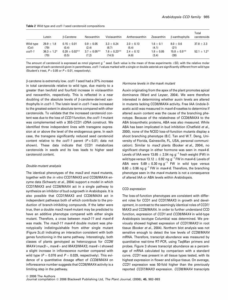
curve. CCD1 was present in all tissue types tested, with its
highest expression in flower and silique tissue. On average,
CCD1 expression was 50-fold higher than the previously
reported CCD7/MAX3 expression. CCD8/MAX4 transcripts
Table 2 Wild type and ccd1-1 seed carotenoid compositions
Lutein b-Carotene Neoxanthin Violaxanthin Antheraxanthin Zeaxanthin b-xanthophyllsTotalcarotenoids
Wild type(Col)
29.9 � 1.8(79)
0.15 � 0.01(0.4)
0.9 � 0.05(2.4)
3.3 � 0.24(8.7)
2.0 � 0.13(5.4)
1.6 � 0.1(4.1)
8.0 � 0.6(21)
37.8 � 2.3
ccd1-1 36.3 � 1.2*(70)
0.28 � 0.02**(0.5)
3.7 � 0.05**(7.2)
7.6 � 0.23**(14.5)
2.4 � 0.12(4.6)
1.8 � 0.05(3.4)
15.8 � 0.5**(30)
52.1 � 1.2*
The amount of carotenoid is expressed as nmol pigment g)1 seed. Each value is the mean of three experiments �SD, with the relative molarpercentage of each carotenoid given in parentheses. ccd1-1 values marked with a single or double asterisk are significantly different fromwild type(Student’s t-test, P < 0.05 or P < 0.01, respectively).
Arabidopsis CCD family 985
ª 2006 The AuthorsJournal compilation ª 2006 Blackwell Publishing Ltd, The Plant Journal, (2006), 45, 982–993
were detected in all tissues tested at levels comparable to
CCD7/MAX3, with the exception of root tissue, where
expression was much higher. CCD8/MAX4 transcript was
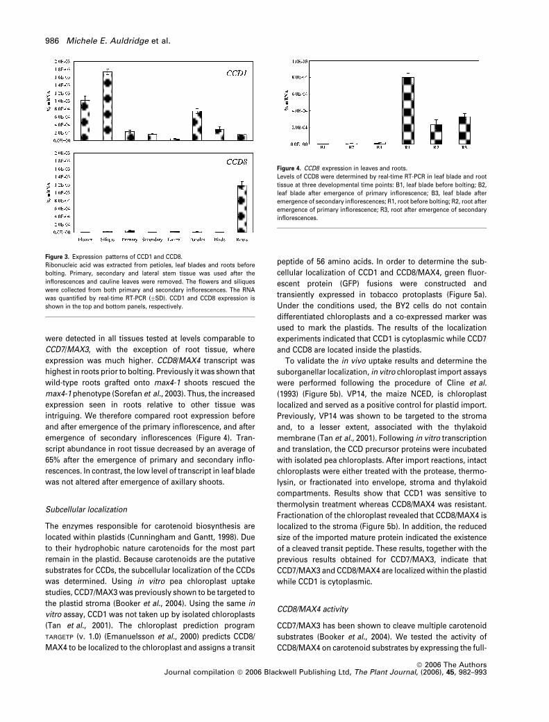
highest in roots prior to bolting. Previously it was shown that
wild-type roots grafted onto max4-1 shoots rescued the
max4-1 phenotype (Sorefan et al., 2003). Thus, the increased
expression seen in roots relative to other tissue was
intriguing. We therefore compared root expression before
and after emergence of the primary inflorescence, and after
emergence of secondary inflorescences (Figure 4). Tran-
script abundance in root tissue decreased by an average of
65% after the emergence of primary and secondary inflo-
rescences. In contrast, the low level of transcript in leaf blade
was not altered after emergence of axillary shoots.
Subcellular localization
The enzymes responsible for carotenoid biosynthesis are
located within plastids (Cunningham and Gantt, 1998). Due
to their hydrophobic nature carotenoids for the most part
remain in the plastid. Because carotenoids are the putative
substrates for CCDs, the subcellular localization of the CCDs
was determined. Using in vitro pea chloroplast uptake
studies, CCD7/MAX3was previously shown to be targeted to
the plastid stroma (Booker et al., 2004). Using the same in
vitro assay, CCD1 was not taken up by isolated chloroplasts
(Tan et al., 2001). The chloroplast prediction program
TARGETP (v. 1.0) (Emanuelsson et al., 2000) predicts CCD8/
MAX4 to be localized to the chloroplast and assigns a transit
peptide of 56 amino acids. In order to determine the sub-
cellular localization of CCD1 and CCD8/MAX4, green fluor-
escent protein (GFP) fusions were constructed and
transiently expressed in tobacco protoplasts (Figure 5a).
Under the conditions used, the BY2 cells do not contain
differentiated chloroplasts and a co-expressed marker was
used to mark the plastids. The results of the localization
experiments indicated that CCD1 is cytoplasmic while CCD7
and CCD8 are located inside the plastids.
To validate the in vivo uptake results and determine the
suborganellar localization, in vitro chloroplast import assays
were performed following the procedure of Cline et al.
(1993) (Figure 5b). VP14, the maize NCED, is chloroplast
localized and served as a positive control for plastid import.
Previously, VP14 was shown to be targeted to the stroma
and, to a lesser extent, associated with the thylakoid
membrane (Tan et al., 2001). Following in vitro transcription
and translation, the CCD precursor proteins were incubated
with isolated pea chloroplasts. After import reactions, intact
chloroplasts were either treated with the protease, thermo-
lysin, or fractionated into envelope, stroma and thylakoid
compartments. Results show that CCD1 was sensitive to
thermolysin treatment whereas CCD8/MAX4 was resistant.
Fractionation of the chloroplast revealed that CCD8/MAX4 is
localized to the stroma (Figure 5b). In addition, the reduced
size of the imported mature protein indicated the existence
of a cleaved transit peptide. These results, together with the
previous results obtained for CCD7/MAX3, indicate that
CCD7/MAX3 and CCD8/MAX4 are localized within the plastid
while CCD1 is cytoplasmic.
CCD8/MAX4 activity
CCD7/MAX3 has been shown to cleave multiple carotenoid
substrates (Booker et al., 2004). We tested the activity of
CCD8/MAX4 on carotenoid substrates by expressing the full-
Figure 3. Expression patterns of CCD1 and CCD8.
Ribonucleic acid was extracted from petioles, leaf blades and roots before
bolting. Primary, secondary and lateral stem tissue was used after the
inflorescences and cauline leaves were removed. The flowers and siliques
were collected from both primary and secondary inflorescences. The RNA
was quantified by real-time RT-PCR (�SD). CCD1 and CCD8 expression is
shown in the top and bottom panels, respectively.
Figure 4. CCD8 expression in leaves and roots.
Levels of CCD8 were determined by real-time RT-PCR in leaf blade and root
tissue at three developmental time points: B1, leaf blade before bolting; B2,
leaf blade after emergence of primary inflorescence; B3, leaf blade after
emergence of secondary inflorescences; R1, root before bolting; R2, root after
emergence of primary inflorescence; R3, root after emergence of secondary
inflorescences.
986 Michele E. Auldridge et al.
ª 2006 The AuthorsJournal compilation ª 2006 Blackwell Publishing Ltd, The Plant Journal, (2006), 45, 982–993
length CCD8/MAX4 with an N-terminal GST (glutathione-
S-transferase) fusion in Escherichia coli strains engineered
to accumulate b-carotene, lycopene or zeaxanthin (Cun-
ningham et al., 1996; Sun et al., 1996). When expression of
CCD8/MAX4 was induced, accumulation of each of these
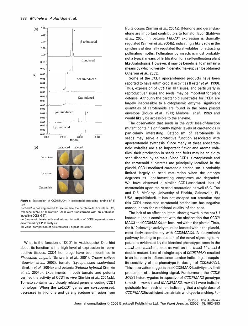
carotenoids was significantly reduced (Figure 6). Despite
this reduction in carotenoid accumulationwewere unable to
identify the cleavage product, either by HPLC analysis of cell
or growth media extracts or by gas chromatography analy-
sis of collected volatiles. It is likely that the products were
further catabolized in E. coli, as has been previously ob-
served by others (von Lintig and Vogt, 2000). Therefore we
were unable to determine the substrate specificities of CCD8/
MAX4. The lack of an accumulating product is likely due to
further metabolism by E. coli. The catabolism of carotenoids
in each of the three E. coli strains does indicate that CCD8/
MAX4 enzyme is capable of acting upon carotenoid sub-
strate(s) in the absence of CCD7/MAX3. Because lycopene
and b-carotene are both intermediates in the synthesis of
zeaxanthin, we cannot rule out that CCD8/MAX4 is only
active on a single carotenoid including, or between, phyto-
ene and lycopene in the biosynthetic pathway. However, the
catabolism of lycopene clearly indicates that the enzyme can
cleave carotenoid substrates other than 10¢-apo-b-carotenal.
Discussion
Carotenoids have critical roles in plant growth, acting as
light-harvesting pigments and preventing oxidative stress
(van den Berg et al., 2000). They are also important sub-
strates for a class of carotenoid cleavage dioxygenases
that synthesize phytohormone apocarotenoids such as
ABA. Recent compelling data support the existence of a
novel, so far unidentified apocarotenoid hormone con-
trolling leaf and lateral shoot growth (Booker et al., 2004;
Sorefan et al., 2003). This apocarotenoid is generated at
least in part by CCD7/MAX3 and/or CCD8/MAX4. Previous
experiments indicated that CCD1 and CCD7/MAX3 have
similar activities, the former oxidatively cleaving multiple
carotenoids symmetrically at the 9,10 and 9¢,10¢ positionsand the latter only at the 9,10 position (Booker et al.,
2004; Schwartz et al., 2001, 2004). Here we have shown
that while loss of CCD7/MAX3 has a major impact on
growth and development, loss of CCD1 has no obvious
morphological effect. While CCD1 is highly expressed in
multiple tissues, the protein is cytoplasmic. In contrast,
CCD7/MAX3 is expressed at much lower levels and the
encoded protein is localized to the inside of plastids, the
predominant sites of carotenoid accumulation.
(a) (b)
Figure 5. Carotenoid cleavage dioxygenases subcellular localization.
(a) CCD–GFP fusions transiently expressed in tobacco BY2 protoplasts (left) compared with a chloroplast marker (center). The merged images are shown on the
right.
(b) Import of in vitro transcribed and translated CCD precursor proteins (pP) into pea chloroplasts were determined and compared with the previously studied
chloroplast imported proteins VP14. Following import, chloroplasts were treated with thermolysin (þTh). Chloroplasts were further fractionated to determine
suborganellar localization of CCD8 to the envelope (E), stroma (S) or thylakoid (T).
Arabidopsis CCD family 987
ª 2006 The AuthorsJournal compilation ª 2006 Blackwell Publishing Ltd, The Plant Journal, (2006), 45, 982–993
What is the function of CCD1 in Arabidopsis? One hint
about its function is the high level of expression in repro-
ductive tissues. CCD1 homologs have been identified in
Phaseolus vulgaris (Schwartz et al., 2001), Crocus sativus
(Bouvier et al., 2003), tomato (Lycopersicon esulentum)
(Simkin et al., 2004a) and petunia (Petunia hybrida) (Simkin
et al., 2004b). Experiments in both tomato and petunia
verified the activity of CCD1 in vivo (Simkin et al., 2004a,b).
Tomato contains two closely related genes encoding CCD1
homologs. When the LeCCD1 genes are co-suppressed,
decreases in b-ionone and geranylacetone emission from
fruits occurs (Simkin et al., 2004a). b-Ionone and geranylac-
etone are important contributors to tomato flavor (Baldwin
et al., 2000). In petunia PhCCD1 expression is diurnally
regulated (Simkin et al., 2004b), indicating a likely role in the
synthesis of diurnally regulated floral volatiles for attracting
pollinating moths. Pollination by insects is most probably
not a typical means of fertilization for a self-pollinating plant
like Arabidopsis. However, it may be beneficial to maintain a
means bywhich diversity in geneticmakeup can be obtained
(Aharoni et al., 2003).
Some of the CCD1 apocarotenoid products have been
reported to have antimicrobial activities (Fester et al., 1999).
Thus, expression of CCD1 in all tissues, and particularly in
reproductive tissues and seeds, may be important for plant
defense. Although the carotenoid substrates for CCD1 are
largely inaccessible to a cytoplasmic enzyme, significant
quantities of carotenoids are found in the outer plastid
envelope (Douce et al., 1973; Markwell et al., 1992) and
would likely be accessible to the enzyme.
The observation that seeds in the ccd1 loss-of-function
mutant contain significantly higher levels of carotenoids is
particularly interesting. Catabolism of carotenoids in
seeds may serve a protective function associated with
apocarotenoid synthesis. Since many of these apocarote-
noid volatiles are also important flavor and aroma vola-
tiles, their production in seeds and fruits may be an aid to
seed dispersal by animals. Since CCD1 is cytoplasmic and
the carotenoid substrates are principally localized in the
plastid, CCD1-mediated carotenoid catabolism is probably
limited largely to seed maturation when the embryo
degreens as light-harvesting complexes are degraded.
We have observed a similar CCD1-associated loss of
carotenoids upon maize seed maturation as well (B.C. Tan
and D.R. McCarty, University of Florida, Gainesville, FL,
USA, unpublished). It has not escaped our attention that
this CCD1-associated carotenoid catabolism has negative
consequences for nutritional quality of the seed.
The lack of an effect on lateral shoot growth in the ccd1-1
knockout line is consistent with the observation that CCD7/
MAX3 andCCD8/MAX4 are localizedwithin the plastid. Thus,
the 9,10 cleavage activity must be located within the plastid,
most likely coordinately with CCD8/MAX4. A biosynthetic
pathway leading to production of the novel signaling com-
pound is evidenced by the identical phenotypes seen in the
max3 and max4 mutants as well as the max3-11 max4-6
doublemutant. Loss of a single copy of CCD8/MAX4 resulted
in an increase in inflorescence number indicating an exquis-
ite sensitivity of the phenotype to dosage of CCD8/MAX4.
This observationsuggests thatCCD8/MAX4activitymay limit
production of a branching signal. Furthermore, the CCD8/
MAX4 heterozygotes irrespective of CCD7/MAX3 genotype
(max3/þ, max4/þ and MAX3/MAX3, max4/þ) were indistin-
guishable from each other, indicating that a single dose of
CCD7/MAX3 is sufficient tomaintainwild-typebranching. For
(a)
(b)
Figure 6. Expression of CCD8/MAX4 in carotenoid-producing strains of E.
coli.
Escherichia coli engineered to accumulate the carotenoids b-carotene (bC),lycopene (LYC) or zeaxanthin (Zea) were transformed with an arabinose-
inducible CCD8-GST.
(a) Carotenoid levels with and without induction of CCD8 expression were
determined by HPLC analysis.
(b) Visual comparison of pelleted cells 3 h post-induction.
988 Michele E. Auldridge et al.
ª 2006 The AuthorsJournal compilation ª 2006 Blackwell Publishing Ltd, The Plant Journal, (2006), 45, 982–993

this reason, we suggest that CCD8/MAX4 activity is likely to
be a key regulated step in the pathway.
We do not yet know the identity of the apocarotenoid
phytohormone. Our previous results indicated that CCD7/
MAX3 has broad substrate specificity, recognizing both
linear and cyclic carotenoid substrates, cleaving them at the
9,10 position to generate two aldehydes (Booker et al.,
2004). Schwartz et al. (2004) determined that for b-carotene,CCD7/MAX3 makes an asymmetric cleavage that generates
a C13 (b-ionone) and a C27 aldehyde (10¢-apo-b-carotenal).They further showed that CCD8/MAX4 cleaves the C27
aldehyde to generate a C18 aldehyde and a C9 dialdehyde.
They did not observe any activity with CCD8/MAX4 alone,
suggesting that CCD8/MAX4 acts sequentially on the CCD7/
MAX3 product(s). Although we have not yet determined the
products, our data indicate that CCD8/MAX4 does act
directly upon carotenoid substrates. Expression of the
enzyme in E. coli led to depletion of carotenoids in strains
synthesizing lycopene, b-carotene or zeaxanthin. Because b-carotene is an intermediate in zeaxanthin synthesis, we
cannot rule out catabolism of b-carotene in the zeaxanthin-
producing strain. However, lack of accumulation of lycopene
clearly indicates a broader substrate specificity than previ-
ously believed. Indeed, in HPLC assays we have observed
depletion of phytoene from a phytoene-accumulating E. coli
strain (data not shown). This suggests that CCD8/MAX4 has
a less stringent substrate requirement than previously
suspected. However, no one has precisely determined
substrate affinities for CCD7 and CCD8 and these enzymes
may have stringent specificities in vivo. Since CCD8/MAX4 is
active in the absence of CCD7/MAX3, the question of order of
action between the two enzymes is still an open one. CCD7/
MAX3 and CCD8/MAX4 may form heterodimers in vivo,
although Schwartz et al. (2004) showed that CCD8/MAX4
cleaved the product of the mouse b-DioxII enzyme, which
also catalyzes 9,10 cleavage of b-carotene. These observa-
tions also raise interesting questions concerning the identity
of the translocated apocarotenoid. If CCD8/MAX4 can act
upon carotenoid substrates in the absence of CCD7/MAX3,
that product may be inaccessible to or unrecognized by
CCD1 since the latter enzyme cannot complement a ccd7/
max3 mutant. Alternatively, CCD1, because it symmetrically
cleaves a wide range of carotenoids, may simply produce an
inactive apocarotenoid. Clearly further experiments addres-
sing the activities of all these enzymes are needed.
Historically, apical dominance was thought to be con-
trolled by the ratio of auxin to cytokinin found within the
plant. It has been postulated that auxins, produced in the
apex of the plant, travel down the stem and inhibit the
growth of axillary meristems. While this is largely true,
recent studies in Arabidopsis (Booker et al., 2004; Sorefan
et al., 2003; Stirnberg et al., 2002), pea (Pisum sativum)
(Beveridge, 2000; Foo et al., 2005; Morris et al., 2001;
Rameau et al., 2002) and petunia (Napoli, 1996; Snowden
et al., 2005) point to a more complex mechanism controlling
branching in plants. The ramosus (rms) mutants in pea
exhibit increased branching but have normal to high auxin
levels and normal auxin transport (Beveridge, 2000; Bever-
idge et al., 1996; Morris et al., 2001; Rameau et al., 2002).
PsRMS1 is orthologous to AtCCD8/MAX4 (Sorefan et al.,
2003). As reported here, the max4-6 mutant, like rms1, also
contains wild-type levels of auxin. These observations lead
to the hypothesis that this branching phenotype is inde-
pendent of auxin signaling. However, in the Arabidopsis
max4-1 mutant auxin sensitivity was decreased (Sorefan
et al., 2003) indicating a potential link to auxin signaling.
Reduced auxin sensitivity was also seen in rms1 and rms2.
Normal sensitivity was regained when rms1 and rms2
shoots were grafted onto wild-type roots, suggesting that
a graft-transmissible signal provided by wild-type roots
restored auxin sensitivity to mutant shoots. How the apoca-
rotenoid signalingmolecule interacts with auxin to influence
branching is clearly complex and remains to be determined.
Two additional mutants,max1 andmax2 (Stirnberg et al.,
2002), have identical phenotypes to max3 and max4 and are
probably involved in the production and perception of the
graft transmissible signal produced by CCD7/MAX3 and
CCD8/MAX4. MAX2, originally identified as ORE9 and later
isolated from an EMS screen for branching mutants, is a
member of the F-box LRR family of proteins (Stirnberg et al.,
2002; Woo et al., 2001). These proteins function in the
ubiquitin-mediated degradation of proteins targeted for
destruction by the proteosome. MAX1 has recently been
identified as a cytochrome P450 (Booker et al., 2005).
Reciprocal grafting experiments among all max plants
indicate that CCD7/MAX3, CCD8/MAX4 and MAX1 are active
in both roots and shoots while MAX2 functions only in the
shoots (Turnbull et al., 2002). The model hypothesizes that
CCD7/MAX3 and CCD8/MAX4 cleave a carotenoid molecule
inside the plastid to make a mobile intermediate that is
subsequently acted on by MAX1 to produce a signal
perceived by MAX2, leading to the inhibition of branching
(Booker et al., 2005). The demonstrated roles for carotenoid
cleavage dioxygenases in Arabidopsis, pea and petunia as
well as the existence of homologous sequences in the
monocots maize and rice (B.C. Tan and D.R. McCarty,
unpublished results) indicate a broadly conserved mechan-
ism for controlling lateral branching in plants.
Experimental procedures
Isolation of loss-of-function mutants
Three publicly available T-DNA populations were used to obtainmutants in this study: the Wisconsin knockout population (Krysanet al., 1999; Weigel et al., 2000), the Syngenta Arabidopsis InsertionLibrary (SAIL) (Sessions et al., 2002) and the Salk Institute GenomicAnalysis Laboratory (Signal) population (Alonso et al., 2003). ccd1-1and max4-6 were obtained from the SAIL population. max3-11 was
Arabidopsis CCD family 989
ª 2006 The AuthorsJournal compilation ª 2006 Blackwell Publishing Ltd, The Plant Journal, (2006), 45, 982–993

obtained from the Signal population andmax4-5was obtained fromthe Wisconsin knockout facility. For each population, PCR was usedto identify a plant homozygous for the T-DNA insert. Gene-specificprimers used in these reactions were the following: CCD1, F 5¢-cagagtgttggatcgttgctggaagaaag-3¢/R 5¢-tcctggagttgttcctgtgaatacca-gac-3¢, CCD7 F 5¢-gctcatgtcttccacaaaatcactcaact-3¢/R 5¢-aaccatga-aaacccatcggaaacgtcaaa-3¢, and CCD8 F 5¢-aaaaccgcatcaaaacttaccg-tcaaact-3¢/R 5¢-ttgcgaattgataggtggaaccagtgaac-3¢. The junctionsbetween the T-DNA inserts and CCDs were sequenced for verifica-tion. Southern analysis using the selectable marker as a probeshowed that ccd1-1 has multiple inserts and max3-11 and max4-6have two tandem inserts (data not shown).
Auxin and abscisic acid measurements
Wild-type andmax4-6 plants were grown on short days (8-h day/16-h night) until their rosettes contained 15–20 leaves, at which timerosettes were frozen individually. Abscisic acid and IAA werequantified following the procedure of Schmelz et al. (2004). Tissuefrom six rosettes was analyzed individually and the measurementswere averaged.
Plant growth conditions and measurements
PlantsweregrownunderCoolWhite andGro-lux (Sylvania, Danvers,MA,USA) bulbs at 50 lmol m)2 sec)1. Temperatures ranged from19to 22�C. Short days consisted of 8 h of light and 16 h of dark, whilelong days consisted of 16 h of light and 8 hof dark.Measurements ofpetiole length and leaf blade lengthwere taken from the sixth leaf onthe rosette. A combined inflorescence number was obtained bycounting every inflorescence, emerging from either the primarymeristem or axillary meristems, 1 week in long days and 2 weeks inshort days after observation of emergence of the primary inflores-cence. For all measurements, data from at least six plants wereaveraged.
Cloning of CCD8
In order to demonstrate complementation, a wild-type copy of CCD8was introduced into themax4-6allele. TheCCD8 cDNA inpBlueScript(KS) was a gift from Steve Schwartz. A single-nucleotide mutationwas found in the cDNA clone and corrected using a Clonetech (PaloAlto, CA, USA) mutagenesis kit. The sequence matched that of theannotated gene in GenBank (At4g32810). CCD8 cDNAwas amplifiedusing the followingprimers F 5¢-caccatggcttctttgatcacaaccaaagc-3¢/R5¢-ttaatctttggggatccagcaaccatg-3¢, put into the Gateway pENTR2Bvector (Invitrogen, Carlsbad, CA, USA) and sequenced. CCD8 cDNAwas recombined from pENTR2B into pDEST15 (Invitrogen) forexpression with a GST tag. CCD8 cDNA was transferred fromCCD8pENTR2B to pDESTOE (Booker et al., 2004) by recombinationfor expression in plants. pDESTOE contains the constitutive FigwortMosaic Virus 35S promoter and the nitric oxide synthase (NOS) ter-minator as well as the plant selection gene NPTII. The plasmid wasintroduced into Agrobacterium tumefaciens and transformed intomax4-6 plants via the floral dipmethod (Clough andBent, 1998). Oneline found to show expression of CCD8 by real-time RT-PCR analysiswas taken to homozygosity.
Real-time RT-PCR
To determine the major sites of CCD expression, tissues for RNAwere harvested from Columbia plants grown in soil on short days
for 2.5 months. Plants were then switched to long days in order topromote flowering. Once plants bolted, the primary inflorescencestem (primary stem minus flowers and cauline leaves), flower andgreen silique tissues were collected. Secondary inflorescence stems(secondary stem minus flowers and cauline leaves) were collectedonce they reached 8 cm in height. For analysis of CCD8 expressionin roots at three different stages of development, roots were col-lected from soil-grown plants prior to bolting, following emergenceof the primary inflorescence, and following emergence of secon-dary inflorescences. Root tissue was carefully rinsed with water toremove soil and frozen in liquid nitrogen. Total RNA was isolated asdescribed (Chang et al., 1993).
All RNAwas DNaseI (Ambion, Austin, TX, USA) treated at 37�C for30 min. Deoxyribonuclease was removed using the RNeasy kit fromQiagen (Valencia, CA, USA). Ribonucleic acid was visualized onagarose gels and quantified by spectrophotometry. An AppliedBiosystems GeneAmp 5700 Real-Time RT-PCR machine was usedwith TaqMan One-Step RT-PCR reagents (Applied Biosystems,Foster City, CA, USA) and reaction conditions were as per themanufacturer’s specifications using 250 ng RNA per reaction in a25 ll reaction volume. The primer/probe pairs were as follows:CCD1 F primer 5¢-acaagagattgacccactccttca-3¢, probe 5¢-FAM-tgctcacccaaaagttgacccggt-TAMRA-3¢, R primer 5¢-tgtttacattcggc-tattcgca-3¢; and CCD8 F primer 5¢-tgataccatctgaaccattcttcgt-3¢, probe5¢-FAM-cctcgacccggtgcaacccat-TAMRA-3¢, R primer 5¢-cgatatcac-cactccatcatcct-3¢. Transcript quantities were determined by com-parison with a standard curve. Transcript for use in the productionof standard curves was synthesized with T7 polymerase in vitro inthe presence of 3H-UTP from CCD1pBK-CMV (linearized with NotI)and CCD8pENTR2B (linearized with XbaI). Quantities were thennormalized to ribosomal RNA, which was detected using theTaqMan Ribosomal RNA Control Reagents kit by Applied Biosys-tems. Tissue expression patterns were determined for three biolo-gical replicates. Figures 3 and 4 show one replicate with error barscorresponding to the duplicate reactions performed for each RNAsample.
Real-time RT-PCR was used to determine whether any CCD1transcript was detectable in the ccd1-1 mutant using 10-day-oldplate-grown Arabidopsis. Seeds were stratified for 4 days at 4�C andthen grown at 22�C under cool white fluorescent lights with a 16-hlight/8-h dark photoperiod. The plants were harvested at the sametime on the 10th day in each biological replicate. Total RNA wasisolated using the Qiagen RNeasy plant kit and on column DNasetreatment was performed with the Qiagen RNase free DNase kit asspecified by the manufacturer. The RNA was then quantified andanalyzed by real-time RT-PCR as described above. The primers andprobe used were designed to span the sixth intron of CCD1, the siteof the T-DNA insertion, which were CCD1 forward primer5¢-atcaccatggaaaacttctagcatt-3¢, probe 5¢-FAM-caggaggcagataag-ccgtacgtcatca-black hole quencher-3¢, CCD1 reverse primer 5¢-gcag-gtctccatcttccaaaac-3¢.
Subcellular localization by in vitro import assays
Transcription of CCDs and Vp14 were controlled by the SP6 pro-moter in the pSP64-PolyA vector (Promega, Madison, WI, USA) andpSMS64 vector (Anderson and Smith, 1986), respectively. The CCD1cDNA in pBK-CMV (Stratagene, La Jolla, CA, USA) was a gift fromB.C. Tan. CCD1pBK-CMV was digested with PstI/SmaI and ligatedinto pSP6-PolyA (Promega). Cloning of CCD8 into the pSP64-PolyAvector was performed by digestion (SalI partial/XbaI) ofCCD8pENTR2B, followed by ligation into pSP6-PolyA. In vitro tran-scription and translation were performed using the coupled tran-scription/translation (TNT) wheat germ extract system by Promega
990 Michele E. Auldridge et al.
ª 2006 The AuthorsJournal compilation ª 2006 Blackwell Publishing Ltd, The Plant Journal, (2006), 45, 982–993

as specified by the manufacturer. A total of 6 lg plasmid DNA wasused in a 100 ll reaction volume and SP6 polymerase was used fortranscription. Translation products were labeled with 3H-leucine.Reactions were incubated for 30 min at 25�C. A 2 ll aliquot of TNTreaction products was set aside and the remaining reaction mix wasused for import into the chloroplast.
Pea chloroplasts were isolated and import assays were per-formed as described (Cline et al., 1993). Chloroplasts were dividedinto two fractions, one for protease treatment with thermolysin andthe other for subfractionation. Chloroplasts were subfractionatedinto a membrane fraction, stromal fraction and thylakoid fraction bydifferential centrifugation. Thermolysin-treated whole chloroplastsand chloroplast subfractions were electrophoresed on 12.5% SDS-polyacrylamide gels. The TNT products were run beside importsamples for size comparisons. Gels were incubated in DMSOfollowed by 2,5-diphenyloxazole (PPO) in DMSO, rinsed with water,dried and exposed to film.
Subcellular localization by in vivo transient assays
The CCDs were put into the Gateway-ready pDESTOE-GFP vector(Moussatche, 2004) by recombination with CCD1pENTRD,CCD7pENTRD or CCD8pENTR2B for C-terminal fusion to GFP. Pro-toplasts used for transfection were isolated from BY2 tobacco cells.BY2 cells were cultured in a modified Murashige–Skoog medium(Duchefa, Roubaix, France). Other additives were as described byNagata et al. (1992), except the further inclusion of 100 mg l)1 myo-inositol. Cell cultures (50 ml in 250 ml Erlenmeyer flasks) were keptin the dark at 25�C under agitation. The cells (2 g) were digested in20 ml solution containing 1.0% (w/v) cellulase 345 (Cayla, Toulouse,France), 0.2% (w/v) pectolyase Y-23 (Seishin Pharmaceutical,Tokyo), 0.6 M mannitol and Tris MES [2-(4-morpholino)-ethanesulfonic acid] 25 mM, pH 5.5, at 37�C for 90 min. Protoplasts weretransfected by a modified polyethylene glycol method as describedby Abel and Theologis (1994) and Negrutiu et al. (1987). Typically,0.2 ml of protoplasts suspension (0.5 · 106 ml)1) were transfectedwith 20 lg sheared salmon sperm carrier DNA and 20 lg of the DNAplasmid carrying the appropriate constructs. Transfected proto-plasts were then incubated for 16 h at 25�C. Co-transfections werecarried out with different CCD constructs fused to GFP and a cRecAgene encoding chloroplast targeted protein fused to modified RedFluorescent Protein (Cao et al., 1997).
Confocal images of transfected protoplasts were acquired with aconfocal laser scanning system (Leica TCS SP2, Leica DM IRBE;Leica Microsystems, Wetzlar, Germany) equipped with an invertedmicroscope (Leica) and a 40·water immersion objective (numericalaperture 0.75). For GFP, the samples were illuminatedwith a 488 nmray line of an argon laser and the emission light collected in the 500–525 nm spectral range. The mRFP fluorescence was observed withthe 543 nm ray line of a helium neon laser for excitation and emittedlight was collected in the 560–660 nm spectral range.
Seed carotenoid determinations
The carotenoid contents of seeds of wild-type and ccd1mutant weredetermined as previously described (Tian et al., 2003).
Complementation of the ccd1-1 mutation
The full-length CCD1 cDNA was cloned into a Gateway entry vector(Invitrogen) and was recombined into a plant transformation des-tination vector under the control of the Figwort Mosaic Virus (FMV)promoter. The DNA was transferred to ccd1-1 plants via the whole
plant dipping method similar to that described by Clough and Bent(1998). Seed germination on medium containing kanamycin(50 lg ml)1) (Fischer Scientific, Fair Lawn, NJ, USA) was used toidentify plants containing the transferred DNA. Real-time PCR wasused tomonitor the expression of CCD1 in the transgenic plants andthose plants with increased CCD1 accumulation were selected. Seedfrom T3 plants containing the transferred DNA was then measuredfor carotenoid content.
CCD8 activity in carotenoid accumulating E. coli strains
BL21AI (Invitrogen) cells were transformed with expression plas-mids for b-carotene, lycopene or zeaxanthin (Cunningham et al.,1996; Sun et al., 1996). Cells with visible expression of these carot-enoids were made chemically competent with calcium chloride andthen transformed with either CCD8pDEST15 or the vector controlpDEST17. Pre-cultures were grown overnight in Luria–Bertani (LB)medium in a rotary shaker at 37�C with 25 lg ml)1 chloramphenicol(carotenoid gene-containing plasmid) and 100 lg ml)1 carbenicillin(pDEST15) or 100 lg ml)1 spectinomycin (pDEST17). Twoml of pre-culture was used to inoculate 100 ml of LBmedium and the cultureswere grown at 37�C in the absence of antibiotics with 0.1% (w/v)glucose, shaking at 300 r.p.m. in the dark until an OD600 1.5 wasreached. The cells were then pelleted by centrifugation at roomtemperature and resuspended in 100 ml fresh LB. The cultures weregrown for 30 min at 25�C in the dark shaking at 300 r.p.m. Theexpression of CCD8-GST was then induced by the addition of 0.1%arabinose. The cultures were grown for an additional 3 h at 25�Cand then harvested by centrifugation. Carotenoids were extractedfrom the pelleted cells by vortexing in 2 ml formaldehyde, followedby extraction in 2 ml methanol and 8 ml ethyl ether. The ethyl etherphase was collected and washed twice with an equal volume ofwater. The organic phase was dried down to 0.5 ml and approxi-mately 100 ll was injected on the HPLC for analysis (Fraser et al.,2000). The injection volume was adjusted for cell density of thecultures at the time of extraction.
Acknowledgements
We are grateful to Dr Steve Schwartz (Michigan State University) forthe CCD8 cDNA clone. We would also like to thank Eric Schmelz(USDA) for his assistance in the ABA and IAA analyses, FrancisCunningham (University of Maryland) for the plasmids used toproduce the carotenoid-accumulating E. coli strains and Dr NemoPeeters (INRA Toulouse) for the cRecA-mRFP construct. This re-search was supported by the Florida Agricultural Experiment Sta-tion and the National Science Foundation (grant no IBN-0115004 toHK and DRM).
References
Abel, S. and Theologis, A. (1994) Transient transformation of Ara-bidopsis leaf protoplasts: a versatile experimental system tostudy gene expression. Plant J. 5, 421–427.
Aharoni, A., Giri, A., Deuerlein, S., Griepink, F., de Kogel, W.,
Verstappen, F., Verhoeven, H., Jongsma, M., Schwab, W. and
Bouwmeester, H. (2003) Terpenoid metabolism in wild type andtransgenic Arabidopsis plants. Plant Cell, 15, 2866–2884.
Alonso, J.M., Stepanova, A.N., Leisse, T.J. et al. (2003) Genome-wide insertional mutagenesis of Arabidopsis thaliana. Science,301, 653–657.
Anderson, S. and Smith, S.M. (1986) Synthesis of the smallsubunit of ribulose-bisphosphate carboxylase from genes
Arabidopsis CCD family 991
ª 2006 The AuthorsJournal compilation ª 2006 Blackwell Publishing Ltd, The Plant Journal, (2006), 45, 982–993

cloned into plasmids containing the SP6 promoter. Biochem. J.240, 709–715.
Baldwin, E.A., Scott, J.W., Shewmaker, C.K. and Schuch, W. (2000)Flavor trivia and tomato aroma: biochemistry and possiblemechanisms for control of important aroma components. Hort-Science, 35, 1013–1021.
van den Berg, H., Faulks, R., Granado, H.F., Hirschberg, J., Olme-
dilla, B., Sandmann, G., Southon, S. and Stahl, W. (2000) Thepotential for the improvement of carotenoid levels in foods andthe likely systemic effects. J. Sci. Food Agri. 80, 880–912.
Beveridge, C.A. (2000) Long-distance signalling and a mutationalanalysis of branching in pea. Plant Growth Reg. 32, 193–203.
Beveridge, C.A., Ross, J.J. and Murfet, I.C. (1996) Branching in pea –action of genes rms3 and rms4. Plant Physiol. 110, 859–865.
Booker, J., Auldridge, M., Wills, S., McCarty, D., Klee, H. and Leyser,
O. (2004) MAX3/CCD7 is a carotenoid cleavage dioxygenase re-quired for the synthesis of a novel plant signaling molecule. Curr.Biol. 14, 1232–1238.
Booker, J., Sieberer, T., Wright, W., Williamson, L., Willett, B.,
Stirnberg, P., Turnbull, C., Srinivasan, M., Goddard, P. and Ley-
ser, O. (2005) MAX1 encodes a Cytochrome P450 family memberthat acts downstream of MAX3/4 to produce a carotenoid-derivedbranch-inhibiting hormone. Dev Cell, 8, 443–449.
Bouvier, F., Suire, C., Mutterer, J. and Camara, B. (2003) Oxidativeremodeling of chromoplast carotenoids: identification of thecarotenoid dioxygenase CsCCD and CsZCD genes involved incrocus secondary metabolite biogenesis. Plant Cell, 15, 47–62.
Cao, J., Combs, C. and Jagendorf, A.T. (1997) The chloroplast-lo-cated homolog of bacterial DNA recombinase. Plant Cell Physiol.38, 1319–1325.
Chang, S., Puryear, J. and Cairney, J. (1993) A simple and efficientmethod for isolating RNA from pine trees. Plant Mol Bio Rep. 11,113–116.
Chatfield, S.P., Stirnberg, P., Forde, B.G. and Leyser, O. (2000) Thehormonal regulation of axillary bud growth in Arabidopsis. PlantJ. 24, 159–169.
Cline, K., Henry, R., Li, C. and Yuan, J. (1993) Multiple pathways forprotein transport into or across the thylakoid membrane. EMBOJ. 12, 4105–4114.
Clough, S.J. and Bent, A.F. (1998) Floral dip: a simplified method forAgrobacterium-mediated transformation of Arabidopsis thaliana.Plant J. 16, 735–743.
Cunningham, F.X. and Gantt, E. (1998) Genes and enzymes ofcarotenoid biosynthesis in plants. Ann. Rev. Plant Physiol. PlantMol. Biol. 49, 557–583.
Cunningham, F., Pogson, B., Sun, A., McDonald, K.A., Della-Penna,
D. and Gantt, E. (1996) Functional analysis of lycopene cyclaseenzymes of Arabidopsis reveals a mechanism for control of cycliccarotenoid formation. Plant Cell, 8, 1613–1626.
Douce, R., Holtz, R.B. and Benson, A.A. (1973) Isolation and prop-erties of the envelope of spinach chloroplasts. J. Biol. Chem. 248,7215–7222.
Emanuelsson, O., Nielsen, H., Brunak, S. and von Heijne, G. (2000)Predicting subcellular localization of proteins based on their N-terminal amino acid sequence. J. Mol. Biol. 300, 1005–1016.
Fester, T., Maier, W. and Strack, D. (1999) Accumulation of secon-dary compounds in barley and wheat roots in response toinoculation with an arbuscular mycorrhizal fungus and co-inoculation with rhizosphere bacteria. Mycorrhiza, 8, 241–246.
Foo, E., Turnbull, C.G. and Beveridge, C.A. (2005) Long-distancesignaling and the control of branching in the rms1 mutant of pea.Plant Physiol. 126, 203–209.
Fraser, P., Pinto, M., Holloway, D. and Bramley, P. (2000) Applicationof high-performance liquid chromatography with photodiode
array detection to the metabolic profiling of plant isoprenoids.Plant J. 24, 551–558.
Iuchi, S., Kobayashi, M., Taji, T., Naramoto, M., Seki, M., Kato, T.,
Tabata, S., Kakubari, Y., Yamaguchi-Shinozaki, K. and Shinozaki,
K. (2001) Regulation of drought tolerance by gene manipulationof 9-cis-epoxycarotenoid dioxygenase, a key enzyme in abscisicacid biosynthesis in Arabidopsis. Plant J. 27, 325–333.
Krysan, P.J., Young, J.C. and Sussman, M.R. (1999) T-DNA as aninsertional mutagen in Arabidopsis. Plant Cell, 11, 2283–2290.
von Lintig, J. and Vogt, K. (2000) Filling the gap in vitamin A re-search. J. Biol. Chem. 275, 11915–11920.
Markwell, J., Bruce, B.D. and Keegstra, K. (1992) Isolation of acarotenoid-containing sub-membrane particle from the chloro-plastic envelope outer membrane of pea (Pisum sativum). J. Biol.Chem. 267, 13933–13937.
Morris, S.E., Turnbull, C.G.N., Murfet, I.C. and Beveridge, C.A. (2001)Mutational analysis of branching in pea. Evidence that Rms1 andRms5 regulate the same novel signal. Plant Physiol. 126, 1205–1213.
Moussatche, P. (2004) The ethylene receptor multigene family: in-sights on expression, localization and function in Arabidopsis andtomato. PhD Thesis. University of Florida, USA.
Nagata, T., Nemoto, Y. and Hasezawa, S. (1992) Tobacco BY-2 cellline as the HeLa cell in the cell biology of higher plants. Int. Rev.Cytol. 132, 1–30.
Napoli, C. (1996) Highly branched phenotype of the petunia dad1-1mutant is reversed by grafting. Plant Phys. 111, 27–37.
Negrutiu, I., Shillito, R., Potrykus, I., Biasini, G. and Sala, F. (1987)Hybrid genes in the analysis of transformation conditions. PlantMol. Biol. 8, 363–373.
Rameau, C., Murfet, I.C., Laucou, V., Floyd, R.S., Morris, S.E. and
Beveridge, C.A. (2002) Pea rms6 mutants exhibit increased basalbranching. Physiol. Plant. 115, 458–467.
Richins, R.D., Scholthof, H.B. and Shepard, R.J. (1987) Sequence ofthe figwort mosaic virus DNA (caulimovirus group).Nucleic AcidsRes. 15, 155–173.
Schmelz, E., Engelberth, J., Tumlinson, J., Block, A. and Alborn, H.
(2004) The use of vapor phase extraction in metabolic profiling ofphytohormones and other metabolites. Plant J. 39, 790–808.
Schwartz, S.H., Tan, B.C., Gage, D.A., Zeevart, J.A.D. and McCarty,
D.R. (1997) Specific oxidative cleavage of carotenoids by VP14 ofmaize. Science, 276, 1872–1874.
Schwartz, S.H., Qin, X. and Zeevaart, J.A.D. (2001) Characterizationof a novel carotenoid cleavage dioxygenase from plants. J. Biol.Chem. 276, 25208–25211.
Schwartz, S.H., Qin, X. and Loewen, M.C. (2004) The biochemicalcharacterization of two carotenoid cleavage enzymes from Ara-bisopsis indicate that a carotenoid-derived compound inhibitslateral branching. J. Biol. Chem. 279, 46940–46945.
Sessions, A., Burke, E., Presting, G. et al. (2002) A high-throughputArabidopsis reverse genetics system. Plant Cell, 14, 2985–2994.
Simkin, A.J., Schwartz, S.H., Auldridge, M., Taylor, M.G. and Klee,
H.J. (2004a) The tomato carotenoid cleavage dioxygenase 1genes contribute to the formation of the flavor volatiles beta-io-none, pseudoionone, and geranylacetone. Plant J. 40, 882–892.
Simkin, A.J., Underwood, B.A., Auldridge, M.E., Loucas, H.M.,
Shibuya, K., Clark, D.G. and Klee, H.J. (2004b) Circadian regula-tion of the PhCCD1 carotenoid dioxygenase controls emission ofbeta-ionone, a fragrance volatile of petunia flowers. Plant. Phys-iol. 136, 3504–3514.
Snowden, K.C., Simkin, A.J., Janssen, B.J., Templeton, K.R., Lou-
cas, H.M., Simons, J.L., Karunairetnam, S., Gleave, A.P., Clark,
D.G. and Klee, H.J. (2005) The decreased apical dominance1Petunia hybrida carotenoid cleavage dioxygenase8 gene affects
992 Michele E. Auldridge et al.
ª 2006 The AuthorsJournal compilation ª 2006 Blackwell Publishing Ltd, The Plant Journal, (2006), 45, 982–993

branch production and plays a role in leaf senescence, rootgrowth, and flower development. Plant Cell, 17, 746–759.
Sorefan, K., Booker, J., Haurogne, K. et al. (2003) MAX4 and RMS1are orthologous dioxygenase-like genes that regulate shootbranching in Arabidopsis and pea. Genes Dev. 17, 1469–1474.
Stirnberg, P., van de Sande, K. and Leyser, H.M.O. (2002)MAX1 andMAX2 control shoot lateral branching in Arabidopsis. Develop,129, 1131–1141.
Sun, Z.R., Gantt, E. and Cunningham, F.X., Jr, (1996) Cloning andfunctional analysis of the beta-carotene hydroxylase of Arabid-opsis thaliana. J. Biol. Chem. 271, 24349–24352.
Tan, B.C., Schwartz, S.H., Zeevaart, J.A.D. and McCarty, D.R. (1997)Genetic control of abscisic acid biosynthesis in maize. Proc. NatlAcad. Sci. USA, 94, 12235–12240.
Tan, B.C., Cline, K. and McCarty, D.R. (2001) Localization and tar-geting of the VP14 epoxy-carotenoid dioxygenase to chloroplastmembranes. Plant J. 27, 372–382.
Tan, B.C., Joseph, L.M., Deng, W.T., Liu, L., Li, Q.B., Cline, K. and
McCarty, D.R. (2003) Molecular characterization of the Arabid-
opsis 9-cis epoxycarotenoid dioxygenase gene family. Plant J. 35,44–56.
Tian, L., Magallanes-Lundback, M., Musetti, V. and DellaPenna, D.
(2003) Functional analysis of b- and e-ring carotenoid hydroxy-lases in Arabidopsis. Plant Cell, 15, 1320–1332.
Turnbull, C.G.N., Booker, J.P. and Leyser, H.M.O. (2002) Micro-grafting techniques for testing long-distance signalling in Ara-bidopsis. Plant J. 32, 255–262.
Ward, S.P. and Leyser, O. (2004) Shoot branching. Curr. Opin. PlantBiol. 7, 73–78.
Weigel, D., Ahn, J.H., Blazquez, M.A. et al. (2000) Activation taggingin Arabidopsis. Plant Physiol. 122, 1003–1013.
Winterhalter, P. and Rouseff, R.L. (eds) (2002) Carotenoid-DerivedAroma Compounds. Washington DC: American Chemical Soci-ety.
Woo, H.R., Chung, K.M., Park, J.H., Oh, S.A., Ahn, T., Hong, S.H.,
Jang, S.K. and Nam, H.G. (2001) ORE9, an F-box protein thatregulates leaf senescence in Arabidopsis. Plant Cell, 13, 1779–1790.
Arabidopsis CCD family 993
ª 2006 The AuthorsJournal compilation ª 2006 Blackwell Publishing Ltd, The Plant Journal, (2006), 45, 982–993