OFF-FARM WORK AND THE ECONOMIC IMPACT OF ADOPTING HERBICIDE-TOLERANT CROPS
Upload
independentCategory
view
0download
0
Aquatic Toxicology, 27 (1993) 61-70 61 © 1993 Elsevier Science Publishers B.V. All rights reserved 0166-445X/93l$06.00
AQTOX 00589
Using the carotenoid biosynthesis inhibiting herbicide, Fluridone, to investigate the ability of
carotenoid pigments to protect algae from the photo- induced toxicity of anthracene
William R. Gala ~ and John P. Giesy
Department of Fisheries and Wildlife, Pesticide Research Center, Institute of Environmental Toxicology, Michigan State University, East Lansing, ML USA
(Received 24 September 1992; revision received 12 March 1993; accepted 13 March 1993)
The ability of carotenoid pigments to protect the green alga, Selenastrum capricornutum, from the photo- induced toxicity of the linear three-ring PAH, anthracene, was investigated. Fluridone, a carotenoid bio- synthesis inhibiting herbicide, was utilized to generate algal cells with different levels of colored carotenoids so that the protection provided by colored carotenoid pigments in algal cells to the photo-induced toxicity of anthracene could be measured.
The percent inhibition of[t4C]bicarbonate incorporation due to the photo-induced toxicity of anthracene was inversely correlated to the concentration of colored carotenoid pigments in S. capricornutum cells. The greater sensitivity of algal cells exposed to anthracene and ultraviolet-A radiation caused by lesser concen- trations of colored carotenoid pigments in the algal cells is in agreement with a type II (singlet oxygen) mechanism, in vivo, for photo-induced toxicity of anthracene. The ability of naturally-occurring carote- holds pigments to protect algal cells from photooxidants may explain the greater resistance of algae to the photo-induced toxicity of anthracene relative to aquatic animals. The importance of the protection provid- ed by carotenoid pigments within algal cells against other toxicants, such as solar ultraviolet radiation, could be assessed utilizing this proposed bioassay system.
Key words: PAH; Algae; Selenastrum capricornutum; Ultraviolet radiation; Fluridone; fl-Carotene
INTRODUCTION
Polycyclic aromatic hydrocarbons (PAH) probably are the most important pho- tosensitizing contaminants in the environment. The photo-induced toxicity of anthra-
Correspondence to." John P. Giesy, Department of Fisheries and Wildlife, Pesticide Research Center, Insti- tute of Environmental Toxicology, Michigan State University, East Lansing, MI 48824, USA.
Ipresent address: Chevron Research and Technology Company, 100 Chevron Way, Richmond, CA 94802- 0627, USA.
62
cene has been observed in algae (Gala and Giesy, 1992), invertebrates (Allred and Giesy, 1985), fishes (Bowling et al., 1983; Oris and Giesy, 1985), amphibians (Kagan et al., 1984), and mammals (Kaidbey and Nonaka, 1984), at concentrations signifi- cantly less than previously thought to be toxic (Giddings, 1979; Eisler, 1987; Neff, 1979). Possibly due to their great concentrations of carotenoid pigments, algae have greater resistance to the photo-induced toxicity of anthracene relative to aquatic animals (Gala and Giesy, 1992; Cody et al., 1984; Oris et al., 1984; Landrum et al., 1986). Protection by carotenoid pigments against the effects of visible light and the photo-induced toxicity of c~-terthienyl has been observed in aquatic invertebrates (Hairston, 1976; Bennett et al., 1986).
Organisms must be able to protect themselves against photo-induced oxidations by endogenous photosensitizers. Carotenoid pigments protect plants by having the ca- pacity to quench triplet chlorophyll and singlet oxygen (Krinsky, 1979). One carote- noid pigment, fl-carotene, is closely associated with chlorophyll in the thylakoid membranes of chloroplasts (Grumbach, 1984) and is the major photoprotection pig- ment in algae and higher plants (Braumann et al., 1984). The capacity of carotenoid pigments to quench singlet oxygen is related to the number of conjugated carbon carbon double bonds in the carotenoid molecule (Foote et al., 1970; Mathis and Kleo, 1973; Mathews-Roth et al., 1974). Carotenoid pigments with at least nine carbon carbon double bonds in conjugation, such as fl-carotene and other colored carotenoid pigments, can quench singlet oxygen because their triplet energies are less than singlet oxygen (Krinsky, 1979).
Manufacturers of herbicides have taken advantage of the importance of the pho- toprotection capacity of carotenoid pigments by developing herbicides which inhibit carotenoid biosynthesis (Britton, 1979). The carotenoid biosynthesis inhibiting herbi- cide, Fluridone, inhibits the synthesis of fl-carotene and other colored carotenoid pigments in algae as well as higher plants (Bartels and Watson, 1978; Sandmann et al., 1984). Concentrations of colored carotenoid pigments, including fl-carotene, in Scenedesmus acutus were only 17% of control concentrations after 48 h exposure to Fluridone (Sandmann et al., 1984). Chlorophyll synthesis was also reduced. Howev- er, this was a secondary response to the deficiency of colored carotenoid pigments in the algal cells (Sandmann et al., 1984).
The objective of this study was to investigate the effects of fl-carotene and other colored carotenoid pigments on the photo-induced toxicity of anthracene to algae. Fluridone was used to decrease the concentrations of colored carotenoid pigments in algal cells exposed to anthracene in the presence of ultraviolet-A (UV-A, 320~,00 nm) radiation so that the protective ability of colored carotenoid pigments in algal cells could be measured.
M A T E R I A L S A N D M E T H O D S
Standard culture and test conditions for the green alga, Selenastrum capricornutum, were used (Gala and Giesy, 1992). Unialgal stock cultures were maintained at 21°C
63
(+ I°C) under continuous illumination (photosynthetically active radiation (PAR, 400-700 nm) of 40/IM/m2/s). Stock cultures were restarted monthly from algal cells stored in liquid nitrogen to keep culture senescence and bacterial contamination to a minimum. The algal culture and test medium was double-strength US, Environment- al Protection Agency algal assay medium (Miller et al., 1978).
Fluridone treatments
Fluridone (99% purity, Eli Lilly Co.) in acetonitrile was added to algal cultures in log growth phase. Acetonitrile, if necessary, was added to each flask, including the control (no Fluridone added), to bring the total volume of acetonitrile added to 100 pl. Each treatment consisted of duplicate 250-ml erlenmeyer flasks containing 50 ml algal culture medium. Due to space limitations, the Fluridone treatments and subse- quent anthracene/UV-A exposures were divided into two series of experiments. In the first series, the concentrations were control, 1.65, 16.5, and 165 pg/1 Fluridone and for the second series, the concentrations were control, 1.65, 3.3, and 33 pg/1 Fluridone. Thus, there were a total of eight possible colored carotenoid levels for the later an- thracene/UV-A exposures.
Nominal concentrations of Fluridone were utilized because the purpose of the Fluridone treatments were to generate algal cells with different concentrations of colored carotenoid pigments for subsequent exposure to anthracene and UV-A radia- tion. Also, concentrations of Fluridone in a range-finding study decreased by less than 30% after 96 h exposure, thus, it was deemed unnecessary to determine actual Fluridone exposure concentrations for this study. After 48 h exposure to 40 ~tm/m2/s PAR, the duplicate flasks were combined and a portion served as the algal inoculum for the anthracene/UV-A exposure. The remainder was utilized for determination of the concentration of total colored carotenoid pigments in the algal cells.
Measurement of colored carotenoid concentrations
The concentrations of total colored carotenoid pigments were determined for each Fluridone treatment by a standard spectrophotometric method (Parsons et al., 1984). Cell density was enumerated and a known volume (60-90 ml) of Fluridone-exposed (or control) algal cultures were filtered through a 0.45-pm membrane filter (47 mm), under less than 0.5 atmospheric vacuum. Each filter was placed in a 15-ml borosili- cate centrifuge tube and 10 ml of 90% acetone was added. The tubes were sealed under nitrogen, vortexed, and the pigments were extracted overnight in the dark at 4°C. The tubes were brought to room temperature and centrifuged for 10 min at 2000 x g to remove cellular and filter debris. The supernatant was decanted into a 1-cm path length cuvette and the absorbance at 750, 510 and 480 nm, relative to a 90% acetone reference blank, was measured by a Gilford 2600 UV/VIS spectrophotome-

64
ter. The absorbance readings at 510 and 480 nm were corrected for turbidity by subtracting the absorbance at 750 nm from the absorbance readings at 510 and 480 nm. The concentration of total colored carotenoid pigments (TCC) was calculated using Eqn. 1 (Parsons et al., 1984),
TCC (as/lg/ml) = 7.6 (A4s0 - 1.49 Asl0) (1)
where m4s 0 and As~0 are the turbidity-corrected absorbances at 480 and 510 nm, respectively. The concentration of total colored carotenoid pigments were reported on a per cell basis (ng colored carotenoid/106 cells).
Anthracene/ UV-A exposures
The experimental design for the exposure to anthracene and UV-A radiation was eight concentrations of colored carotenoid pigments x two anthracene treatments, with six replicate (16 x 125 mm, borosilicate) test tubes per treatment combination. The two anthracene treatments were no anthracene added (acetonitrile solvent con- trol) and anthracene added (target conc. = 35/~g/1 anthracene). All treatment combi- nations were concurrently exposed to 375/~W/cm 2 UV-A radiation and 40/uM/m2/s PAR for 8 h. The lighting apparatus has been previously described (Gala and Giesy, 1992). The source of the UV-A radiation and PAR were F40 BLB (General Electric) blacklight fluorescent bulbs and Chroma F40C50 (General Electric) white fluorescent bulbs, respectively. The UV-A source was covered by a sheet of mylar (DuPont) to eliminate toxic UV-B radiation wavelengths shorter than 315 nm. The PAR source was covered by a sheet of Plexiglass UF-3 (Rohm and Haas), which eliminated all wavelengths less than 390 nm. UV-A radiation was quantified using the UV-A (365 + 36 nm) probe of the Macam Photometrics Model UV-103 radiometer. PAR was quantified using the LI-192s quantum sensor coupled to a LiCor LI-150 integra- tor. The UV-A radiation and PAR intensities used were approximately 6% and 2%, respectively, of the maximum summer intensities measured at the surface of Lake Michigan (Gala and Giesy, 1991).
Nominal concentrations of anthracene were utilized because anthracene loss kinet- ics were predictable from previous studies (Gala and Giesy, 1992) and the 5-ml test volume was insufficient to allow subsampling for anthracene determination. Each tube received 4.5 ml of algal medium to which anthracene (in acetonitrile) or only acetonitrile was added using a repipetor. Then, an algal inoculum of 0.5 ml from the eight different concentrations of colored carotenoid pigments was added to form the 16 (8 x 2) treatment combinations. Initial cell density ranged from 18 39 x 104 cells/ ml. The final acetonitrile concentration in all of the test tubes was 20% of the acetoni- trile concentration that caused no effect on [14C]bicarbonate incorporation in previ- ous studies (Gala and Giesy, 1992).
The effects of the concentration of colored carotenoid pigments in algal cells on the

65
photo-induced toxicity of anthracene were determined by measuring [~4C]bicarbonate incorporation on a per cell basis. This simple indicator of toxicity was chosen because it was the most resistant to the photo-induced toxicity of anthracene and it is the cellular function that would be expected to be most affected by the protective effects of carotenoid pigments (Gala and Giesy, 1992). After 4 and 8 h exposure to anthra- cene and UV-A radiation, three tubes from each treatment combination were re- moved for determination of [~4C]bicarbonate incorporation. Cell density was enumer- ated for each tube using a hemocytometer. The [~4C]bicarbonate incorporation meth- od described in Gala and Giesy (1992) was utilized. The tubes were spiked with approximately 0.2 pCi [~4C]bicarbonate and were incubated for 2 h in PAR lighting (40 50 pM/m2/s) at 21°C (+ 2°C). The entire contents of each test tube were poured into a labelled scintillation vial, acidified and bubbled for 5 min to remove unfixed [~4C]bicarbonate. Ten ml of Safety Solve scintillation solvent (R.P.I.) was added to each vial. All [~4C]bicarbonate incorporation values were reported on a per cell basis.
An analysis of variance was performed to determine if Fluridone had any effect on cell counts and [~4C]bicarbonate incorporation on a per cell basis. Because Fluridone significantly reduced both cell counts and [~4C]bicarbonate incorporation at the two greatest Fluridone concentrations (33 and 165 pg/1), relative to the no Fluridone, no anthracene control, the t-test was used to determine the level of significance between the difference in the mean incorporation of [14C]bicarbonate and cell density for the two anthracene treatments at each concentration of colored carotenoid pigments after 4 and 8 h exposure to anthracene and UV-A radiation.
The percent inhibition of [~4C]bicarbonate incorporation for each concentration of colored carotenoid pigments was calculated by dividing the [~4C]bicarbonate incorpo- ration (DPM/104 cells) in the anthracene treatment by [~4C]bicarbonate incorporation in the no anthracene treatment and subtracting the resultant value from 100%. The relationship between the concentration of colored carotenoid pigments in the algal cells and the percent inhibition of [~4C]bicarbonate incorporation due to the photo- induced toxicity of anthracene was determined by correlation and regression analysis. All statistics were performed using statistical routines contained in SAS (SAS Insti- tute, 1987).
RESULTS
Concentrations of colored carotenoid pigments in S. capricornutum cells were re- duced after 48 h exposure, relative to the no Fluridone treatments, to concentrations of Fluridone greater than 3.3/tg/1 (Table 1). A slight increase in the concentration of colored carotenoid pigments was observed in algal cells exposed to the smallest con- centration (1.65 ¢tg/l) of Fluridone. The mean coefficient of variation for three repli- cate measurements of the concentration of colored carotenoid pigments were deter- mined in a pilot study to be less than 8%. Therefore, differences in concentrations of

66
colored carotenoid pigments in the algal cells of less than 20% are probably not significant.
There was no effect on cell density due to the photo-induced toxicity of anthracene, except for the 100.6 ng colored carotenoid/106 cells treatment after 4 h exposure to anthracene and UV-A radiation (Table 1). Because this reduction in cell density disappeared by 8 h it was not considered to be biologically significant.
After 4 h exposure to anthracene and UV-A radiation, there was no significant inhibition of [14C]bicarbonate incorporation (on a per cell basis) due to the photo- induced toxicity of anthracene, except at the 106.6 ng colored carotenoid/106 cells treatment (Table 1). This was not considered to be biologically significant because [~4C]bicarbonate incorporation recovered by 8 h in this treatment. The absence of inhibition of [~4C]bicarbonate incorporation due to the photo-induced toxicity of
TABLE 1 The effect of the concentration of colored carotenoid pigments in S. capricornutum cells on the photo- induced toxicity of anthracene (ANTH). Cell density is reported as 104 cells/ml and [~4C]bicarbonate incor- poration is expressed on a per cell basis (DPM/104 cells). Values represent mean of three replicates with one standard deviation of the mean in parentheses. Astericks (*) denote means that are significantly different (P < 0.05) from the respective no anthracene control as determined by the t-test.
4 h UV-A exposure 8 h UV-A exposure
Colored Fluridone ANTH = 0 ¢tg/l ANTH = 35 ¢tg/1 ANTH = 0 pg/1 ANTH = 35 pg/l carote- (¢tg/l) noid (ng/ cell DPM/ cell DPM/ cell DPM/ cell DPM/ 106 cells) density x 104 density x 104 density x 104 density x 104
(x 104) cells (x 104) cells (x 104) cells (x 104) cells
117.4 1.65 26.8 202.8 24.6 194.6 ~ 28.8 135.6 31.8 139.5 (4.6) (40.5) (2.2) (17.8) (2.0) (31.2) (5.3) (23.8)
115.7 0 26.9 201.3 27.8 173.9 28.0 171.3 25.8 142.6 (4.9) (28.9) (3.0) (22.9) (5.0) (27.0) (1.0) (7.3)
76.9 16.5 29.2 184.0 29.0 150.4 33.8 132.6 29.5 109.6" (4.6) (28.8) (3.6) (30.1) (2.8) (3.5) (3.0) (12.9)
37.4 165 21.6 26.4 15.7 30.6 20.7 50.9 22.9 19.0" (4.0) (5.0) (1.9) (4.0) (5.2) (14.5) (1.0) (12.5)
106.6 1.65 32.5 90.2 30.8 77.5* 40.2 72.9 30.8 67.5 (1.0) (2.7) (2.8) (4.4) (11.2) (17.1) (4.8) (14.0)
100.6 0 40.2 76.6 31.8" 79.4 42.7 77.8 38.7 59.9* (1.0) (2.5) (5.0) (13.4) (2.6) (4.7) (4.5) (6.2)
89.1 3.3 48.5 62.8 43.5 60.6 44.0 69.9 42.2 48.9* (11.1) (15.8) (11.3) (17.1) (6.9) (8.9) (5.1) (3.2)
27.8 33 23.8 20.0 27.7 10.6" 23.8 28.3 25.7 11.6* (1.5) (4.3) (2.0) (2.3) (2.3) (1.2) (3.5) (1.1)
tMean of two replicates.
67
1 0 0
z 80 O_
0 0_ o~ 6 0 0 [3 z ( D
4 0
h 0 Z 0 2 0 F--
3 2 Z
0
I I
Y = 79 .26 - 0 .627 . (X )
r 2 = 0.861
I I I
g
J I I L I , I ~ l 2 0 4 0 6 0 8 0 1 O0 1 2 0
COLORED CAROTENOID ( n g / l O 6 c e l l s )
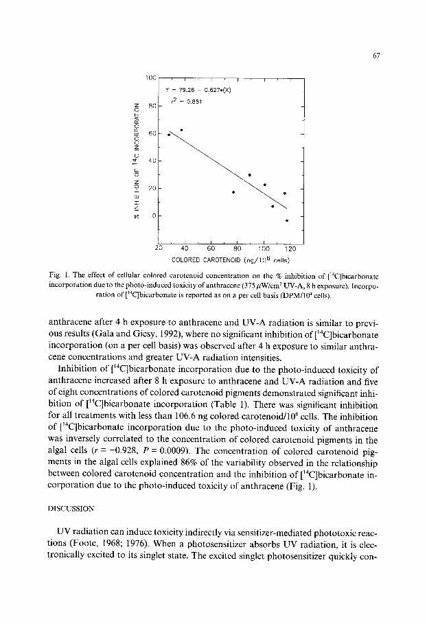
Fig. 1. The effect o f ce l lular co lored caroteno id concentra t ion on the % inhibi t ion o f [ '4C]bicarbonate
i n c o r p o r a t i o n due to the p h o t o - i n d u c e d toxic i ty o f an thracene (375/IW/cm 2 UV-A, 8 h exposure) . Incorpo-
rat ion o f [~4C]bicarbonate is reported as on a per cell basis (DPM/104 cells).
anthracene after 4 h exposure to anthracene and UV-A radiation is similar to previ- ous results (Gala and Giesy, 1992), where no significant inhibition of ['4C]bicarbonate incorporation (on a per cell basis) was observed after 4 h exposure to similar anthra- cene concentrations and greater UV-A radiation intensities.
Inhibition of ['4C]bicarbonate incorporation due to the photo-induced toxicity of anthracene increased after 8 h exposure to anthracene and UV-A radiation and five of eight concentrations of colored carotenoid pigments demonstrated significant inhi- bition of ['4C]bicarbonate incorporation (Table 1). There was significant inhibition for all treatments with less than 106.6 ng colored carotenoid/106 cells. The inhibition of [~4C]bicarbonate incorporation due to the photo-induced toxicity of anthracene was inversely correlated to the concentration of colored carotenoid pigments in the algal cells (r =-0.928, P = 0.0009). The concentration of colored carotenoid pig- ments in the algal cells explained 86% of the variability observed in the relationship between colored carotenoid concentration and the inhibition of ['4C]bicarbonate in- corporation due to the photo-induced toxicity of anthracene (Fig. 1).
DISCUSSION
UV radiation can induce toxicity indirectly via sensitizer-mediated phototoxic reac- tions (Foote, 1968; 1976). When a photosensitizer absorbs UV radiation, it is elec- tronically excited to its singlet state. The excited singlet photosensitizer quickly con-

68
verts to its longer-lived triplet state. Photo-induced toxicity can result from basically two types of sensitizer-mediated phototoxic reactions. In type I reactions, a triplet photosensitizer interacts with a substrate directly via hydrogen atom, electron, or energy transfer, thereby generating free radicals (Grossweiner and Zwicker, 1961). The reduced photosensitizer can also react with oxygen producing superoxide, which can dismutate to hydrogen peroxide (Freeman and Crapo, 1982). Free radicals, su- peroxide and hydrogen peroxide have all been implicated in lipid peroxidation which can lead to membrane destabilization and subsequent toxicity (Freeman and Crapo, 1982; Foote, 1976). Type II reactions involve the transfer of excitation energy from the triplet photosensitizer to molecular oxygen thereby generating singlet oxygen (Foote, 1968). Singlet oxygen can oxidize enzymes, RNA and other subcellular struc- tures and macromolecules resulting in photo-induced toxicity (Foote, 1976).
The dominance of type I or II mechanisms depends on the photosensitizer involved in the reaction. Sensitizers that are readily oxidized, such as phenols and amines, or reduced, such as quinones, will react via type 1 reactions, especially at small concen- trations of oxygen (Grossweiner and Zwicker, 1961; Foote, 1976). Compounds not so easily reduced or oxidized, including thiophene compounds (Arnason et al., 1981; Kagan et al., 1984), photosensitizing dyes (Jahnke and Frenkel, 1978; Foote et al., 1970), psoralens (Joshi and Pathak, 1983), and polycyclic aromatic hydrocarbons (Foote, 1976; Sinha and Chignell, 1983), favor type II reactions. Indirect evidence, consisting of chemical trapping, kinetic and quenching studies (Jahnke and Frenkel, 1978; Foote et al., 1970; Pooler and Valenzeno, 1979; Sinha and Chignell, 1983), overwhelmingly supports the intermediacy of singlet oxygen in most photosensitized reactions.
Carotenoid pigments are important antioxidants in algae and higher plants (Krinsky, 1979) and, thus, should protect algal and plant cells from the toxic action of anthracene and UV-A radiation. The photo-induced toxicity of PAH has been found to proceed via a type II (singlet oxygen) mechanism, in vitro (Foote, 1976; Sinha and Chignell, 1983). Colored carotenoid pigments, which have nine or more conjugated carbon~arbon double bonds, are efficient quenchers of singlet oxygen (Foote et al., 1970; Mathis and Kleo, 1973; Mathews-Roth et al., 1974). The capacity of an algal cell to quench singlet oxygen and protect itself from the photo-induced toxicity of anthracene should be directly related to the concentration of colored carot- enoid pigments contained within the algal cells. The significant inverse relationship for the concentration of colored carotenoid pigments in S. capricornutum cells and the inhibition of [t4C]bicarbonate incorporation due to the photo-induced toxicity of anthracene is in agreement with a type lI (singlet oxygen) mechanism for the photo- induced toxicity of anthracene to algae, in vivo.
The greater resistance of algae to the photo-induced toxicity of PAH, relative to animals, appears to be related to the great concentration of colored carotenoid pig- ments, such as fl-carotene, present in algal cells. Colored carotenoid pigments can protect algal cells by quenching singlet oxygen generated from photo-excited PAH.
69
Additional investigations to assess the utility of using algal cells whose carotenoid biosynthesis has been inhibited, as a bioassay system to determine the mechanism of toxicity of other PAH and photosensitizers are needed. The importance of the protec- tion provided by carotenoid pigments within algal cells against other toxicants, such as solar UV radiation, could be assessed utilizing this proposed bioassay system.
ACKNOWLEDGEMENTS
We wish to thank Drs. Zabik, Leavitt and Miller and the staff of the Pesticide Research Center for their support during the course of this project and Dr. Addison (ELANCO) for supplying us with technical grade Fluridone. This research was sup- ported by a cooperative agreement between the EPA Great Lakes National Program Office and Michigan State University.
REFERENCES
Allred, P.M. and J.P. Giesy, 1985. Solar radiation-induced toxicity of anthracene to Daphniapulex. Envi- ron. Toxicol. Chem. 4, 219-226.
Arnason, T., J.R. Stein, E. Graham, C.-K. Wat, G.H.N. Towers and J. Lam, 1981. Phototoxicity to selected marine and freshwater algae of polyacetylenes from species in the Asteraceae. Can. J. Bot. 59. 54-58.
Bartels, P.G. and C.W. Watson, 1978. Inhibition of carotenoid synthesis by fluridone and norflurazon. Weed Sci. 26, 198-203.
Bennett, W.E., J.L. Maas, S.A. Sweeney and J. Kagan, 1986. Phototoxicity in aquatic organisms: The protecting effect of beta-carotene. Chemosphere 15, 781 786.
Bowling, J.W., G.J. Leversee, P.F. Landrum and J.P. Giesy, 1983. Acute mortality of anthracene-contam- inated fish exposed to sunlight. Aquat. Toxicol. 3, 79 90.
Braumann, T.H., T.H. Gropper, I. Damn and L.H. Grimme, 1984. Function of chlorophylls and carote- noids in thylakoid membranes. Pigment bleaching in relation to PS-I and PS-II activity of subchloroplast particles prepared with digitonin. In: Advances in Photosynthesis Research, Volume II, edited by C. Sybesma. Kluwer Academic Publishers Group, Boston, pp. 137-140.
Britton, G., 1979. Carotenoid biosynthesis a target for herbicide activity. Z. Naturforsch. 34c, 979-985. Cody, T.E., M.J. Radike and D. Warshawsky, 1984. The phototoxicity of benzo[a]pyrene in the green alga
Selenastrum capricornutum. Environ. Res. 35, 122 132. Eisler, R., 1987. Polycyclic aromatic hydrocarbon hazards to fish, wildlife, and invertebrates: a synoptic
review. US Fish Wildl. Serv. Biol. Rep. 85 (1.11), US Fish and Wildlife Service, Laurel, MD. Foote, C.S., 1968. Mechanisms of photosensitized oxidation. Science 162, 963 970. Foote, C.S., 1976. Photosensitized oxidation and singlet oxygen: Consequences in biological systems. Free
Rad. Biol. 2, 86-133. Foote, C.S., Y.C. Chang and R.W. Denny, 1970. Chemistry of singlet oxygen. X. Carotenoid quenching
parallels biological protection. J. Am. Chem. Soc. 92, 5216-5218. Freeman, B.A. and J.D. Crapo, 1982. Biology of disease: Free radicals and tissue injury. Lab. Invest. 47,
412426.
Gala, W.R. and J.P. Giesy, 1991. Effects of ultraviolet radiation on the primary production of natural phytoplankton assemblages in Lake Michigan. Ecotoxicol. Environ. Safety 22, 345 361.
Gala, W.R. and J.P. Giesy, 1992. Photo-induced toxicity of anthracene to the green alga, Selenastrum
70
capricornutum. Arch. Environ. Contam. Toxicol. 23, 316 323. Giddings, J.M., 1979. Acute toxicity to Selenastrum capricornutum of aromatic compounds from coal
conversion. Bull. Environ. Contain. Toxicol. 23, 360 364. Grossweiner, L.I. adn E.F. Zwicker, 1961. Transient measurements of photochemical processes in dyes. I.
The photosensitized oxidation of phenol by cosin and related dyes. J. Chem. Phys. 34, 1141 1147. Grumbach, K.H., 1984. Does the chloroplast envelope contain carotenoids and quinones in vivo? Physiol.
Plant. 60, 180 186. Hairston, N.G., 1976. Photoprotection by carotenoid pigments in the copepod Diaptomus nevadensis. Proc.
Natl. Acad. Sci. USA 73, 971 974. Jahnke, L.S. and A. Frenkel, 1978. Photooxidation of epinephrine sensitized by methylene blue cvidence
for the involvement of singlet oxygen and superoxide. Photochem. Photobiol. 28, 517 523. Joshi, P.C. and M.A. Pathak, 1983. Production of singlet oxygen and superoxide radicals by psoralens and
their biological significance. Biochem. Biophys. Res. Commun. 112, 638 646. Kagan, J., P.A. Kagan and H.E. Bushe, 1984. Light-dependent toxicity of c~-terthienyl and anthracene
toward late embryonic stages ofRanapipiens. J. Chem. Ecol. 10, 1115 1122. Kaidbey, K.H. and S. Nonaka, 1984. Action spectrum for anthracene-induced photosensitization of hu-
man skin. Photochem. Photobiol. 39, 375 378. Krinsky, N.I., 1979. Carotenoid protection against oxidation. Pure Appl. Chem. 51,649 660. Landrum, P.F., J.P. Giesy, J.T. Oris and P.M. Allred, 1986. The photoinduced toxicity of polycyclic
aromatic hydrocarbons to aquatic organisms. In: Oil and Freshwater: Chemistry, Biology, Technology, edited by J.H. Vandermeulen and S. Hrudey. Pergamon Press, New York, pp. 304- 318.
Mathis, P. and J. Kleo, 1973. The triplet state offl-carotene and of analog polyenes of different length. Photochem. Photobiol. 18, 343 346.
Matthews-Roth, M.M., T. Wilson, E. Fujimori and N.I. Krinsky, 1974. Carotenoid chromophore length and protection against photosensitization. Photochem. Photobiol. 19, 217 222.
Miller, W.E., J.C. Greene and T. Shiroyama, 1978. The Selenastrum capricornutum Printz algal assay bottle test. EPA 600/9-78-018, U.S. Environmental Protection Agency, Corvallis, OR.
Neff, J.M., 1979. Polycyclic Aromatic Hydrocarbons in the Aquatic Environment. Applied Science Publ. Ltd. London.
Oris, J.T. and J.P. Giesy, 1985. The photoenhanced toxicity of anthracene to juvenile sunfish (Lepomis spp.) Aquat. Toxicol. 6, 133 146.
Oris, J.T., J.P. Giesy, P.M. Allred, D.F. Grant and P.F. Landrum, 1984. Photoinduced toxicity of anthra- cene in aquatic organisms: An environmental perspective. In: The Biosphere: Problems and Solutions, edited by T.N. Veziroglu. Elsevier Science Publishers BV, Amsterdam, pp. 639 658.
Parsons, T.R., Y. Maita and C.M. Lalli, 1984. A Manual of Chemical and Biological Methods for Seawater Analysis. Plenum Press, New York, pp. 101 104.
Pooler, J.P. and D.P. Valenzeno, 1979. The role of singlet oxygen in photooxidation of excitable cell membranes. Photochem. Photobiol. 30, 581 584.
Sandmann, G., I.E. Clarke, P.M. Bramley and P. Boger, 1984. Inhibition of phytoene desaturase the mode of action of certain bleaching herbicides. Z. Naturforsch. 39c, 443-449.
SAS Institute, 1987. SAS/STAT Guide for Personal Computers, Version 6 Edition. SAS Institute Inc., Cary, NC.
Sinha, B. and C.F. Chignell, 1983. Binding of anthracene to cellular macromolecules in the presence of light. Photochem. Photobiol. 37, 33 37.
Copyright © 2022 FDOKUMEN