Studies of glycosaminoglycan composition and biologic activity of VesselS, a hypolipidemic agent
13
Atherosclerosis, 31 (1978) 217-229 @ Elsevier/North-Holland Scientific Publishers, Ltd. STUDIES OF GLYCOSAMINOGLYCAN COMPOSITION AND BIOLOGIC ACTIVITY OF VESSEL@, A HYPOLIPIDEMIC AGENT BHANDARU RADHAKRISHNAMURTHY, HAROLD A. RUIZ, SATHANUR R. SRINIVASAN, WILLIAM PREAU, EDWARD R. DALFERES, Jr. and GERALD S. BERENSON Departments of Medicine and Biochemistry, Louisiana State University Medical Center, 1542 Tulane Avenue, New Orleans, LA 70112 (U.S.A.) (Received 17 April, 1978) (Revised, received 5 June, 1978) (Accepted 21 June, 1978) Summary Heparin-like glycosaminoglycans (GAG) were isolated from commercial Vessel’ and their biologic properties studied. Vessel@ was found to be a mix- ture of chondroitin sulfates, dermatan sulfate and heparin-like GAG. Chondroi- tin sulfates and dermatan sulfate in Vessel@ were hydrolyzed by chondroitinase ABC and the residual Vessel@ was fractionated on a Dowex-1 Cl- column elut- ing with a stepwise-increasing concentration of NaCl (1.2--4.0 M). The major fractions eluted at 1.6 M and 1.8 M NaCl were tentatively identified by chemi- cal analysis as heparin-like GAG with somewhat lower sulfate content than standard heparin. Both fractions had lipoprotein lipase-releasing activity and anticoagulant activity similar to heparin, but 1.6 M NaCl fraction had a third of the anticoagulant activity of standard heparin. The 1.8 M NaCl fraction com- plexed with serum lipoproteins similarly to heparin. In preliminary studies cho- lesterol-fed rabbits treated with Vessel@ exhibited somewhat less atherosclero- sis than controls. --- - -_- Key words: Anticoagulant activity - GAG complexes - GAG from Vessel@ - Hypolipi- demic agent - Lipoprotein lipase-releasing activity - Serum lipoprotein Introduction Numerous clinical and experimental studies suggest a relationship between serum lipid and lipoproteins and the development of atherosclerosis. A variety Supported by funds from The National Heart, Lung. and Blood Institute of the USPHS (HL02942. 21 G-$9) and the Spwialized Center of Kesearch - Arteriosclerosis (HL15103).
-
Upload
independent -
Category
Documents
-
view
4 -
download
0
Transcript of Studies of glycosaminoglycan composition and biologic activity of VesselS, a hypolipidemic agent

Atherosclerosis, 31 (1978) 217-229 @ Elsevier/North-Holland Scientific Publishers, Ltd.
STUDIES OF GLYCOSAMINOGLYCAN COMPOSITION AND BIOLOGIC ACTIVITY OF VESSEL@, A HYPOLIPIDEMIC AGENT
BHANDARU RADHAKRISHNAMURTHY, HAROLD A. RUIZ, SATHANUR R. SRINIVASAN, WILLIAM PREAU, EDWARD R. DALFERES, Jr. and GERALD S. BERENSON
Departments of Medicine and Biochemistry, Louisiana State University Medical Center, 1542 Tulane Avenue, New Orleans, LA 70112 (U.S.A.)
(Received 17 April, 1978) (Revised, received 5 June, 1978) (Accepted 21 June, 1978)
Summary
Heparin-like glycosaminoglycans (GAG) were isolated from commercial Vessel’ and their biologic properties studied. Vessel@ was found to be a mix- ture of chondroitin sulfates, dermatan sulfate and heparin-like GAG. Chondroi- tin sulfates and dermatan sulfate in Vessel@ were hydrolyzed by chondroitinase ABC and the residual Vessel@ was fractionated on a Dowex-1 Cl- column elut- ing with a stepwise-increasing concentration of NaCl (1.2--4.0 M). The major fractions eluted at 1.6 M and 1.8 M NaCl were tentatively identified by chemi- cal analysis as heparin-like GAG with somewhat lower sulfate content than standard heparin. Both fractions had lipoprotein lipase-releasing activity and anticoagulant activity similar to heparin, but 1.6 M NaCl fraction had a third of the anticoagulant activity of standard heparin. The 1.8 M NaCl fraction com- plexed with serum lipoproteins similarly to heparin. In preliminary studies cho- lesterol-fed rabbits treated with Vessel@ exhibited somewhat less atherosclero- sis than controls. --- - -_- Key words: Anticoagulant activity - GAG complexes - GAG from Vessel@ - Hypolipi-
demic agent - Lipoprotein lipase-releasing activity - Serum lipoprotein
Introduction
Numerous clinical and experimental studies suggest a relationship between serum lipid and lipoproteins and the development of atherosclerosis. A variety
Supported by funds from The National Heart, Lung. and Blood Institute of the USPHS (HL02942.
21 G-$9) and the Spwialized Center of Kesearch - Arteriosclerosis (HL15103).

218
of compounds, which include Atromid-S, cholestyramme, and several naturally occurring carbohydrate macromolecules, have been used to reduce serum lipid levels. Heparin, a glycosaminoglycan (GAG) with fat-clearing activity, has been used to inhibit the development of atherosclerot.ic lesions in experimental ani- mals [l--4], and Engelberg et al. [ 51 and others [ 6,7] have used heparin in the treatment of human coronary artery disease. Other GAG, like chondroitin 6-sulfate and heparan sulfate, were also used to inhibit the development of atherosclerosis in experimental animals and to reduce cardiovascular incidences in patients with previous myocardial infarction [ 81. Because of high anticoagu- lant activity and other complications, like decalcification, heparin is not recom- mended for long-term therapy. Attempts therefore have been made to isolate heparin-like GAG that possess high lipoprotein lipase-releasing ability with mini- mal anticoagulant activity. Bianchini et al. [9] isolated a crude heparinoid from pig duodenal mucosa and pancreas and Crinos Laboratory, Como Villa Guardia, Italy, marketed it under the name Ateroid@ as an hypolipidemic agent with low anticoagulant activity. We [lo] have recently studied the GAG composi- tion of Ateroid@ and observed that its major GAG was heparin, although hepa- ran sulfate, dermatan sulfate and hyaluronic acid were present in considerable amounts. The heparin fraction isolated from Rteroid@ exhibited lipoprotein lipase-releasing activity similar to mucosal heparin but possessed only a six- teenth of the anticoagulant activity of the mucosal preparation.
Vessel@ , a product similar to Ateroid@, has been marketed in Europe since 1973. The therapeutic indications of the drug as described by Alfa Farmaceu- tici, Bologna, Italy, include alteration of blood lipid levels, peripheral athero- sclerotic arteriopathy and thromboembolic syndromes [ 11 I. Since the precise chemistry of Vessel@ is not known, we have studied its GAG composition and biologic activity in an effort to understand its hypolipidemic and antiathero- genie properties.
Materials and methods
Vessel@ in 0.15 M NaCl solution was obtained from Alfa Farmaceutici, Bologna, Italy, through courtesy of Professor C.R. Sir-tori, University of Milan, Italy. Heparin (160 units/mg) was donated by the Upjohn Company (Kalama- zoo, MI). Chondroitinase-ABC was purchased from Miles Laboratories, Inc. (Elkhart, IN) and thrombin from the Upjohn Company.
Analytical methods We determined uranic acid by the method of Bitter and Muir [ 121 and hexos-
amine by the method of Boas [13] omitting the use of resin. The method of Dodgson and Spencer [14] as modified by Muir [15] was used for the determi- nation of total sulfate and Lagunoff and Warren’s [16] method for N-sulfate estimation,
Gas-liquid chromatography techniques [ 17-201 as described previously were used to differentiate glucuronic acid from iduronic acid and glucosamine from galactosamine, to estimate N-acetyl groups and to determine total sulfate in samples that were available in small quantities.
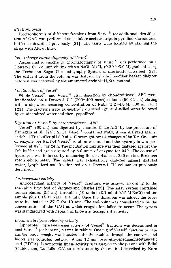
Electrophoresis Electrophoresis of different fractions from Vessel@ for additional identifica-
tion of GAG was performed on cellulose acetate strips in pyridine-formic acid buffer as described previously [21]. The GAG were located by staining the strips with Alcian Blue.
ion-exchange chromatography of Vessel@ Automated ion-exchange chromatography of Vessel@ was performed on a
Dowex-1 Cl- column eluting with a NaCl-MgCl, (0.2 M--3.0 M) gradient using the Technicon Sugar Chromatography System as previously described [22]. The effluent from the column was dialyzed by a hollow-fiber beaker dialyzer before it was analyzed by the automated orcinol-H,SO, method.
Fractionation of Vessel@ Whole Vessel@ and Vessel@ after digestion by chondroitinase-ABC were
fractionated on a Dowex-1 Cl- (200-400 mesh) column (50 X 1 cm) eluting with a stepwise-increasing concentration of NaCl (1.2-4.0 M, 500 ml each) [ 231. The fractions were exhaustively dialyzed against distilled water followed by demineralized water and then lyophilized.
Digestion of Vessel@ by chrondroitinase-ABC Vessel@ (60 ml) was digested by chondroitinase-ABC by the procedure of
Yamagata et al. [24]. Since Vessel@ contained NaCl, it was dialyzed against enriched Tris buffer pH 8.0 at 4°C overnight over 4 changes of buffer. One unit of enzyme per 6 ml of Vessel@ solution was used and the hydrolysis was per- formed at 37°C for 24 h. The incubation mixture was then dialyzed against the Tris buffer and again digested by 5.0 units of enzyme for 24 h. The course of hydrolysis was followed by measuring the absorbance at 230 nm in a Beckman spectrophotometer. The digest was exhaustively dialyzed against distilled water, lyophilized and fractionated on a Dowex-1 Cl- column as previously described.
Anticoagulant activity Anticoagulant activity of Vessel @ fractions was assayed according to the
thrombin time test of Jacques and Charles [25]. The assay system contained human plasma (0.5 ml), thrombin (10 units in 0.1 ml of 0.15 M NaCl) and the sample plus 0.15 M NaCl (0.4 ml). Once the thrombin was added, the tubes were incubated at 37°C for 10 min. The end-point was considered to be the concentration of the GAG at which coagulation failed to occur. The system was standardized with heparin of known anticoagulant activity.
Lipoprotein lipase-releasing activity Lipoprotein lipase-releasing activity of Vessel@ fractions was determined in
post-Vessel@ (or heparin) plasma in rabbits. One mg of Vessel@ fraction or hep- arm/kg body weight was injected into the rabbits through the ear vein and blood was collected between 9 and 12 min over ethylenediaminetetraacetic acid (EDTA). Lipoprotein lipase activity was assayed in the plasma with Ediol (Calbiochem, La Jolla, CA) as a substrate by the method described by Korn
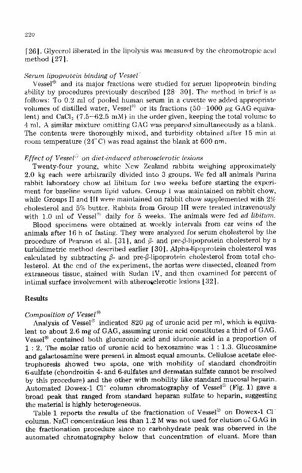
[ 261. Glycerol liberated in the lipolysis was measured by the chromotropic acid method [ 271.
Serum lipoprotein binding of Vessel” Vessel@ and its major fractions were studied for serum lipoprot,ein binding
ability by procedures previously described [28-m-30]. The method in brief is as follows: To 0.2 ml of pooled human serum in a cuvette we added appropriate volumes of distilled water, Vessel” or its fractions (50-1000 pg GAG equiva- lent) and CaCl, (7.5562.5 mM) in the order given, keeping the total volume to 4 ml. A similar mixture omitting GAG was prepared simultaneously as a blank. The contents were thoroughly mixed, and turbidity obtained after 15 min at, room temperature (24°C) was read against the blank at 600 nm.
Effect of Vessel” on diet-induced atherosclerotic Lesions Twenty-four young, white New Zealand rabbits weighing approximately
2.0 kg each were arbitrarily divided into 3 groups. We fed all animals Purina rabbit laboratory chow ad libitum for two weeks before starting the experi- ment for baseline serum lipid values. Group I was maintained on rabbit chow, while Groups II and III were maintained on rabbit chow supplemented with 2% cholesterol and 5% butter. Rabbits from Group III were treated intravenously with 1.0 ml of Vessel@ daily for 5 weeks. The animals were fed ad libitum.
Blood specimens were obtained at weekly intervals from ear veins of the animals after 16 h of fasting. They were analyzed for serum cholesterol by the procedure of Pearson et al. [ 311, and p- and pre-P-lipoprotein cholesterol by a turbidimetric method described earlier [ 301. Alpha-lipoprotein cholesterol was calculated by subtracting /3- and pre-@lipoprotein cholesterol from total cho- lesterol. At the end of the experiment, the aortas were dissected, cleaned from extraneous tissue, stained with Sudan IV, and then examined for percent of intimal surface involvement with atherofclerotic lesions [ 321.
Results
Composition of Vessel @ Analysis of Vessel@ indicated 820 pg of uranic acid per ml, which is equiva-
lent to about 2.6 mg of GAG, assuming uranic acid constitutes a third of GAG. Vessel@ contained both glucuronic acid and iduronic acid in a proportion of 1 : 2. The molar ratio of uranic acid to hexosamine was 1 : 1.3. Glucosamine and galactosamine were present in almost equal amounts. Cellulose acetate elec- trophoresis showed two spots, one with mobility of standard chondroitin 6-sulfate (chondroitin 4- and 6-sulfates and dermatan sulfate cannot be resolved by this procedure) and the other with mobility like standard mucosal heparin. Automated Dowex-1 Cl- column chromatography of Vessel@ (Fig. 1) gave a broad peak that ranged from standard heparan sulfate to heparin, suggesting the material is highly heterogeneous.
Table 1 reports the results of the fractionation of Vessel@ on Dowex-1 Cl- column. NaCl concentration less than 1.2 M was not used for elution of GAG in the fractionation procedure since no carbohydrate peak was observed in the automated chromatography below that concentration of eluant. More than

221
CHROlATOGRAPHIC PROFILE OF VESSELR AND A MIXTURE OF STANDARD GLYCOSRMINOGLYCANS
2 1 HOURS
Fig. 1. Automated Dowex-1 Cl- column chromatographic profile of Vessel@ superimposed on a chro- matogram of standard glycosaminoglycans, hyaluronic acid (HA). heparan sulfate (HS), chondroitin 4- and B-sulfates (CS), dermatan sulfate (DS), and heparin (Hep). The conditions of chromatogr+phy are dis- cussed in the text. The broad peak of Vessel@ suggests its heterogeneity
50% of the total GAG eluted in 1.6 M NaCl. Considerable amounts of GAG were also present in 1.4 M and 1.8 M fractions. The analysis of GAG of each of these fractions is shown in Table 2. The fractions vary in total sulfate and N-sulfate contents. In general, fractions having higher sulfate contents eluted from the column at higher concentrations of NaCl. Except the 1.2 M NaCl frac- tion, all fractions contained both glucuronic acid and iduronic acid, and glucos- amine and galactosamine in varied proportions. The amount of galactosamine was greater than glucosamine in 1.4 M NaCl fraction, and they were almost equal in 1.6 M NaCl fraction. Galactosamine was also present in 1.8 M and 2.0 M NaCl fractions.
The presence of large amounts of galactosamine in these fractions suggests
TABLE 1
FRACTIONATION OF VESSEL@ ON A DOWEX-1 Cl- COLUMN ELUTING WITH STEPWISE-IN- CREASING CONCENTRATIONS OF NaCJ
Fraction NaCJ Total uranic acid a (M) (me)
1.2 b 0.73 1.4 1.00 1.6 4.48 1.8 1.52 2.0 0.61 4.0 0.22
% of total uranic acid
8.5 11.8 52.3 17.7
7.1 2.5
a 10 ml of Vessel@ was fractionated; 105% uronate was recovered. b No detectable uronate material eluted from the column with NaCJ of less than 1.2 M concentration.

222
TABLI’ 2
ANALYSIS OF DOWEX-1 Cl-- COL,UMN FRACTIONS OF VESSEL*;
Fraction
NaCl
(M)
UA” Total N-SO,, K-Ace- GlcUA : GlcN
SOJ tvl b 1dU.A ‘1 GalN b
(mole per mole hcxosaminc)
1.2 1.2 1.5 0.67 0.34 100 : 0 100 : 0 1.4 1.1 1.6 0.5fi 0.32 84 : 16 37 : 63
1.6 1.2 1.8 0.71 0.24 51 : 49 56 : 44
1.8 1.3 1.8 0.77 O.lfi 34 : 66 80 : 20
2.0 1.3 2.6 0.83 0.12 47 : ,53 89 : 11
HS d
HS + CS + DS
Hl?P + CS + DS
HEP
HEP
a Abbreviations: UA = uranic acid; GlcUA = glucuronic acid: IdUA = iduronic acid; GlcN = glucosamine;
GalN = galactosamine; HS = heparan sulfate: CS = chrondroitin 4- and 6-sulfates: DS = dennatan sulfate;
HEP = heparin.
b Determined by gas-liquid chromatography.
c Electrophoresis was performed in pyridine----formate buffer. and in this system HS. CS and DS cannot be
resolved.
d Identified as HS since no galactosamine was present in the fraction.
that they contain considerable portions of chondroitin sulfat.es and dermatan sulfate, and these GAG are known to have no lipoprotein lipase-releasing activ- ity. Therefore Vessel@ was digested with chondroitinase-ABC and then frac- tionated on Dowex-1 Cl- column to obtain the heparin-like GAG free of chon- droitin sulfates. Analysis of the chondroitinase---ABC-digested Vessel@ after dialysis indicated that 52% of the uranic acid material was hydrolyzed by this enzyme. Table 3 shows the results of the fractionation of Vessel@ after hydrol- ysis by the enzyme.
Most of the GAG eluted at 1.6 M and 1.8 M NaCl as in the previous fraction- ation (Table 1). The analyses of these fractions are reported in Table 4. There was a slight increase in total hexosamine content in most of the fractions (com- pare Table 2), but a decrease in total sulfate content. No difference was ob- served in N-sulfate content between these fractions and those obtained from Vessel@ without hydrolysis by the enzyme. N-acetyl content decreased in all fractions after digestion of Vessel@ by the enzyme. All of these fractions were devoid of galactosamine, suggesting the absence of chondroitin sulfates and
TABLE 3
FRACTIONATION OF VESSEL@ HYDROLYZED BY CHONDROITINASE-ABC ON DOWEX-1 Cl-
COLUMN ~--..._--__
Fraction NaCl Total uranic acid a % of total uranic acid
(M) (mg1
1.2 1.65 9.4
1.4 1.‘71 9.8
1.6 7.80 44.6
1.8 5.66 32.3
2.0 0.62 3.5
4.0 0.05 0.3
a 60 ml Vessel@ was hydrolyzed by the enzyme, dialyzed against distilled water and the nondialyzable
GAG fractionated on a Dowex-1 Cl- column: 92% uronate placed on the column was recovered.
223
TABLE 4
ANALYSIS OF DOWEX-1 Cl- COLUMN FRACTIONS OF VESSEL@ HYDROLYZED BY CHONDROI-
TINASES
Fraction UA
NaCl (M)
Total
SO4
N-SO4 N-Ace-
tYI
GlcUA :
IdUA
GlcN :
GalN
Electrophoretic
identification
(mole per mole of hexosamine)
1.2 1.5 1.1 0.65 0.28 100 : 0 100 : 0 HS
1.4 1.6 1.0 0.70 0.21 93 : 7 100 : 0 HS + HEP
1.6 1.6 1.6 0.70 0.18 61 : 39 100 : oa HS + HEP
1.8 1.3 2.1 0.80 0.09 52 : .1R 100:oa HEP
2.0 1.4 2.3 0.75 0.08 54 : 4fJ 10o:oa HEP
a Contains trace of galactosamine less than 0.5%.
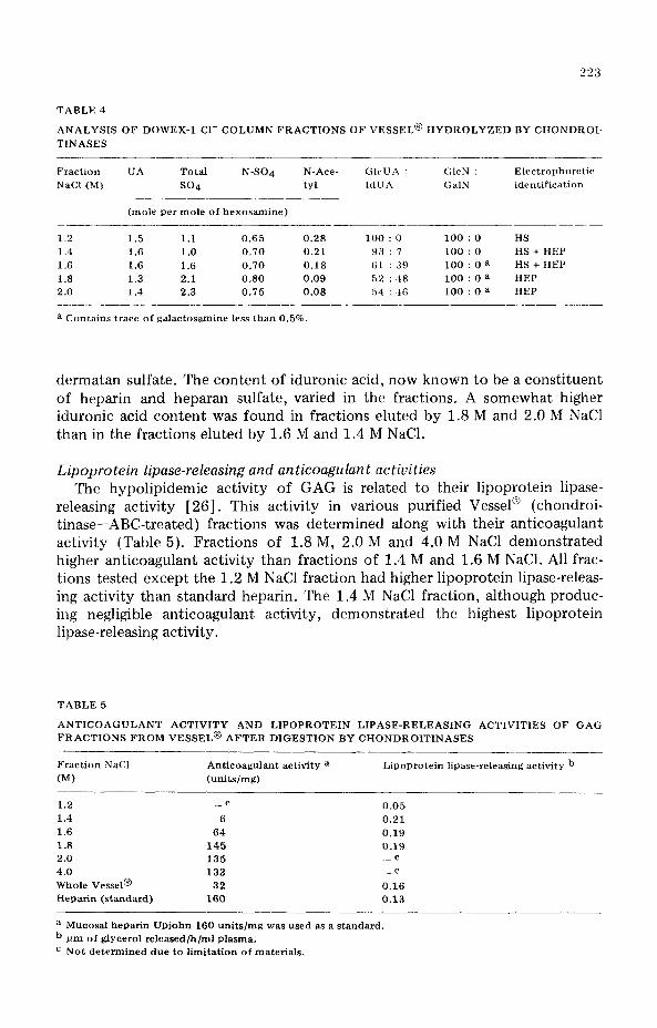
dermatan sulfate. The content of iduronic acid, now known to be a constituent of heparin and heparan sulfate, varied in the fractions. A somewhat higher iduronic acid content was found in fractions eluted by 1.8 M and 2.0 M NaCl than in the fractions eluted by 1.6 M and 1.4 M NaCl.
Lipoprotein lipase-releasing and anticoagulant activities The hypolipidemic activity of GAG is related to their lipoprotein lipase-
releasing activity [26]. This activity in various purified Vessel@ (chondroi- tinase-ABC-treated) fractions was determined along with their anticoagulant activity (Table 5). Fractions of 1.8 M, 2.0 M and 4.0 M NaCl demonstrated higher anticoagulant activity than fractions of 14 M and 1.6 M NaCl. All frac- tions tested except the 1.2 M NaCl fraction had higher lipoprotein lipase-releas- ing activity than standard heparin. The 1.4 M NaCl fraction, although produc- ing negligible anticoagulant activity, demonstrated the highest lipoprotein lipase-releasing activity.
TABLE 5
ANTICOAGULANT ACTIVITY AND LIPOPROTEIN LIPASE-RELEASING ACTIVITIES OF GAG
FRACTIONS FROM VESSEL@ AFTER DIGESTION BY CHONDROITINASES
Fraction NaCl Anticoagulant activity a Lipoprotein Iipase-releasing activity b
(M) (units/mg)
1.2 _c 0.05 1.4 6 0.21 1.6 64 0.19 1.8 145 0.19 2.0 135 c
4.0 133 c
Whole Vessel@ 32 0.16
Heparin (standard) 160 0.13
a Mucosal heparin Upjohn 160 units/mg was used as a standard.
b /un of glycerol released/h/ml plasma.
c Not determined due to limitation of materials.

224
Lipoprotein binding activity Another biologic property of Vessel”’ that we studied was its abihty to form
insoluble complexes with serum low density and very low density lipoproteins (LDL, VLDL). GAG form insoluble complexes selectively with serum LDL and VLDL in the presence of Ca++. Of various GAG, heparin has been found to have the greatest ability to form such complexes in vitro [ 281.
The degree of binding of G,4G with serum LDL and VLDL was measured in terms of the turbidity produced by such interactions. The nature and concen- tration of GAG as well as the concentration of Cat+ affect the reaction [SS]. The complex formation of Vessel” and some of its fractions at various concen- trations of GAG (50-1000 pg) in the presence of 62.5 mM Ca” is shown in Fig. 2. Except 1.8 M NaCl fraction free of chondroitin sulfates and dermatan sulfate (1.8 M NaCl-CS), all fractions and whole Vessel@ showed a more or less linear relationship between GAG concentration and turbidity, suggesting a lack of saturation of lipoprotein binding sites by GAG. The 1.8 M NaClCS fraction produced maximum turbidity at a relatively low concentration of GAG (250 pg) and behaved like standard heparin (not shown in the figure) at the concentrations studied. The turbidity produced at different concentrations of Ca” (7.5-62.5 mM) at a GAG concentration of 250 pg in the system is shown in Fig. 3. Whole Vessel”’ produced about 50% less turbidity than stan-
LIPOPROTEIN BINDING OF VESSEL? FRACTIONS AT
VARIOUS CONCENTRATIONS 625mM Co’+
100 200 300 400 500 600 700 800 900 1000
GAG, ug
Fig. 2. Serum low and very low density lipoprotein binding of Vessel@ and its fractions at various con- centrations in the presence of 62.5 mM of Ca++. Fractionation of whole Vessel@ and of Vessel@ after digestion by chondroitinase-ABC (-CS) was performed on a Dowex-1 Cl- column eluting with stepwise- increasing concentration of NaCl (1.2-4.0 M). The major fractions 1.6 M and 1.8 M NaCl were studied for lipoproteins binding at various concentrations. Although not shown in the figure, standard heparin behaved like 1.8 M NaCIGS fraction in producing maximum turbidity at 250 pg GAG concentration. Whole Vessel@ and other fractions did not reach maxima even at 1000 fig GAG concentration.

LIPOPROTEIN BINDING OF VESSEt? FRACTIONS AT VARIOUS CONCENTRATIONS OF Ca” AND 250 pg GAG
Co Cl,, mhj
Fig. 3. Serum low and very low density lipoproteins binding of Vessel@ and its fractions at a fixed con- centration of GAG (250 pg) but with varied concentration of Ca++ in the system. Like standard mucosal heparin 1.8 M NaCl-CS fraction needed only 12.5 mM of Ca++ whole Vessel@ and other fractions needed more Ca++.
to produce maximum turbidity whereas
dard heparin. Of the various fractions of Vessel@ the degree of insoluble com- plex formation (turbidity) was similar to heparin only for 1.8 M NaCl-CS frac- tion. While standard heparin and the 1.8 M NaCl-CS fraction of Vessel@ required an optimal level of 12.5 mM of Caf+ for maximum insoluble complex formation, the other fractions required twice that amount. Unfortunately, due to limitation of materials we could not perform similar studies on 1.2 M and 1.4 M NaCl fractions.
Effect of Vessel@ on the inhibition of diet-induced atherosclerotic lesions All but one rabbit in Group I (control) remained in good health throughout
the experiment. The animals in Groups II (high cholesterol diet) and Group III (high cholesterol diet and Vessel@ treatment) exhibited mean weight gains of 0.55 kg and 0.53 kg, respectively, while those in Group I showed a mean weight gain of 0.31 kg over the course of the study.
The mean baseline serum cholesterol values of rabbits was about 75 mg/lOO ml and there was little or no difference among individual rabbits during the pre-experimental period. A week after the start of the experiment, rabbits in Group II and Group III developed mean serum cholesterol levels of 1,000 mg/ 100 ml and reached a value of 2,500 mg/lOO ml by the end of the 5th week, whereas the controls had mean cholesterol values of 80 mg/lOO ml throughout the experimental period. The rise in total cholesterol observed in Groups II and III was due mainly to cholesterol carried in the fl- and pre-fl-lipoproteins (hepa- rin-Ca”-precipitable fractions) although a smaller rise also occurred in the

226
TABLE 6
PERCENTAGE OF INTIMAL SURFACE COVERED WITH FATTY STREAKS OF AORTAS FROM
RABBITS TREATED WITH VESSEL@ a ___ _~~ ._~ ___. -. ~.
Control diet High cholesterol diet High cholesterol diet with Vrssrl’~” __.-
Animal b Thoracic Abdominal Animal Thor&c Abdominal Animal c Thoracic Abdonunal
number aorta aorta number aorta aorta number aorta aorta -~-
9
10
12
13
20
23
24
0.3 0.1
5
7
15
18
21
22
21
28
12 5
10 2
6 1
7 1
4 0
33 0
10 2
3 1
10.6 1.5 8.0 1.1
a Evaluated visually by grossly detectable sudanophilic lesions.
b One animal died during the experiment.
C One aorta accidently cut into pieces and could not be properly stained and evaluated.
a-lipoprotein fraction. The mean value for p- and pre-/3-lipoprotein cholesterol for Groups II and III was 2,000 mg/lOO ml of serum at the termination of the experiment. Although individual variations within the groups were observed, the mean values of the groups were similar.
The percent of intimal surface involvement with atherosclerotic lesions ob- served after staining the aorta with Sudan IV is reported in Table 6. Although there were wide variations within the groups the mean surface involvement with lesions in aortas of rabbits fed a high cholesterol diet with Vessel@ treat- ment was slightly less than in the aortas from rabbits fed a high cholesterol diet without Vessel@ treatment. Control rabbits, except one, did not have any lesions.
Although it is important to know the effect of Vessel@ on tissue decalcifica- tion, we did not determine the calcium content of various organs of rabbits treated with Vessel@ in this study.
Discussion
It is evident from this study that Vessel @ is not a single component but a mixture of several GAG with heparin or heparin-like materials constituting 50% of the total GAG. The other major GAG in Vessel@ are condroitin sulfates and/or dermatan sulfate. Since these isomeric chondroitin sulfates do not con- tribute to lipoprotein lipase activity, no attempt was made to quantitate them individually. In their total sulfate and N-sulfate contents, 1.2 M and 1.4 M NaCl fractions resemble heparan sulfate, whereas 1.8 M and 2.0 M NaCl fractions appear similar to heparin. The predominant 1.6 M NaCl fraction could be either heparan sulfate, degraded heparin or a mixture of both.
It is interesting to note that 1.4 M NaCl fraction with low total sulfate con- tent had the highest lipoprotein lipase-releasing activity with insignificant anti- coagulant activity. Anticoagulant activity appears to vary with the molecular
227
size. Cifonelli [33] observed that high molecular weight preparations of com- mercial heparin had higher anticoagulant activity than low molecular weight preparations. Chains containing lo--l1 repeating disaccharide units appear optimum for demonstrating maximum activity. Dietrich [35] prepared a hexa- saccharide from heparin by bacterial degradation. The hexasaccharide resem- bles heparin in chemical composition but differs in anticoagulant activity. Whether the low molecular weight preparations exhibit the same lipoprotein lipase activity like the b.igh molecular weight heparin preparation is not known. The results presented in this study suggest that anticoagulant activity and lipo- protein lipase activity are independent, since 1.4 M NaCl with the lower sulfate content had higher lipoprotein lipase activity than fractions with the higher sulfate contents. Whether the structural requirement of heparin or heparan sul- fate for lipoprotein lipase-releasing activity depends on molecular size, sulfate content or glucuronic acid to iduronic acid proportion is yet to be determined.
GAG, particularly sulfated GAG, are known to form insoluble complexes with serum LDL and VLDL. This phenomenon has been implicated in the sequestration of serum lipoprotein in the arterial wall and subsequent develop- ment of atherosclerotic lesions. Experimental evidence for this hypothesis has been recently presented by the isolation of lipoprotein--GAG complexes from human atherosclerotic lesions [3,5,36] as well as from lesions from experi- mental animals [37]. To further help us characterize Vessel@ and its fractions, we isolated the various GAG fractions and studied the lipoprotein interaction in vitro. The 1.8 M NaCl-CS fraction was found to behave like heparin.
The preliminary observations of the inhibitory effect of Vessel@ on the development of diet-induced atherosclerotic lesions suggest its potential as an antiatherogenic agent. Although Vessel@ did not reduce serum cholesterol levels, it lowered the surface involvement of intima by atherosclerotic lesions by 20%. Horlick and Duff [2] noted retardation of atherogenesis by heparin in choles- terol-fed rabbits, although the total serum cholesterol levels were not effected by heparin treatment in that study. It is possible that the high amount of cho- lesterol used in the experimental diets simply overwhelmed any effect the drug may have had on serum cholesterol levels. This possible masking effect could be overcome by using lower levels of dietary cholesterol. A heparan sulfate prepa- ration from beef lung [38] has been shown to inhibit atherosclerotic lesion for- mation in rabbits fed a 2% cholesterol diet with simultaneous reduction in serum cholesterol levels, but the heparan sulfate preparation was administered to the animals through a catheter into the alkaline portion of the proximal bowel to prevent degradation of sulfate groups in the stomach. This could have caused better absorption of the compound and resulted in lower serum choles- terol levels.
Although our work on Vessel@ . 1s not extensive enough to show very signifi- cant alternations of vascular lesions, there is accumulated evidence of the im- portant role of GAG in atherogenesis. Heparin and certain other naturally occurring GAG have the potential for inhibition of atherosclerotic lesions. Sev- eral mechanisms might be considered.
Circulating GAG interact with serum LDL and VLDL [39] and could com- pete with arterial GAG. This competitive interaction might inhibit the seques- t,ration of lipoproteins with arterial GAG. The recent hypothesis proposed by
228
Zilversmit [40] also should be considered. Zilversmit suggests that atherogent>-- sis may result from the release of cholesterol-rich fragments in proximity to thcb arterial endothelium when the lipoproteins are degraded by arterial lipoprotein lipase. Like serum LDL and VLDL, lipoprotein lipase 1411 binds hcparin, and it is likely that the GAG act as a bridge linking lipoproteins and lipoprotein lipase to the arterial surface. When injected, heparin might release 110th lipopro- teins and lipoprotein lipase by competing for binding sites at the intimal sur- face. This might avoid surface lipolysis and local accumulation of 1)otcntially injurious cholesterol-rich lipoprotein fragments and fatty acids and thereby inhibit the development of atherosclerotic lesions. Another mechanism con- cerns the affect on platelet aggregation, a role which remains to be determined.
Acknowledgements
The authors wish to thank Professor C.R. Sirtori, University of Milan, Italy, and Dr. G. Biavati, Alfa Farmaceutici, Italy, for kindly providing Vessel@; Dr. William Newman III for evaluation of the rabbit aortas for atherosclerotic lesions; Mr. W.S. Brooks III and Ms. L.L. Wells for technical assistance and Ms. Caroline Andrews for editing the manuscript.
References
1 Constantinides. P., Szasz, G. and Harder, F., Retardation of atheromatosis and adrenal enlargement by heparin in the rabbit, Arch. Path., 56 (1953) 36.
2 Horlick, L. and Duff, G.L., Heparin in experimental cholesterol atherosclerosis in the rabbit, Part 1 (Effect of heparin on the serum lipids and the development of atherosclerosis), Arch. Path., 57 (1954) 417.
3 Meng, H.C. and Davis, W.S.. Effects of heparin on development of atherosclerosis and fatty liver, Arch. Path., 60 (1955) 276.
4 Rowsell, H.C., Murphy. E.A. and Mustard, J.F., Heparin dosage and atherogenesis in the rabbit, Arch. Path., 80 (1965) 63.
5 Engelberg, H., Kuhn, R. and Steinman, M., Controlled study of the effect of intermittent heparin therapy on the course of human coronary atherosclerosis, Circulation, 13 (1956) 489.
6 Bottiger, L.E., Carlson, L.A.. Engstedt, L. and Ore, L., Effect of long-term heparin treatment in ischaemic heart disease, J. Atheroscler. Res., 5 (1965) 253.
7 B&tiger, L.E., Carlson, L.A., Engstedt, L. and 0x0, L., Long-term heparin treatment in ischaemic heart disease - Effects on clinical condition and plasma lipids, Acta Med. Stand., 182 (1967) 245.
8 Morrison, L.M. and Schjeide, O.A., Coronary Heart Disease and the Mucopolysacchtides (GIYcoS- aminoglycans), Charles C. Thomas, Springfield, IL, 1974.a
9 Bianchini, P.. Osima, B., Casetta. R., Bramanti, G. and Mostacci, C., Un nuovo eparinoide Pancreatic0 attivo sul metabolismo lipidico, Giornale Dell’ Arteriosclerosi, 3 (1967), 327.
10 Seethanathan, P., Dalferes, Jr., E.R., Radhakrishnamurthy, B., Victor, R. and Berenson, G.S., Glvcos- aminoglvcans from Ateroid@ and bovine duodenal mucosa and pancreas, Mol. Cell. Biochem., 8 (1975) 177.
11 Biavati, G.. Personal communication. 12 Bitter, T. and Muir, H.M., A modified uranic acid carbazole reaction, Anal. Biochem.. 4 (1962) 330. 13 Boas, N.F., Method for determination of hexosamines in tissues, J. Biol. Chem., 204 (1953) 553. 14 Dodgson, K. and Spencer, B.. Studies on sulfatases. Part 5 (The determination of inorganic sulfate in
the study of sulfates). Biochem. J., 55 (1953) 436. 15 Muir, H., The nature of the link between protein and carbohydrate of chondroitin sulfate complex
from hyaline cartilage, Biochem. J., 69 (1958) 195. 16 Lagunoff, D. and Warren, G., Determination of 2-deoxy-2-sulfoamino hexose content of muco~olv-
saccharides, Arch. Biochem. Biophys., 99 (1962) 396. 17 Radhakrishnamwthy. B., Dalferes, Jr.. E.R. and Berenson, G.S.. Determination of hexosamines by
gas-liquid chromatography, Anal. Biochem., 17 (1966) 545. 18 Radhakrishnamurthy, B., Dalferes, Jr., E.R. and Berenson, G.S., Analysis of gIYcoSaminOglYcanS by

229
gas--liquid chromatography and the nature of hexuronic acids in heparin, Anal. Biochem., 24 (1968) 397.
19 Radhakrishnamurthy, B., Dalferes, Jr.. E.R. and Berenson, G.S., Determination of Nacetyl groups in glvcosaminoglycans by gas-liquid chromatography, Anal. Biochem., 26 (1968) 61.
20 Srinivasan, S.R., Radhakrishnamurthy, B., Dalferes, Jr., E.R. and Berenson, G.S., Determination of sulfate in glycosaminoglycans by gas--liquid chromatography, Anal. Biochem., 35 (1970) 398.
21 Matalon, R. and Dorfman, A., Hurler’s syndrome - Biosynthesis of acid mucopolysaccharides in tis- sue culture, Proc. Nat. Acad. Sci. (U.S.A.), 56 (1966) 1310.
22 Radhakrishnamurthy, B., Dalferes, Jr.. E.R., Ruiz. H.A. and Berenson. G.S.. Determination of aorta glycosaminoglycans by automated ion-exchange chromatography, Anal. Biochem., 82 (1977) 445.
23 Schiller, S., Shover, G.A. and Dorfman, A., A method for the separation of acid mucopolysaccha- rides - Its application to the isolation of heparin from the skin of rats, J. Biol. Chem., 236 (1961) 983.
24 Yamagata, T., Saito, H., Habuchi. 0. and Suzuki, S., Purification and properties of bacterial chondroi- tinases and chondrosulfatases, J. Biol. Chem., 243 (1968) 1523.
25 Jacques, L.B. and Charles, A.F., The assay of heparin, Quart. J. Pharm., 14 (1941) 415. 26 Kom. E.D.. Lipoprotein lipase (clearing factor). In: S.P. Colowick and N.O. Kaplan (Eds.). Methods in
Ewymology, Vol. 5. Academic Press, New York, NY, 1962, p. 542. 27 Hanahan. D.J. and Olley, J.N.. Chemical nature of monophosphoinositides, J. Biol. Chem., 231
(1958) 813. 28 Srinivasan, S.R., Lopez-S. A.. Radhakrishnamurthy, B. and Berenson. G.S., ComPlexing of serum
pm-P- and ~-lipoproteins and acid mucopolysaccharides, Atherosclerosis, 12 (1970) 321. 29 Srinivsssn, S.R.. Lopez-S. A.. Radhakrishnamurthy, B. and Berenson, G.S., A simplified technique for
semiquantitative, clinical estimation of serum p- and pre-P-lipoproteins. Angiologica, 7 (1970) 344. 30 Berenson, G.S., Srinivasan, S.R., Pargaonkar, P.S., Radhakrishnamurthy, B. and Dalferes. Jr.. E.R.,
Clinical application of an indirect method for quantitating serum lipoproteins, Clin. Chim. Acta. 36 (1972) 175.
31 Pearson, S.. St.erns and McGavaek, T.H., A rapid. accurate method for the determination of total cho- lesterol in serum, Anal. Chem., 25 (1953) 813.
32 Guzman, M.A., McMahan, C.A., McGill, Jr.. H.C.. Strong, J.P., Tejada, C., Restrepo. C., Eggen, D.A., Robertson, W.B. and Solberg, L.A., Selected methodologic aspects of the international atherosclerosis project, Lab. Invest., 18 (1968) 479.
33 Cifonelli, J.A., Relation of chemical structure of heparin to its anticoagulant activity. In: R.A. Bradshaw and S. Wessler (Eds.), Heparin - Structure, Function and Clinical Applications (Advances in Experimental Medicine and Biology), Plenum Press, New York, NY. 1974, p. 95.
34 Dietrich, C.P., Novel heparin degradation products, Biochem. J., 108 (1968) 647. 35 Srinivasan, S.R., Dolan, P.. Radhakrishnamurthy, B. and Berenson, G.S.. Isolation of lipoprotein-acid
mucopolysaccharide complexes from fatty streaks of human aortas, Atherosclerosis, 16 (1972) 95. 36 Srinivasan, S.R ., Dolan, P., Radhakrishnamurthy, B. and Berenaon, G.S., Lipoprotein-acid mucopoly-
saccharide complexes of human atherosclerotic lesions, Biochem. Biophys. Acta, 338,(1975) 58. 37 Mawhinney, T.P., Augustyn, J.M. and Fritz, K.F., Isolation and characterization of glycosamino-
glycans--lipoprotein complexes from aortas of hypercholesterolemic rabbits, Fed. Proc., 34 (1975) 873.
38 Grossman, B.J.. Cifonelli, 3.A. and Ozoa, A.K., Inhibition of atherosclerosis in cholesterol-fed rabbits by a heparitin sulfate, Atherosclerosis, 13 (1971) 103.
39 Nakashima, Y.. DiFerrante. N., Jackson, R.L. and Pownall. H.J.. The interaction of human plasma glycosaminoglycans with plasma lipoproteins, J. Biol. Chem., 250 (1975) 5386.
40 Zilversmit, D.B., A proposal linking atherogenesis of the interaction of endothelial lipoprotein lipase with triglyceride-rich lipoproteins, Circ. Res.. 33 (1973) 633.
41 Olivecrona, T. and Egelrud, T., Evidence for an ionic binding of lipoprotein lipase to heparin, Bio- chcm. Biophys. Res. Commun., 43 (1971) 524.