Molecular docking of the highly hypolipidemic agent α-asarone with the catalytic portion of HMG-CoA...
6
Molecular docking of the highly hypolipidemic agent a-asarone with the catalytic portion of HMG-CoA reductase Jose ´ Luis Medina-Franco, a Fabia ´n Lo ´ pez-Vallejo, a Sergio Rodrı ´guez-Morales, a Rafael Castillo, a, * Germa ´n Chamorro b and Joaquı ´n Tamariz c, * a Departamento de Farmacia, Facultad de Quı ´mica, Universidad Nacional Auto ´noma de Me ´xico, Circuito Exterior, Ciudad Universitaria, 04510 Me ´xico, D.F., Mexico b Laboratorio de Toxicologı ´a Preclı ´nica, Escuela Nacional de Ciencias Biolo ´ gicas, Instituto Polite ´cnico Nacional. Prol. Carpio y Plan de Ayala, 11340 Me ´ xico, D.F., Mexico c Departamento de Quı ´mica Orga ´ nica, Escuela Nacional de Ciencias Biolo ´ gicas, Instituto Polite ´cnico Nacional. Prol. Carpio y Plan de Ayala, 11340 Me ´ xico, D.F., Mexico Received 27 September 2004; revised 3 November 2004; accepted 15 December 2004 Abstract—Docking experiments using a number of published crystal structures of HMG-CoA reductase with the potent hypocho- lesterolemic agent a-asarone are described. The results indicate that a-asarone binds in the enzymeÕs active site. The methoxy groups play a key role in the binding and probably also in its biological activity, as shown by extensive SAR studies reported for analogues of a-asarone. The docking results will be valuable for the structure-based design of novel hypolipidemic agents. Ó 2004 Elsevier Ltd. All rights reserved. Research in new lipid-lowering drugs is very active because hypercholesterolemia 1 and high levels of serum LDL-cholesterol 2 have been generally recognized to contribute significantly to the progression of atheroscle- rosis. Cholesterol biosynthesis in the body is mainly regulated in the liver by the enzyme 3-hydroxy-3- methylglutaryl-coenzyme A (HMG-CoA) reductase (HMGR). 3 Inhibition of this enzyme has proven to be the most efficient therapy for hyperlipidemia, since the enzymatic transformation of HMG-CoA to mevalonate represents one of the key steps in the metabolic pathway toward the biosynthesis of isoprenoids and sterols, such as cholesterol. 4 Among the most effective hypocholeste- rolemic drugs for clinical use today are the statins, which possess an HMG-like moiety linked to a hydrophobic decalin core. 5–7 Synthetic statin-like compounds includ- ing an HMG-like moiety have shown significant hypo- cholesterolemic activity. 8–10 The structures of the catalytic portion of human HMG- CoA reductase (HMGR) in complex with the substrate and six statins have been recently determined. 11,12 The enzyme forms tetramers and has four actives sites formed by residues of two monomers. The HMG-bind- ing pocket is characterized by the so-called cis loop (res- idues 682–694). 11 A proposed catalytic mechanism suggests that the residues Leu-691 and Glu-559 partici- pate directly in the reduction of the substrate HMG- CoA (Fig. 1). 13 a-Asarone (Fig. 1) is the main biological active compo- nent of the bark extract of Guatteria gaumeri Greenman (Annonaceae), 14 a medicinal plant utilized in Mexico to treat hypercholesterolemia and cholelithiasis. 15 In view of its remarkable hypolipidemic activity, 16,17 and more recently of its use as a potential antithrombotic, 18 anti- microbial, insecticidal, nematicidal, and antifeedant agent, 19 a-asarone has attracted widespread interest. The hypolipidemic action mechanism of a-asarone has recently been established in a rat model as an inhibitory effect on hepatic HMGR. 20 The stimulation of bile secretion was found as an additional mechanism on reducing the serum cholesterol levels and on its associ- ated cholelitholytic activity. Although these results sug- gest that a-asarone inhibits cholesterol biosynthesis in parallel with the statin mechanism, 12 there is no 0960-894X/$ - see front matter Ó 2004 Elsevier Ltd. All rights reserved. doi:10.1016/j.bmcl.2004.12.046 Keywords: a-Asarone; Docking; HMG reductase; Hypolipidemia. * Corresponding authors. Tel.: +52 55 5729 6300/62411; fax: +52 55 5396 3503 (J.T.); e-mail: [email protected] Bioorganic & Medicinal Chemistry Letters 15 (2005) 989–994
-
Upload
independent -
Category
Documents
-
view
4 -
download
0
Transcript of Molecular docking of the highly hypolipidemic agent α-asarone with the catalytic portion of HMG-CoA...
Bioorganic & Medicinal Chemistry Letters 15 (2005) 989–994
Molecular docking of the highly hypolipidemic agent a-asaronewith the catalytic portion of HMG-CoA reductase
Jose Luis Medina-Franco,a Fabian Lopez-Vallejo,a Sergio Rodrıguez-Morales,a
Rafael Castillo,a,* German Chamorrob and Joaquın Tamarizc,*
aDepartamento de Farmacia, Facultad de Quımica, Universidad Nacional Autonoma de Mexico, Circuito Exterior,
Ciudad Universitaria, 04510 Mexico, D.F., MexicobLaboratorio de Toxicologıa Preclınica, Escuela Nacional de Ciencias Biologicas,
Instituto Politecnico Nacional. Prol. Carpio y Plan de Ayala, 11340 Mexico, D.F., MexicocDepartamento de Quımica Organica, Escuela Nacional de Ciencias Biologicas,
Instituto Politecnico Nacional. Prol. Carpio y Plan de Ayala, 11340 Mexico, D.F., Mexico
Received 27 September 2004; revised 3 November 2004; accepted 15 December 2004
Abstract—Docking experiments using a number of published crystal structures of HMG-CoA reductase with the potent hypocho-lesterolemic agent a-asarone are described. The results indicate that a-asarone binds in the enzyme�s active site. The methoxy groupsplay a key role in the binding and probably also in its biological activity, as shown by extensive SAR studies reported for analoguesof a-asarone. The docking results will be valuable for the structure-based design of novel hypolipidemic agents.� 2004 Elsevier Ltd. All rights reserved.
Research in new lipid-lowering drugs is very activebecause hypercholesterolemia1 and high levels of serumLDL-cholesterol2 have been generally recognized tocontribute significantly to the progression of atheroscle-rosis. Cholesterol biosynthesis in the body is mainlyregulated in the liver by the enzyme 3-hydroxy-3-methylglutaryl-coenzyme A (HMG-CoA) reductase(HMGR).3 Inhibition of this enzyme has proven to bethe most efficient therapy for hyperlipidemia, since theenzymatic transformation of HMG-CoA to mevalonaterepresents one of the key steps in the metabolic pathwaytoward the biosynthesis of isoprenoids and sterols, suchas cholesterol.4 Among the most effective hypocholeste-rolemic drugs for clinical use today are the statins, whichpossess an HMG-like moiety linked to a hydrophobicdecalin core.5–7 Synthetic statin-like compounds includ-ing an HMG-like moiety have shown significant hypo-cholesterolemic activity.8–10
The structures of the catalytic portion of human HMG-CoA reductase (HMGR) in complex with the substrate
0960-894X/$ - see front matter � 2004 Elsevier Ltd. All rights reserved.doi:10.1016/j.bmcl.2004.12.046
Keywords: a-Asarone; Docking; HMG reductase; Hypolipidemia.* Corresponding authors. Tel.: +52 55 5729 6300/62411; fax: +52 55
5396 3503 (J.T.); e-mail: [email protected]
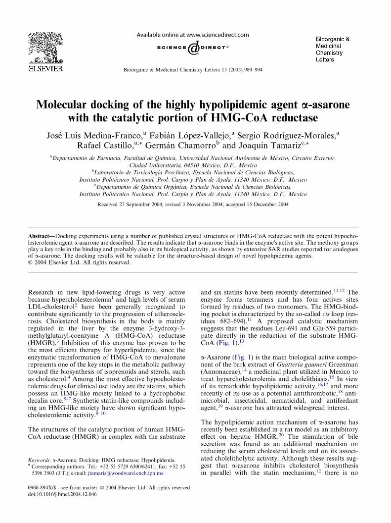
and six statins have been recently determined.11,12 Theenzyme forms tetramers and has four actives sitesformed by residues of two monomers. The HMG-bind-ing pocket is characterized by the so-called cis loop (res-idues 682–694).11 A proposed catalytic mechanismsuggests that the residues Leu-691 and Glu-559 partici-pate directly in the reduction of the substrate HMG-CoA (Fig. 1).13
a-Asarone (Fig. 1) is the main biological active compo-nent of the bark extract of Guatteria gaumeri Greenman(Annonaceae),14 a medicinal plant utilized in Mexico totreat hypercholesterolemia and cholelithiasis.15 In viewof its remarkable hypolipidemic activity,16,17 and morerecently of its use as a potential antithrombotic,18 anti-microbial, insecticidal, nematicidal, and antifeedantagent,19 a-asarone has attracted widespread interest.
The hypolipidemic action mechanism of a-asarone hasrecently been established in a rat model as an inhibitoryeffect on hepatic HMGR.20 The stimulation of bilesecretion was found as an additional mechanism onreducing the serum cholesterol levels and on its associ-ated cholelitholytic activity. Although these results sug-gest that a-asarone inhibits cholesterol biosynthesisin parallel with the statin mechanism,12 there is no
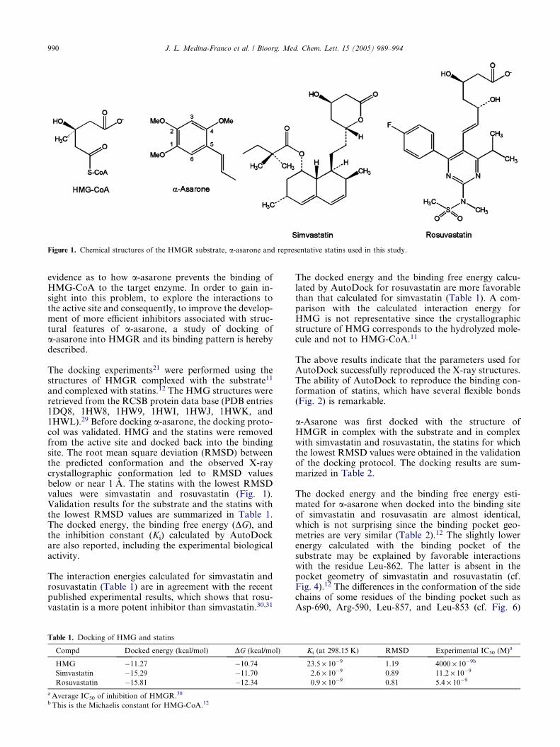
Figure 1. Chemical structures of the HMGR substrate, a-asarone and representative statins used in this study.
990 J. L. Medina-Franco et al. / Bioorg. Med. Chem. Lett. 15 (2005) 989–994
evidence as to how a-asarone prevents the binding ofHMG-CoA to the target enzyme. In order to gain in-sight into this problem, to explore the interactions tothe active site and consequently, to improve the develop-ment of more efficient inhibitors associated with struc-tural features of a-asarone, a study of docking ofa-asarone into HMGR and its binding pattern is herebydescribed.
The docking experiments21 were performed using thestructures of HMGR complexed with the substrate11
and complexed with statins.12 The HMG structures wereretrieved from the RCSB protein data base (PDB entries1DQ8, 1HW8, 1HW9, 1HWI, 1HWJ, 1HWK, and1HWL).29 Before docking a-asarone, the docking proto-col was validated. HMG and the statins were removedfrom the active site and docked back into the bindingsite. The root mean square deviation (RMSD) betweenthe predicted conformation and the observed X-raycrystallographic conformation led to RMSD valuesbelow or near 1 A. The statins with the lowest RMSDvalues were simvastatin and rosuvastatin (Fig. 1).Validation results for the substrate and the statins withthe lowest RMSD values are summarized in Table 1.The docked energy, the binding free energy (DG), andthe inhibition constant (Ki) calculated by AutoDockare also reported, including the experimental biologicalactivity.
The interaction energies calculated for simvastatin androsuvastatin (Table 1) are in agreement with the recentpublished experimental results, which shows that rosu-vastatin is a more potent inhibitor than simvastatin.30,31
Table 1. Docking of HMG and statins
Compd Docked energy (kcal/mol) DG (kcal/mol)
HMG �11.27 �10.74Simvastatin �15.29 �11.70Rosuvastatin �15.81 �12.34
a Average IC50 of inhibition of HMGR.30
b This is the Michaelis constant for HMG-CoA.12
The docked energy and the binding free energy calcu-lated by AutoDock for rosuvastatin are more favorablethan that calculated for simvastatin (Table 1). A com-parison with the calculated interaction energy forHMG is not representative since the crystallographicstructure of HMG corresponds to the hydrolyzed mole-cule and not to HMG-CoA.11
The above results indicate that the parameters used forAutoDock successfully reproduced the X-ray structures.The ability of AutoDock to reproduce the binding con-formation of statins, which have several flexible bonds(Fig. 2) is remarkable.
a-Asarone was first docked with the structure ofHMGR in complex with the substrate and in complexwith simvastatin and rosuvastatin, the statins for whichthe lowest RMSD values were obtained in the validationof the docking protocol. The docking results are sum-marized in Table 2.
The docked energy and the binding free energy esti-mated for a-asarone when docked into the binding siteof simvastatin and rosuvasatin are almost identical,which is not surprising since the binding pocket geo-metries are very similar (Table 2).12 The slightly lowerenergy calculated with the binding pocket of thesubstrate may be explained by favorable interactionswith the residue Leu-862. The latter is absent in thepocket geometry of simvastatin and rosuvastatin (cf.Fig. 4).12 The differences in the conformation of the sidechains of some residues of the binding pocket such asAsp-690, Arg-590, Leu-857, and Leu-853 (cf. Fig. 6)
Ki (at 298.15 K) RMSD Experimental IC50 (M)a
23.5 · 10�9 1.19 4000 · 10�9b
2.6 · 10�9 0.89 11.2 · 10�9
0.9 · 10�9 0.81 5.4 · 10�9

Table 2. Results of docking experiments of a-asarone with the
catalytic portion of human HMGR
Binding pocket Docked energy
(kcal/mol)
DG(kcal/mol)
Ki(at 298.15 K)
HMG-CoA �6.60 �6.02 3.9 · 10�5
Simvastatin �6.29 �5.68 6.8 · 10�5
Rosuvastatin �6.24 �5.66 7.1 · 10�5
Figure 2. Comparison between the binding position of rosuvastatin
found within the crystal structure (yellow) and the conformation
predicted by AutoDock (red).
Figure 3. Docking model derived for a-asarone with the catalyticportion of HMGR. The optimized complex of a-asarone with thebinding pocket of the substrate is shown. Hydrogen bonds are
displayed as yellow dashes. Representative residues within 3.6 A of a-asarone are shown. Residues from one monomer are orange and those
from the other monomer are purple. Nonpolar hydrogens are omitted
for clarity except the hydrogens involved in hydrogen bonding.
J. L. Medina-Franco et al. / Bioorg. Med. Chem. Lett. 15 (2005) 989–994 991
may also contribute to this difference. The much lessfavorable binding free energy calculated for a-asaronecompared with the free energy of binding calculatedfor simvastatin and rosuvastatin (cf. Tables 1 and 2) isquite noteworthy. This is in good agreement with theexperimental HMGR inhibition trend determined fora-asarone and the statins that show inhibition constants(Ki) and IC50 in the nanomolar range for rosuvastatinand simvastatin30 and IC50 in the millimolar range fora-asarone.20 These observations suggest that the bindingmode predicted for a-asarone is reasonable.
AutoDock found one main binding mode for a-asaronein all binding pockets used. a-Asarone binds in the ac-tive site of HMGR occupying the region where the 3-hydroxymethylglutaryl moiety of the substrate binds(Fig. 3).
According to the derived docking model, a-asaroneinteracts with two monomers of the HMGR tetramer.Polar interactions with residues Ser-684, Asp-690, Lys-691, and Lys-692 of the cis loop and with Arg-590 ofone monomer are observed (Fig. 3). The C-4 methoxygroup of a-asarone makes polar contacts with Glu-559of the second monomer and hydrogen bonds wereformed with the side chains of Lys-691 and Asn-755.Additional hydrogen bonds were formed between C-1and C-2 methoxy groups and Ser-684 and Arg-590(Fig. 3). The propenyl side chain of a-asarone is close
to Glu-559, Leu-562, His-752, Asn-755, and Leu-853,all residues being of one monomer. The aromatic ringis close to Leu-853 and Leu-862, with which van derWaals contacts are made. These interactions seem tobe responsible for the affinity of a-asarone with HMGR,thus sterically preventing the substrate from binding.Similar conclusions were obtained when the dockingexperiments were conducted using the structures ofHMGR in complex with compactin, fluvastatin, ceriva-statin, and atorvastatin.
The interactions of a-asarone with the catalytic site ofHMGR resemble the interactions observed in the sub-strate and statins complexes. The C-4 methoxy groupof a-asarone occupies a region similar to the C-5 car-bonyl oxygen of HMG-CoA and to the C-5 hydroxylgroup of statins, the latter interacting with the sidechains of Lys-691 and Glu-559, which seem to partici-pate directly in the catalysis (Fig. 4).13 According tothe mechanism of catalysis proposed for the reductionof HMG-CoA,13 which suggests a Glu-559 protonatedunder physiological conditions, the C-4 oxygen of themethoxy group in a-asarone may also form a hydrogenbond with the carboxylic group of Glu-559.
The C-1 and C-2 methoxy groups of a-asarone have alsosimilar interactions to those of the C-1 carboxylategroup of both the substrate and the statins, includinghydrogen bonds with Ser-684 and interactions with thepolar side chains of Lys-692 and Lys-735.11,12 The C-1methoxy group is located in the binding region of thebutyryl group of simvastatin and the fluorophenyl groupof rosuvastatin (Fig. 4b and c, respectively). The methylgroup of the C-1 methoxy group makes van der Waalscontacts with the side chain of Leu-857. The propenylside chain of a-asarone is located near the hydrophobicgroup of the statins.

Figure 4. Comparison of the predicted binding mode of a-asarone with the actual binding mode of (a) the substrate; (b) simvastatin, and (c)rosuvastatin. Representative residues within 3.6 A of a-asarone are shown. Residues are colored as in Figure 3. Nonpolar hydrogens are omitted forclarity.
992 J. L. Medina-Franco et al. / Bioorg. Med. Chem. Lett. 15 (2005) 989–994
The above observations suggest that the interactions ofthe C-1, C-2, and C-4 oxygen atoms on a-asarone arevery important in the a-asarone binding to HMGRand play a similar role to that of the HMG-like moietypresent in the substrate and the statins (Fig. 5). This is inagreement with the experimental observation that thetrimethoxybenzene core seems to be fundamental forthe hypocholesterolemic activity of a-asarone ana-logues.32 The docking model also helps to visualize, atthe molecular level, the experimental results that indi-cate that the side chain of a-asarone should be at themeta and para positions of the C-1 and C-2 methoxygroups, respectively.33
Figure 5. (a) HMG-like moiety of the substrate (in bold). (b) Structure
fragment of a-asarone (in bold) that play a similar role to that of theHMG-like moiety in the binding to HMGR.
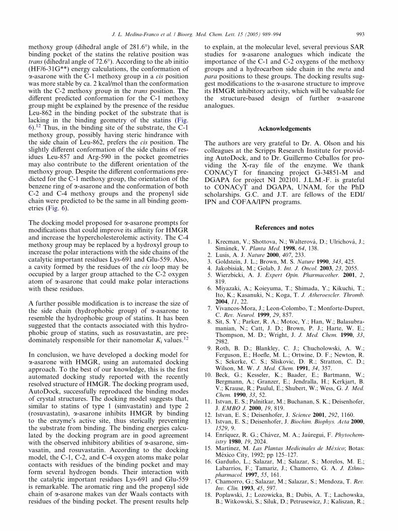
Figure 6. Predicted binding conformations for the C-1 methoxy group
of a-asarone when docked into the binding pocket of the substrate(yellow) and into the binding pocket of rosuvastatin (cyan). Repre-
sentative residues are shown. Nonpolar hydrogens are omitted for
clarity.
AutoDock found two possible binding conformationsfor the C-1 methoxy group depending on the bindingsite. In the binding pocket of the substrate, the methoxygroup was in a cis position relative to the vicinal C-2
J. L. Medina-Franco et al. / Bioorg. Med. Chem. Lett. 15 (2005) 989–994 993
methoxy group (dihedral angle of 281.6�) while, in thebinding pocket of the statins the relative position wastrans (dihedral angle of 72.6�). According to the ab initio(HF/6-31G**) energy calculations, the conformation ofa-asarone with the C-1 methoxy group in a cis positionwas more stable by ca. 2 kcal/mol than the conformationwith the C-2 methoxy group in the trans position. Thedifferent predicted conformation for the C-1 methoxygroup might be explained by the presence of the residueLeu-862 in the binding pocket of the substrate that islacking in the binding geometry of the statins (Fig.6).12 Thus, in the binding site of the substrate, the C-1methoxy group, possibly having steric hindrance withthe side chain of Leu-862, prefers the cis position. Theslightly different conformation of the side chains of res-idues Leu-857 and Arg-590 in the pocket geometriesmay also contribute to the different orientation of themethoxy group. Despite the different conformations pre-dicted for the C-1 methoxy group, the orientation of thebenzene ring of a-asarone and the conformation of bothC-2 and C-4 methoxy groups and the propenyl sidechain were predicted to be the same in all binding geom-etries (Fig. 6).
The docking model proposed for a-asarone prompts formodifications that could improve its affinity for HMGRand increase the hypercholesterolemic activity. The C-4methoxy group may be replaced by a hydroxyl group toincrease the polar interactions with the side chains of thecatalytic important residues Lys-691 and Glu-559. Also,a cavity formed by the residues of the cis loop may beoccupied by a larger group attached to the C-2 oxygenatom of a-asarone that could make polar interactionswith these residues.
A further possible modification is to increase the size ofthe side chain (hydrophobic group) of a-asarone toresemble the hydrophobic group of statins. It has beensuggested that the contacts associated with this hydro-phobic group of statins, such as rosuvastatin, are pre-dominately responsible for their nanomolar Ki values.
12
In conclusion, we have developed a docking model fora-asarone with HMGR, using an automated dockingapproach. To the best of our knowledge, this is the firstautomated docking study reported with the recentlyresolved structure of HMGR. The docking program used,AutoDock, successfully reproduced the binding modesof crystal structures. The docking model suggests that,similar to statins of type 1 (simvastatin) and type 2(rosuvastatin), a-asarone inhibits HMGR by bindingto the enzyme�s active site, thus sterically preventingthe substrate from binding. The binding energies calcu-lated by the docking program are in good agreementwith the observed inhibitory abilities of a-asarone, sim-vasatin, and rosuvastatin. According to the dockingmodel, the C-1, C-2, and C-4 oxygen atoms make polarcontacts with residues of the binding pocket and mayform several hydrogen bonds. Their interaction withthe catalytic important residues Lys-691 and Glu-559is remarkable. The aromatic ring and the propenyl sidechain of a-asarone makes van der Waals contacts withresidues of the binding pocket. The present results help
to explain, at the molecular level, several previous SARstudies for a-asarone analogues which indicate theimportance of the C-1 and C-2 oxygens of the methoxygroups and a hydrocarbon side chain in the meta andpara positions to these groups. The docking results sug-gest modifications to the a-asarone structure to improveits HMGR inhibitory activity, which will be valuable forthe structure-based design of further a-asaroneanalogues.
Acknowledgements
The authors are very grateful to Dr. A. Olson and hiscolleagues at the Scripps Research Institute for provid-ing AutoDock, and to Dr. Guillermo Ceballos for pro-viding the X-ray file of the enzyme. We thankCONACyT for financing project G-34851-M andDGAPA for project NI 202101. J.L.M.-F. is gratefulto CONACyT and DGAPA, UNAM, for the PhDscholarships. G.C. and J.T. are fellows of the EDI/IPN and COFAA/IPN programs.
References and notes
1. Krecman, V.; Shottova, N.; Walterova, D.; Ulrichova, J.;Simanek, V. Planta Med. 1998, 64, 138.
2. Lusis, A. J. Nature 2000, 407, 233.3. Goldstein, J. L.; Brown, M. S. Nature 1990, 343, 425.4. Jakobisiak, M.; Golab, J. Int. J. Oncol. 2003, 23, 2055.5. Wierzbicki, A. J. Expert Opin. Pharmacother. 2001, 2,819.
6. Miyazaki, A.; Koieyuma, T.; Shimada, Y.; Kikuchi, T.;Ito, K.; Kasanuki, N.; Koga, T. J. Atheroescler. Thromb.2004, 11, 22.
7. Vivancos-Mora, J.; Leon-Colombo, T.; Monforte-Dupret,C. Rev. Neurol. 1999, 29, 857.
8. Sit, S. Y.; Parker, R. A.; Motoc, Y.; Han, W.; Balasubra-manian, N.; Catt, J. D.; Brown, P. J.; Harte, W. E.;Thompson, M. D.; Wright, J. J. Med. Chem. 1990, 33,2982.
9. Roth, B. D.; Blankley, C. J.; Chucholowski, A. W.;Ferguson, E.; Hoefle, M. L.; Ortwine, D. F.; Newton, R.S.; Sekerke, C. S.; Sliskovic, D. R.; Stratton, C. D.;Wilson, M. W. J. Med. Chem. 1991, 34, 357.
10. Beck, G.; Kesseler, K.; Baader, E.; Bartmann, W.;Bergmann, A.; Granzer, E.; Jendralla, H.; Kerkjart, B.V.; Krause, R.; Paulul, E.; Shubert, W.; Wess, G. J. Med.Chem. 1990, 33, 52.
11. Istvan, E. S.; Palnitkar, M.; Buchanan, S. K.; Deisenhofer,J. EMBO J. 2000, 19, 819.
12. Istvan, E. S.; Deisenhofer, J. Science 2001, 292, 1160.13. Istvan, E. S.; Deisenhofer, J. Biochim. Biophys. Acta 2000,
1529, 9.14. Enrıquez, R. G.; Chavez, M. A.; Jauregui, F. Phytochem-
istry 1980, 19, 2024.15. Martınez, M. Las Plantas Medicinales de Mexico; Botas:
Mexico City, 1992; pp 125–127.16. Garduno, L.; Salazar, M.; Salazar, S.; Morelos, M. E.;
Labarrios, F.; Tamariz, J.; Chamorro, G. A. J. Ethno-pharmacol. 1997, 55, 161.
17. Chamorro, G.; Salazar, M.; Salazar, S.; Mendoza, T. Rev.Inv. Clin. 1993, 45, 597.
18. Poplawski, J.; Lozowicka, B.; Dubis, A. T.; Lachowska,B.; Witkowski, S.; Siluk, D.; Petrusewicz, J.; Kaliszan, R.;

994 J. L. Medina-Franco et al. / Bioorg. Med. Chem. Lett. 15 (2005) 989–994
Cybulski, J.; Strzalkowska, M.; Chilmonczyk, Z. J. Med.Chem. 2000, 43, 3671.
19. Momin, A. A.; Nair, M. G. J. Agric. Food Chem. 2002, 50,4475.
20. Rodrıguez-Paez, L.; Juarez-Sanchez, M.; Antunez-Solıs,J.; Baeza, I.; Wong, C. Phytomedicine 2003, 10, 397.
21. Automated docking was used to locate the appropriatebinding orientation and conformation of a-asarone withHMGR. The powerful genetic algorithm method imple-mented in the program AutoDock 3.022 was employed.The structures of a-asarone and the proteins wereprepared using Sybyl 6.8.23 All water, adenosine-5 0-diphosphate, and 2,3-dihydroxy-1,4-dithiobutane mole-cules when present were removed from the original RCSBprotein data base files. Polar hydrogen atoms were addedand Kollman charges,24 atomic solvation parameters andfragmental volumes were assigned to the protein usingAutoDock tools (ADT). For docking calculations, Gastei-ger–Marsili partial charges25 were assigned to the ligandsand nonpolar hydrogen atoms were merged. All torsionswere allowed to rotate during docking. The auxiliaryprogram AutoGrid generated the grid maps. Each gridwas centered at the crystal structure of the substrate orthe corresponding statin. The grid dimensions were23 · 23 · 23 A3 with points separated by 0.375 A. Thegrid dimensions were large enough to cover the substrate,the statins and the NADP(H) binding sites. Lennard–Jones parameters 12–10 and 12–6, supplied with theprogram, were used for modeling H-bonds and van derWaals interactions, respectively. The distance-dependentdielectric permittivity of Mehler and Solmajer26 was usedfor the calculation of the electrostatic grid maps. For allligands, random starting positions, random orienta-tions, and torsions were used. The translation, quaternion,and torsion steps were taken from default values inAutoDock. The Lamarckian genetic algorithm and thepseudo-Solis and Wets methods were applied for minimi-zation, using default parameters. The number of dockingruns was 100. The population in the genetic algorithm was50, the energy evaluations were 250,000, and the maxi-mum number of iterations 27,000. The complexes of a-asarone with HMGR resulting from molecular docking
were further structurally optimized with the Tripos forcefield.27 During minimization, atoms within 6 A from theligand were free to move (other atoms were fixed). Forcomplex optimization, a-asarone with Mulliken chargesobtained from ab initio (HF/6-31G**) single-point energycalculation was used. For the protein, Kollman all chargesencoded in Sybyl 6.8 were employed. The ab initiocalculations were conducted with Spartan�02.28
22. Morris, G. M.; Goodsell, D. S.; Halliday, R. S.; Huey, R.;Hart, W. E.; Belew, R. K.; Olson, A. J. J. Comp. Chem.1998, 19, 1639.
23. Sybyl, version 6.8; Tripos, Inc.: St. Louis, MO, 2001.24. Weiner, S. J.; Kollman, P. A.; Case, D. A.; Singh, U. C.;
Ghio, C.; Alagona, G.; Profeta, S.; Weiner, P. J. Am.Chem. Soc. 1984, 106, 765.
25. Gasteiger, J.; Marsili, M. Tetrahedron 1980, 36, 3219.26. Mehler, E. L.; Solmajer, T. Protein Eng. 1991, 4, 903.27. Purcel, W. P.; Singer, J. A. J. Chem. Eng. Data 1967, 12,
235–246. Details of the implementation are given in Sybyl6.6 Force Field Manual; Tripos: St. Louis, MO, 1999; p 80.
28. Kong, J.; White, C. A.; Krylov, A. I.; Sherrill, C. D.;Adamson, R. D.; Furlani, T. R.; Lee, M. S.; Lee, A. M.;Gwaltney, S. R.; Adams, T. R.; Ochsenfeld, C.; Gilbert, A.T. B.; Kedziora, G. S.; Rassolov, V. A.; Maurice, D. R.;Nair, N.; Shao, Y.; Besley, N. A.; Maslen, P. E.;Dombroski, J. P.; Daschel, H.; Zhang, W.; Korambath,P. P.; Baker, J.; Byrd, E. F. C.; Van Vooris, T.; Oumi, M.;Hsu, C.-P.; Ishikawa, N.; Florian, J.; Warshel, A.;Johnson, B. G.; Gill, P. M. W.; Head-Gordon, M.; Pople,J. A. J. Comput. Chem. 2000, 21, 1532.
29. Brookhaven Protein Data Bank. http://www.rcsb.org.30. McTaggart, F.; Buckett, L.; Davidson, R.; Holdgate, G.;
McCormick, A.; Scheneck, D.; Smith, G.; Warwick, M.Am. J. Cardiol. 2001, 87(suppl.), 28B.
31. Holdgate, G. A.; Ward, W. H. J.; McTaggart, F. Biochem.Soc. Trans. 2003, 31, 528.
32. Cruz, A.; Garduno, L.; Salazar, M.; Martınez, E.;Jimenez-Vazquez, H. A.; Dıaz, F.; Chamorro, G.; Tama-riz, J. Arzneimittel-Forsch. 2001, 51, 535.
33. Cruz, M. C.; Salazar, M.; Garciafigueroa, Y.; Hernandez,D.; Dıaz, F.; Chamorro, G.; Tamariz, J. Drug Dev. Res.2003, 60, 186.