Clinical Biologic Pathophysiologies of Women's Sexual Dysfunction

The Heparin-binding Domain of IGFBP-2 Has Insulin-likeGrowth Factor Binding-independent Biologic Activity in theGrowing Skeleton*□S
Received for publication, October 12, 2010, and in revised form, January 30, 2011 Published, JBC Papers in Press, March 3, 2011, DOI 10.1074/jbc.M110.193334
Masanobu Kawai‡§, Anne C. Breggia‡, Victoria E. DeMambro‡, Xinchun Shen¶, Ernesto Canalis�, Mary L. Bouxsein**,Wesley G. Beamer‡‡, David R. Clemmons¶, and Clifford J. Rosen‡1
From the ‡Center for Clinical and Translational Research, Maine Medical Center Research Institute, Scarborough, Maine 04074,the §Department of Bone and Mineral Research, Osaka Medical Center and Research Institute for Maternal and Child Health,Izumi, Osaka 594-1101, Japan, the ¶Department of Medicine, University of North Carolina, School of Medicine, Chapel Hill,North Carolina 27599, the �Department of Research, Saint Francis Hospital & Medical Center, Hartford, Connecticut 06105,the **Orthopaedic Biomechanics Laboratory, Beth Israel Deaconess Medical Center and Harvard Medical School,Boston, Massachusetts 02215, and ‡‡The Jackson Laboratory, Bar Harbor, Maine 04609
Insulin-like growth factor-binding protein 2 (IGFBP-2) is amember of a family of six highly conserved IGFBPs that are car-riers for the insulin-like growth factors (IGFs). IGFBP-2 levelsrise during rapid neonatal growth and at the time of peak boneacquisition. In contrast, Igfbp2�/� mice have low bone massaccompanied by reduced osteoblast numbers, low bone forma-tion rates, and increased PTEN expression. In the current study,we postulated that IGFBP-2 increased bonemass partly throughthe activity of its heparin-binding domain (HBD). We synthe-sized a HBD peptide specific for IGFBP-2 and demonstrated invitro that it rescued the mineralization phenotype of Igfbp2�/�
bonemarrow stromal cells and calvarial osteoblasts. Consistentwith its cellular actions, the HBD peptide ex vivo stimulatedmetacarpal periosteal expansion. Furthermore, administrationofHBDpeptide to Igfbp2�/� mice increased osteoblast number,suppressed marrow adipogenesis, restored trabecular bonemass, and reduced bone resorption. Skeletal rescue in theIgfbp2�/� mice was characterized by reduced PTEN expressionfollowed by enhancedAkt phosphorylation in response to IGF-Iand increased �-catenin signaling through two mechanisms: 1)stimulation of its cytosolic accumulation and 2) increased phos-phorylation of serine 552.We conclude that theHBDpeptide ofIGFBP-2 has anabolic activity by activating IGF-I/Akt and�-catenin signaling pathways. These data support a growingbody of evidence that IGFBP-2 is not just a transport protein butrather that it functions coordinately with IGF-I to stimulategrowth and skeletal acquisition.
Insulin-like growth factor-binding protein-2 (IGFBP-2)2 is amember of a highly conserved family of six IGFBPs that circu-
late or reside locally in the extracellular space including thebone marrow microenvironment (1–3). IGFBP-2 has highaffinity for IGF-I/IGF-II and is believed to regulate IGF bio-availability in the pericellular environment. IGFBP-2 is the sec-ondmost abundant circulating IGFBPs and is expressed in sev-eral mammalian tissues including the skeleton (2). Growingevidence demonstrates that increased IGFBP-2 levels are asso-ciated with reduced adipose tissue mass and improved glucosemetabolism both in human and mouse models (4–6). Hed-backer et al. (5) recently reported that IGFBP-2 exhibits acuteglucose lowering properties in several diabetic mouse modelsand that IGFBP-2maymediate someof themetabolic activity ofleptin. However, the role of IGFBP-2 in skeletal homeostasis isless clear and is dependent on the experimental model system.Generally, IGFBP-2 has been shown to inhibit IGF-I actions invitro (3). Also, overexpression of Igfbp2 inhibits growth hor-mone-stimulated chondrocytic expansion in growth hormonetransgenic mice (7). However, Khosla and co-workers (8, 9)reported that IGFBP-2 markedly stimulated bone formation inpatients with hepatitis C-associated osteosclerosis, and theadministration of IGFBP-2 � IGF-II prevented bone loss inimmobilized rats. Furthermore, we recently showed thatIgfbp2�/� mice had markedly impaired bone formation,reduced trabecular bone volume fraction, and altered micro-architecture and low bone turnover, suggesting that IGFBP-2might be extremely important within the skeletal milieu (10).In addition to its IGF-I binding dependent actions, IGFBP-2
and other IGFBPs have been shown to stimulate biologicresponses that are independent of their abilities to bind to IGFs(3). Similarly Lodish and co-workers (11) recently demon-strated that IGFBP-2 alone could stimulate hematopoietic stemcells ex vivo, although the mechanism of its action was notdescribed. Previously, IGFBP-2 was reported to bind to extra-cellular matrices (12, 13). More recently several lines of evi-dence show that the heparin-binding domain (HBD) plays a
* This work was supported, in whole or in part, by National Institutes of HealthGrant AR021707 (to E. C.) and National Institutes of Arthritis, Musculoskel-etal, and Skin Disorders Grant AR153853 (to C. J. R.).
□S The on-line version of this article (available at http://www.jbc.org) containssupplemental Tables S1 and S2 and Figs. S1–S3.
1 To whom correspondence should be addressed: Maine Medical CenterResearch Institute, 81 Research Dr., Scarborough, ME 04074-7205. Tel.: 207-885-8100; Fax: 207-885-8174; E-mail: [email protected].
2 The abbreviations used are: IGFBP, insulin-like growth factor-binding pro-tein; ALP, alkaline phosphatase; PPAR-�, peroxisome proliferator-activated
receptor-�; HBD, heparin-binding domain; BMSC, bone marrow stromalcell; COB, calvarial osteoblast; �MEM, �-minimum essential medium; PEG-HBD, pegylated HBD; aBMD, areal bone mineral density; BrdUrd, bromode-oxyuridine; BW, body weight; aBMC, areal bone mineral content; PTEN,phosphatase and tensin homolog.
THE JOURNAL OF BIOLOGICAL CHEMISTRY VOL. 286, NO. 16, pp. 14670 –14680, April 22, 2011© 2011 by The American Society for Biochemistry and Molecular Biology, Inc. Printed in the U.S.A.
14670 JOURNAL OF BIOLOGICAL CHEMISTRY VOLUME 286 • NUMBER 16 • APRIL 22, 2011
role in IGFBP-2 function. Russo et al. (14) reported thatIGFBP-2was proteolytically cleaved and that smaller fragmentsthat include HBD still possessed the capacity to bind to extra-cellular matrix and with low affinity to IGF-I. They also dem-onstrated that IGFBP-2 stimulated the proliferation and meta-static behavior of neuroblastoma cells through the HBD (15).Interestingly, zebrafish IGFBP-2 lacks the HBD and cannotbind to any extracellular matrix (16). Taken together, theseresults suggest that IGFBP-2 binding to extracellular matrix islikely to be dependent on the HBD.PTEN (phosphatase and tensin homolog deleted on chromo-
some 10) is a lipid phosphatase that opposes IGF-I signaling bydephosphorylating phosphatidylinositol 3,4,5-trisphosphate tophosphatidylinositol 4,5-diphosphate (17). Mice lacking PTENonly in osteoblasts showed progressively increasing bone masswith age, indicating the important role of PTEN and the PI3K/Akt signaling pathway in bonemaintenance (18). There is also anegative correlation between IGFBP-2 and PTEN expression incertain types of tumor cells (19–21). Moreover, we reportedthat PTEN expression was enhanced in osteoblasts derivedfrom Igfbp2�/� bone marrow stromal cells (10). Interestingly,Perks et al. (20) suggested that IGFBP-2 suppression of PTENwasmediated in an IGF binding-independent manner. BecauseAkt activation is a critical factor regulating bone mass (22, 23),we speculated that IGFBP-2 could enhance Akt signaling bysuppressing PTEN expression independent of IGF-I bindingand that the HBD is involved in the down-regulation of PTEN.IGFBP-2 contains two putative HBDs: one HBD in the C
terminus domain shares the sequence similarity with otherHBD containing IGFBPs such as IGFBP-3 and IGFBP-5 (3). Onthe other hand, theHBD in the linker region is unique and is notpresent in other IGFBPs. Based on the unique sequence of theIGFBP-2 HBD subsequently referred to simply as the HBD, wegenerated a peptide corresponding to the one in the linkerregion and tested whether the HBD peptide affected skeletalturnover.We found that theHBDpeptide has an anabolic effecton the skeleton through suppression of PTEN in osteoblastsand that this effect was mediated by enhanced Akt and�-catenin signaling. Moreover, there was significant synergybetween the HBD and IGF-I in respect to enhancement ofpAKT and �-catenin production.
EXPERIMENTAL PROCEDURES
Mice—Generation of the original mixed background strainB6;129-Igfbp2�tm1Jep�, which we refer to as Igfbp2�/� mice,has been described previously (10, 24). The original mice werebackcrossed onto C57BL/6J background for 10 generations.Igfbp2�/� mice were C57BL/6J controls. All of the experimen-tal studies were performed with male mice. All of the animalstudies were reviewed and approved by the Institutional Ani-mal Care and Use Committee of Maine Medical CenterResearch Institute.Cell Culture—MC3T3-E1 cells were purchased from ATCC
andmaintained in�MEMcontaining 10% FCS neonatal calvar-ial osteoblasts were collected from 7–10-day-old mice. Briefly,calvariae were digested five times with collagenase P and tryp-sin. The cells released from digests 2–5 were collected asprimary calvarial osteoblasts andmaintained inDMEMsupple-
mented with 10% FCS and nonessential amino acids. Osteo-blastogenesis of primary calvarial osteoblasts was analyzed bytreating cells with 4mMof�-glycerophosphate and 50�g/ml ofascorbic acid in �MEM with 10% FCS. Bone marrow stromalcells (BMSCs) were harvested from femurs and tibias of8-week-old mice. BMSCs were grown in �MEM containing10% FCS. Osteoblastogenesis of BMSCs was induced by thetreatment with osteogenic medium consisting of �MEM con-taining 10% FCS, 8 mM �-glycerophosphate, and 50 �g/mlascorbic acid.Metacarpal Culture—Metacarpals were isolated from
Igfbp2�/� mice from 1-day-old mice and incubated in DMEMcontaining 0.5% BSA, 50�g/ml ascorbic acid, and 1mM �-glyc-erol phosphate for 10 days. Stimulants were added to culturemedium from day 1. At day 10, metacarpals were incubated inmedium containing calcein (500 ng/ml) for 2 h for staining ofcalcium deposition, and images were obtained. The bones werethen fixed with 4% phosphonoformic acid, embedded in paraf-fin, and processed for Alcian blue and von Kossa doublestaining.Real Time RT-PCR—Total RNA was prepared using a
RNeasymini kit (Qiagen). cDNAwas generated using a randomhexamer and reverse transcriptase (Superscript III; Invitrogen)according to themanufacturer’s instructions. Quantification ofmRNA expression was carried out using an iQ SYBR GreenSupermix in a iQ5 thermal cycler and detection system (Bio-Rad). GAPDHwas used as an internal standard control gene forall quantification. The primer sequences are available uponrequest.Western Blot Analysis—To prepare whole cell lysates, the
cells were solubilized in radioimmune precipitation assaybuffer (50 mM Tris, 150 mM NaCl, 1 mM EDTA, 1% NonidetP-40, 0.25% sodium deoxycholate, 2 �g/ml aprotinin, 2 �g/mlleupeptin, 2 �g/ml pepstatin A, 0.5 mM phenylmethylsulfonylfluoride, and 1mM dithiothreitol). To isolate the cytosolic frac-tion, the cells were solubilizedwith hypotonic buffer containingprotease and phosphatase inhibitors (10 mM Tris, 0.2 mM
MgCl2, pH 7.4) and kept on ice for 10min. The cell lysates weremixedwith sucrose buffer (final concentrations: 25mMsucrose,0.1mMEDTA) and centrifuged at 20,000� g for 1 h. The super-natant contained the cytosolic fraction. To prepare the mem-brane fraction, the cells were solubilized with PBS containingprotease and phosphatase inhibitors. The lysates were frozen at�80 °C for 1 h and thawed at room temperature. After threecycles, they were centrifuged at 13,000 � g for 25 min. Thepellet was resuspendedwithmembrane protein isolation buffer(20mMTris-HCl, 150mMNaCl, 1mMEDTA, 1mMEGTA, and1% Triton X-100, pH 7.5) containing protease and phosphataseinhibitors. Equal amounts of sample were separated by SDS-PAGE and transferred electrophoretically to nitrocellulosemembranes. The membranes were blocked in 5% BSA in Tris-buffered saline. Thereafter, the membranes were immuno-blotted with anti-PTEN (Cell Signaling), anti-Akt (Cell Signal-ing), anti-Ser(P)473-Akt (Cell Signaling), anti-�-catenin (BDTransduction Laboratories, 610153), anti-Ser(P)552-�-catenin(Cell Signaling), or anti-�-actin (Santa Cruz) and developedwith horseradish peroxidase-coupled anti-mouse or rabbit IgG
Heparin-binding Domain of IGFBP-2 Is Anabolic to Growing Bone
APRIL 22, 2011 • VOLUME 286 • NUMBER 16 JOURNAL OF BIOLOGICAL CHEMISTRY 14671

antibodies, followed by enhancement with SuperSignal WestDura extended duration substrate antibodies (Pierce).Immunoprecipitation with Akt1 and Akt2—The cells were
starved with serum-free medium overnight and then exposedto 0 or 50 ng/ml IGF-I for 10 min. The cells were lysed withradioimmune precipitation assay buffer. The cell lysates werecentrifuged at 14,000 � g for 10 min at 4 °C. They were immu-noprecipitated with either anti-Akt1 antibody (1:500) or anti-Akt2 antibody (1:500) overnight at 4 °C. The immunoprecipi-tateswere immobilized using proteinA-Sepharose beads for 2 hat 4 °C and washed three times with the same buffer. The pre-cipitated proteins were eluted in 40 �l of 2� Laemmli samplebuffer, boiled for 5 min, and separated using 10% SDS-PAGE.The proteins were then transferred to Immobilon-P mem-branes. The blots were incubated overnight at 4 °C with theindicated antibodies. The proteins were visualized usingenhanced chemiluminescence (Pierce).Generation of Heparin-binding Domain Peptide—The
synthetic peptide containing the heparin-binding domain(amino acids 196–199) and nine additional amino acids ofmouse IGFBP-2, 188KHLSLEEPKKLRP200 (referred to asHBD peptide), and control peptide for HBD peptide(AALSLEEPAALAP) were synthesized by the Protein Chemis-try Core Facility at the University of North Carolina. Purity andsequence confirmation were determined by mass spectrome-try. The synthetic peptide for heparin-binding domain ofIGFBP5 (201RKGFYKRKQCKPSRGRKR218)was also generatedusing the same method as mentioned above. The IGFBP-2-HBD peptide was pegylated as follows: 10 mg of peptide wasmixed with 380 �g of methoxy PEG maleimide (20,000 kDa)(1:3 molar ratio peptide to PEG) (Jenkem Biotechnology) in 4.0ml of 0.05 MNaPO4, pH 7.0. Following an overnight incubationat 4 °C, cysteine was added to a final concentration of 17 mM toblock untreated sites. To remove the nonpegylated peptide andcysteine, the mixture was desalted using a Zebra Desalt spincolumn (Thermo Scientific) following the manufacturer’sinstructions. Pegylation was verified by SDS-PAGE analysiswith Coomassie staining.In Vitro IGFBP-2 Binding Assay—The cells were starvedwith
serum-free medium overnight and then exposed to 0 or 50ng/ml IGF-I for 10 min. They were suspended in 100 �l of PBS(pH 7.4) and incubated with biotinylated BP2 (final concentra-tion, 28 nM) and/or HBD (140 nM) for 4 h at 4 °C. The cells werewashed three times with PBS and lysed with radioimmune pre-cipitation assay buffer. The cell lysates were centrifuged at14,000 � g for 10 min at 4 °C. The proteins from supernatantswere separated by 12.5% of SDS-PAGE and detected usingHRP-conjugated avidin.In vivo treatment of Igfbp2�/� Mice with Pegylated (PEG)
HBD Peptide—Igfbp2�/� mice were administered with PBS orPEG-HBD peptide (50 �g/day) intraperitoneally five times/week from 6 to 9 weeks of age. Dual energy x-ray absorptiom-etry was performed before the initiation of treatment. Themicewere injected with 20 mg/kg calcein intraperitoneally 7 and 2days before sample collection.Dual Energy X-ray Absorptiometry—Dual energy x-ray
absorptiometry for whole body and femoral areal bone mineraldensity (aBMD, g/cm2) and body composition exclusive of
the head were performed using the PIXImus (GE-Lunar) asdescribed previously (10).Micro-computed Tomography—Microarchitecture of distal
trabecular bone and midshaft cortical bone were analyzed withfemurs and vertebrae (L5) by high resolution microcomputedtomography (MicroCT40; Scanco Medical AG, Switzerland).Approximately 100 CT slices were measured just proximal tothe distal growth plate, using an isotropic voxel size of 12 �m.Midshaft cortical bone properties were analyzed in a similarfashion using 18 CT slices obtained at the midfemoraldiaphysis.Bone Histomorphometry—In vivo histomorphometry differ-
ences were analyzed between Igfbp2�/� mice treated with PBSor PEG-HBD peptide at 9 weeks of age. The mice were injectedwith 20 mg/kg calcein intraperitoneally 7 and 2 days beforesample collection. The femurs were analyzed as described pre-viously (10).Femoral Biomechanics—Femoral biomechanical properties
were compared by three-point bending between Igfbp2�/�
mice treated with PBS or PEG-HBD peptide at 9 weeks of age.Load was applied at a constant rate (0.05 mm/s) until failure.We measured maximum load (newtons), bending stiffness(newtons/mm), and work-to-failure (newtons/mm) from theload displacement curve and computed the apparent elasticmodulus and ultimate strength using the relevant midfemo-ral cross-sectional geometry measured from microCT.In Vivo BrdUrd Assay—BrdUrd (100 �g/g of BW) and FdU
(12 �g/g of BW) were injected intraperitoneally in 2-week-oldmalemice. 2 h after of injection femurswere collected and fixedin 4% phosphonoformic acid overnight at 4 °C. The femurswere decalcified using 15% EDTA (pH 7.4) for 5 days and sec-tioned. Endogenous peroxidase was blocked with 0.3%H2O2 inPBS, and the sectionswere incubatedwith amousemonoclonalanti-BrdUrd antibody (1:200; Sigma B8434) overnight at 4 °C.The sections were then incubated with a biotinylated goat anti-mouse secondary antibody followed by the incubation withstreptavidin-biotinylated HRP complex and visualized with3,3�-diaminobenzidine.Statistical Analysis—All of the data are expressed as the
means � S.E. The results were analyzed for statistically signif-icant differences using Student’s t test or analysis of variancefollowed by Bonferroni multiple comparison post hoc test. Sta-tistical significance was set at p � 0.05.
RESULTS
The Heparin-binding Domain of IGFBP-2 Stimulates inVitro Osteoblastogenesis—Previously, we reported that maleIgfbp2�/� mice exhibited an osteopenic phenotype with lowbone formation and that osteoblastogenesis was impaired inBMSCs from Igfbp2�/� mice compared with wild-type cells.Interestingly, male Igfbp2�/� mice have a profound reductionin osteoblast number that accounts for the reduction in bonevolume fraction in the tibia and femur, although the percentageof osteoblast apoptosis by TUNEL staining did not differbetween Igfbp2�/� and Igfbp2�/�. To investigate whether theHBD of IGFBP-2 could be responsible for the phenotype of theIgfbp2�/� mouse, we generated a synthetic peptide (the HBDpeptide) that does not bind IGFs and that contains the HBD of
Heparin-binding Domain of IGFBP-2 Is Anabolic to Growing Bone
14672 JOURNAL OF BIOLOGICAL CHEMISTRY VOLUME 286 • NUMBER 16 • APRIL 22, 2011

IGFBP-2, and tested its effect on osteoblastogenesis in vitro.To confirm the specificity of the HBD peptide, we also made acontrol peptide and analyzed its effect on osteogenesis. Thecontrol peptide did not enhance mineralization of Igfbp2�/�
COBs compared with PBS-treated cells, whereas the HBD pep-tide stimulated COB mineralization (Fig. 1A). Based on this,PBS was used as controls for further studies. To prolong the
half-life of the HBD peptide such that it was feasible to under-take in vivo studies, pegylation of the HBD peptide was per-formed (PEG-HBD). Igfbp2�/� COBs and BMSCs were cul-tured in osteogenic medium in the presence of PBS, thePEG-HBD peptide, or the HBD peptide. Both PEG-HBD andHBD peptide enhanced osteogenesis as measured by alkalinephosphatase andAlizarin Red staining (Fig. 1,A–C). Consistent
FIGURE 1. The heparin-binding domain of IGFBP-2 enhances osteoblastogenesis in vitro and ex vivo. A, Igfbp2�/� COBs were cultured with osteogenicmedium with PBS, control peptide (2 �g/ml), or the HBD peptide (2 �g/ml). Osteoblastogenesis was evaluated by Alizarin Red staining. B–D, Igfbp2�/� COBsand BMSCs were treated with osteogenic medium with PEG-HBD peptide (2 �g/ml). ALP staining (day 7) and Alizarin Red staining (day 14) were performed (B).Alizarin Red staining positive area was quantified using NIH Image (C). Expression of ALP, OC, and Runx2 was analyzed by real time RT-PCR using BMSCs at day14 (n � 3) (D). E–L, Igfbp2�/� metacarpals were treated with osteogenic medium with PBS or control peptide (2 �g/ml) (E–H). Igfbp2�/� metacarpals weretreated with osteogenic medium with IGFBP-2 (200 ng/ml) or the HBD peptide (2 �g/ml) in the presence or absence of IGF-1 (20 ng/ml) (I–L). At day 10, thebones were treated with 500 ng/ml of calcein for 2 h. The calcein-incorporated area was visualized (E and I), and the longitudinal length of the calcein-incorporated area was quantified (F and J). Metacarpals were fixed with 4% phosphonoformic acid and subjected to Alcian blue and von Kossa double staining(G and K). The length and width of the whole metacarpals were quantified (H and L). The figures shown represent at least three independent experiments. Thevalues are expressed as the means � S.E. (n � 3– 4). *, p � 0.05; a, p � 0.05 versus PBS; b, p � 0.01 versus PBS; c, p � 0.001 versus PBS; d, p � 0.01 versus HBDpeptide; e, p � 0.05 versus IGF-I; f, p � 0.05 versus HBD peptide; ns, not significant.
Heparin-binding Domain of IGFBP-2 Is Anabolic to Growing Bone
APRIL 22, 2011 • VOLUME 286 • NUMBER 16 JOURNAL OF BIOLOGICAL CHEMISTRY 14673

with this,ALP,Runx2 (Runt-related transcription factor 2), andOC (osteocalcin) expression was enhanced in BMSCs treatedwith PEG-HBD- versus PBS-treated cells (Fig. 1D). Thus, therewas no difference between the pegylated form of HBD and theHBD itself with respect to induction of osteogenesis.The Heparin-binding Domain of IGFBP-2 Stimulates Perios-
teal Expansion of Metacarpals ex Vivo—To further understandthe anabolic effect of HBD on the skeleton, we next collectedmetacarpals from 1-day-old mice, incubated them with osteo-genic medium in the absence of fetal calf serum, and tested theeffects of the HBD peptide on osteogenesis. Bone accrual wasevaluated by the longitudinal length of the calcein-incorpo-rated area as well as the width and length of the wholemetacar-pals. The control peptide had no effect on the calcein-incorpo-rated area, and the length and width of the whole metacarpalswere comparable with themetacarpals treated with PBS (Fig. 1,E–H). Hence PBS was used as the control for further studies.IGFBP-2 and the HBD peptide significantly enhanced the cal-cein-incorporated area of metacarpals (Fig. 1, I and J). IGF-Ialso stimulated expansion of the calcein-incorporated area to asimilar extent as IGFBP-2 and the HBD peptide (Fig. 1, I and J).When metacarpals were treated with IGF-I together with theHBD peptide, the metacarpals displayed further enhancementof the calcein-incorporated area (Fig. 1, I and J). In addition,IGFBP-2 and HBD peptide also increased the width of thewhole metacarpals, implying that IGFBP-2 has an importantrole in periosteal expansion (Fig. 1, K and L).Because other IGFBPs possess HBD regions as well as
IGFBP-2,we analyzedwhether the osteogenic effect of theHBDis specific for this region in IGFBP-2 or whether the HBD inother forms of IGFBPs also had this effect. For this purpose, weprepared a peptide that contained the HBD region of IGFBP-5(BP5-HBD) inwhich the amino acid sequence is quite similar tothe HBD in the C-terminal region of IGFBP-2 and determinedits effect on osteogenesis. We cultured Igfbp2-deficient COBsin the presence of BP5-HBD and found that BP5-HBD did notshow any enhancement of differentiation or mineralizationcomparedwith PBS-treated cells (Fig. 2A). Second,we collectedmetacarpals and cultured them in osteogenic medium with orwithout BP5-HBD. BP5-HBD had no effect on the calcein-in-corporated area, length, or width of whole metacarpals (Fig. 2,B–E). These data suggest that the effect of the HBD on skeletalacquisition is likely specific to IGFBP-2.The HBD of IGFBP-2 Increases Bone Mass in Growing
Igfbp2�/� Mice—Next, we tested whether the HBD peptidecould enhance bone mass in vivo. After calculating the half-lifeof PEG-HBD (data not shown) in vivo, we administered 50�g ofPEG-HBD five times per week for 3 weeks to Igfbp2 �/� micestarting at 6 weeks of age. Because administration of pegylatedcontrol peptide in female C57BL/6J mice did not show any dif-ference in terms of the change in aBMD compared with thetreatment with PBS, PBS was used as a control in the in vivostudy (supplemental Fig. S1). We first analyzed whether PEG-HBD could reverse the bone loss observed in the Igfbp2�/�
male mice. At base line, aBMD and areal bone mineral content(aBMC)/BW did not differ between Igfbp2�/� mice treatedwith PBS or PEG-HBD (supplemental Table S1). As expected,Igfbp2�/� mice treated with PBS had higher trabecular bone
volume by microCT compared with PBS-treated Igfbp2�/�
mice (Table 1). In Igfbp2�/� mice, 3 weeks of treatment withPEG-HBD enhanced aBMD and aBMC/BW compared withPBS, although it did not reach a statistical significance (supple-mental Table S1). MicroCT revealed a higher trabecular bonevolume fraction and greater trabecular thickness in both thedistal femur and L5 vertebrae of Igfbp2�/� mice treated withPEG-HBD- versus PBS-treated Igfbp2�/� mice. (Fig. 3A andTables 1 and 2). At the femoral midshaft, cortical thickness wasnot affected by PEG-HBD treatment, but total bone area was
FIGURE 2. The heparin-binding domain of IGFBP-5 does not show anyeffects on in vitro osteoblastogenesis or periosteal expansion of meta-carpals ex vivo. A, Igfbp2�/� primary calvarial osteoblasts were treated withosteogenic medium with or without the heparin-binding domain of IGFBP-5(BP5-HBD) (2 �g/ml). Osteoblastogenesis was evaluated by Alizarin Red stain-ing. B–E, Igfbp2�/� metacarpals were treated with osteogenic medium withor without BP5-HBD (2 �g/ml) for 10 days. The bones were labeled with cal-cein (500 ng/ml), and the calcein-incorporated area was visualized (B). Thelongitudinal length of calcein-incorporated area was quantified (C). Alcianblue and Von Kossa double staining was performed (D), and the length andwidth of whole metacarpals were quantified (E). The figures shown representat least three independent experiments. The values are expressed as themeans � S.E. (n � 3). ns, not significant.
TABLE 1MicroCT analysis of femurs from PBS-treated IGFBP2�/� and PBS orPEG-HBD peptide-treated IGFBP2�/� male miceThe values are expressed as the means � S.E.
PBS PEG-HBD
Igfbp2�/�
(n � 8)Igfbp2�/�
(n � 12)Igfbp2�/�
(n � 6)
MidshaftCortical thickness (mm) 0.198 � 0.007 0.197 � 0.003 0.200 � 0.006Bone area (mm2) 0.889 � 0.047 0.868 � 0.018 0.903 � 0.024Total area (mm2) 1.970 � 0.062 1.955 � 0.036 2.076 � 0.031bBone area/total area 0.449 � 0.011 0.444 � 0.006 0.435 � 0.014
Distal femurBone volume/total volume 0.143 � 0.017 0.095 � 0.007a 0.125 � 0.012cTrabecular number (1/mm) 4.86 � 0.16 4.29 � 0.09a 4.47 � 0.15Trabecular thickness (mm) 0.047 � 0.003 0.041 � 0.001 0.048 � 0.002dTrabecular spacing (mm) 0.200 � 0.007 0.230 � 0.005a 0.221 � 0.008
a p � 0.01 versus Igfbp2�/� with PBS.b p � 0.05 versus Igfbp2�/� with PBS.c p � 0.05 versus Igfbp2�/� with PBS.dp � 0.01 versus Igfbp2�/� with PBS.
Heparin-binding Domain of IGFBP-2 Is Anabolic to Growing Bone
14674 JOURNAL OF BIOLOGICAL CHEMISTRY VOLUME 286 • NUMBER 16 • APRIL 22, 2011

increased in PEG-HBD-treated Igfbp2�/� mice versus PBS-treated Igfbp2�/� mice (Table 1). This is consistent with aneffect of PEG-HBD on periosteal expansion that was also notedin the metacarpal assay. The cortical bone changes were asso-ciated with a modest albeit nonsignificant increase in strengthparameters analyzed by a three-point bending assay (supple-mental Fig. S2). In addition, 3 weeks of treatment with PEG-HBD did not have any effect on the width and morphology ofgrowth plate, which is consistent with the ex vivo metacarpalculture findings that the HBD did not affect the length of wholemetacarpals (supplemental Fig. S3).Dynamic histomorphometry revealed that osteoblast
number per bone perimeter was increased 33% in PEG-HBD-treated Igfbp2�/� mice versus PBS-treated Igfbp2�/�
mice, whereas osteoclast number showed a trend toward adecrease in the PEG-HBD-treated Igfbp2�/� mice versusPBS-treated Igfbp2�/� mice (Table 3). Consistent with theincrease in osteoblast number in HBD-treated mice, in vivoBrdUrd labeling of the femur in Igfbp2 �/� mice revealedsignificantly enhanced proliferation of osteoblasts in theHBD-treated Igfbp2�/� group compared with PBS-treatedIgfbp2�/� mice; furthermore the degree of BrdUrd labelingin the HBD-treated group was the same as that of PBS-treated Igfbp2�/� mice (Fig. 3B).Consistent with the decreased number of osteoclasts, serum
TRAP5b levels were significantly decreased in PEG-HBD-
treated Igfbp2�/� mice (Fig. 3C). However, PEG-HBD or HBDpeptide did not directly affect osteoclastogenesis in Igfbp2�/�
bone marrow cells (data not shown), although PEG-HBD sup-pressed Rankl (receptor activator of NF-�B ligand) expressionwithout changing Opg (osteoprotegerin) expression inIgfbp2�/� COBs (Fig. 3,D and E). The number of bonemarrowadipocytes was reduced in PEG-HBD-treated Igfbp2�/� mice,althoughwhole body adipositywas unchanged (Fig. 3F and sup-plemental Table S1). Not surprisingly, bone marrow Ppar�expression was significantly reduced in COBs treated withPEG-HBD (Fig. 3G).We previously reported that female Igfbp2 �/� mice exhib-
ited a different phenotype frommale Igfbp2�/� mice in that notrabecular bone differences were noted in Igfbp2�/� femalemice compared with littermate controls (10). Based on theseobservations, we analyzed whether PEG-HBD still had a posi-tive effect on skeletal mass in female Igfbp2�/�mice even in theabsence of a skeletal phenotype. At base line, whole body aBMDand aBMC/BW were not different between female Igfbp2�/�
mice treated with PBS or PEG-HBD (supplemental Table S2).Interestingly, PEG-HBD significantly increased whole body
FIGURE 3. The heparin-binding domain of IGFBP-2 enhances bone mass of Igfbp2�/� mice. Male Igfbp2�/� mice were treated with PBS, and maleIgfbp2�/� mice were treated with PBS or PEG-HBD domain from 6 to 9 weeks (A, C, and F). Two-week-old male Igfbp2�/� mice were treated with PBS, andIgfbp2�/� mice were treated with PBS or PEG-HBD (15 �g) for 3 days (B). A, microCT image of distal femur at 9 weeks old. B, BrdUrd staining was performed usingthe distal femur, and the percentage of BrdUrd positive osteoblasts was calculated (n � 3). C, serum Trap5b levels were analyzed by ELISA in Igfbp2�/� mice at9 weeks old (n � 6 –9). D, Igfbp2�/� COBs were treated with or without PEG-HBD for 2 h. E, Igfbp2�/� BMSCs were cultured with osteogenic medium with PBSor PEG-HBD for 2 weeks. Expression of Opg and Rankl was analyzed by real time RT-PCR (n � 3). F, adipocyte number was counted just proximal to the distalgrowth plate of femur and normalized by the total area (N.Ad/T.Ar) in Igfbp2�/� mice at 9 weeks old (n � 6). G, Igfbp2�/� COBs were treated with PBS orPEG-HBD for 24 h in serum-free medium. Expression of Pparg was analyzed by real time PCR (n � 3). The figures shown represent at least three independentexperiments. The values are expressed as the means � S.E. *, p � 0.001; **, p � 0.01; †, p � 0.05.
TABLE 2MicroCT analysis of vertebrae (L5) from male Igfbp2�/� treated withPBS and male Igfbp2�/� treated with PBS or PEG-HBDThe values are expressed as the means � S.E.
PBS PEG-HBD
Igfbp2�/�
(n � 7)Igfbp2�/�
(n � 9)Igfbp2�/�
(n � 6)
Bone volume/total volume 0.314 � 0.020 0.278 � 0.010 0.305 � 0.012aTrabecular number (1/mm) 6.15 � 0.36 5.77 � 0.11 5.70 � 0.10Trabecular thickness (mm) 0.052 � 0.002 0.049 � 0.001 0.054 � 0.002bTrabecular spacing (mm) 0.155 � 0.009 0.160 � 0.004 0.161 � 0.004
a p � 0.09 versus Igfbp2�/� with PBS.b p � 0.05 versus Igfbp2�/� with PBS.
TABLE 3Histomorphometric analysis of femur from male Igfbp2�/� micetreated with PBS or PEG-HBDThe values are expressed as the means � S.E. OS, osteoid surface; BS, bone surface;ObS, osteoblast surface; Nob, osteoblast number; BPm, bone perimeter; ES, erosionsurface; OcS, osteoclast surface; Noc, osteoclast number; MAR, mineral appositionrate; BFR, bone formation rate.
Igfbp2�/�
PBS (n � 5) PEG-HBD (n � 5)
OS/BS (%) 2.38 � 1.11 3.56 � 0.44ObS/BS (%) 11.5 � 1.03 16.26 � 1.42aNob/BPm(/mm) 12.31 � 0.52 16.3 � 1.4aES/BS (%) 17.11 � 0.93 14.79 � 01.58OcS/BS (%) 11.02 � 0.87 9.14 � 0.90Noc/BPm (per mm) 5.19 � 0.37 4.37 � 0.39MAR (�m/day) 0.873 � 0.0078 0.91 � 0.0453Bone formation rate/surface/day
(�m3/�m2/day)0.0796 � 0.0164 0.080 � 0.0191
a p � 0.05 versus Igfbp2�/� with PBS.
Heparin-binding Domain of IGFBP-2 Is Anabolic to Growing Bone
APRIL 22, 2011 • VOLUME 286 • NUMBER 16 JOURNAL OF BIOLOGICAL CHEMISTRY 14675

aBMD and aBMC/BW compared with PBS treatment (supple-mental Table S2). MicroCT confirmed that the skeletal pheno-type in response to PEG-HBD was very similar to what wasobserved in Igfbp2�/� males, i.e. enhanced cortical bone sizeand increased trabecular bone volume (Table 4). However,unlike the Igfbp2�/� male mice, the increase in trabecular vol-ume fraction was associated with increased trabecular numberrather than thickness. These lines of evidence imply that theHBD not only rescues the low bonemass phenotype of growingIgfbp2�/� male mice but also possesses an intrinsic anaboliceffect during skeletal acquisition.The Heparin-binding Domain of IGFBP-2 Suppresses PTEN
Expression and Stimulates IGF-I/Akt Signaling—There is evi-dence of a negative association between IGFBP-2 and PTENexpression in human cancer cells such as glioblastoma andprostate cancer (19–21, 25). In addition, we have shown thatPTEN expression was enhanced in Igfbp2-deficient osteo-blasts (10). These findings led us to speculate that IGFBP-2negatively regulates PTEN expression and enhances IGF-Isignaling in osteoblasts. First, we analyzed whether IGFBP-2suppressed PTEN expression using primary Igfbp2�/� COBsand found that full-length IGFBP-2 suppressed PTENexpression in these cells (Fig. 4, A and B). The control pep-tide had no effect on PTEN expression (Fig. 4, C and D), butmuch like IGFBP-2, the HBD peptide and PEG-HBD alsoinduced a reduction in PTEN expression in a dose- and time-dependent manner (Fig. 4, C–H). Second, we asked whetherthe reduction in PTEN expression resulted in enhanced IGF-I/Akt signaling.Western blot analysis for phosphorylation ofAkt at serine 473 (Ser(P)473-Akt) revealed that IGF-I-stimu-lated Akt activation was impaired in Igfbp2�/� COBs com-pared with Igfbp2�/� COBs (Fig. 4, I and J). Furthermore,Ser(P)473-Akt was enhanced in Igfbp2�/� COBs treated withPEG-HBD versus PBS-treated controls (Fig. 4, K and L). Theeffects of PEG-HBD on PTEN suppression and Akt phos-phorylation by IGF-I were weaker than those of full-lengthIGFBP-2 protein (Fig. 4M). To examine which Akt isoform isinvolved in this process, we analyzed the expression of Akt1and Akt2 in COBs. As shown in Fig. 4N, both Akt1 and Akt2are expressed in this cell type. Interestingly, although Ser473phosphorylation of Akt1 by IGF-I was impaired in Igfbp2�/�
COBs, Akt2 phosphorylation by IGF-I was not changed afterbeing adjusted by the expression of total Akt2 (Fig. 4N).Finally, we investigatedwhether the heparin-binding domain
of IGFBP-2 is required for the suppression of PTEN expressionby IGFBP-2. For this purpose, we performed an in vitro bindingassay to test whether IGFBP-2 can interact with COBs usingbiotinylated IGFBP-2 and found that IGFBP-2 binds to the cellsurface of COBs, whichwas further enhanced in the presence ofIGF-I (Fig. 4O). In addition, the binding of IGFBP-2 to the cellsurface of COBs was partially blocked in the presence of PEG-HBD (Fig. 4O). These data indicate that IGFBP-2 enhancesIGF-I-stimulated Akt activation by suppressing PTEN expres-sion in part through its HBD, thereby further activating Akt1.The Heparin-binding Domain of IGFBP-2 Stimulates IGF-I
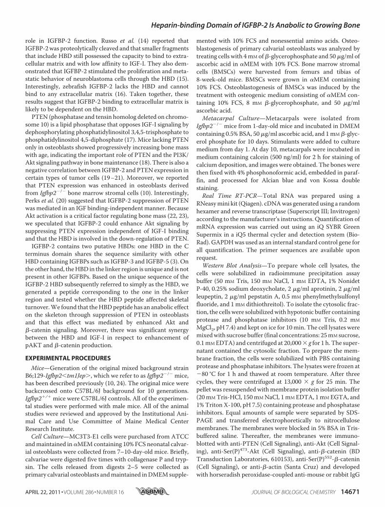
Induced Cytosolic �-Catenin Accumulation and Ser552 Phos-phorylation of �-Catenin—Several lines of evidence demon-strate cross-talk between IGF-I signaling and �-catenin signal-ing in tumor cell lines (26–29). Thus, we hypothesized thatIGF-I signaling would stimulate �-catenin signaling and thatHBDpeptide could enhance�-catenin signaling by suppressingPTEN expression in osteoblastic cells. We analyzed whetherIGF-I affects �-catenin accumulation in the cytosol of osteo-blastic cells. Western blot analysis revealed that IGF-I en-hanced cytosolic accumulation of�-catenin associatedwith thereduced expression ofmembrane �-catenin inMC3T3-E1 cells(Fig. 5A). In addition, cytosolic accumulation of �-catenin inresponse to IGF-I was enhanced in Igfbp2�/� COBs treatedwith PEG-HBD compared with PBS-treated control cells (Fig.5, B and C). This suggests that the HBD peptide is involved inIGF-I/�-catenin signaling possibly by suppressing PTENexpression. Ser552 phosphorylation of �-catenin, which can beinduced by Akt, has been shown to be important for dissocia-tion of �-catenin from membrane and relocalization to cyto-plasm and nucleus (30–32). Based on this observation, weasked whether IGF-I stimulated Ser552 phosphorylation in�-catenin and whether the PEG-HBD facilitates this responsein osteoblastic cells. As shown in Fig. 5 (D–F), IGF-I stimulated�-catenin phosphorylation on Ser552, and this response wasenhanced in cells treated with PEG-HBD. Because activation of�-catenin is important for bone formation, the osteogeniceffect of the HBD peptide might be mediated by IGF-I/�-catenin signaling in osteoblasts (33).
DISCUSSION
In this study, we report that IGFBP-2 has anabolic propertiesin the growing skeleton and that part of its biologic effect ismediated through the HBD in an IGF binding-independentmanner. IGFBP-2 levels are very high during neonatal growthand again during pubertal bone acquisition. IGF-I is also a crit-ical factor for bone accrual, and it can induce IGFBP-2 expres-sion in bone cells (34). Previous studies have shown that excessIGFBP-2 relative to IGF-I could inhibit the biologic activity ofIGF-I (3). In linewith this, an excess of IGFBP-2 has been shownto suppress osteoblast proliferation in part by antagonizingIGF-I action. In vivo, genetic models overexpressing Igfbp2have reduced bodymass and bone size (7, 35). In contrast, Kho-sla and co-workers (8, 9) reported that IGF-II in complex withIGFBP-2 (used in equimolar amounts) had a high affinity for
TABLE 4MicroCT analysis of femurs from female Igfbp2�/� treated with PBS orPEG-HBD peptideThe values are expressed as the means � S.E.
Igfbp2�/�
PBS (n � 10) PEG-HBD (n � 4)
MidshaftCortical thickness (mm) 0.176 � 0.003 0.188 � 0.003aBone area (mm2) 0.726 � 0.018 0.791 � 0.012Total area (mm2) 1.744 � 0.038 1.830 � 0.039bBone area/total area 0.417 � 0.006 0.433 � 0.008
Distal femurBone volume/total volume 0.038 � 0.001 0.049 � 0.006cTrabecular number (1/mm) 2.99 � 0.08 3.35 � 0.12cTrabecular thickness (mm) 0.035 � 0.001 0.036 � 0.002Trabecular spacing (mm) 0.338 � 0.011 0.298 � 0.011c
a p � 0.06 versus Igfbp2�/� with PBS.b p � 0.05 versus Igfbp2�/� with PBS.c p � 0.05 versus Igfbp2�/� with PBS.
Heparin-binding Domain of IGFBP-2 Is Anabolic to Growing Bone
14676 JOURNAL OF BIOLOGICAL CHEMISTRY VOLUME 286 • NUMBER 16 • APRIL 22, 2011

bone matrix, stimulated osteoblast proliferation, and could pre-vent disuse osteoporosis. Palermo et al. (34) also showed thatlower concentrations of IGFBP-2-stimulated IGF-II inducedALPexpression in rat tibial osteoblasts. In this study, we expand ourinsights into the physiologic role of IGFBP-2 and provide new evi-dence that the heparin-binding domain of IGFBP-2may enhancebonemass principally by stimulating osteoblast proliferation.
Initially we characterized the skeletal phenotype of maleIgfbp2�/� mice and found decreased skeletal mass, reducednumbers of osteoblasts, and low bone turnover (10). Thesefindings suggested that physiological concentrations ofIGFBP-2 were required for recruitment of osteoblasts andproper bone acquisition. Because forming a protein complexwith IGFBP-2 is critical for stabilization of IGF-I in the pericel-
FIGURE 4. The heparin-binding domain of IGFBP-2 suppresses PTEN expression. A and B, Igfbp2�/� COBs were treated with PBS, control peptide, or HBDpeptide overnight, and whole cell lysate was collected. PTEN expression was analyzed using Western blot analysis (A), and quantitative analysis was performed(B). C–H, Igfbp2�/� COBs were isolated and serum-starved overnight with PBS, IGFBP-2 (C and D) or PEG-HBD at the indicated concentration or period (E–H).Whole cell lysates were collected, and expression of PTEN was analyzed by Western blot analysis. Expression levels of PTEN were quantified by normalizing tothe expression levels of �-actin. I–L, Igfbp2�/� or Igfbp2�/� COBs were serum-starved overnight and treated with IGF-I (100 ng/ml) for 15 min (I and J). Igfbp2�/�
COBs were serum-starved with PBS or PEG-HBD overnight and treated with IGF-I (100 ng/ml) for 15 min (K and L). Whole cell lysates were collected, andexpression of Ser(P)473-Akt (pAkt) and Akt was analyzed by Western blot analysis. Expression levels of Ser(P)473-Akt were quantified by normalizing to theexpression levels of Akt. M, Igfbp2�/� COBs were exposed to IGF-I (50 ng/ml), IGFBP-2 (200 ng/ml), or the PEG-HBD (2 �g/ml) for 4 h. The lysates were preparedand immunoblotted for total PTEN or pAkt. Immunoblotting for �-actin is shown as a loading control. N, COBs were isolated from Igfbp2�/� and Igfbp2�/� mice.Duplicate cultures were exposed to IGF-I (50 ng/ml) for 10 min. The lysates were immunoprecipitated for Akt1 or Akt2 and then immunoblotted for pAkt or therespective Akt1 and Akt2 proteins. O, Igfbp2�/� COBs were incubated with IGF-I (50 ng/ml), IGFBP-2 (200 ng/ml), or the PEG-HBD (2 �g/ml) in the variouscombinations shown. They were also exposed to biotinylated IGFBP-2. Cell lysates were prepared and analyzed by SDS-PAGE with immunoblotting using HRPavidin. The figures shown represent at least three independent experiments. The values are expressed as the means � S.E. (n � 3– 4). *, p � 0.001; **, p � 0.01;†, p � 0.05; IP, immunoprecipitation; IB, immunoblot.
Heparin-binding Domain of IGFBP-2 Is Anabolic to Growing Bone
APRIL 22, 2011 • VOLUME 286 • NUMBER 16 JOURNAL OF BIOLOGICAL CHEMISTRY 14677
lular space, an anabolic action for IGFBP-2 was certainly con-ceivable, particularly when the anabolic effects of IGF-I wereconsidered within that same context. Furthermore, emergingevidence suggests that IGFBP-2 could function at least partiallyindependently of IGF-I binding to stimulate cell proliferationthrough theHBDdomain thatmediates IGFBP-2 binding to theextracellular matrix (13–16). Consistent with this tenet, wedemonstrated that IGFBP-2 binding to the cell surface waspartially blocked in the presence of the HBD peptide. Impor-tantly, IGFBP-2 binding to the cell surfacewas greatly enhancedin the presence of IGF-I, suggesting that the native proteinmayhave undergone a conformational change following IGF-Iassociation.Our finding that the HBD suppresses PTEN in osteoblasts
and enhances osteoblast proliferation provides a possiblemechanism by which IGFBP-2 could regulate skeletal mass, incoordination with IGF-I. Thismay be particularly relevant dur-ing stages of rapid bone growth such as the neonatal and ado-lescent period when the recruitment of osteoblast precursors iscritical. However, sorting the IGF-I-dependent and indepen-dent activities of intact IGFBP-2 is challenging. Clearly, IGF-Isignaling is a prerequisite for skeletal acquisition. Furthermore,we and others have shown thatmodulation of the IGF signalingpathway results in alterations in skeletal homeostasis (18,36–41). Therefore, an anabolic effect of the HBD on the skele-ton could in part be mediated by enhanced IGF-I-inducedactivation of the PI3K/Akt pathway as a result of PTEN sup-pression. In addition, the increase in osteoblast number inHBD-treated Igfbp2�/� mice could also be due to this mecha-nistic change because enhanced PI3K/Akt signaling in osteo-
blasts is involved in cell proliferation. In fact, Liu et al. (18)showed that the loss of PTEN in osteoblasts in vivo resulted invery high bone mass and increased osteoblast number.Although administration of the HBD rescued the low osteo-
blast number in Igfbp2�/� mice, osteoclast number was furtherdiminished by administration of HBD. This observation is con-sistent with the observed increase in bone mass but suggeststhat bone turnover is not accelerated by HBD treatment.Indeed, mineral apposition rate and bone formation rateshowed minimal changes in Igfbp2�/� mice treated with HBD.In contrast to the HBD, IGF-I signaling has been shown todirectly stimulate osteoclastogenesis (42), and our prior datasuggest that physiological levels of IGFBP-2 in the skeletalmicroenvironment are required for osteoclastogenesis. It isvery possible that IGFBP-2 acts to maintain IGF-I concentra-tions within the pericellular niche but that the HBD, whichlacks the IGF-I-binding domain, may play a distinct role byinhibiting bone resorption when supraphysiologic concentra-tions are administered. Because the HBD does not directly sup-press osteoclast differentiation in vitro, the underlying mecha-nism whereby the HBD decreases bone resorption is likelyindirect and due to decreased Rankl expression in osteoblasts.A reduction in PTEN expression has been shown to be asso-
ciated with increased �-catenin signaling. Inactivation ofGSK3� (glycogen synthase kinase 3) by Akt has been proposedto be the key pathway by which Akt activation enhances�-catenin stabilization, whereas several lines of evidence dem-onstrate the important role of Ser552 phosphorylation in�-catenin activation (30–32). �-Catenin is directly phosphor-ylated byAkt, and Ser552 phosphorylation is important for cyto-
FIGURE 5. The heparin-binding domain of IGFBP-2 stimulates cytosolic accumulation and Ser552 phosphorylation of �-catenin by IGF-I. A, MC3T3-E1cells were serum-starved overnight and treated with IGF-I at the indicated concentration for 6 h. Expression of �-catenin was analyzed by Western blot analysisusing membrane and cytosolic fraction. Pan-cadherin and �-actin was used as a loading control for membrane and cytosolic fraction, respectively. B and C,Igfbp2�/� COBs were serum-starved overnight with PBS or PEG-HBD and treated with IGF-I (100 ng/ml) for 6 h. Expression of �-catenin was analyzed byWestern blot analysis using cytosolic fraction (B) and quantified by normalizing to the expression levels of �-actin (C). D, MC3T3-E1 cells were serum-starvedovernight and treated with IGF-I at the indicated concentration for 15 min. Expression of Ser(P)552-�-catenin, �-catenin, Ser(P)473-Akt (pAkt), and Akt wasanalyzed by Western blot analysis using whole cell lysates. E and F, Igfbp2�/� COBs were serum-starved overnight with PBS or PEG-HBD and treated with IGF-I(100 ng/ml) for 15 min. Expression of Ser(P)552-�-catenin was analyzed by Western blot analysis using whole cell lysates (E) and quantified by normalizing to theexpression levels of �-catenin (F). The figures shown represent at least three independent experiments. The values are expressed as the means � S.E. (n � 3).*, p � 0.01; **, p � 0.05.
Heparin-binding Domain of IGFBP-2 Is Anabolic to Growing Bone
14678 JOURNAL OF BIOLOGICAL CHEMISTRY VOLUME 286 • NUMBER 16 • APRIL 22, 2011

solic and nuclear accumulation of �-catenin, resulting inincreased �-catenin transcriptional activity (30). He et al. (18)reported that the loss of PTEN resulted in an excess of intestinalstem cells by stimulating �-catenin activity in part throughSer552 phosphorylation (31). In the current study, we demon-strated that IGF-I enhanced �-catenin accumulation in cyto-plasm and phosphorylated Ser552 of �-catenin in osteoblasticcells, and this response was further enhanced by addition of theHBD. Taken together, these lines of evidence support our find-ings of a synergistic effect of IGFBP-2 with IGF-I on �-cateninsignaling and suggest that the increased bone formation notedin Igfbp2�/� mice treated with PEG-HBD is related to activa-tion of this signaling pathway.In the PEG-HBD-treated Igfbp2�/� mice, cell proliferation
(as measured by BrdUrd labeling) and osteoblast number byhistomorphometry were increased and were accompanied byreduced number of marrow adipocytes. Mesenchymal cell allo-cation is an important step in regulating bonemass andmarrowadiposity.Numerous factors are involved in this process includ-ing Wnt/�-catenin signaling and the transcription factor,PPAR-� (43). Activation of Wnt/�-catenin signaling favorsosteoblastogenesis over adipogenesis in part by either seques-tering PPAR-� or suppressing PPAR-� transcriptional activity(44–46). Because HBD enhances �-catenin stability that isinduced by IGF-I, the switch of mesenchymal cells toward theosteogenic lineage might explain the increased cell prolifera-tion as well as osteoblast number and reduced adipocyte num-ber in bone marrow from Igfbp2�/� mice treated withPEG-HBD.Nevertheless, the mechanism by which the HBD suppresses
PTEN expression has not been defined. One possibility residesin the binding of HBD with the integrin receptor. Maile et al.(47) reported that heparin-binding domain of vitronectinbound to a cysteine loop region of �3 integrin and modulated�V�3 integrin signaling in smooth muscle cells. Furthermore,Perks et al. (20) reported that PTEN down-regulation byIGFBP-2 might be mediated by an integrin receptor, althoughthey did not demonstrate an interaction between IGFBP-2 anda specific integrin receptor or whether theHBDwas involved inthis interaction. Further studies are needed to define specificsites of IGFBP-2 binding leading to activation of two criticalintracellular signaling pathways.Our results using supraphysiologic concentration of the
HBD cannot be used to reach a definitive conclusion regardingthe role of physiologic concentrations of IGFBP-2 in boneacquisition, particularly relative to IGF-I. However, it does sug-gest that this binding protein has unique properties that mightexplain the high circulating levels of IGFBP-2 during periods ofrapid growth such as the first year of life and during puberty, atime when serum IGF-I levels are also high. Importantly, HBDincreased bone mass even in growing female mice that do nothave alterations in bone mineral density (10). Further analysesare needed to determine whether HBD also has an anaboliceffect in older mice. Notwithstanding, we found that the HBDin IGFBP-2 has an anabolic effect on the skeleton of growingmice by targeting osteoblast recruitment through suppressionof PTEN expression and activation of �-catenin signaling.These data support the tenet that IGFBP-2, which is induced by
IGF-I, could act synergistically with IGF-I to promote cell pro-liferation through its unique heparin-binding domain in thelinker region of the molecule. Our studies lend further supportto recent work from the Lodish laboratory (11) demonstratingthe strong proliferative properties of IGFBP-2 in hematopoieticstem cells and provide a potential mechanism for IGFBP-2activity in some neoplasms.In summary, we show that the HBD in IGFBP-2 has an ana-
bolic effect on the skeleton, and this effect is mediated by thesuppression of PTEN expression and involves activation of�-catenin signaling. These lines of evidence add to our growingknowledge regarding the physiologic role of IGFBP-2 relative toIGF-I in several tissues and provide insights into the cross-talkbetween two signaling networks that have been shown to beessential for bone growth and maintenance.
REFERENCES1. Hwa, V., Oh, Y., and Rosenfeld, R. G. (1999) Endocr. Rev. 20, 761–7872. Jones, J. I., and Clemmons, D. R. (1995) Endocr. Rev. 16, 3–343. Firth, S. M., and Baxter, R. C. (2002) Endocr. Rev. 23, 824–8544. Hu, D., Pawlikowska, L., Kanaya, A., Hsueh, W. C., Colbert, L., Newman,
A. B., Satterfield, S., Rosen, C., Cummings, S. R., Harris, T. B., and Ziv, E.(2009) J. Am. Geriatr. Soc. 57, 1213–1218
5. Hedbacker, K., Birsoy, K., Wysocki, R. W., Asilmaz, E., Ahima, R. S., Fa-rooqi, I. S., and Friedman, J. M. (2010) Cell Metab. 11, 11–22
6. Wheatcroft, S. B., Kearney, M. T., Shah, A. M., Ezzat, V. A., Miell, J. R.,Modo, M., Williams, S. C., Cawthorn, W. P., Medina-Gomez, G., Vidal-Puig, A., Sethi, J. K., and Crossey, P. A. (2007) Diabetes 56, 285–294
7. Hoeflich, A.,Nedbal, S., Blum,W. F., Erhard,M., Lahm,H., Brem,G., Kolb,H. J., Wanke, R., and Wolf, E. (2001) Endocrinology 142, 1889–1898
8. Conover, C. A., Johnstone, E.W., Turner, R. T., Evans, G. L., John Ballard,F. J., Doran, P. M., and Khosla, S. (2002) Growth Horm. IGF Res. 12,178–183
9. Khosla, S., Hassoun, A. A., Baker, B. K., Liu, F., Zein, N. N., Whyte, M. P.,Reasner, C.A.,Nippoldt, T. B., Tiegs, R.D., Hintz, R. L., andConover, C.A.(1998) J. Clin. Invest. 101, 2165–2173
10. DeMambro, V. E., Clemmons,D. R., Horton, L.G., Bouxsein,M. L.,Wood,T. L., Beamer, W. G., Canalis, E., and Rosen, C. J. (2008) Endocrinology149, 2051–2061
11. Zhang, C. C., Kaba, M., Iizuka, S., Huynh, H., and Lodish, H. F. (2008)Blood 111, 3415–3423
12. Arai, T., Busby, W., Jr., and Clemmons, D. R. (1996) Endocrinology 137,4571–4575
13. Russo, V. C., Bach, L. A., Fosang, A. J., Baker, N. L., and Werther, G. A.(1997) Endocrinology 138, 4858–4867
14. Russo, V. C., Rekaris, G., Baker, N. L., Bach, L. A., and Werther, G. A.(1999) Endocrinology 140, 3082–3090
15. Russo, V. C., Schutt, B. S., Andaloro, E., Ymer, S. I., Hoeflich, A., Ranke,M. B., Bach, L. A., and Werther, G. A. (2005) Endocrinology 146,4445–4455
16. Duan, C., Ding, J., Li, Q., Tsai, W., and Pozios, K. (1999) Proc. Natl. Acad.Sci. U.S.A. 96, 15274–15279
17. Kawai, M., and Rosen, C. J. (2009) Pediatr. Nephrol. 24, 1277–128518. Liu, X., Bruxvoort, K. J., Zylstra, C. R., Liu, J., Cichowski, R., Faugere,M. C.,
Bouxsein, M. L., Wan, C., Williams, B. O., and Clemens, T. L. (2007) Proc.Natl. Acad. Sci. U.S.A. 104, 2259–2264
19. Mehrian-Shai, R., Chen, C. D., Shi, T., Horvath, S., Nelson, S. F., Reichardt,J. K., and Sawyers, C. L. (2007) Proc. Natl. Acad. Sci. U.S.A. 104,5563–5568
20. Perks, C. M., Vernon, E. G., Rosendahl, A. H., Tonge, D., and Holly, J. M.(2007) Oncogene 26, 5966–5972
21. Levitt, R. J., Georgescu, M. M., and Pollak, M. (2005) Biochem. Biophys.Res. Commun. 336, 1056–1061
22. Sakata, T., Wang, Y., Halloran, B. P., Elalieh, H. Z., Cao, J., and Bikle, D. D.(2004) J. Bone Miner. Res 19, 436–446
Heparin-binding Domain of IGFBP-2 Is Anabolic to Growing Bone
APRIL 22, 2011 • VOLUME 286 • NUMBER 16 JOURNAL OF BIOLOGICAL CHEMISTRY 14679

23. Shoba, L. N., and Lee, J. C. (2003) J. Cell. Biochem. 88, 1247–125524. Wood, T. L., Rogler, L. E., Czick, M. E., Schuller, A. G., and Pintar, J. E.
(2000)Mol. Endocrinol. 14, 1472–148225. Cohen, P., Peehl, D.M., Stamey, T. A.,Wilson, K. F., Clemmons, D. R., and
Rosenfeld, R. G. (1993) J. Clin. Endocrinol. Metab. 76, 1031–103526. Jin, T., George Fantus, I., and Sun, J. (2008) Cell Signal. 20, 1697–170427. Desbois-Mouthon, C., Cadoret, A., Blivet-Van Eggelpoel, M. J., Bertrand,
F., Cherqui, G., Perret, C., and Capeau, J. (2001) Oncogene 20, 252–25928. Playford, M. P., Bicknell, D., Bodmer, W. F., and Macaulay, V. M. (2000)
Proc. Natl. Acad. Sci. U.S.A. 97, 12103–1210829. Amin, S., Riggs, B. L., Melton, L. J., 3rd, Achenbach, S. J., Atkinson, E. J.,
and Khosla, S. (2007) J. Bone Miner. Res. 22, 799–80730. Fang, D., Hawke, D., Zheng, Y., Xia, Y., Meisenhelder, J., Nika, H., Mills,
G. B., Kobayashi, R., Hunter, T., and Lu, Z. (2007) J. Biol. Chem. 282,11221–11229
31. He, X. C., Yin, T., Grindley, J. C., Tian, Q., Sato, T., Tao,W.A., Dirisina, R.,Porter-Westpfahl, K. S., Hembree, M., Johnson, T., Wiedemann, L. M.,Barrett, T. A., Hood, L.,Wu, H., and Li, L. (2007)Nat. Genet. 39, 189–198
32. Taurin, S., Sandbo, N., Qin, Y., Browning, D., and Dulin, N. O. (2006)J. Biol. Chem. 281, 9971–9976
33. Glass, D. A., 2nd, and Karsenty, G. (2006) Curr. Top. Dev. Biol. 73, 43–8434. Palermo, C., Manduca, P., Gazzerro, E., Foppiani, L., Segat, D., and Bar-
reca, A. (2004) Am. J. Physiol. Endocrinol. Metab. 286, E648–E65735. Eckstein, F., Pavicic, T., Nedbal, S., Schmidt, C., Wehr, U., Rambeck, W.,
Wolf, E., and Hoeflich, A. (2002) Anat. Embryol. 206, 139–14836. Fujita, T., Azuma, Y., Fukuyama, R., Hattori, Y., Yoshida, C., Koida, M.,
Ogita, K., and Komori, T. (2004) J. Cell Biol. 166, 85–95
37. Peng, X. D., Xu, P. Z., Chen,M. L., Hahn-Windgassen, A., Skeen, J., Jacobs,J., Sundararajan, D., Chen,W. S., Crawford, S. E., Coleman, K. G., andHay,N. (2003) Genes Dev. 17, 1352–1365
38. Liu, J. P., Baker, J., Perkins, A. S., Robertson, E. J., and Efstratiadis, A. (1993)Cell 75, 59–72
39. Yakar, S., Rosen, C. J., Beamer, W. G., Ackert-Bicknell, C. L., Wu, Y., Liu,J. L., Ooi, G. T., Setser, J., Frystyk, J., Boisclair, Y. R., and LeRoith, D. (2002)J. Clin. Invest. 110, 771–781
40. Zhang,M., Xuan, S., Bouxsein,M. L., von Stechow,D., Akeno,N., Faugere,M. C., Malluche, H., Zhao, G., Rosen, C. J., Efstratiadis, A., and Clemens,T. L. (2002) J. Biol. Chem. 277, 44005–44012
41. Zhao, G., Monier-Faugere, M. C., Langub, M. C., Geng, Z., Nakayama, T.,Pike, J.W., Chernausek, S. D., Rosen, C. J., Donahue, L. R.,Malluche,H.H.,Fagin, J. A., and Clemens, T. L. (2000) Endocrinology 141, 2674–2682
42. Wang, Y., Nishida, S., Elalieh, H. Z., Long, R. K., Halloran, B. P., and Bikle,D. D. (2006) J. Bone Miner. Res. 21, 1350–1358
43. Gesta, S., Tseng, Y. H., and Kahn, C. R. (2007) Cell 131, 242–25644. Liu, J., Wang, H., Zuo, Y., and Farmer, S. R. (2006) Mol. Cell. Biol. 26,
5827–583745. Bennett, C. N., Longo, K. A., Wright, W. S., Suva, L. J., Lane, T. F., Han-
kenson, K. D., and MacDougald, O. A. (2005) Proc. Natl. Acad. Sci. U.S.A.102, 3324–3329
46. Ross, S. E., Hemati, N., Longo, K. A., Bennett, C. N., Lucas, P. C., Erickson,R. L., and MacDougald, O. A. (2000) Science 289, 950–953
47. Maile, L. A., Aday, A. W., Busby, W. H., Sanghani, R., Veluvolu, U., andClemmons, D. R. (2008) J. Cell. Biochem. 105, 437–446
Heparin-binding Domain of IGFBP-2 Is Anabolic to Growing Bone
14680 JOURNAL OF BIOLOGICAL CHEMISTRY VOLUME 286 • NUMBER 16 • APRIL 22, 2011
Copyright © 2022 FDOKUMEN