Fibronectin matrix assembly requires distinct contributions from Rho kinases I and -II

Structural Basis of Rnd1 Binding to Plexin Rho GTPaseBinding Domains (RBDs)*□S
Received for publication, October 22, 2010, and in revised form, May 20, 2011 Published, JBC Papers in Press, May 24, 2011, DOI 10.1074/jbc.M110.197053
Hui Wang‡1, Prasanta K. Hota§1,2, Yufeng Tong‡, Buren Li‡, Limin Shen‡, Lyudmila Nedyalkova‡,Susmita Borthakur§, SoonJeung Kim§, Wolfram Tempel‡, Matthias Buck§¶�3, and Hee-Won Park‡**4
From the ‡Structural Genomics Consortium and **Department of Pharmacology, University of Toronto, Toronto, Ontario M5G 1L5,Canada and the Departments of §Physiology and Biophysics, ¶Neuroscience, and �Pharmacology, Case Western Reserve UniversitySchool of Medicine, Cleveland, Ohio 44106
Plexin receptors regulate cell adhesion, migration, and guid-ance. The Rho GTPase binding domain (RBD) of plexin-A1and -B1 can bind GTPases, including Rnd1. By contrast,plexin-C1 and -D1 reportedly bind Rnd2 but associate withRnd1 only weakly. The structural basis of this differentialRnd1 GTPase binding to plexin RBDs remains unclear. Here,we solved the structure of the plexin-A2 RBD in complex withRnd1 and the structures of the plexin-C1 and plexin-D1 RBDsalone, also compared with the previously determinedplexin-B1 RBD.Rnd1 complex structure. The plexin-A2RBD�Rnd1 complex is a heterodimer, whereas plexin-B1 and-A2 RBDs homodimerize at high concentration in solution,consistent with a proposed model for plexin activation.Plexin-C1 and -D1 RBDs are monomeric, consistent withmajor residue changes in the homodimerization loop. Inplexin-A2 and -B1, the RBD �3-�4 loop adjusts its conforma-tion to allow Rnd1 binding, whereas minimal structuralchanges occur in Rnd1. The plexin-C1 and -D1 RBDs lackseveral key non-polar residues at the corresponding GTPasebinding surface and do not significantly interact with Rnd1.Isothermal titration calorimetry measurements on plexin-C1and -D1 mutants reveal that the introduction of non-polarresidues in this loop generates affinity for Rnd1. Structure
and sequence comparisons suggest a similar mode of Rnd1binding to the RBDs, whereas mutagenesis suggests that theinterface with the highly homologous Rnd2 GTPase is differ-ent in detail. Our results confirm, from a structural perspec-tive, that Rnd1 does not play a role in the activation ofplexin-C1 and -D1. Plexin functions appear to be regulated bysubfamily-specificmechanisms, some of which involve differ-ent Rho family GTPases.
Members of the plexin family of transmembrane receptorsbind to semaphorin guidance cues and perform critical func-tions in axonal growth cone migration during neural develop-ment (1–3), regulate blood vessel patterning in cardiovasculardevelopment (4, 5), are responsible for invasive growth of met-astatic cancer cells (6, 7), and play a role in the immuneresponse (8, 9). Vertebrate plexins can be classified into foursubfamilies (A–D). Type A plexins transduce class 3 sema-phorin signaling via the neuropilin co-receptor, whereasplexin type B receptors mediate class 4 semaphorin-inducedgrowth cone collapse in hippocampal neurons and inhibitionof cellular migration in various other cell types (10, 11). Dif-ferent from type A and B plexins, plexin-C1 is a receptor forsemaphorins that stimulate cytokine expression, inducing aproinflammatory response in monocytes (12). In dendriticcells, plexin-C1 stimulation by the Vaccinia virus sema-phorin mimic, A39R, suppresses membrane extensions thatare required for phagocytosis; this impairs dendritic cellmigration, ultimately limiting the ability of dendritic cells tofunction as efficient antigen-presenting cells (13). In con-trast to subfamily A and B plexins, which are predominantlyexpressed in neuronal systems, plexin-D1 is detected in thevascular endothelium, as well as in neuronal systems, andplays an essential role in the formation of the vasculatureduring early embryogenesis (14). In endothelial cells,plexin-D1 associates with neuropilin to transduce multiplesemaphorin signals (5, 15). Furthermore, plexin-D1 isexpressed at high levels in a wide range of human tumortissues (16), whereas other plexins, such as plexin-B1, func-tion as tumor suppressors in some settings. Because the sig-naling pathways are diverse and additional mechanisms ofplexin action probably remain to be discovered, structuralcharacterization of plexins will elucidate the structure-func-tion relationships and the potential of these receptors astherapeutic targets.
* This work was supported, in whole or in part, by National Institutes ofHealth Grants R01GM73071, K02HL084384, and R01GM092851 (toM. B.). This work was supported in part by the Structural Genomics Con-sortium, a registered charity (number 1097737) that receives fundsfrom the Canadian Institutes for Health Research, the Canadian Foun-dation for Innovation, Genome Canada through the Ontario GenomicsInstitute, GlaxoSmithKline, Karolinska Institutet, the Knut and AliceWallenberg Foundation, the Ontario Innovation Trust, the Ontario Min-istry for Research and Innovation, Merck & Co., Inc., the NovartisResearch Foundation, the Swedish Agency for Innovation Systems, theSwedish Foundation for Strategic Research, and the Wellcome Trust.
□S The on-line version of this article (available at http://www.jbc.org) containssupplemental Table S1 and Fig. S1.Author’s Choice—Final version full access.
The atomic coordinates and structure factors (codes 3Q3J, 3KUZ, and 3H6N)have been deposited in the Protein Data Bank, Research Collaboratory forStructural Bioinformatics, Rutgers University, New Brunswick, NJ(http://www.rcsb.org/).
1 Both authors contributed equally to this work.2 A postdoctoral fellow of the American Heart Association, Great Rivers
Affiliate.3 To whom correspondence may be addressed: Case Western Reserve Univer-
sity, School of Medicine, Robbins Blvd., Rm. E607, 10900 Euclid Ave., Cleve-land, OH 44106-4970. E-mail: [email protected].
4 To whom correspondence should be addressed: 101 College St., MaRSSouth Tower, Rm. 737, Toronto, Ontario M5G1L7, Canada. E-mail: [email protected].
THE JOURNAL OF BIOLOGICAL CHEMISTRY VOL. 286, NO. 29, pp. 26093–26106, July 22, 2011Author’s Choice © 2011 by The American Society for Biochemistry and Molecular Biology, Inc. Printed in the U.S.A.
JULY 22, 2011 • VOLUME 286 • NUMBER 29 JOURNAL OF BIOLOGICAL CHEMISTRY 26093
by guest on July 1, 2015http://w
ww
.jbc.org/D
ownloaded from
The four plexin subfamilies share a similar domain structure(1). The intracellular region of the plexins exhibits sequencesimilarity to the GTPase-activating protein (GAP)5 domain ofp120 RasGAP (17, 18). The uniqueness of the GAP domain liesin that this part of plexins is split into two segments by a regionof �200 residues. This intervening domain is overall well con-served and has been identified as the binding location of severalRho GTPases, including Rnd1 (19–21), and hereafter isreferred to as the Rho GTPase binding domain (RBD).Rho family GTPases play important roles in the regulation of
the cell’s actin cytoskeleton and mediate the repulsive andattractive effects of guidance molecules (22). The Rnd proteinsare a distinct subgroup of Rho GTPases. Rnd1 and Rnd3 lackintrinsic GTPase activity due to amino acid substitutions atcritical positions, whereas Rnd2may have partial GTPase activ-ity (23). It has been established that three Rho GTPases (i.e.Rac1, Rnd1, and RhoD), when in the active GTP bound form,directly associate with the RBDs of A and B family plexins (19,21). Moreover, it has been reported recently that Rnd2 isinvolved in the Sema3E/plexin-D1-induced inhibition of axonoutgrowth in cortical neurons (24). However, the mechanismsby which Rho GTPases regulate plexin function remain to befully elucidated, particularly in regard to the role of the Rnd1GTPase in plexin signaling.Recent studies have revealed the atomic details of the entire
intracellular domain of plexin-B1 and -A3 (25, 26). In bothstructures, the two RasGAP homologous segments are inter-twined and form a compact fold, similar to that seen in otherRasGAPs, whereas the RBD domain forms an independentfolding unit and is linked to the coreGAPdomain via a so-called“coupling loop” (25). Rnd1 binding has been shown to be essen-tial for the GAP activity of plexin-A1 and -B1 (15, 17, 19), sug-gesting the importance of the RBD domain and its interactionwith Rho family GTPases.Here we report the structures of the RBDs of plexin-C1 and
plexin-D1 as well as that of plexin-A2 in complex with Rnd1.The analysis of the binding interfaces of the plexin-A2 and alsoof the previously published plexin-B1 RBD�Rnd1 complex andthe structure-based alignment of other plexin RBDs reveal theprimary factors in the protein-protein interaction betweenplexin RBDs and Rnd1. These results provide insight into thespecificity of Rnd1 binding and suggest a differential use of thisGTPase in the activation mechanism of plexins.
EXPERIMENTAL PROCEDURES
Protein Expression and Purification—The genes encodingthe RBDs of human plexin-A2 (residues 1490–1600), plexin-B1(residues 1746–1852), plexin-C1 (residues 1198–1305),plexin-D1 (residues 1553–1678), and Rnd1 GTPase (residues5–200) and Rnd2 (residues 1–227) were amplified by PCR andsubcloned into the expression vector pET28-MHL, whichencodes an N-terminal His6 tag followed by the tobacco etchvirus protease cleavage site. All recombinant proteins were
overexpressed in Escherichia coli strain BL21(DE3) (Strat-agene) upon overnight induction with 1.0mM isopropyl 1-thio-�-D-galactopyranoside at 16 °C. Proteins were purified usingnickel-NTAaffinity chromatography and gel filtration chroma-tography using Superdex 75 (GE Healthcare). Rnd1 and Rnd2GTPases were loaded with non-hydrolyzable nucleotide byovernight incubation with 5 mM GMPPNP. Protein sampleswere stored at 4 °C in gel filtration buffer.Crystallization—Proteins were crystallized using the sitting
drop vapor diffusion method. Equal volumes (0.5 �l) of proteinsolution and reservoir solution were mixed for crystallization.Plexin-A2 RBD and Rnd1weremixed in a 1:1 ratio in the bufferused for protein purification. The crystal of the complex grew in25.5% polyethylene glycol 3350 with 0.2 M MgCl2 and 0.1 M
HEPES at pH 7.5. Plexin-C1 RBD was crystallized using an insitu proteolysis method (27). A 0.5-�l protein sample contain-ing chymotrypsin (1:100, w/w) was mixed with 0.5 �l of wellsolution containing 0.1 M HEPES (pH 7.5), 2.0 M (NH4)2SO4,and 0.2 M NaCl. The plexin-D1 RBD was overexpressed inE. coli using a prepacked M9 selenomethionine growthmedium kit (Medicilon) and the N-terminal His6 tag wasremoved by tobacco etch virus protease cleavage before crystal-lization. The crystal of the plexin-D1 RBD was grown in 0.1 M
sodium cacodylate buffer (pH 5.2) containing 1.39 M sodiumcitrate and 15 mM tris(2-carboxyethyl)phosphine, using the insitu proteolysis method with chymotrypsin (1:100, w/w). Allcrystals grew to mountable size in 3–5 days.Data Collection and Structure Determination—Diffraction
data were collected at beam line 19-ID at the Advanced PhotonSource (Argonne, IL) with a Quantum-315 CCD detector(ADSC) and reduced with the HKL3000 suite (28). The struc-ture of the plexin-A2 RBD�Rnd1 complex was solved by molec-ular replacement with MOLREP (29) using models of Rnd1(PDB code 2REX) (25) and murine plexin-A3 RBD (PDB code3IG3) (26) as starting models. Initial phases for the plexin-D1structure were obtained by single-wavelength anomalous dif-fraction, using the programs SHELX (30), MLPHARE (31), andDM (32) through the HKL3000 interface. The structure ofplexin-C1 was solved by molecular replacement with the pro-gram PHASER (33) and a composite searchmodel of plexin-D1(described here) and the mouse plexin-A3 RBD coordinates(26) (PDB code 3IG3). ARP/wARP was used for automatedmodel building, PHENIX (34) and REFMAC (35) for coordi-nate/B-factor refinement, COOT (36) for manual modeladjustments, and MOLPROBITY (37) for validation ofgeometry.Analytical Gel Filtration Assay—Analytical gel filtration was
performed at 4 °C using a Superdex75 10/300 GL column (GEHealthcare). The column was run in 20 mM Tris/HCl, pH 7.5,0.5 M NaCl, 5 mM MgCl2, and 5% (v/v) glycerol at a flow rate of0.5 ml/min. GMPPNP (5 mM) was added to all of the proteinsamples for internal reference and binding factor. Each injec-tion contained a 200-�l protein sample with a concentration of�5 mg/ml, and the protein absorbance was monitored at 280nm. For gel filtration to study the dimerization behavior of theplexin-A2 RBD, we used 500 �l of protein at concentrations upto 3 mM. The protein absorbance of the concentrated sampleswas followed at 254 nm.
5 The abbreviations used are: GAP, GTPase-activating protein; RBD, RhoGTPase binding domain; GMPPNP, 5�-guanylyl imidodiphosphate; ITC, iso-thermal titration calorimetry; PRAM, plexin-specific Rho GTPase associa-tion motif; PDB, Protein Data Bank.
Structural Insights into the Affinity of Rnd1 for Plexins
26094 JOURNAL OF BIOLOGICAL CHEMISTRY VOLUME 286 • NUMBER 29 • JULY 22, 2011
by guest on July 1, 2015http://w
ww
.jbc.org/D
ownloaded from

Isothermal Titration Calorimetry—Binding interactions ofplexin RBDs and Rnd1 or Rnd2were performed at 25 °C using aMicroCal iTC200 microcalorimeter. The experiments werecarried out in 50 mM phosphate buffer, pH 7.0, 50 mM NaCl, 4mM MgCl2, 0.5 mM tris(2-carboxyethyl)phosphine. The PlexinRBDswere kept in the syringe (volume 40�l, concentration 400�M), whereas the Rnd1 or Rnd2 was placed in the cell (volume250 �l, concentration 40 �M). The interval between injectionswas 120 s. The data were analyzed in Origin 7.0, and the binaryequilibrium association constant (Ka � 1/Kd) was fitted (whereKd is equal to [GTPase][plexin]/[GTPase-plexin complex]).
In order to confirm the differences in binding affinity for keyinteractions, we used a second technique, plasmon surface res-onance. The method and data are shown in the supplementalmaterial.
RESULTS
Structure of the Plexin-B1 RBD in Complex with Rnd1—Wepreviously solved and reported the structure of the plexin-B1RBD in complex with Rnd1 at a resolution of 2.3 Å (PDB code2REX) (25). However, the interface had not been analyzed. Theplexin-B1 RBD in the complex forms a homodimer by the C2symmetric antiparallel �-sheet (�5-�6), with a �-cation inter-action betweenTrp1830 andArg1832, in an asymmetric unit (Fig.
1A). A similar homodimerization interface has been observedin the structure of the Rnd1-free form of plexin-B1 RBD (21).Rnd1 binding to the plexin-B1 RBD appears to promote
ordering of a flexible region, the loop �3-�4, in the plexin-B1RBD-Rnd1 interface in the crystal (Fig. 1, B and C). In the freeRBD, the B-factors in this loop are �20% above those seen forC� atoms in the regular secondary structure. In the boundform, they are similar to those of the �-helices and �-strandregions, whereas a rotational movement of the �3-�4 loopappears to create a surface for optimal Rnd1 binding. The RBD�1-�2 loop, which is not directly involved in the binding ofRnd1, was also ordered in the crystal. By contrast, in the freeform of the plexin-B1 RBD, the corresponding loop is disor-dered (Fig. 1C). However, NMR data and molecular dynamicscalculations on RBD-GTPase complexes suggest that this loopremains highly flexible in solution (38).6 The conformation of aregion on the opposite side of the RBD that contains the 310-helices �1 and �2 was pushed away from the main structure,although, by comparisonwith other RBD structures, thismove-ment may be independent of the ordering of the two loopregions mentioned above.
6 P. K. Hota, L. Zhang, and M. Buck, unpublished data.
FIGURE 1. Structures of the complex of plexin-B1 RBD and Rnd1. A, ribbon diagram of the plexin-B1 RBD�Rnd1 complex homodimer/heterotetramer. Theasymmetric unit consists of plexin-B1 RBD chains A (cyan) and C (green) (residues 1746 –1852), forming a homodimer, and Rnd1 chains B (violet) (residues13–188) and D (hot pink) (residues 7–188), bound on opposite sides to the RBD dimer. The homodimer interface is stabilized by a �-cation interaction betweenTrp1830 and Arg1832 (21). B, one unit of the plexin-B1 RBD (cyan) and Rnd1 (violet) complex. Several of the � strands (�1–�7) and two �-helices (�1 and �2) arelabeled in plexin-B1 RBD. C, superimposition of the RBDs of the plexin-B1 RBD�Rnd1 complex (cyan) with the free plexin-B1 RBD (green) illustrates conforma-tional changes that take place in the �1-�2 loop, the �3-�4 loop, and the �1-�2 region upon complex formation with Rnd1.
Structural Insights into the Affinity of Rnd1 for Plexins
JULY 22, 2011 • VOLUME 286 • NUMBER 29 JOURNAL OF BIOLOGICAL CHEMISTRY 26095
by guest on July 1, 2015http://w
ww
.jbc.org/D
ownloaded from

In addition, Rnd1 binding caused an appreciable twisting ofthe conformation of the�-hairpin (�5 and�6 and the interven-ing loop), weakening the dimerization interaction, which is partof the mechanism that maintains monomer-dimer equilibriumin plexin-B1 signaling (25). In the complex, twoRnd1moleculesbind to opposite sides of the plexin-B1 RBD homodimer inter-face (Fig. 1A). An equimolar ratio of the two proteins is seen insolution, based on gel filtration data, which is consistent withprevious observations that Rnd1 binding weakens the RBDdimerization (21, 39).Rnd1 binding to the plexin-B1 RBD buries a solvent-accessi-
ble surface area of �1900 Å2 as calculated using NACCESS (byS. J. Hubbard and J. M. Thornton available on theWorldWideWeb). Binding involves twelve residues of plexin-B1 located instrands �3 and �4, a short �2-helix, and a loop toward thedimerization �-hairpin as well as nine residues of Rnd1 located
in the �2 and �3 strands, the 310-helices �1 and �2, and one�-helix of the GTPase (Fig. 2,A and B). The composition of theheterodimer interface residues indicates that the core interfaceis hydrophobic. Mainly, Trp1807 and Leu1815 of the plexin-B1RBD interact with Phe47 and Val77 of Rnd1; the interactingresidues are surrounded by Ser1809, Gly1813, Tyr1839, andLys1840 of the plexin-B1 RBD and Trp66, Tyr73, Tyr74, Asn76,and Leu80 of Rnd1 (Fig. 2A). Fold-X calculations (41) substitut-ing residues at the interface for Ala (or Ser in case the residue isAla already) predict seven hot spots (with substitution energygreater than 1 kcal/mol) on the side of the plexin-B1 RBD(Ser1809, Gly1810, Gly1813, His1814, Leu1815, Val1822, andThr1823)and six hot spots on the side of Rnd1 (Phe47, Trp66, Tyr73, Tyr74,Val77, and Leu80).Several hydrogen bonds were also observed in the interface
(Fig. 2B). The side-chain amide nitrogen atom of Asn76 of Rnd1
FIGURE 2. Interface of the plexin-B1 RBD�Rnd1 complex. A, transparent surface representation of the plexin-B1 RBD�Rnd1 complex. The plexin-B1 RBD andRnd1 are shown in cyan and violet, respectively. The �3 and �4 strands and helix �2 of the plexin-B1 RBD are located in the interface, as are the �2 and �3strands, the 310-helices �1 and �2, and one �-helix of Rnd1. The residues in the interface are represented by a stick model. The close-up view of the interface isshown on the right, where the hydrophobic core is highlighted by a gray transparent circle. B, amino acid interactions at the interface are shown in a LIGPLOTdiagram (53). Hydrogen bonds are presented as dashed lines; interatomic distances are in angstroms. “Radiating” curves indicate hydrophobic contactsbetween the corresponding atoms or residues and the surrounding residues. C, two areas of the interface contact are shown, illustrating that the three residuesof Rnd1, Phe47, Glu48, and Tyr114, are flipped toward plexin-B1 (shown in cyan), as seen in the superimposition of the bound (magenta for complex withplexin-B1) and the free (blue) structures of Rnd1.
Structural Insights into the Affinity of Rnd1 for Plexins
26096 JOURNAL OF BIOLOGICAL CHEMISTRY VOLUME 286 • NUMBER 29 • JULY 22, 2011
by guest on July 1, 2015http://w
ww
.jbc.org/D
ownloaded from
forms a hydrogen bond to the main-chain carboxyl oxygenatom of His1838 of the plexin-B1 RBD (2.8 Å), and their sidechains stack on each other. The side chain of Glu48 of Rnd1forms hydrogen bonds to themain-chain amides of Val1811 (3.0Å) and Ala1812 (2.7 Å) of the plexin-B1 RBD, maintaining therotated conformation of the �3-�4 loop. The side chain ofTrp66 of Rnd1 forms a T-shaped interaction with one face ofHis1814 in the plexin-B1 RBD, whereas the main chain of theordered �1-�2 loop interacts with the other face of His1814 inplexin-B1 RBD. His1814 is also part of a network of hydrogenbonds that includes Glu1806 and Arg1808.
Minor conformational changes are observed in Rnd1 uponbinding to plexin-B1 RBD (Fig. 2C) in comparison with thestructure of free homodimeric Rnd1 (PDB code 2CLS). In thecomplex the side chain of Phe47 of Rnd1 is flipped towardTrp1807 of the plexin-B1 RBD, whereas the rotation of the�3-�4 loop of plexin-B1 RBD (residues Ser1809–Gly1813)exposes the same side chain of Trp1807 so that it can form a�-�interactionwith the side chain of Phe47 of Rnd1. The side-chainrotation of Tyr114 is obvious because it forms hydrophobic con-tacts with Val1822 and with the side-chain methyl group ofThr1823 of plexin-B1. The movement of the side chain of Rnd1Glu48 is also observed because it forms hydrogen bonds to the�3-�4 loop of plexin-B1 RBD.Structure of the Plexin-A2 RBD in Complex with Rnd1—The
structure of the plexin-A2 RBD was solved in complex withRnd1 at a resolution of 2.0 Å (PDB code 3Q3J) (Fig. 3A andTable 1). In the crystal structure, the plexin-A2 RBD does notform a homodimer seen in the plexin-B1 RBD (PDB code2REX) (25). In fact, the equivalent region of the plexin-B1homodimerization loop is disordered in the plexin-A2�Rnd1complex (the electron density of residues 1575–1578, whichinclude the equivalent Trp and Arg residues, is very poor). Theelectron density surrounding these residues arises from regionsof symmetry-related molecules in the crystal lattice that areremote from their own putative dimerization segment. Thus,this region is not involved in a dimer formation in the crystal;however, free plexin-A2 RBD homodimerizes at high proteinconcentration in solution (see Fig. 6B). These findings are againconsistentwith aweakeneddimerization of theRBDonce aRhofamily GTPase is bound.The RBD has the same ubiquitin fold as other family mem-
bers and the structure has an r.m.s. deviation of 1.00 and 0.93Å,as calculated using SSM (29), to the Rnd1 bound plexin-B1 andto the unbound plexin-A3 RBD in the intracellular regions ofplexin-A3 (26), respectively. In the plexin-A2 complex, theRBD-Rnd1 interface closely resembles that of the plexin-B1complex. Similarly to the observationmade above for the RBD-B1�Rnd1 complex, loop �3-�4 is also bent away from Rnd1 inthe complex with RBD-A2 (Fig. 3C) and has relatively lowB-factors. In another part of the protein, and different from theplexin-B1 RBD�Rnd1 complex, the RBD-A2 �1-�2 loop is threeresidues shorter than in the RBD of plexin-B1 but is not movedtoward Rnd1 and has high B-factors. Another loop region, fol-lowing the RBD �1 helix and opposite to the binding interface,is disordered in the RBD-A2�Rnd1 complex.Rnd1 binding to the plexin-A2 RBD buries a surface area of
�1300 Å2 according to NACCESS, which is considerably
smaller than for the plexin-B1 RBD�Rnd1 complex, in part dueto missing side chains in the A2 complex (e.g. in the �3-�4loop). According to the crystal structure, binding involves eightresidues on the side of the plexin-A2 RBD in direct contactswith Rnd1. On the side of Rnd1, eleven residues are involved(Fig. 3, B and D). These Rnd1 residues are located in the sameregions as in the plexin-B1 RBD-Rnd1 interaction, and manymake the same contacts with the A2 RBD. Accordingly, hydro-phobic side chains of Trp1555 and Val1563 of plexin-A2 interactwith Phe47 and Val77 of Rnd1, and this non-polar patch is sur-rounded by Gln1557, Ala1561, Tyr1587, and Gln1588 on the side ofplexin andbyTrp66, Tyr73, Tyr74, Ans76, andLeu80 of Rnd1 (Fig.3A).Fold-X calculations (41) utilizing an Ala (or in the case of a
preexisting Ala, a Ser) scan predict six hot spot residues on theside of the RBD (Trp1555, Arg1559, Ala1561, Val1563, Ile1570, andThr1571) and four hot spot residues on the side of Rnd1 (Phe47,Tyr74, Val77, and Leu80) as having substitution energy differ-ences greater than 1 kcal/mol. It should be noted that althoughall four of the Rnd1 hot spot residues are involved in both com-plexes, only four of the seven hot spot RBD-A2 residues are hotspots in the case of the plexin-B1 RBD�Rnd1 complex, suggest-ing that despite a similarity of the overall features, some of thedetailed energetic contributions of residues to the complex dif-fer considerably between the twoRBDproteins. In addition, therelative contribution of RBD and Rnd1 residues to the interfaceappears to be different in the two complexes. Specifically, twomore Rnd1 residues, Trp66 and Tyr73, as well as one less RBDresidue are involved in the case of the complex plexin-B1RBD�Rnd1 compared with the complex with plexin-A2.A hydrogen bond connects the Rnd1Asn76 side-chain amino
group and the plexin-A2 RBD main-chain carbonyl oxygen ofHis1586, as it does in the plexin-B1 RBD�Rnd1 structure. Thereis less hydrogen bonding across the interface, however, com-pared with the plexin-B1 complex (Fig. 3B, compare with Fig.2B). The Rnd1 Glu48 side chain is farther away from the �3-�4turn of the plexin-A2 RBD and does not form hydrogen bondswith the main-chain segment. In fact, this region may be moreflexible in the plexin-A2 complex because side chains ofArg1559and Ile1560 at the equivalent position (Val1811 and Ala1812 inplexin-B1) do not have electron density beyond C�. On theother hand, Fold-X calculations (where the full side chains arebuilt in) suggest that Arg1559 and Ala1561 have a considerableeffect on the stability of the complex and, thus, may be respon-sible for the rotation of the loop region away from the interac-tion with Rnd1. His1814 in the RBD-B1 has been replaced byArg1562. The hydrocarbonportion of its side chainweakly inter-acts with Trp66 of Rnd1, whereas the guanidinium moietyof Arg1562 participates in a salt bridge networkwithGlu1554 andArg1556, a �/�/� charge motif that appears conserved acrossall plexin-A and -B family members, with the exception ofplexin-B2.As in the case of plexin-B1 RBD binding, several minor con-
formational changes occur in Rnd1 (Fig. 3D). The same obser-vations are made for the plexin-A2 RBD�Rnd1 complex as weremade for the plexin-B1 RBD upon binding to Rnd1 regarding aside-chain flip of Rnd1 Phe47 and Tyr114. In the case of Tyr114,one of the interaction partners is changed from a Val1822 in
Structural Insights into the Affinity of Rnd1 for Plexins
JULY 22, 2011 • VOLUME 286 • NUMBER 29 JOURNAL OF BIOLOGICAL CHEMISTRY 26097
by guest on July 1, 2015http://w
ww
.jbc.org/D
ownloaded from

plexin-B1 to Ile1570 in plexin-A2. Themost significant change isthat the side chain of Glu48 is not hydrogen-bonded to the�3-�4 loop in the plexin-A2 RBD�Rnd1 complex.Structure of the Plexin-C1 RBD—We solved the structure of
plexin-C1 RBD at 2.3Å (residues 1198–1305, PDB code 3KUZ)(Table 1). Superimposition of the plexin-C1 RBD with the freeplexin-B1 RBD structure (PDB code 2R2O) gives a root meansquare deviation of 1.5 Å over 94 aligned residues (29). Theplexin-C1 RBD consists of a central �-helix, two short 310-heli-ces, and a five-stranded�-sheet, presenting a ubiquitin-like fold
(Fig. 4A). In the core region, a five-stranded �-sheet has a left-handed twist, forming a groovewhere the central�-helix (�1) isbound. The central �-helix of the plexin-C1 RBD structure,whose equivalent in the plexin-B1 intracellular region isinvolved in RBD binding to the RasGAP domain, is longer byfour residues than that of the plexin-B1 RBD.Additional differences between this and the plexin-B1 RBD
are observed in twoother regions (Fig. 4B). First, the�1-�2 loop(Glu1208–Arg1218) is ordered in the crystal and is bound to theelongated helix �1 as well as to its C-terminal loop region
FIGURE 3. Interface of the plexin-A2 RBD�Rnd1 complex. In part similar to Fig. 2A, a transparent surface representation of plexin-A2 RBD�Rnd1 complex isshown, with the interface displayed in an expanded view. B, LIGPLOT diagram of contacts at the interface. Rnd1 residues Trp66 and Tyr114 are omitted for clarity(but are shown in D). C, superimposition of the RBDs of the plexin-A2 RBD�Rnd1 complex (gold) and free plexin-B1 (green) illustrates conformational changesthat take place. D, superposition of two regions in contact showing perturbations similar to those of the Rnd1 side-chain structure upon association withplexin-A2 RBD as seen in the case of plexin-B1 RBD binding. Gold, RBD residues; blue, free Rnd1; magenta, bound Rnd1 residues.
Structural Insights into the Affinity of Rnd1 for Plexins
26098 JOURNAL OF BIOLOGICAL CHEMISTRY VOLUME 286 • NUMBER 29 • JULY 22, 2011
by guest on July 1, 2015http://w
ww
.jbc.org/D
ownloaded from

through hydrogen bonds. In contrast, the corresponding�1-�2loop of the plexin-B1 RBD is invisible in the free form andbecomes ordered in the complex with Rnd1, as noted above.Second, the region of plexin-C1 RBD (Leu1270–Ile1289), whichcorresponds to the homodimerization �-hairpin of theplexin-B1RBD, is not engaged in dimerization. Several residuesinvolved in homodimerization of the plexin-B1 RBD are differ-ent in the plexin-C1 RBD. In particular, Ile1283 of plexin-C1RBD replaces Trp1830 of plexin-B1 RBD, eliminating an impor-tant �-stacking interaction and, thereby, reducing the possibil-ity of homodimerization through this region. This is consistentwith themonomeric state of plexin-C1 in solution, as shown bygel filtration data and also by NMR (data not shown). In theasymmetric unit of the crystal, we observe that twomolecules ofthe plexin-C1 RBD are linked by an intermolecular disulfidebond through Cys1227. Disulfide bonds are not generally foundin cytosolic proteins due to the reducing environment. There-fore, the dimerization of plexin-C1 RBD via the intermoleculardisulfide bond appears to be a crystallization artifact.Structure-based sequence analysis shows that plexin-C1 has
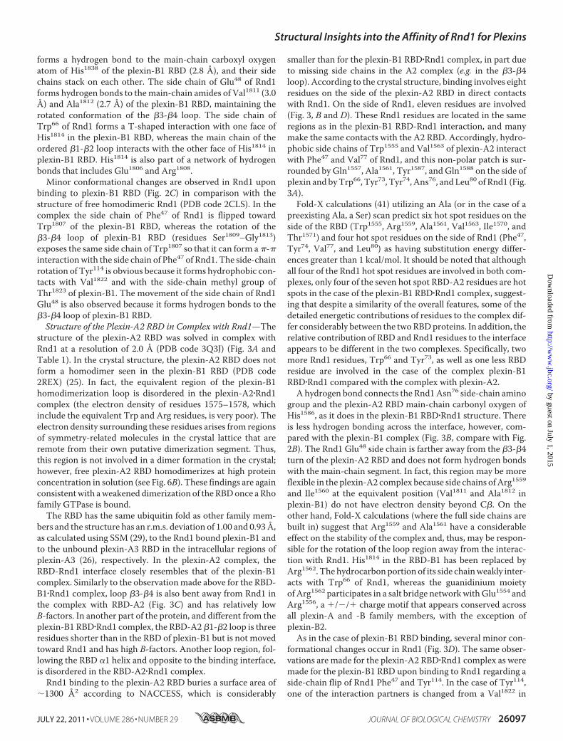
unique residues at the putative Rnd1 binding site. All sidechains are different from plexin-B1 with the exception of tworesidues (His1291 and Tyr1292) in helix �2 (Fig. 5A). The differ-ence in sequence results in a series of repulsive effects ifplexin-C1 were to interact with Rnd1 with the same interfaceas plexin-B1. Briefly, Trp1807 of plexin-B1 is replaced by Leu1260in plexin-C1; the presence of leucine at this site precludes the�-� interactionwith Phe47 of Rnd1 and results in the additionalloss of several hydrophobic contacts. In addition to the changes
in the hydrophobic interface, the shorter turn between the �3and �4 strands of plexin-C1may result in significant steric hin-drance involvingGlu48 of Rnd1. The long side chains ofMet1262
and Arg1265 of plexin-C1 in this location, compared with thoseof Ser1809 and Gly1813 in plexin-B1, also could prevent bindingto Rnd1. These changes are likely to be responsible for theabsence of binding of Rnd1 to the plexin-C1 RBD, as demon-strated by the gel filtration chromatography and isothermaltitration calorimetry (see below).Structure of the Plexin-D1 RBD—The structure of the
plexin-D1RBD (residues 1553–1678) was solved at a resolutionof 2.0 Å (Table 1). The final model of the plexin-D1 RBD con-tains residues 1553–1656 with 22 residues invisible at the Cterminus of the recombinant protein (PDB code 3H6N). Theplexin-D1 RBD also adopts a ubiquitin-like fold (Fig. 4C) withthe r.m.s. deviation to the plexin-B1 RBD of 1.6 Å over 97aligned residues (29).The �5-�6 segment of the plexin-D1 RBD has a conforma-
tion similar to the homo-dimerization �-hairpin loop ofplexin-B1 RBD as a result of interactions between the antipar-allel �5 and �6 strands and a tight turn conformation connect-ing these two strands. Unlike plexin-B1, however, theplexin-D1 RBD appears to be monomeric by gel filtration andNMR (data not shown). In the plexin-B1 RBD homodimer, theTrp1830 residue of one molecule interacts with an arginine res-idue of the second molecule through a �-cation interaction,which is critical for homodimerization (23). In contrast, Trp1830
is replaced by arginine in the plexin-D1 RBD, abolishing the
TABLE 1Statistics for x-ray diffraction data and structure refinement of the plexin-C1 and plexin-D1 RBDs and the plexin-A2�Rnd1 complex
Plexin-C1 Plexin-D1 Plexin-A2�Rnd1
PDB code 3KUZ 3H6N 3Q3JDiffraction dataUnit cell parameters (Å) a � 72.25 a � 81.78 a � 59.02
b � 72.25 b � 27.05 b � 67.13c � 116.24 c � 52.65 c � 145.29
Unit cell parameters (degrees) � � 114.06.0Space group P43212 C2 I222No. of copies/asymmetric unit 2 1 2Resolution range (Å)a 20–2.30 (2.34–2.30) 30–2.00 (2.03–2.00) 40–1.97 (2.00–19.7)Unique reflections 14,277 (697) 7,245 (381) 20,734 (1,018)Data completeness (%) 99.6 (98.3) 99.5 (99.7) 99.9 (100.0)Rsym (%)b 18.7 (96.3) 8.6 (49.9) 7.0 (96.4)Redundancy 9.0 (6.2) 4.4 (3.6) 7.2 (7.2)Average I/�(I) 13.2 (1.5) 28.1 (2.6) 36.3 (2.4)
Refinement statisticsRwork (%)c/Rfree (%) 25.0/28.0 24.3/27.6 21.1/24.8No. of atomsProtein 1,608 775 2073Ligand/ion 0 2 33Water 44 11 38
r.m.s. deviation from idealBond length (Å) 0.015 0.012 0.013Bond angles (degrees) 1.5 1.1 1.2Average B-factors (Å2)Protein 24.6 38.7 39.0Ligand/ion N/A 52.5 26.9Water 21.1 40.3 34.7
Ramachandran plot (%) (55)Most favored 94.8 96.8 91.9Additional allowed 5.2 3.2 8.1Generously allowed None None NoneDisallowed None None None
a Numbers in parentheses are for the outer shell.bRsym � ��I � �I��/�I.c Rwork � ��Fo� � �Fc�/��Fo�, where Fo and Fc are the observed and calculated structure factors, respectively. Rfree was calculated as Rwork by using 3.8% of the data selected inthin resolution shells with SFTOO.
Structural Insights into the Affinity of Rnd1 for Plexins
JULY 22, 2011 • VOLUME 286 • NUMBER 29 JOURNAL OF BIOLOGICAL CHEMISTRY 26099
by guest on July 1, 2015http://w
ww
.jbc.org/D
ownloaded from

critical �-cation interaction, thus preventing homodimeriza-tion (Fig. 5A).Sequence alignments show that five of the plexin-B1 RBD
residues that interact with Rnd1 are conserved in the plexin-D1RBD (e.g. tryptophan in the hydrophobic interface and the tri-peptide motif HYK in helix �2) (Fig. 5A). In contrast, the morehydrophilic nature of the�3-�4 loopof plexin-D1may interferewith Rnd1 binding. Val1811 and Ala1812 of plexin-B1 arereplaced with Ser1614 and Ser1615 in plexin-D1. In a structuralalignment with Rnd1 in the same position as it is found in theplexin-B1 RBD�Rnd1 complex, Ser1614 would have a steric clashwith Rnd1 Glu48, whereas the replacement of Ala1812 ofplexin-B1 with Thr1615 in plexin-D1 results in steric hindranceof the interaction with Rnd1 Arg49. The replacement of Leu1815
of plexin-B1 by Tyr1618 in plexin-D1 could promote a �-�interaction of Tyr1618 with Phe47 of Rnd1, but the bulky sidechain of the tyrosine can also provide steric hindrance. Consis-tent with this idea, an oncogenic form of plexin-B1 that con-tains a mutation of Leu1815 to a bulky phenylalanine residuecauses a substantial reduction in affinity for Rnd1 (42). Further-more, the side-chain hydroxyl group of plexin-D1 Tyr1618 islikely to interfere with the hydrophobic nature of this interfaceregion. Both steric hindrance and weakened hydrophobicity
would negatively affect the formation of the plexin-D1RBD�Rnd1 complex.Analysis of Binding of Rnd1 in Vitro by Size Exclusion Chro-
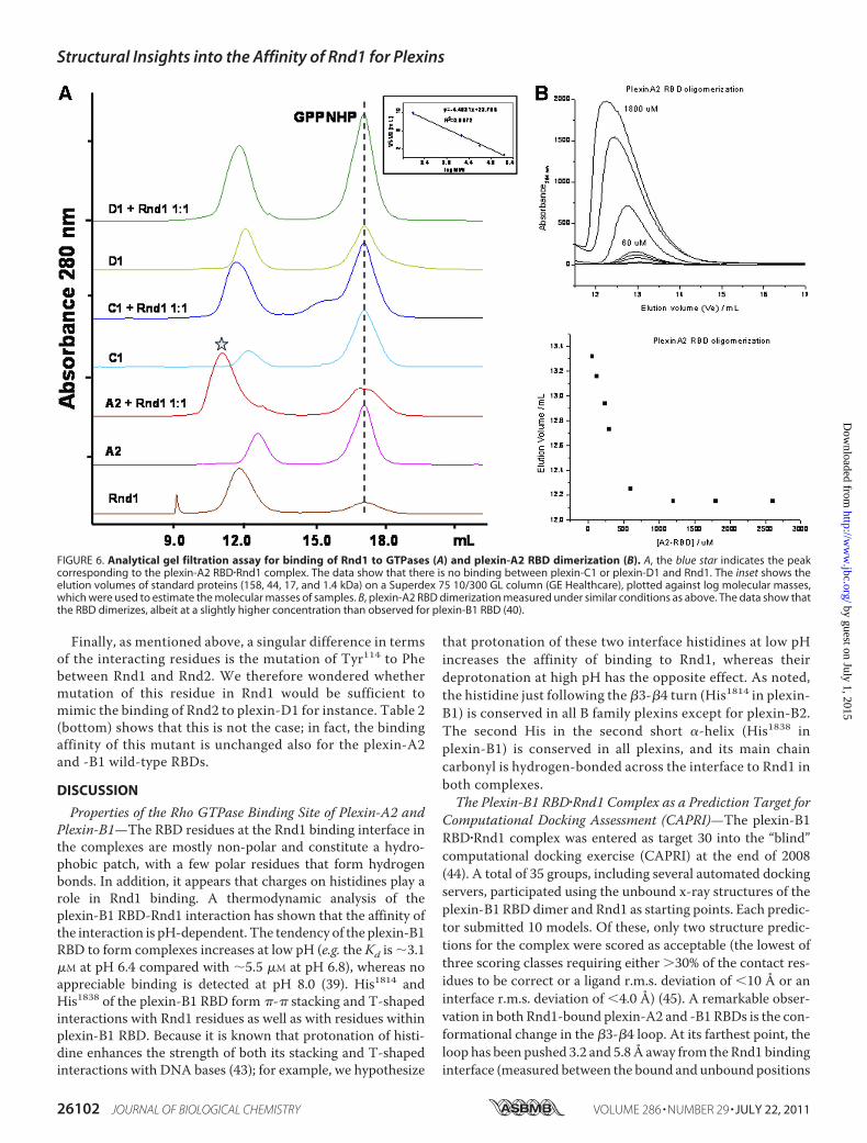
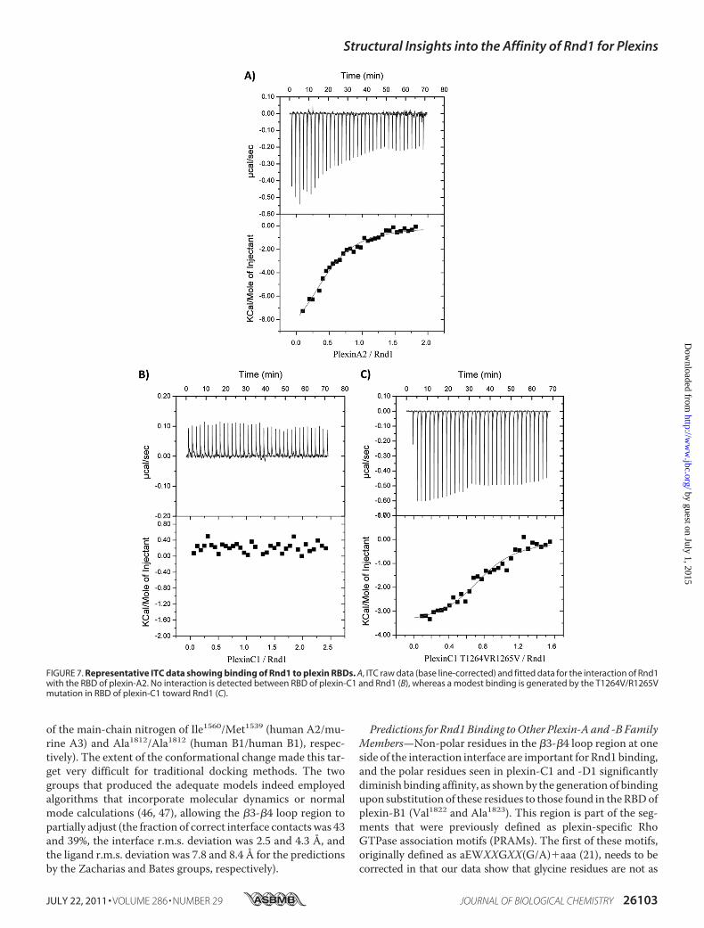
matography and Isothermal Titration Calorimetry—We exam-ined the binding of plexin RBDs (plexin-A2, plexin-B1, plexin-C1, and plexin-D1) to Rnd1 by gel filtration chromatography.The data show that, in the presence of the non-hydrolyzableGTP analog GMPPNP, plexin-A2 and plexin-B1 RBDs bind toRnd1, whereas plexin-C1 and plexin-D1 RBDs do not interactwith Rnd1 (Fig. 6A). The isothermal titration calorimetry mea-surements also show that plexin-C1 and plexin-D1 do notinteract with Rnd1, whereas plexin-A2 interacts with Rnd1with a very similar binding affinity (Kd � 7.0 �M) as comparedwith plexin-B1 (Kd � 5.5 �M) (Fig. 7 and Table 2, top). Withinthe confines of the quantitative differences typically observed insuch measurements, we confirmed the relative differences inbinding behavior of the RBDs and keymutants (see below) witha second technique, surface plasmon resonance spectroscopy(see supplemental material).Experimental Verification of the Structure-based Predictions—
The mutation of residue Trp1807, Asp1810, Ala1812, Gly1813,His1814, Leu1815, or Val1822 in plexin-B1 RBD to the corre-sponding residue in plexin-C1 or plexin-D1 RBD highly desta-bilizes the plexin-B1 RBD�Rnd1 complex, as shown by compu-tational modeling with FoldX, suggesting the importance ofthese side chains (results not shown). In order to test these andthe other structure-based predictions experimentally, wemutated several residues in plexin-B1, such as Leu1815 to Lys orTyr (the equivalent residues in the plexin-C1 and -D1 RBDs), inorder to demonstrate that these changes abolish or significantlyweaken the plexin-B1 RBD-Rnd1 interaction (Table 2, top).Indeed, binding to the L1815K is no longer detectable by ITC,and binding of L1815Y is almost 3-fold weaker compared withwild type.Furthermore, we mutated residues in plexin-C1 and
plexin-D1 to the corresponding plexin-B1 residues and showeda substantial gain in binding affinity for Rnd1. Specifically, wegenerated RBD-D1 Y1618L, S1614V/T1615A, and S1613G/Q1616A mutants and RBD-C1 K1267L and T1264V/R1265Vmutants, reverting key residues to their equivalents in plexin-B1, and measured possible binding interactions with Rnd1(Table 2, top). As noted above, neither wild type RBDs ofplexin-C1 nor -D1 bind to Rnd1 with affinities detectable byITC. Remarkably, the single and doublemutations, Y1618L andS1614V/T1615A, are sufficient to generate a plexin-D1 RBD-Rnd1 interaction with a modest binding affinity, detectable byITC (Kd � 5–20 �M, compared with 5.5 �M for the plexin-B1RBD-Rnd1 interaction). A K1267L rescue mutant generatesweak binding affinity for Rnd1 in plexin-C1, and a similar dou-blemutant, T1264V/R1265V, yields a proteinwith strong affin-ity for Rnd1 (Kd 2.6 �M), despite the fact that Lys1267 at theinterface would still be weakening an interaction. The resultsuggests that not all interactions that are seen in the plexin-B1and -A2 RBD complexes with Rnd1 are equally important andthat hydrophobic contacts between the RBD �3-�4 loop andRnd1 play a major role in complex formation.Binding of the RBDs to Rnd2, a close homologue of Rnd1
(with 62% sequence identity and 83% similarity over residues
FIGURE 4. Crystal structure of plexin-C1 and -D1 RBD. A, ribbon diagram ofplexin-C1 RBD. B, superimposition of the plexin-C1 RBD (green) and the free(blue) and bound (cyan) forms of the plexin-B1 RBD. C, plexin-D1 RBD.D, superimposition of the plexin-D1 RBD (gold) and the free (blue) and bound(cyan) forms of the plexin-B1 RBD in a ribbon diagram.
Structural Insights into the Affinity of Rnd1 for Plexins
26100 JOURNAL OF BIOLOGICAL CHEMISTRY VOLUME 286 • NUMBER 29 • JULY 22, 2011
by guest on July 1, 2015http://w
ww
.jbc.org/D
ownloaded from
5–180) was also examined by ITC (Table 2, middle). Severalmutations were studied in order to explore differences in theinteraction between RBDs and Rnd2, compared with Rnd1.First, we tested the affinity for the four wild type RBD proteins,and ITC showed weaker binding of Rnd2 to plexin-A2 and -B1comparedwithRnd1. Similarly to Rnd1, no bindingwas evidentby ITC to the plexin-C1wild typeRBD.However, the plexin-D1wild type RBD showed a weak affinity for Rnd2 (whereas noaffinity to Rnd1 was detected). Surprisingly, L1815K andL1815Y mutations in plexin-B1 did not weaken the interactionwith Rnd2 as strongly as for Rnd1. Similarly, Y1618L barely
increased binding of the plexin-D1 RBD protein to Rnd2,whereas this rescuemutation had a strong effect for Rnd1 bind-ing. By contrast, the K1267Lmutation rescued Rnd2 binding inplexin-C1 similarly as with Rnd1, and the plexin-C1 and -D1RBD�3-�4 loopmutants also showed similar increases in affin-ity for Rnd2. In conclusion then, compared with Rnd1 binding,the central �-strand 4 location (Leu1815 in plexin-B1) does notplay as critical a role for Rnd2 binding to the plexin-D1 RBD,suggesting that the mode of Rnd2 binding is somewhat differ-ent from that for the plexin-A1 and -B1 RBD�Rnd1 complexeswhose structures we obtained so far.
FIGURE 5. Structure-based sequence alignments. Structure-based sequence alignment of human plexin RBDs (A) and Rnd1 to -3 and Rac1 to -3 Rho GTPase(B). Alignments were generated using ESPript 2.1 (54). Sequence similarities are highlighted in yellow, and sequence identities are shown as white letters on a redbackground. Secondary structural elements (arrows for �-strands and coils for �-helices) are indicated at the top. Blue and green triangles indicate residues at theplexin-A2 and -B1 RBD�Rnd1 complex interface, respectively, as discussed under “Results.”
Structural Insights into the Affinity of Rnd1 for Plexins
JULY 22, 2011 • VOLUME 286 • NUMBER 29 JOURNAL OF BIOLOGICAL CHEMISTRY 26101
by guest on July 1, 2015http://w
ww
.jbc.org/D
ownloaded from
Finally, as mentioned above, a singular difference in termsof the interacting residues is the mutation of Tyr114 to Phebetween Rnd1 and Rnd2. We therefore wondered whethermutation of this residue in Rnd1 would be sufficient tomimic the binding of Rnd2 to plexin-D1 for instance. Table 2(bottom) shows that this is not the case; in fact, the bindingaffinity of this mutant is unchanged also for the plexin-A2and -B1 wild-type RBDs.
DISCUSSION
Properties of the Rho GTPase Binding Site of Plexin-A2 andPlexin-B1—The RBD residues at the Rnd1 binding interface inthe complexes are mostly non-polar and constitute a hydro-phobic patch, with a few polar residues that form hydrogenbonds. In addition, it appears that charges on histidines play arole in Rnd1 binding. A thermodynamic analysis of theplexin-B1 RBD-Rnd1 interaction has shown that the affinity ofthe interaction is pH-dependent. The tendency of the plexin-B1RBD to form complexes increases at low pH (e.g. the Kd is �3.1�M at pH 6.4 compared with �5.5 �M at pH 6.8), whereas noappreciable binding is detected at pH 8.0 (39). His1814 andHis1838 of the plexin-B1 RBD form �-� stacking and T-shapedinteractions with Rnd1 residues as well as with residues withinplexin-B1 RBD. Because it is known that protonation of histi-dine enhances the strength of both its stacking and T-shapedinteractions with DNA bases (43); for example, we hypothesize
that protonation of these two interface histidines at low pHincreases the affinity of binding to Rnd1, whereas theirdeprotonation at high pH has the opposite effect. As noted,the histidine just following the �3-�4 turn (His1814 in plexin-B1) is conserved in all B family plexins except for plexin-B2.The second His in the second short �-helix (His1838 inplexin-B1) is conserved in all plexins, and its main chaincarbonyl is hydrogen-bonded across the interface to Rnd1 inboth complexes.The Plexin-B1 RBD�Rnd1 Complex as a Prediction Target for
Computational Docking Assessment (CAPRI)—The plexin-B1RBD�Rnd1 complex was entered as target 30 into the “blind”computational docking exercise (CAPRI) at the end of 2008(44). A total of 35 groups, including several automated dockingservers, participated using the unbound x-ray structures of theplexin-B1 RBD dimer and Rnd1 as starting points. Each predic-tor submitted 10 models. Of these, only two structure predic-tions for the complex were scored as acceptable (the lowest ofthree scoring classes requiring either 30% of the contact res-idues to be correct or a ligand r.m.s. deviation of 10 Å or aninterface r.m.s. deviation of 4.0 Å) (45). A remarkable obser-vation in both Rnd1-bound plexin-A2 and -B1 RBDs is the con-formational change in the �3-�4 loop. At its farthest point, theloop has been pushed 3.2 and 5.8Å away from the Rnd1 bindinginterface (measured between the bound and unbound positions
FIGURE 6. Analytical gel filtration assay for binding of Rnd1 to GTPases (A) and plexin-A2 RBD dimerization (B). A, the blue star indicates the peakcorresponding to the plexin-A2 RBD�Rnd1 complex. The data show that there is no binding between plexin-C1 or plexin-D1 and Rnd1. The inset shows theelution volumes of standard proteins (158, 44, 17, and 1.4 kDa) on a Superdex 75 10/300 GL column (GE Healthcare), plotted against log molecular masses,which were used to estimate the molecular masses of samples. B, plexin-A2 RBD dimerization measured under similar conditions as above. The data show thatthe RBD dimerizes, albeit at a slightly higher concentration than observed for plexin-B1 RBD (40).
Structural Insights into the Affinity of Rnd1 for Plexins
26102 JOURNAL OF BIOLOGICAL CHEMISTRY VOLUME 286 • NUMBER 29 • JULY 22, 2011
by guest on July 1, 2015http://w
ww
.jbc.org/D
ownloaded from
of the main-chain nitrogen of Ile1560/Met1539 (human A2/mu-rine A3) and Ala1812/Ala1812 (human B1/human B1), respec-tively). The extent of the conformational change made this tar-get very difficult for traditional docking methods. The twogroups that produced the adequate models indeed employedalgorithms that incorporate molecular dynamics or normalmode calculations (46, 47), allowing the �3-�4 loop region topartially adjust (the fraction of correct interface contacts was 43and 39%, the interface r.m.s. deviation was 2.5 and 4.3 Å, andthe ligand r.m.s. deviation was 7.8 and 8.4 Å for the predictionsby the Zacharias and Bates groups, respectively).
Predictions for Rnd1Binding toOther Plexin-A and -B FamilyMembers—Non-polar residues in the �3-�4 loop region at oneside of the interaction interface are important for Rnd1 binding,and the polar residues seen in plexin-C1 and -D1 significantlydiminish binding affinity, as shownby the generation of bindingupon substitution of these residues to those found in theRBDofplexin-B1 (Val1822 and Ala1823). This region is part of the seg-ments that were previously defined as plexin-specific RhoGTPase association motifs (PRAMs). The first of these motifs,originally defined as aEWXXGXX(G/A)�aaa (21), needs to becorrected in that our data show that glycine residues are not as
FIGURE 7. Representative ITC data showing binding of Rnd1 to plexin RBDs. A, ITC raw data (base line-corrected) and fitted data for the interaction of Rnd1with the RBD of plexin-A2. No interaction is detected between RBD of plexin-C1 and Rnd1 (B), whereas a modest binding is generated by the T1264V/R1265Vmutation in RBD of plexin-C1 toward Rnd1 (C).
Structural Insights into the Affinity of Rnd1 for Plexins
JULY 22, 2011 • VOLUME 286 • NUMBER 29 JOURNAL OF BIOLOGICAL CHEMISTRY 26103
by guest on July 1, 2015http://w
ww
.jbc.org/D
ownloaded from

important as the non-polar (aliphatic) nature of themid-loop res-idues. Also, theTrp residue in�3 is not strictly required. Thus, thefirst PRAMshouldbe redefined as aEaXXX(a/R)aXX(a/R/K)XL, atleast for Rnd protein binding, where a indicates aliphatic residues,X represents anunknownpreference/any residue,� is a positivelycharged residue, and (a/b/c) indicates a preference for amino acida, b, or c.In light of this change, the following predictions can bemade
about the Rnd1 binding affinity of the plexin-A and -B families.As mentioned above, the plexin-A3 structure and sequencematches the plexin-A2 RBD closely (74% BLAST sequenceidentity over the length of the model; Fig. 5A). Several key res-idues of the revised first PRAMare structurally conserved in theplexin-A family members. Specifically, the turn �3-�4sequenceRIARV in the plexin-A2RBD is replaced byRMTRI inplexin-A3, whereas it is similar in the plexin-A1 RBD with asequence of RMARI. In all cases, the two Arg residues areexpected to point away from the interface because their sidechains have partial hydrophobic character, and the second Argforms a charged triad with the highly conserved Glu and Argthat surround the conserved Trp on �-strand 3 of the RBDstructure. As stipulated above, the second and third positionsalso have aliphatic character. The exception to this is thesequence of the plexin-A4 RBD, which reads SGARM in thisregion. No information exists yet on Rnd1 binding to plexin-A4, and our analysis predicts that binding is possibly weakened.In the plexin-B1 RBD�Rnd1 complex, the Val1811 and His1814
side chains of the plexin-B1 VAGHL sequence also point awayfrom the Rnd1 binding interface. In plexin-B3, the correspond-ing sequence reads LAGHL, again highly conducive for binding,whereas in plexin-B2, there is a point deletion at the first posi-tion, and the remainder reads STAQ. This is a significant
departure from the motif, probably leading to reduced Rnd1binding, although residues in other regions, such as Leu1519,may compensate (see discussion on Rdn1/Rnd2 binding toplexin-C1 and -D1 RBDs below).In summary, plexins have evolved a region at the edge of the
Rnd1 binding interface that shows significant sequence varia-tions in some familymembers and between families. The struc-tural and mutagenesis data show that the character of the�3-�4 loop appears to have a profound effect on Rnd bindingaffinity.Absence of Rnd1 Binding and Weak Binding of Rnd2 by the
RBDs of Plexin-C1 and -D1—The lack of non-polar residues inkey positions of the �3-�4 loop of the plexin-C1 and -D1 RBDsexplains why Rnd1 neither binds to these two plexins nor playsa regulatory role in their activities. On the other hand, it hasbeen reported that Rnd2 binding to plexin-D1 is required forSema3E-induced inhibition of axonal outgrowth in corticalneurons (24). Table 2 (middle) shows that GMPPNP-loadedRnd2 indeed binds the plexin-D1RBDweakly (Kd� 16.6�M). Itshould be noted that the structure of Rnd2 has not yet beendetermined. Based on the analysis of interface residues in theplexin-B1 and -A2 RBD�Rnd1 complexes, only one residue dif-fers between Rnd1 and Rnd2 in the region of contact with theRBDs; Tyr114 of Rnd1 is replaced by Phe120 in Rnd2 (Fig. 5B).We made this mutation in Rnd1 in order to mimic Rnd2, butthe modified GTPase showed no ITC-detectable interactionwith either the RBD of plexin-C1 or -D1, whereas overall bind-ing affinity with plexin-B1 was unperturbed, as shown in Table2 (bottom). (Interestingly, the changes in enthalpy and entropycontributions for this interaction suggest an increase in burialof hydrophobic area relative to wild type, whereas enthalpy isdecreased, possibly due to lack of a hydrogen bond to Tyr114).
TABLE 2Thermodynamic parameters for the association of Rnd1/2 with RBDs derived from ITC measurementsUncertainty in measurement is given in parentheses. NI, no interaction.
Ka Kd �H �G T�S
105 M�1 �M kcal/mol kcal/mol kcal/molRnd1-GTPPlexin-A2 WT 1.4 (0.1) 7.0 (0.7) �8.0 (0.7) �7.0 (0.1) �1.0 (0.1)Plexin-B1 WT 1.8 (0.1) 5.5 (0.6) �6.7 (0.3) �7.1 (0.1) �0.5 (0.1)L1815K NI NI NI NI NIL1815Y 0.7 (0.5) 14.1 (8) �1.4 (0.2) �6.6 (0.3) �5.2 (0.4)Plexin-C1 WT NI NI NI NI NIK1267L 1.0 (0.8) 10.0 (5.5) �1.9 (0.7) �6.8 (0.4) �4.9 (0.4)T1264V/R1265V 3.6 (0.8) 2.6 (0.4) �3.4 (0.1) �7.6 (0.2) �4.2 (0.5)Plexin-D1 WT NI NI NI NI NIY1618L 1.9 (0.6) 5.2 (1.8) �3.1 (0.1) �7.2 (0.1) �4.1 (0.2)S1614V/T1615A 0.5 (0.35) 20 (14) �1.3 (0.2) �6.4 (0.3) �7.7 (0.3)S1613G/Q1616A NI NI NI NI NI
Rnd2-GMPPNPPlexin-A2 WT 0.9 (0.4) 10.7 (4.1) �1.2 (0.2) �6.7 (0.3) �5.5 (0.3)Plexin-B1 WT 0.4 (0.4) 20 �0.2 (0.2) �6.3 (0.4) �6.1 (0.4)L1815K 0.8 (0.2) 20.0 (3.6) �3.2 (1.2) �6.5 (0.2) �3.5 (0.3)L1815Y 0.9 (0.4) 9.8 (3.9) �1.6 (0.5) �6.7 (0.4) �5.0 (0.4)Plexin-C1 WT NI NI NI NI NIK1267L 0.9 (0.7) 11.1 (5.1) �0.6 (0.4) �6.7 (0.3) �6.1 (0.4)T1264V/R1265V 1.2 (0.4) 8.3 (2.1) �1.5 (0.4) �6.9 (0.3) �5.4 (0.3)Plexin-D1 WT 0.6 (0.4) 16.6 (12) �0.4 (0.2) �6.5 (0.4) �6.1 (0.5)Y1618L 0.7 (0.2) 14.2 (3.2) �1.6 (0.4) �6.6 (0.2) �5.0 (0.3)S1614V/T1615A 1.3 (0.4) 7.7 (2.5) �1.4 (0.2) �6.9 (0.2) �5.5 (0.2)
Rnd1-GTP Y114FPlexin-A2 WT 1.0 (0.2) 10.0 (2.4) �4.1 (0.1) �6.7 (0.3) �2.7 (0.2)Plexin-B1 WT 1.8 (0.5) 5.5 (1.5) �2.5 (0.3) �6.7 (0.1) �4.2 (0.1)Plexin-C1 WT NI NI NI NI NIPlexin-D1 WT NI NI NI NI NI
Structural Insights into the Affinity of Rnd1 for Plexins
26104 JOURNAL OF BIOLOGICAL CHEMISTRY VOLUME 286 • NUMBER 29 • JULY 22, 2011
by guest on July 1, 2015http://w
ww
.jbc.org/D
ownloaded from
Thus, theTyr-to-Phe change fails to explainhowRnd2canbindto theplexin-D1RBDbecause,baseduponthemodelderived fromthe plexin-B1 RBD�Rnd1 complex, the RBD region that wouldcontact theRnd2GTPase in the region of Phe114 is actually largelyhydrophilic (plexin-C1 and -D1 present a Ser and Asp, respec-tively; by contrast, these residues are Val1822 in the plexin-B1 andIle1570 in the plexin-A2 RBD). Two neighboring residues, Glu1820and Glu1825, in plexin-B1 have been replaced by Leu and Val,respectively, in plexin-C1 andby Ile andVal in plexin-D1. (Glu1820is also replaced by a Leu in plexin-B2.)Another example is mutation L1815K at the center of strand
�4 of the plexin-B1 RBD, made to mimic plexin-C1. This abro-gated binding to Rnd1, but the mutant still weakly binds toRnd2. Similarly, themutation Y1618L (the same�4 site), whichgenerated binding in the plexin-D1 RBD for Rnd1, has littleeffect on the affinity of the same protein for Rnd2. This findingsuggests that the site is not as important for Rnd2 binding and,again, implies that the mode of Rnd2 binding to plexin RBDs islikely to be different at least in some detail from that of Rnd1;not all interactions at the interfacewill be the same. At the sametime, essentially identical interface contacts on the side of Rnd1are observed when this GTPase interacts with the two differentRBDs of plexin-A2 and -B1, strongly suggesting that these inde-pendent crystal structures are not an artifact.Recently, it has been found that the modes of interaction
even involving homologous proteins can be considerably differ-ent (42).7 In fact, there are several residues in the switch 1 and 2regions, outside the putative binding region, that differ betweenRnd1 and Rnd2. These could bind directly in a slightly differentbindingmode, as postulated above, ormay communicate a con-formational change to the residues that are directly bound.There is considerable evidence for longer range determinantsfor GTPase functional specificity, based upon mutagenesisstudies (48), that would support the latter hypothesis.The current model of plexin-B1 activation involves a shift in
the equilibrium between a state of the intracellular proteinregion that is dimerized through theRBDand one that is not. Thelatter state is stabilizedbyRnd1binding to theplexin-B1RBD(25).However, because a dimeric form of the plexin-D1 RBD is notobserved, the RBD dimer-monomer equilibrium model may notapply to plexin-D1 activation by Rnd2. Instead, Rnd2 binding tothe plexin-D1 RBD could induce a conformational change in adifferent region that affects the interactions between the RBD andthe GAP region, helping to activate the receptor.The binding affinity of either Rnd1 or Rnd2 to the wild type
plexin-C1RBD is veryweak (Kd 50�Mas estimated from ITCand also confirmed by surface plasmon resonance), consistentwith cell-based studies that suggest that this interaction may notbe required for theactivationofGAPfunction inplexin-C1 (24). Inaddition, plexin-C1 occurs as a monomer in solution, suggestingthat a mechanism that involves destabilization of RBD dimers, asseen for plexin-B1, is not applicable. This implies a different acti-vationmechanism forplexin-C1 (e.g.one that requires thebindingof yet to be identified agonist proteins).Functional Implications for Rnd1 GTPase Binding to Plexins—
At present, it is known that Rnd1 binding is required for certain
functions of the plexin-A1 and -B1 receptors. The structuresreported here, together with the analysis of the interface, pre-dict that also plexin-B3 and plexin-A3 will bind to Rnd1.Whether (or howwell) the RBDs of plexin-B2 and -A4 bind thisGTPase is presently unknown. It is possible that otherGTPases,including Rnd2, substitute for Rnd1 in order to stimulate plexinfunction. Such specificity is not unexpected; For instance, goodbinding of RhoD and weak binding of Rac1 were seen forplexin-A1 (19), whereas in plexin-B1, these affinities arereversed (21). It should be noted that the plexin RBD-RndGTPase interactions are of lower affinity compared with othereffector-small Rho GTPase interactions. However, modestaffinities have been observed in other systems, such as for theWASP-Cdc42 interaction with a Kd of 3–7 �M (49). In the caseof Rnd1 (50) and of the plexin-B1 receptor, both partners arelargely membrane-associated, and it is known that proteins canbe concentrated by their membrane localization up to severalthousand-fold over their density in the cytoplasm (51, 52),mak-ing these binding affinities physiologically relevant. In addition,protein preorientation, reduced entropy, and allosteric effectscan enhance complex formation at membranes. Further work isneeded to expand the present study into how other Rho and RasGTPases interact in detail with plexins. This knowledge, togetherwith measurements of active GTPase levels and their co-localiza-tion with the receptor in the cell, will ultimately lead to a detailedunderstanding of the plexin-mediated signaling mechanisms indifferent developmental and also pathogenic settings.In conclusion, all RBDs structurally characterized thus far
exhibit a typical ubiquitin-like fold that, except for loop regions,is highly conserved structurally. The comparative analysis ofthe plexin-A2 and -B1 RBD�Rnd1 complexes shows that thebinding interface is formed mainly by the RBD �4 strand and asecond short �-helix, with a conformational adjustment of the�3-�4 loop.Ourmutagenesis studies show that the character ofresidues in this latter, less well conserved loop and of a residueat the center of strand �4 is critical for Rnd1 binding but less sofor Rnd2 binding. In fact, it was possible to recover Rnd1 bind-ing in the plexin-C1 and -D1 RBD by making this loop morenon-polar. We also show that the plexin-A2 RBD tends todimerize, whereas dimerization is absent in plexin-C1 and -D1RBDs. This is consistent with the previous finding that Rnd1destabilizes RBD dimers but does not play a role in the activa-tion of plexin-C1 and -D1. Overall, then, evidence is mountingthat plexin functions are regulated by subfamily-specific mech-anisms, some of which involve different Rho family GTPases.
Acknowledgments—We thankShufenCao for assistancewithRnd1andRnd2 protein preparation and Drs. Christina Kiel and Luis Serrano(EMBL-CRG, Barcelona) for introducing us to Fold-X. Crystallographicdata shown in this report were derived fromwork performed at ArgonneNational Laboratory, Structural Biology Center, beam line 19ID, at theAdvanced Photon Source. Data collection at The National Institute ofGeneral Medical Sciences and National Cancer Institute CollaborativeAccess Team, beam line 23ID-B, was supported by NCI Grant Y1-CO-1020 and NIGM Grant Y1-GM-1104 from the National Institutes ofHealth.Use of theAdvancedPhoton Sourcewas supported by theUnitedStates Department of Energy, Office of Science, Office of Basic EnergySciences, under Contract DE-AC02–06CH11357.
7 P. K. Hota and M. Buck, manuscript in preparation.
Structural Insights into the Affinity of Rnd1 for Plexins
JULY 22, 2011 • VOLUME 286 • NUMBER 29 JOURNAL OF BIOLOGICAL CHEMISTRY 26105
by guest on July 1, 2015http://w
ww
.jbc.org/D
ownloaded from

REFERENCES1. Tamagnone, L., Artigiani, S., Chen, H., He, Z., Ming, G. I., Song, H., Che-
dotal, A., Winberg, M. L., Goodman, C. S., Poo, M., Tessier-Lavigne, M.,and Comoglio, P. M. (1999) Cell 99, 71–80
2. Negishi, M., Oinuma, I., and Katoh, H. (2005) Cell Mol. Life Sci. 62,1363–1371
3. Patel, B. N., and Van Vactor, D. L. (2002) Curr. Opin. Cell Biol. 14,221–229
4. Gitler, A. D., Lu, M. M., and Epstein, J. A. (2004) Dev. Cell 7, 107–1165. Serini, G., Valdembri, D., Zanivan, S., Morterra, G., Burkhardt, C., Cacca-
vari, F., Zammataro, L., Primo, L., Tamagnone, L., Logan, M., Tessier-Lavigne, M., Taniguchi, M., Puschel, A. W., and Bussolino, F. (2003) Na-ture 424, 391–397
6. Giordano, S., Corso, S., Conrotto, P., Artigiani, S., Gilestro, G., Barberis,D., Tamagnone, L., and Comoglio, P. M. (2002)Nat. Cell Biol. 4, 720–724
7. van Beijnum, J. R., Petersen, K., andGriffioen, A.W. (2009) Front. Biosci. 1,216–225
8. O’Connor, B. P., and Ting, J. P. (2008) Immunol. Res. 41, 217–2229. Moretti, S., Procopio, A., Boemi, M., and Catalano, A. (2006) Curr. Neu-
rovasc. Res. 3, 295–30510. Takahashi, T., Fournier, A., Nakamura, F., Wang, L. H., Murakami, Y.,
Kalb, R. G., Fujisawa, H., and Strittmatter, S. M. (1999) Cell 99, 59–6911. Okuno, T., Nakatsuji, Y., Moriya, M., Takamatsu, H., Nojima, S., Takega-
hara, N., Toyofuku, T., Nakagawa, Y., Kang, S., Friedel, R. H., Sakoda, S.,Kikutani, H., and Kumanogoh, A. (2010) J. Immunol. 184, 1499–1506
12. Comeau, M. R., Johnson, R., DuBose, R. F., Petersen, M., Gearing, P.,VandenBos, T., Park, L., Farrah, T., Buller, R. M., Cohen, J. I., Strockbine,L. D., Rauch, C., and Spriggs, M. K. (1998) Immunity 8, 473–482
13. Walzer, T., Galibert, L., and De Smedt, T. (2005) Eur. J. Immunol. 35,391–398
14. Gu, C., Yoshida, Y., Livet, J., Reimert, D. V., Mann, F., Merte, J., Hender-son, C. E., Jessell, T. M., Kolodkin, A. L., and Ginty, D. D. (2005) Science307, 265–268
15. Toyofuku, T., Zhang, H., Kumanogoh, A., Takegahara, N., Suto, F., Kamei,J., Aoki, K., Yabuki, M., Hori, M., Fujisawa, H., and Kikutani, H. (2004)Genes Dev. 18, 435–447
16. Roodink, I., Verrijp, K., Raats, J., and Leenders, W. P. (2009) BMC Cancer9, 297–313
17. Oinuma, I., Ishikawa, Y., Katoh, H., and Negishi, M. (2004) Science 305,862–865
18. Kruger, R. P., Aurandt, J., and Guan, K. L. (2005) Nat. Rev. Mol. Cell Biol.6, 789–800
19. Zanata, S. M., Hovatta, I., Rohm, B., and Puschel, A.W. (2002) J. Neurosci.22, 471–477
20. Driessens,M.H., Hu,H., Nobes, C. D., Self, A., Jordens, I., Goodman, C. S.,and Hall, A. (2001) Curr. Biol. 11, 339–344
21. Tong, Y., Chugha, P., Hota, P. K., Alviani, R. S., Li, M., Tempel, W., Shen,L., Park, H. W., and Buck, M. (2007) J. Biol. Chem. 282, 37215–37224
22. Puschel, A. W. (2007) Adv. Exp. Med. Biol. 600, 12–2323. Fujita, H., Katoh, H., Ishikawa, Y., Mori, K., and Negishi, M. (2002) J. Biol.
Chem. 277, 45428–4543424. Uesugi, K., Oinuma, I., Katoh, H., and Negishi, M. (2009) J. Biol. Chem.
284, 6743–675125. Tong, Y., Hota, P. K., Penachioni, J. Y., Hamaneh, M. B., Kim, S., Alviani,
R. S., Shen, L., He, H., Tempel,W., Tamagnone, L., Park, H.W., and Buck,M. (2009) J. Biol. Chem. 284, 35962–35972
26. He, H., Yang, T., Terman, J. R., and Zhang, X. (2009) Proc. Natl. Acad. Sci.U.S.A. 106, 15610–15615
27. Dong, A., Xu, X., Edwards, A. M., Chang, C., Chruszcz, M., Cuff, M.,Cymborowski, M., Di Leo, R., Egorova, O., Evdokimova, E., Filippova, E.,Gu, J., Guthrie, J., Ignatchenko, A., Joachimiak, A., Klostermann, N., Kim,
Y., Korniyenko, Y.,Minor,W.,Que,Q., Savchenko,A., Skarina, T., Tan, K.,Yakunin, A., Yee, A., Yim, V., Zhang, R., Zheng, H., Akutsu, M., Arrow-smith, C., Avvakumov, G. V., Bochkarev, A., Dahlgren, L. G., Dhe-Pa-ganon, S., Dimov, S., Dombrovski, L., Finerty, P., Jr., Flodin, S., Flores, A.,Graslund, S., Hammerstrom, M., Herman, M. D., Hong, B. S., Hui, R.,Johansson, I., Liu, Y., Nilsson, M., Nedyalkova, L., Nordlund, P., Nyman,T., Min, J., Ouyang, H., Park, H. W., Qi, C., Rabeh, W., Shen, L., Shen, Y.,Sukumard, D., Tempel, W., Tong, Y., Tresagues, L., Vedadi, M., Walker,J. R., Weigelt, J., Welin, M., Wu, H., Xiao, T., Zeng, H., and Zhu, H. (2007)Nat. Methods 4, 1019–1021
28. Minor, W., Cymborowski, M., Otwinowski, Z., and Chruszcz, M. (2006)Acta Crystallogr. D Biol. Crystallogr. 62, 859–866
29. Vagin, A., and Teplyakov, A. (2010) Acta Crystallogr. D Biol. Crystallogr.66, 22–25
30. Schneider, T. R., and Sheldrick, G. M. (2002) Acta Crystallogr. D Biol.Crystallogr. 58, 1772–1779
31. Otwinowski, Z. (1991) Proceedings of the CCP4 Study Weekend (Evans,P. R., and Leslie, A. G. W., eds) pp. 80–86, Daresbury Laboratory, War-rington, U.K.
32. Cowtan, K. D., and Main, P. (1996) Acta Crystallogr. D Biol. Crystallogr.52, 43–48
33. McCoy, A. J., Grosse-Kunstleve, R.W., Storoni, L. C., andRead, R. J. (2005)Acta Crystallogr. D Biol. Crystallogr. 61, 458–464
34. Adams, P. D., Grosse-Kunstleve, R. W., Hung, L. W., Ioerger, T. R., Mc-Coy, A. J., Moriarty, N.W., Read, R. J., Sacchettini, J. C., Sauter, N. K., andTerwilliger, T. C. (2002) Acta Crystallogr. D Biol. Crystallogr. 58,1948–1954
35. Murshudov, G. N., Vagin, A. A., and Dodson, E. J. (1997)Acta Crystallogr.D Biol. Crystallogr. 53, 240–255
36. Emsley, P., and Cowtan, K. (2004)Acta Crystallogr. D Biol. Crystallogr. 60,2126–2132
37. Davis, I.W.,Murray, L.W., Richardson, J. S., and Richardson, D. C. (2004)Nucleic Acids Res. 32,W615–W619
38. Bouguet-Bonnet, S., and Buck, M. (2008) J. Mol. Biol. 377, 1474–148739. Hota, P. K., and Buck, M. (2009) Protein Sci. 18, 1060–107140. Tong, Y., Hughes, D., Placanica, L., and Buck, M. (2005) Structure 13,
7–1541. Schymkowitz, J., Borg, J., Stricher, F., Nys, R., Rousseau, F., and Serrano, L.
(2005) Nucleic Acids Res. 33,W382–W38842. Tong, Y., Hota, P. K., Hamaneh, M. B., and Buck, M. (2008) Structure 16,
246–25843. Churchill, C. D., and Wetmore, S. D. (2009) J. Phys. Chem. B 113,
16046–1605844. Janin, J. (2010) Proteins 78, 3067–307245. Lensink, M. F., and Wodak, S. J. (2010) Proteins 78, 3085–309546. Fiorucci, S., and Zacharias, M. (2010) Proteins 78, 3131–313947. Li, X., Moal, I. H., and Bates, P. A. (2010) Proteins 78, 3189–9648. Heo, W. D., and Meyer, T. (2003) Cell 113, 315–32849. Buck, M., Xu, W., and Rosen, M. K. (2004) J. Mol. Biol. 338, 271–28550. Nobes, C. D., Lauritzen, I.,Mattei,M. G., Paris, S., Hall, A., andChardin, P.
(1998) J. Cell Biol. 141, 187–19751. Giesen, P. L., Willems, G. M., Hemker, H. C., and Hermens, W. T. (1991)
J. Biol. Chem. 266, 18720–1872552. Goryachev, A. B., and Pokhilko (2006) PLoS Comput. Biol. 12, e17253. Wallace, A. C., Laskowski, R. A., andThornton, J.M. (1995) Protein Eng. 8,
127–13454. Gouet, P., Robert, X., and Courcelle, E. (2003) Nucleic Acids Res. 31,
3320–332355. Laskowski, R. A., MacArthur, M. W., Moss, D. S., and Thornton, J. M.
(1993) J. Appl. Cryst. 26, 283–291
Structural Insights into the Affinity of Rnd1 for Plexins
26106 JOURNAL OF BIOLOGICAL CHEMISTRY VOLUME 286 • NUMBER 29 • JULY 22, 2011
by guest on July 1, 2015http://w
ww
.jbc.org/D
ownloaded from

Tempel, Matthias Buck and Hee-Won ParkSusmita Borthakur, SoonJeung Kim, WolframBuren Li, Limin Shen, Lyudmila Nedyalkova, Hui Wang, Prasanta K. Hota, Yufeng Tong, Rho GTPase Binding Domains (RBDs)Structural Basis of Rnd1 Binding to PlexinProtein Structure and Folding:
doi: 10.1074/jbc.M110.197053 originally published online May 24, 20112011, 286:26093-26106.J. Biol. Chem.
10.1074/jbc.M110.197053Access the most updated version of this article at doi:
.JBC Affinity SitesFind articles, minireviews, Reflections and Classics on similar topics on the
Alerts:
When a correction for this article is posted•
When this article is cited•
to choose from all of JBC's e-mail alertsClick here
Supplemental material:
http://www.jbc.org/content/suppl/2011/05/24/M110.197053.DC1.html
http://www.jbc.org/content/286/29/26093.full.html#ref-list-1
This article cites 54 references, 16 of which can be accessed free at
by guest on July 1, 2015http://w
ww
.jbc.org/D
ownloaded from
Copyright © 2022 FDOKUMEN