Tunneling spectroscopy for ferromagnet/superconductor junctions

Epithelial junctions and Rho family GTPases:the zonular signalosome
Sandra Citi1,2,*, Diego Guerrera1, Domenica Spadaro1, and Jimit Shah1
1Department of Cell Biology; University of Geneva; Geneva, Switzerland; 2Institute of Genetics and Genomics of Geneva; University of Geneva; Switzerland
Keywords: junctions, Rho, Rac, Cdc42, cytoseleton, epithelium
Abbreviations: AJ, adherens junction; AMOT, angiomotin; AMPK, Adenosine Monophosphate-Activated Protein Kinase; APC, ade-nomatous poliposis coli; Cdc42, cell division cycle 42; CD2AP, CD2-associated protein; CGN, cingulin; CGNL1, paracingulin; Dbl,
diffuse B-cell lymphoma; DLC, deleted in liver cancer; EPLIN, epithelial protein lost in neoplasm; ERK, extracellular regulatedkinase; FERM, four.point.one, ezrin, radixin, moesin; FGD5, FYVE, RhoGEF and PH domain containing 5; GEF, guanine nucleo-tide exchange factor; GAP, GTPase activating protein; GST, glutathione -S- transferase; JAM D junctional adhesion molecule; MCF-7, Michigan Cancer Foundation - 7; MDCK, Madin Darby Canine Kidney; MgcRacGAP, male germ cell racGAP; MKLP1, mitotickinesin-like protein-1; MRCK, myotonic dystrophy-related Cdc42-binding kinase; PA, puncta adhaerentia; PAK, p21-activated
kinase; PATJ, Pals1 associated tight junction protein; PCNA, proliferating cell nuclear antigen; PDZ, Post synaptic density protein(PSD95), Drosophila, disc large tumour suppressor (DlgA), and zonula occludens-1; PLEKHA7, pleckstrin homology domain con-taining, family A member 7; RICH-1, RhoGAP interacting with CIP4 homologues; ROCK, Rho-associated protein kinase; SH3BP1,(SH3 domain 490 binding protein-1); Tbx-3, T-box-3; Tiam, Tumor invasion and metastasis; TJ, tight junction; WASP, Wiskott-Aldrich Syndrome Protein; WAVE, WASP family Verprolin-homologous protein; ZA, zonula adhaerens; ZO, zonula occludens;
ZONAB, (ZO-1)–associated nucleic acid binding protein.
The establishment and maintenance of epithelial cell-celljunctions is crucially important to regulate adhesion, apico-basal polarity and motility of epithelial cells, and ultimatelycontrols the architecture and physiology of epithelial organs.Junctions are supported, shaped and regulated bycytoskeletal filaments, whose dynamic organization andcontractility are finely tuned by GTPases of the Rho family,primarily RhoA, Rac1 and Cdc42. Recent research hasidentified new molecular mechanisms underlying the cross-talk between these GTPases and epithelial junctions. Here webriefly summarize the current knowledge about theorganization, molecular evolution and cytoskeletal anchoringof cell-cell junctions, and we comment on the most recentadvances in the characterization of the interactions betweenRho GTPases and junctional proteins, and their consequenceswith regards to junction assembly and regulation of cellbehavior in vertebrate model systems. The concept of“zonular signalosome” is proposed, which highlights the closefunctional relationship between proteins of zonular junctions(zonulae occludentes and adhaerentes) and the control ofcytoskeletal organization and signaling through Rho GTPases,transcription factors, and their effectors.
Introduction
Cell-cell junctions provide epithelial tissues with mechanicaland functional integrity, by playing an essential role in cell-celladhesion and formation of barriers between distinct body com-partments. One key feature of junctions is their association withhighly ordered cytoskeletal networks of actin, microtubules andintermediate filaments. Since Rho GTPases are major regulatorsof the polymerization, organization and mechanics of the cyto-skeleton, the interplay between Rho GTPase activity and theorganization of junctions is of fundamental importance in epithe-lial morphogenesis and physiology. In this review we attempt toaddress the complexity of this regulation, going from basic con-cepts about the organization, evolution and cytoskeletal anchor-ing of cell-cell junctions, to the most recent exciting findingsabout the role of GEFs and GAPs at junctions, and their mecha-nisms of regulation.
The Organization and Molecular Evolution of theEpithelial Apical Junctional Complex
Epithelial tissues are at the boundary between the organismand the external environment, and form the first barrier to theentry of pathogens and toxins.1,2 In addition, they separate inter-nal body compartments, thus allowing the maintenance ofhomeostatic specialized functions, which depend on polarizedsecretion and absorption, and maintenance of gradients acrossepithelia.
To form efficient barriers, epithelial tissues must display spe-cific architectural characteristics, such as being formed by at leastone continuous layer of closely packed cells, and show a
© Sandra Citi, Diego Guerrera, Domenica Spadaro, and Jimit Shah*Correspondence to: Sandra Citi; Email: [email protected]: 11/06/2013; Revised: 06/02/2014; Accepted: 09/05/2014http://dx.doi.org/10.4161/21541248.2014.973760
This is an Open Access article distributed under the terms of the CreativeCommons Attribution-Non-Commercial License (http://creativecommons.org/licenses/by-nc/3.0/), which permits unrestricted non-commercial use,distribution, and reproduction in any medium, provided the original work isproperly cited. The moral rights of the named author(s) have been asserted.
www.landesbioscience.com 1Small GTPases
Small GTPases 5:4, 1--15; November 1, 2014; Published with license by Taylor & Francis Group, LLCREVIEW
Dow
nloa
ded
by [U
nive
rsité
de
Gen
ève]
at 0
5:48
18
Dec
embe
r 201
4
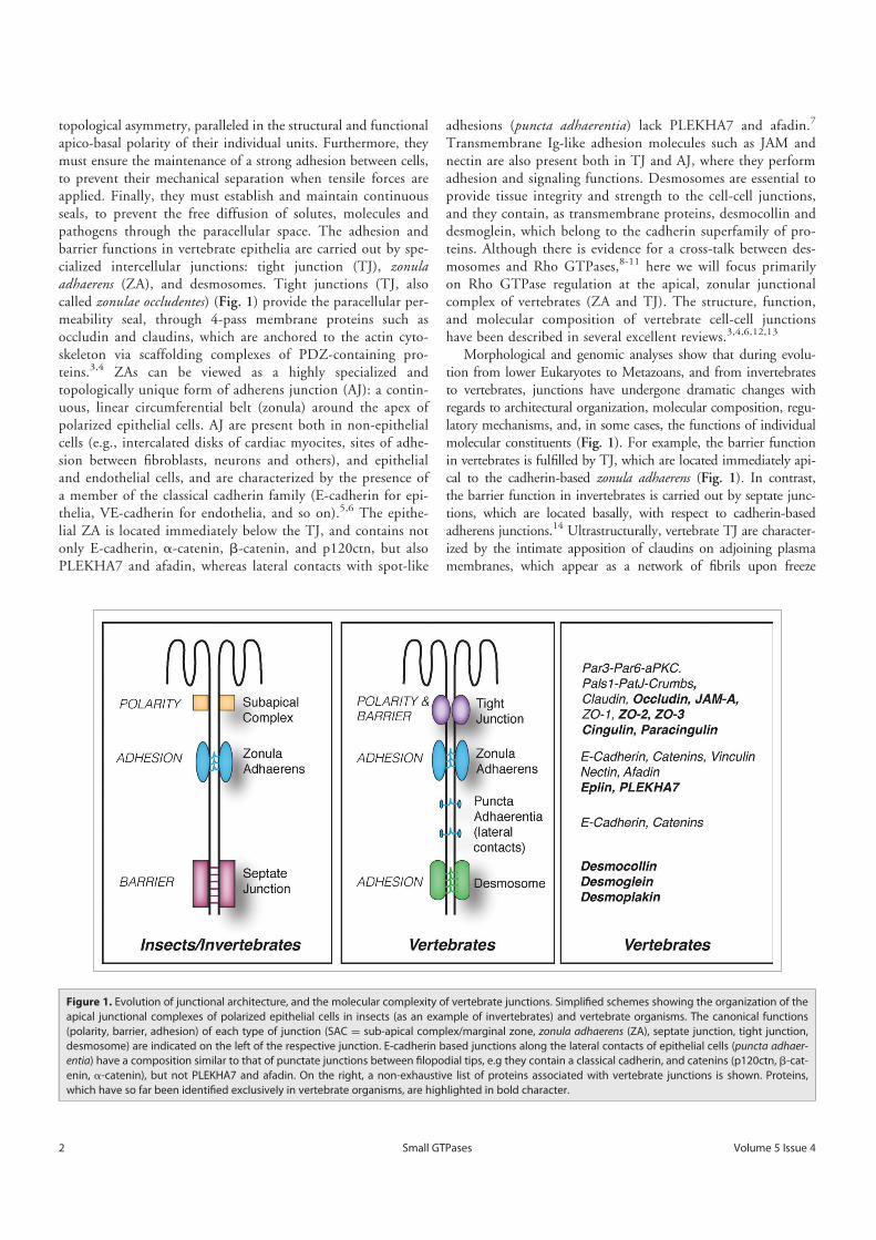
topological asymmetry, paralleled in the structural and functionalapico-basal polarity of their individual units. Furthermore, theymust ensure the maintenance of a strong adhesion between cells,to prevent their mechanical separation when tensile forces areapplied. Finally, they must establish and maintain continuousseals, to prevent the free diffusion of solutes, molecules andpathogens through the paracellular space. The adhesion andbarrier functions in vertebrate epithelia are carried out by spe-cialized intercellular junctions: tight junction (TJ), zonulaadhaerens (ZA), and desmosomes. Tight junctions (TJ, alsocalled zonulae occludentes) (Fig. 1) provide the paracellular per-meability seal, through 4-pass membrane proteins such asoccludin and claudins, which are anchored to the actin cyto-skeleton via scaffolding complexes of PDZ-containing pro-teins.3,4 ZAs can be viewed as a highly specialized andtopologically unique form of adherens junction (AJ): a contin-uous, linear circumferential belt (zonula) around the apex ofpolarized epithelial cells. AJ are present both in non-epithelialcells (e.g., intercalated disks of cardiac myocites, sites of adhe-sion between fibroblasts, neurons and others), and epithelialand endothelial cells, and are characterized by the presence ofa member of the classical cadherin family (E-cadherin for epi-thelia, VE-cadherin for endothelia, and so on).5,6 The epithe-lial ZA is located immediately below the TJ, and contains notonly E-cadherin, a-catenin, b-catenin, and p120ctn, but alsoPLEKHA7 and afadin, whereas lateral contacts with spot-like
adhesions (puncta adhaerentia) lack PLEKHA7 and afadin.7
Transmembrane Ig-like adhesion molecules such as JAM andnectin are also present both in TJ and AJ, where they performadhesion and signaling functions. Desmosomes are essential toprovide tissue integrity and strength to the cell-cell junctions,and they contain, as transmembrane proteins, desmocollin anddesmoglein, which belong to the cadherin superfamily of pro-teins. Although there is evidence for a cross-talk between des-mosomes and Rho GTPases,8-11 here we will focus primarilyon Rho GTPase regulation at the apical, zonular junctionalcomplex of vertebrates (ZA and TJ). The structure, function,and molecular composition of vertebrate cell-cell junctionshave been described in several excellent reviews.3,4,6,12,13
Morphological and genomic analyses show that during evolu-tion from lower Eukaryotes to Metazoans, and from invertebratesto vertebrates, junctions have undergone dramatic changes withregards to architectural organization, molecular composition, regu-latory mechanisms, and, in some cases, the functions of individualmolecular constituents (Fig. 1). For example, the barrier functionin vertebrates is fulfilled by TJ, which are located immediately api-cal to the cadherin-based zonula adhaerens (Fig. 1). In contrast,the barrier function in invertebrates is carried out by septate junc-tions, which are located basally, with respect to cadherin-basedadherens junctions.14 Ultrastructurally, vertebrate TJ are character-ized by the intimate apposition of claudins on adjoining plasmamembranes, which appear as a network of fibrils upon freeze
Figure 1. Evolution of junctional architecture, and the molecular complexity of vertebrate junctions. Simplified schemes showing the organization of theapical junctional complexes of polarized epithelial cells in insects (as an example of invertebrates) and vertebrate organisms. The canonical functions(polarity, barrier, adhesion) of each type of junction (SAC D sub-apical complex/marginal zone, zonula adhaerens (ZA), septate junction, tight junction,desmosome) are indicated on the left of the respective junction. E-cadherin based junctions along the lateral contacts of epithelial cells (puncta adhaer-entia) have a composition similar to that of punctate junctions between filopodial tips, e.g they contain a classical cadherin, and catenins (p120ctn, b-cat-enin, a-catenin), but not PLEKHA7 and afadin. On the right, a non-exhaustive list of proteins associated with vertebrate junctions is shown. Proteins,which have so far been identified exclusively in vertebrate organisms, are highlighted in bold character.
2 Volume 5 Issue 4Small GTPases
Dow
nloa
ded
by [U
nive
rsité
de
Gen
ève]
at 0
5:48
18
Dec
embe
r 201
4
fracture. Insect septate junctions show extracellular electron-dense“septa” bridging the opposite plasma membranes, rather than clau-din-based fibrils.14 In vertebrates, TJ correspond topologically tothe physical “fence” separating apical from lateral plasma mem-brane domains, which maintains apico-basal polarity (Fig. 1).Instead, the fence in invertebrates is not the septate junction, butthe subapical complex (SAC)/marginal zone, which is apical to theZA, and morphologically distinct from TJ (Fig. 1). Evolutionarilyconserved polarity complexes confer either apical identity (Par3-Par6-apKC and Crumbs-Pals1-PatJ complexes), or basolateralidentity (Scribble-Dlg-Lgl complex) to the plasma membrane, andare segregated at the level of the TJ in vertebrates and the subapicalcomplex (SAC)/marginal zone in invertebrates (Fig. 1).3,14,15 Atthe molecular level, the number of isoforms and/or family mem-bers for most junctional proteins is considerably larger in verte-brates, providing for increased molecular complexity andredundancy. For example, although cadherin and catenins areshared between insect and vertebrate AJ, invertebrates do notexpress many classical cadherin isoforms, and lack desmosomalcadherins, desmosomes and intermediate filaments.15,16 Strikingly,epithelial cells of lower Eukaryotes, such as the amoeba Dyctioste-lium discoideum, achieve adhesion and polarity in the absence ofany cadherin, whereas in metazoans E-cadherin is criticallyrequired for cell-adhesion, embryonic development, and the gener-ation of apico-basal polarity.17,18 Claudins, the transmembraneproteins responsible for the barrier of TJ to ions, are highly diver-gent in their sequence from invertebrates to vertebrates, and thefamily includes over 20 members in vertebrates, whereas only 5and 3 members, respectively, have been described so far in C. ele-gans or Drosophila.14 A ZO-1 homolog has been identified in Dro-sophila, but the ZO family in vertebrates comprises alsoZO-2 and ZO-3, which have partially redundant functions withZO-1.19,20 Knock-out of the components of the Par3-Par6-aPKCcomplex in invertebrates has dramatic consequences on epithelialand neuronal morphogenesis, but can have only tissue-specific andmore subtle effects in mice.21 The lateral polarity protein Lgl actsas a canonical tumor suppressor protein in Drosophila, but not inmice.22 Over 70 GEFs and 60 GAPs have been described in Ver-tebrates, whereas only about 10 Rho GEFs and GAPs combinedhave so far been identified in Drosophila.23 In summary, althoughinvertebrate model systems are useful to establish some generalprinciples, vertebrate cells and organisms are required to under-stand the remarkably more complex organization of vertebratejunctions and their signaling and regulatory mechanisms. In thisreview, we focus on vertebrate model systems.
Assembly and Anchoring of the Cytoskeleton atEpithelial Apical Junctional Complexes
To understand the relationships between Rho GTPase regula-tion and assembly of vertebrate junctions, it is necessary to exam-ine how the actomyosin and microtubule cytoskeletons, whichare major targets of Rho GTPase effectors, functionally andstructurally interact with AJ and TJ.
The circumferential, junction-associated bundle of actinmicrofilaments and nonmuscle myosin in the brush border ofpolarized epithelial cells was described three decades ago.24 Theactomyosin cytoskeleton regulates the distribution, stability, clus-tering and endocytosis of cadherin at the cell membrane,25 andthe thickness of the bundle is related to the greater mechanicaltensions applied to the ZA, compared to weaker forces applied tolateral AJ complexes.26 Contraction of the ZA-associated actomy-osin ring causes apical constriction, which is crucial to supportmorphogenetic changes in developing embryos.27 In addition,the contractile apical actomyosin ring is critical for the regulationof TJ integrity and barrier function.28
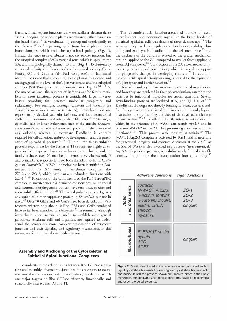
How actin and myosin are structurally connected to junctions,and how they are regulated in their polymerization, assembly andactivities by junctional molecules are crucial questions. Severalactin-binding proteins are localized at AJ and TJ (Fig. 2).12,29
E-cadherin, although not directly binding to actin, acts as a scaf-fold for cytoskeleton-associated protein complexes, and plays aninstructive role by marking the sites of de novo actin filamentpolymerization.30,31 E-cadherin directly interacts with cortactin,which in the presence of N-WASP can recruit Arp2/3 and itsactivator WAVE2 to the ZA, thus promoting actin nucleation atjunctions.30,32 This process also requires a-actinin.33 TheWAVE2-Arp2/3 complex is activated by Rac1, and is necessaryfor junctional integrity and contractile tension at the ZA.34 Atthe ZA, N-WASP is also involved in a putative “non-canonical,"Arp2/3-independent pathway, to stabilize newly formed actin fil-aments, and promote their incorporation into apical rings.35
Figure 2. Proteins implicated in the organization and junctional anchor-ing of cytoskeletal filaments. For each type of cytoskeletal filament (actinand microtubules) the proteins shown are involved either in their poly-merization, bundling, and anchoring to junctions, based on biochemicaland/or cell biological evidence.
www.landesbioscience.com 3Small GTPases
Dow
nloa
ded
by [U
nive
rsité
de
Gen
ève]
at 0
5:48
18
Dec
embe
r 201
4
This is an example of a new role played at zonular junctions by aprotein, beyond its classical activity. Nucleation of actin filamentsby the Arp2/3 complex gives rise to an extensive array ofbranched actin filaments, but mature apical junctions are charac-terized by the presence of bundled actin filaments. Several actin-binding proteins can influence microfilament organization anddynamics at the ZA. Formins, for example, have been implicatedin the formation of junctional actin bundles in some celltypes.36,37 a-catenin, which has an evolutionarily conserved rolein organizing the cortical actin cytoskeleton,17,38,39 suppressesactin polymerization by the Arp2/3 complex, while stabilizingand bundling actin filaments. The affinities of interaction ofmonomeric a-catenin with actin and vinculin are dramaticallyincreased when tensile forces are applied to junctions, through amolecular stretching mechanism, indicating that monomerica-catenin bound to b-catenin can directly link F-actin to thecadherin complex in vivo,40,41 although this is not observed invitro.42 EPLIN (Epithelial Protein Lost In Neoplasm) is recruitedto junctions by a-catenin, and it inhibits actin depolymerization,and crosslinks actin filaments.43,44 Afadin associates with thecytoplasmic domain of nectins and JAM,45 is recruited to the ZAthrough an interaction with a-catenin, and directly interacts withactin filaments.46 Afadin is a major organizer of the apical junc-tional complex, and is essential for the development of apico-basal polarity in vertebrate embryogenesis.47,48 Finally, myosin IIis an essential component of the contractile bundle associatedwith the ZA, and its positioning is regulated by Shroom, andactin-binding protein which interacts with the Rho effectorkinase ROCK,49 and is regulated by the FERM domain proteinLulu.50 Recent studies have addressed the role of different actinand myosin isoforms at epithelial junctions. Depletion studiesshow that both b- and g- actin isoforms, though differently dis-tributed, are essential for TJ barrier function and junction assem-bly, whereas b-actin is selectively involved in the establishmentof apico-basal cell polarity.51 Concerning myosins, myosin IIA isthe most important in regulating cell morphology and cell-celladhesion, whereas myosin IIB has more subtle roles in actin fila-ment dynamics.52,53
Besides AJ, TJ are also structurally and functionally linked tothe actin cytoskeleton (Fig. 2). Actin has multiple potential part-ners at TJ, including the ZO proteins (ZO-1, ZO-2, ZO-3),occludin and cingulin (Fig. 2).54-56 Cells depleted of ZO-1 showdefects in the barrier to larger solutes, and changes in the junction-associated actin, indicating that ZO-1 forms a stabilizing linkbetween the barrier and the junctional actomyosin.57,58 In con-trast, depletion of ZO-2 does not lead to either actin reorganiza-tion or altered permeability to larger molecules,59 whereasdepletion of both ZO-1 and ZO-2 leads to a dramatic expansionof the actomyosin belt associated with AJ.60 Since ZO-1 interactsdirectly or indirectly with several actin-binding proteins, includ-ing a-catenin and cortactin,19,61,62 and with GEFs for Rac1 andRhoA,63,64 some of the phenotypes of these knock-down modelsmay be dependent on these interactions, although this remains tobe determined. Cingulin is so far the only TJ protein for which anactin-bundling activity has been described in vitro.56 However,cingulin depletion or overexpression in MDCK cells does not
result in dramatic changes in actin organization or barrier func-tion,65-67 suggesting functional redundancies with other proteins.
Microtubules show a polarized distribution in epithelial cells,and associate with the apical junctional complex.68 Recent stud-ies show that E-cadherin is connected to the minus ends ofmicrotubules through a complex containing p120ctn, PLE-KHA7, paracingulin and nezha (CAMSAP3)69,70 (Fig. 2).Microtubule anchoring confers stability to apical junctions,69,71
and also indirectly stabilizes TJ barrier function, by enhancingthe accumulation of E-cadherin and associated proteins at theZA.72 Exogenous PLEKHA7 can accumulate at lateral contactspuncta adhaerentia, probably through its interaction withp120ctn, but this does not result in increased recruitment ofmicrotubule minus ends, suggesting that microtubule anchoringrequires a specialized molecular environment that occurs only atthe ZA.72 Interaction of microtubule plus ends with cadherin-based junctions involves dynein, which interacts with b-cate-nin,73 APC,74 and the spectroplakin ACF7.75 Recent experi-ments indicate that a planar apical network of microtubules isanchored to TJ through cingulin, and this interaction is regulatedby adenosine monophosphate protein kinase (AMPK)-mediatedphosphorylation of cingulin 76 (Fig. 2). There is an importantcross-talk between the actin and microtubule cytoskeletons. Forexample, the formin mDia is involved both in linear actin poly-merization and microtubule stabilization,77 and microtubulescan both sequester Rho GEFs that control actin organiza-tion,78,79 and associate with the centralspindlin complex, whichplays roles not only in mitotic spindle organization and cytokine-sis, but also in the control of Rho and Rac activity at junc-tions.29,80 In summary, cell-cell junctions are critical sites ofanchoring and organization of cytoskeletal filaments, throughspecific adaptor and regulatory molecules.
The Involvement of RhoA, Rac1 and Cdc42 inEpithelial Junction Assembly and Regulation
The cytoskeleton is essential for the establishment, mainte-nance, remodeling and disassembly of apical junctions, and thisprocess is regulated by Rho family GTPases and their effectors.The first studies addressed the role of Rho GTPases in junctionregulation by exogenously expressing either the Rho inhibitor C3transferase, or dominant negative (DN) or constitutively active(CA) mutants of Rho GTPases. This lead to loss of barrier andfence functions of TJ, inhibition or perturbation of junctionassembly, and was in some cases associated with disrupted locali-zation of junctional proteins, depending on expression levels ofmutant proteins.81-85 The observation that DN and CA mutantshave similar effects is consistent with the notion that catalyticcycling between active and inactive states, rather than a perma-nent “on” or “off” state, is essential for the proper functioning ofRho GTPases. Thus, mutant phenotypes may similarly affect theRho GTPases functional output, by binding to and sequesteringtargets and effectors. In summary, correct junction assembly andfunction requires a finely tuned balance in the activities of RhoA,Rac1 and Cdc42.
4 Volume 5 Issue 4Small GTPases
Dow
nloa
ded
by [U
nive
rsité
de
Gen
ève]
at 0
5:48
18
Dec
embe
r 201
4
Rac1 and Cdc42 are essential in the initial formation of junc-tions, following engagement of adhesion receptors at primordialjunctions,31,86,87 by promoting the polymerization of actin fila-ments in lamellipodia and filopodia, through activation of theArp2/3 complex by WAVE2.29 A second crucial role of Cdc42 isto promote the formation of the Par6-aPKC-Par3 complex, thusallowing the establishment of apico-basal polarity and segregationof apical TJ.3 The role of Cdc42 in polarity was first discoveredin the yeast Saccharomyces cerevisiae, where a Cdc42 mutationresulted in inhibition of polarized budding.88 In vertebrate cells,aPKC activity is required for the establishment of TJ, but not fortheir maintenance, whereas the role of Cdc42 in the regulation ofmature TJ appears to depend on cell type. For example, in endo-thelial cells Cdc42 does not play a significant role in regulatingjunctional actin organization and barrier function,89 but in other,non-endothelial cells, regulation of Cdc42 is necessary for TJmaintenance.90,91 Evidence from invertebrate models indicatesthat at steady state the Cdc42-Par6-aPKC axis acts by limitingRhoA activity, and thus junctional tension at AJ.92 The role ofN-WASP, a target of Cdc42 and Rac1, in regulating junctionarchitecture and cortical tension has also been demonstrated invertebrate model systems.63 N-WASP can also be targeted bypathogens, to promote cell-to-cell spreading.93
RhoA plays a fundamental role both in the establishment andmaintenance of AJ and TJ through 2 major effectors: Rho-associ-ated protein kinase (ROCK) and Diaphanous-related formin-1(Dia).94 mDia nucleates linear actin polymerization at the AJ,36
and can sense and generate mechanical forces on actin fila-ments,95 whereas ROCK promotes the bundling of actin fila-ments and the contractility of actomyosin, by enhancing thephosphorylation of nonmuscle myosin light chains.96 Thesefunctions are critical to maintain tension at apical junctions,inhibit cadherin endocytosis, and establish and maintain TJ bar-riers.92,97,98 A physiological balance between mDia and ROCKactivities is required to maintain ZAs, since decreased Dia orincreased ROCK activation can induce the transition from belt-like ZA to punctate PA.94,99 Indeed, ROCK activation can alsobe a major mechanism of junction disruption, triggered by cyto-kines and other exogenous stimuli.100 Therefore the fine-tuningof the activation of RhoA effectors in space and time is a criticalfactor in the regulation of junction assembly and stability.
New Insights Into the Molecular MechanismsUnderlying the Spatio-temporal Regulation of Rho
Family GTPases at Junctions
The biological impact of Rho family GTPases criticallydepends on the precise site and timing of their activation. Thus,understanding how Rho GTPases control junction assemblyrequires the identification of the molecular mechanisms that reg-ulate Rho GTPase activity at junctions.
The spatial and temporal control of Rho GTPases is coordi-nated by GEFs and GAPs, which activate and deactivate RhoGTPases by promoting either the exchange of GDP for GTP, orGTP hydrolysis, respectively. GEFs and GAPs interact with
adaptor proteins, which recruit them to defined subcellular sites,and/or modulate their activity, for example by phosphorylation(see section on “Regulation of regulators”). In a previous review,we summarized the interactions of vertebrate junctional proteinswith GEFs and GAPs implicated in the regulation of RhoA,Rac1, Rap1 and Cdc42.101 Here, we will focus on more recentstudies, which have provided additional insights into the cross-talk between junctional proteins and Rho family GTPases, andframe them into a dynamic view of the involvement of GEFs andGAPs in the different steps of junction formation.
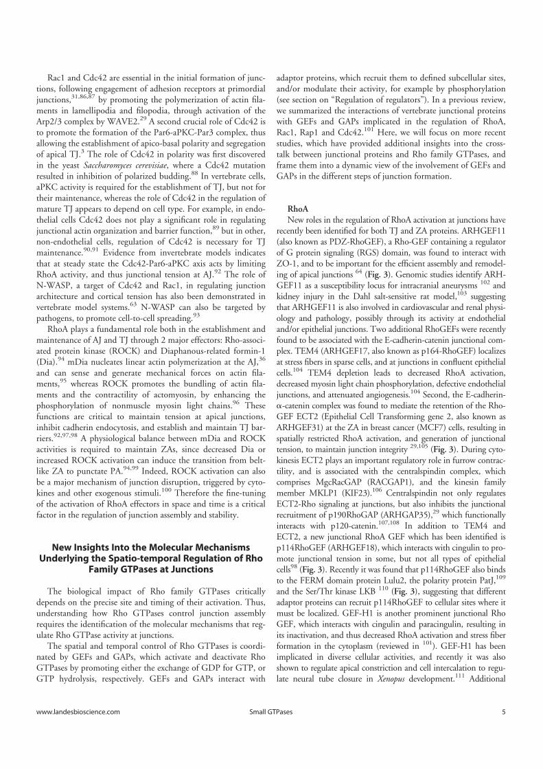
RhoANew roles in the regulation of RhoA activation at junctions have
recently been identified for both TJ and ZA proteins. ARHGEF11(also known as PDZ-RhoGEF), a Rho-GEF containing a regulatorof G protein signaling (RGS) domain, was found to interact withZO-1, and to be important for the efficient assembly and remodel-ing of apical junctions 64 (Fig. 3). Genomic studies identify ARH-GEF11 as a susceptibility locus for intracranial aneurysms 102 andkidney injury in the Dahl salt-sensitive rat model,103 suggestingthat ARHGEF11 is also involved in cardiovascular and renal physi-ology and pathology, possibly through its activity at endothelialand/or epithelial junctions. Two additional RhoGEFs were recentlyfound to be associated with the E-cadherin-catenin junctional com-plex. TEM4 (ARHGEF17, also known as p164-RhoGEF) localizesat stress fibers in sparse cells, and at junctions in confluent epithelialcells.104 TEM4 depletion leads to decreased RhoA activation,decreased myosin light chain phosphorylation, defective endothelialjunctions, and attenuated angiogenesis.104 Second, the E-cadherin-a-catenin complex was found to mediate the retention of the Rho-GEF ECT2 (Epithelial Cell Transforming gene 2, also known asARHGEF31) at the ZA in breast cancer (MCF7) cells, resulting inspatially restricted RhoA activation, and generation of junctionaltension, to maintain junction integrity 29,105 (Fig. 3). During cyto-kinesis ECT2 plays an important regulatory role in furrow contrac-tility, and is associated with the centralspindin complex, whichcomprises MgcRacGAP (RACGAP1), and the kinesin familymember MKLP1 (KIF23).106 Centralspindin not only regulatesECT2-Rho signaling at junctions, but also inhibits the junctionalrecruitment of p190RhoGAP (ARHGAP35),29 which functionallyinteracts with p120-catenin.107,108 In addition to TEM4 andECT2, a new junctional RhoA GEF which has been identified isp114RhoGEF (ARHGEF18), which interacts with cingulin to pro-mote junctional tension in some, but not all types of epithelialcells98 (Fig. 3). Recently it was found that p114RhoGEF also bindsto the FERM domain protein Lulu2, the polarity protein PatJ,109
and the Ser/Thr kinase LKB 110 (Fig. 3), suggesting that differentadaptor proteins can recruit p114RhoGEF to cellular sites where itmust be localized. GEF-H1 is another prominent junctional RhoGEF, which interacts with cingulin and paracingulin, resulting inits inactivation, and thus decreased RhoA activation and stress fiberformation in the cytoplasm (reviewed in 101). GEF-H1 has beenimplicated in diverse cellular activities, and recently it was alsoshown to regulate apical constriction and cell intercalation to regu-late neural tube closure in Xenopus development.111 Additional
www.landesbioscience.com 5Small GTPases
Dow
nloa
ded
by [U
nive
rsité
de
Gen
ève]
at 0
5:48
18
Dec
embe
r 201
4

RhoGEFs which have been implicated in epithelial apical constric-tion during morphogenesis are Trio,112 and ARHGEF11.113
Regarding Rho GAPs, indirect roles in regulating junctionshave been found for the unconventional myosins Myo9a andMyo9b, large single-headed motor molecules that comprise a N-terminal actin binding domain, and a tail with a Rho GAPdomain.114,115 Depletion and overexpression studies show thatboth Myo9a and Myo9b regulate collective epithelial cell migra-tion and wound healing, by down-regulating RhoA activity, andthus reducing localized cytoskeletal tension at the leading edge oflamellipodia, thus stabilizing nascent cell-cell contacts. However,assembly of junctions in non-migrating cells is not affected by
Myo9a-depletion, suggesting that this myosin may be importantonly for dynamic junctions.114 In another study, knockdown ofMyo9a was reported to disrupt TJ,116 similarly to what observedfollowing Myo9b depletion in Caco2 intestinal cells.115 Interest-ingly, polymorphisms in the gene encoding the Myo9b heavychain are linked to several forms of inflammatory bowel dis-ease,117,118 and Myo9b function may be implicated in pathogen-esis both through defective cell migration of sub-mucosalimmune cells, and a leaky TJ barrier. Another Rho GAP that hasrecently been implicated in the maintenance of cell adhesion isDLC1 (Deleted in Liver Cancer 1), which acts as a GAP forRhoA, RhoC, and, to a lesser extent, for Cdc42. Exogenous
Figure 3. Crosstalk between junctions and Rho GTPases during the biogenesis of epithelial junctions. Simplified schemes showing sequential steps in theformation and maturation of the apical junctional complex (TJ and ZA) in epithelial cells, from primordial contact (top) to mature junction (bottom), andthe proteins involved. Legends for graphical objects are shown in box (top left). Green and red arrows/lines indicate activation and inhibition, respec-tively. The main effects of Rho GTPase regulation on cytoskeletal organization and function are summarized on the sides of each scheme. Proteins andprotein interactions depicted here are derived from studies on different model systems, so they do not necessarily occur together, but are grouped inone scheme for the sake of summarizing them. See text for additional details.
6 Volume 5 Issue 4Small GTPases
Dow
nloa
ded
by [U
nive
rsité
de
Gen
ève]
at 0
5:48
18
Dec
embe
r 201
4
DLC1 interacts with a-catenin at AJ, and suppresses invasionand metastasis by up-regulating E-cadherin expression, in a Rho-dependent manner.119 Another member of the DLC family ofRhoGAP proteins, DLC3, is localized at AJ in breast cancer cellswhen exogenously expressed, and is essential for E-cadherin-mediated maintenance of cell-cell contacts120 (Fig. 3).
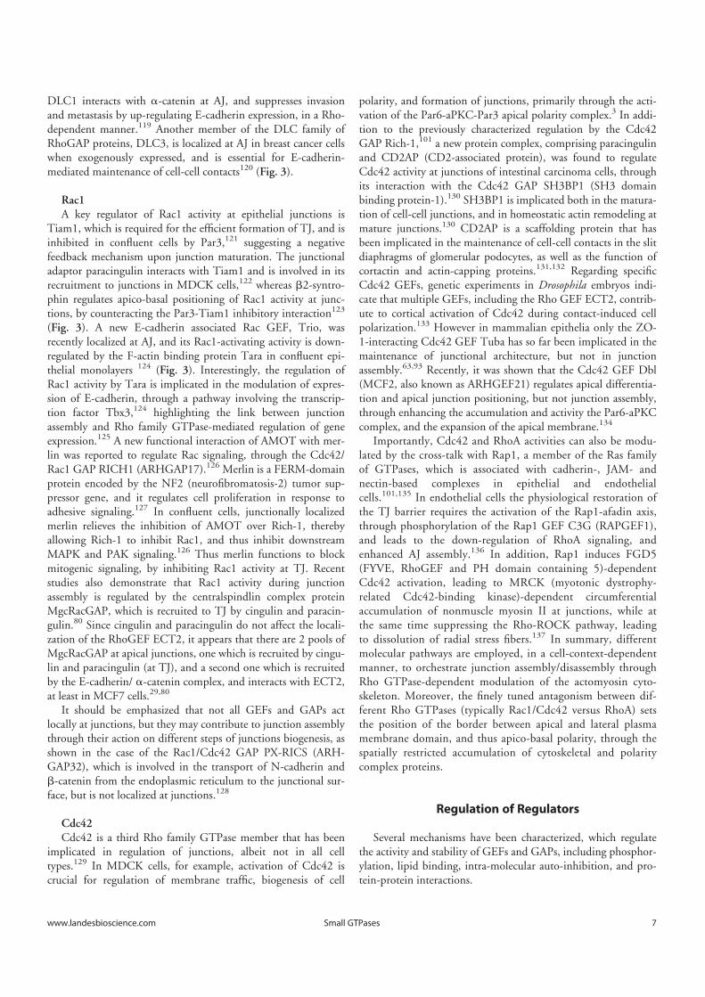
Rac1A key regulator of Rac1 activity at epithelial junctions is
Tiam1, which is required for the efficient formation of TJ, and isinhibited in confluent cells by Par3,121 suggesting a negativefeedback mechanism upon junction maturation. The junctionaladaptor paracingulin interacts with Tiam1 and is involved in itsrecruitment to junctions in MDCK cells,122 whereas b2-syntro-phin regulates apico-basal positioning of Rac1 activity at junc-tions, by counteracting the Par3-Tiam1 inhibitory interaction123
(Fig. 3). A new E-cadherin associated Rac GEF, Trio, wasrecently localized at AJ, and its Rac1-activating activity is down-regulated by the F-actin binding protein Tara in confluent epi-thelial monolayers 124 (Fig. 3). Interestingly, the regulation ofRac1 activity by Tara is implicated in the modulation of expres-sion of E-cadherin, through a pathway involving the transcrip-tion factor Tbx3,124 highlighting the link between junctionassembly and Rho family GTPase-mediated regulation of geneexpression.125 A new functional interaction of AMOT with mer-lin was reported to regulate Rac signaling, through the Cdc42/Rac1 GAP RICH1 (ARHGAP17).126 Merlin is a FERM-domainprotein encoded by the NF2 (neurofibromatosis-2) tumor sup-pressor gene, and it regulates cell proliferation in response toadhesive signaling.127 In confluent cells, junctionally localizedmerlin relieves the inhibition of AMOT over Rich-1, therebyallowing Rich-1 to inhibit Rac1, and thus inhibit downstreamMAPK and PAK signaling.126 Thus merlin functions to blockmitogenic signaling, by inhibiting Rac1 activity at TJ. Recentstudies also demonstrate that Rac1 activity during junctionassembly is regulated by the centralspindlin complex proteinMgcRacGAP, which is recruited to TJ by cingulin and paracin-gulin.80 Since cingulin and paracingulin do not affect the locali-zation of the RhoGEF ECT2, it appears that there are 2 pools ofMgcRacGAP at apical junctions, one which is recruited by cingu-lin and paracingulin (at TJ), and a second one which is recruitedby the E-cadherin/ a-catenin complex, and interacts with ECT2,at least in MCF7 cells.29,80
It should be emphasized that not all GEFs and GAPs actlocally at junctions, but they may contribute to junction assemblythrough their action on different steps of junctions biogenesis, asshown in the case of the Rac1/Cdc42 GAP PX-RICS (ARH-GAP32), which is involved in the transport of N-cadherin andb-catenin from the endoplasmic reticulum to the junctional sur-face, but is not localized at junctions.128
Cdc42Cdc42 is a third Rho family GTPase member that has been
implicated in regulation of junctions, albeit not in all celltypes.129 In MDCK cells, for example, activation of Cdc42 iscrucial for regulation of membrane traffic, biogenesis of cell
polarity, and formation of junctions, primarily through the acti-vation of the Par6-aPKC-Par3 apical polarity complex.3 In addi-tion to the previously characterized regulation by the Cdc42GAP Rich-1,101 a new protein complex, comprising paracingulinand CD2AP (CD2-associated protein), was found to regulateCdc42 activity at junctions of intestinal carcinoma cells, throughits interaction with the Cdc42 GAP SH3BP1 (SH3 domainbinding protein-1).130 SH3BP1 is implicated both in the matura-tion of cell-cell junctions, and in homeostatic actin remodeling atmature junctions.130 CD2AP is a scaffolding protein that hasbeen implicated in the maintenance of cell-cell contacts in the slitdiaphragms of glomerular podocytes, as well as the function ofcortactin and actin-capping proteins.131,132 Regarding specificCdc42 GEFs, genetic experiments in Drosophila embryos indi-cate that multiple GEFs, including the Rho GEF ECT2, contrib-ute to cortical activation of Cdc42 during contact-induced cellpolarization.133 However in mammalian epithelia only the ZO-1-interacting Cdc42 GEF Tuba has so far been implicated in themaintenance of junctional architecture, but not in junctionassembly.63,93 Recently, it was shown that the Cdc42 GEF Dbl(MCF2, also known as ARHGEF21) regulates apical differentia-tion and apical junction positioning, but not junction assembly,through enhancing the accumulation and activity the Par6-aPKCcomplex, and the expansion of the apical membrane.134
Importantly, Cdc42 and RhoA activities can also be modu-lated by the cross-talk with Rap1, a member of the Ras familyof GTPases, which is associated with cadherin-, JAM- andnectin-based complexes in epithelial and endothelialcells.101,135 In endothelial cells the physiological restoration ofthe TJ barrier requires the activation of the Rap1-afadin axis,through phosphorylation of the Rap1 GEF C3G (RAPGEF1),and leads to the down-regulation of RhoA signaling, andenhanced AJ assembly.136 In addition, Rap1 induces FGD5(FYVE, RhoGEF and PH domain containing 5)-dependentCdc42 activation, leading to MRCK (myotonic dystrophy-related Cdc42-binding kinase)-dependent circumferentialaccumulation of nonmuscle myosin II at junctions, while atthe same time suppressing the Rho-ROCK pathway, leadingto dissolution of radial stress fibers.137 In summary, differentmolecular pathways are employed, in a cell-context-dependentmanner, to orchestrate junction assembly/disassembly throughRho GTPase-dependent modulation of the actomyosin cyto-skeleton. Moreover, the finely tuned antagonism between dif-ferent Rho GTPases (typically Rac1/Cdc42 versus RhoA) setsthe position of the border between apical and lateral plasmamembrane domain, and thus apico-basal polarity, through thespatially restricted accumulation of cytoskeletal and polaritycomplex proteins.
Regulation of Regulators
Several mechanisms have been characterized, which regulatethe activity and stability of GEFs and GAPs, including phosphor-ylation, lipid binding, intra-molecular auto-inhibition, and pro-tein-protein interactions.
www.landesbioscience.com 7Small GTPases
Dow
nloa
ded
by [U
nive
rsité
de
Gen
ève]
at 0
5:48
18
Dec
embe
r 201
4

The Rho GEF GEF-H1 can be sequestered either by bindingto microtubules in the cytoplasm,78,138 or by binding to cingulinand paracingulin at epithelial junctions.66,122,139 Phosphoryla-tion of GEF-H1 occurs at several different sites and has multipleeffects on GEF-H1 activity. In Jurkat cells, phosphorylation bythe Rac1 effector PAK1 leads to GEF-H1 binding to 14-3-3, andassociation of the complex with microtubules, resulting in inhibi-tion of GEF-H1 activity.140 Another member of the PAK kinasefamily, PAK4, induces dissociation of GEF-H1 from microtu-bules in fibroblasts, and switching of substrate specificity, fromRho to Rac1.141 In COS cells, phosphorylation by Par1b, amember of the conserved Par/MARK serine/threonine kinasefamily, leads to dissociation of GEF-H1 from microtubules, andmicrotubule destabilization.142,143 GEF-H1 phosphorylation canbe cell-cycle dependent, since at early stages of mitosis in Helacells GEF-H1 is phosphorylated by Aurora A kinase, whereas attelophase it is dephosphorylated, to allow RhoA activation, cleav-age furrow formation, and ingression during cytokinesis.144 InHT1080 (fibrosarcoma) and LK2 (lung squamous cell carci-noma) cells phosphorylation by ERK enhances the guanidineexchange activity of GEF-H1.145 In LLC-PK1 kidney tubularcells, GEF-H1 is involved in the sequential activation of Rac1and RhoA, through TNF-a induced phosphorylation, whichactivates Rac1, followed by a signaling cascade that results inERK phosphorylating GEF-H1, leading to RhoA activation.146
ERK signaling also leads to inhibition of GEF-H1 through phos-phorylation in MDA-MB-231 breast cancer cells, thus regulatingcell motility and invasiveness.147 In summary, the specific mecha-nisms of GEF-H1 regulation appear largely determined by thecell context-dependent expression of interacting partners.
The Rho activator ECT2 is a key regulator of cytokinesis, andis subjected to cell-cycle-dependent regulation. ECT2 firstbecomes active in prophase, when it is phosphorylated by Cdk1,and exported from the nucleus into the cytoplasm, to activateRhoA and induce the formation of a mechanically stiff androunded metaphase cortex.148 Phosphorylation on a different siteis required for catalytic activity and interaction with polo-likekinase, leading to stimulation of RhoA activity and SRE-regu-lated transcription.149 In anaphase ECT2 associates with the cen-tralspindlin complex, and is targeted to the equatorial membranethrough a mechanism that requires a pleckstrin homologydomain and a polybasic cluster that bind to phosphoinositide lip-ids.150 Targeting of ECT2 to the equatorial membrane is the keystep to initiate cleavage furrow formation during cytokinesis.Upon completion of mitosis, ECT2 undergoes ubiquitin-depen-dent degradation, indicating that ECT2 is a bona fide cell-cycle-regulated protein.151
Another GEF for which a putative phosphoinositide lipid-mediated recruitment to the membrane has been proposed isTiam1, a Rac1-specific GEF.152 The guanine nucleotideexchange activity of Tiam1 is enhanced by different inositolphospholipids and other lipids.153,154 Tiam1 is a substrate forthe Src kinase, and phosphorylation of residue Y384 of Tiam1,which occurs preferentially at AJ, triggers its degradation, leadingto AJ disruption and increased cell migration.155 Tiam1 alsointeracts with 14-3-3 proteins when phosphorylated on serine
residues, and although phosphorylation does not affect Tiam1activity, it is required for Tiam1 proteolytic degradation.155,156
Tiam1 phosphorylation by protein kinase C and by calcium-cal-modulin kinase II have been described in activated fibroblasts,this latter leading to increased guanidine exchange activity towardRac1 in vitro.157 Finally, protein kinase-D-mediated phosphory-lation of the Rho GEF Syx reduces its junctional targeting,through binding to 14-3-3 proteins.158
Phosphorylation also regulates GAPs, to activate or inhibittheir activity, or affect their stability. p190RhoGAP is regulatedby phosphorylation on Tyr, Ser and Thr residues, and by bindingto phospholipids. Activation of Src by different pathways (EGF,integrin, PKC, and cadherin engagement) leads to phosphoryla-tion of p190RhoGAP on Y1105, resulting in enhanced GAPactivity, inhibition of RhoA and stress fiber disassembly.159,160
Recruitment of active p190RhoGAP to cadherin throughp120ctn leads to local suppression of RhoA activity, which isessential for AJ formation.107,161 ERK mediated phosphorylationon different Ser and Thr residues in the C-terminal part of theprotein suppresses the GAP activity of p190RhoGAP duringfocal adhesion formation.162 Interaction of a polybasic region(PBR) of p190RhoGAP with phospholipids can switch substratespecificity of p190RhoGAP, from RhoA to Rac1, and this inter-action is antagonized by phosphorylation on Ser1221 andThr1226.163 Substrate specificity of RICH-1 is regulated in plate-lets by Src-mediated phosphorylation, either to inhibit activity onRho/Rac or to activate GAP activity toward Cdc42.164 MgcRac-GAP is regulated by binding to PRC1, which inhibits GAP activ-ity toward Cdc42, thus allowing spindle formation duringmitosis, and by Aurora B-mediated phosphorylation and PP2A-mediated dephosphorylation, which affect substrate specificityand interaction with ECT2.165-167 The Rho GAP activity ofDLC1 can be inhibited either by phosphorylation, which favorsinteraction with 14-3-3 and exclusion from focal adhesions,168 orby intramolecular autoinhibition, which is mediated by a SAMdomain, and modulated by EGF signaling, through tensin3.169
A Dynamic View of the Cross-talk Between RhoGTPases and Junction Assembly
The process that leads to the formation of epithelial cell-celljunctions is highly regulated in space and time, and results fromthe coordinated interactions between Rho GTPases, their GEFand GAP regulators, and junctional molecules (Fig. 3). Uponformation of primordial contacts, accumulation of E-cadherinand Ig-like adhesion molecules (JAMs, nectins) is driven by and,at the same time, stimulates Rac1-dependent actin polymeriza-tion at the submembrane cortex, in positive feedback loop thatfurther promotes the accumulation of adhesion molecules at newjunctions (Fig. 3). In this initial phase, the activities of Rac1/Cdc42, and the Ras-like GTPase Rap1 play a key role, both togenerate the cytoskeletal scaffold upon which to build the newjunction, and to expand the junctional surface, through cortac-tin/N-WASP/WAVE2/Arp2/3-mediated actin polymerization,and directed targeting of membrane vesicles. In order for
8 Volume 5 Issue 4Small GTPases
Dow
nloa
ded
by [U
nive
rsité
de
Gen
ève]
at 0
5:48
18
Dec
embe
r 201
4
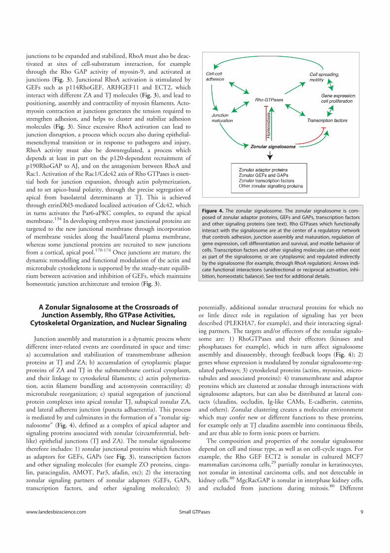
junctions to be expanded and stabilized, RhoA must also be deac-tivated at sites of cell-substratum interaction, for examplethrough the Rho GAP activity of myosin-9, and activated atjunctions (Fig. 3). Junctional RhoA activation is stimulated byGEFs such as p114RhoGEF, ARHGEF11 and ECT2, whichinteract with different ZA and TJ molecules (Fig. 3), and lead topositioning, assembly and contractility of myosin filaments. Acto-myosin contraction at junctions generates the tension required tostrengthen adhesion, and helps to cluster and stabilize adhesionmolecules (Fig. 3). Since excessive RhoA activation can lead tojunction disruption, a process which occurs also during epithelial-mesenchymal transition or in response to pathogens and injury,RhoA activity must also be downregulated, a process whichdepends at least in part on the p120-dependent recruitment ofp190RhoGAP to AJ, and on the antagonism between RhoA andRac1. Activation of the Rac1/Cdc42 axis of Rho GTPases is essen-tial both for junction expansion, through actin polymerization,and to set apico-basal polarity, through the precise segregation ofapical from basolateral determinants at TJ. This is achievedthrough ezrinDbl3-mediated localized activation of Cdc42, whichin turns activates the Par6-aPKC complex, to expand the apicalmembrane.134 In developing embryos most junctional proteins aretargeted to the new junctional membrane through incorporationof membrane vesicles along the basal/lateral plasma membrane,whereas some junctional proteins are recruited to new junctionsfrom a cortical, apical pool.170-174 Once junctions are mature, thedynamic remodelling and functional modulation of the actin andmicrotubule cytoskeletons is supported by the steady-state equilib-rium between activation and inhibition of GEFs, which maintainshomeostatic junction architecture and tension (Fig. 3).
A Zonular Signalosome at the Crossroads ofJunction Assembly, Rho GTPase Activities,
Cytoskeletal Organization, and Nuclear Signaling
Junction assembly and maturation is a dynamic process wheredifferent inter-related events are coordinated in space and time:a) accumulation and stabilization of transmembrane adhesionproteins at TJ and ZA; b) accumulation of cytoplasmic plaqueproteins of ZA and TJ in the submembrane cortical cytoplasm,and their linkage to cytoskeletal filaments; c) actin polymeriza-tion, actin filament bundling and actomyosin contractility; d)microtubule reorganization; e) spatial segregation of junctionalprotein complexes into apical zonular TJ, subapical zonular ZA,and lateral adherens junction (puncta adhaerentia). This processis mediated by and culminates in the formation of a “zonular sig-nalosome” (Fig. 4), defined as a complex of apical adaptor andsignaling proteins associated with zonular (circumferential, belt-like) epithelial junctions (TJ and ZA). The zonular signalosometherefore includes: 1) zonular junctional proteins which functionas adaptors for GEFs, GAPs (see Fig. 3), transcription factorsand other signaling molecules (for example ZO proteins, cingu-lin, paracingulin, AMOT, Par3, afadin, etc); 2) the interactingzonular signaling partners of zonular adaptors (GEFs, GAPs,transcription factors, and other signaling molecules); 3)
potentially, additional zonular structural proteins for which noor little direct role in regulation of signaling has yet beendescribed (PLEKHA7, for example), and their interacting signal-ing partners. The targets and/or effectors of the zonular signalo-some are: 1) RhoGTPases and their effectors (kinases andphosphatases for example), which in turn affect signalosomeassembly and disassembly, through feedback loops (Fig. 4); 2)genes whose expression is modulated by zonular signalosome-reg-ulated pathways; 3) cytoskeletal proteins (actins, myosins, micro-tubules and associated proteins): 4) transmembrane and adaptorproteins which are clustered at zonulae through interactions withsignalosome adaptors, but can also be distributed at lateral con-tacts (claudins, occludin, Ig-like CAMs, E-cadherin, catenins,and others). Zonular clustering creates a molecular environmentwhich may confer new or different functions to these proteins,for example only at TJ claudins assemble into continuous fibrils,and are thus able to form ionic pores or barriers.
The composition and properties of the zonular signalosomedepend on cell and tissue type, as well as on cell-cycle stages. Forexample, the Rho GEF ECT2 is zonular in cultured MCF7mammalian carcinoma cells,29 partially zonular in keratinocytes,not zonular in intestinal carcinoma cells, and not detectable inkidney cells.80 MgcRacGAP is zonular in interphase kidney cells,and excluded from junctions during mitosis.80 Different
Figure 4. The zonular signalosome. The zonular signalosome is com-posed of zonular adaptor proteins, GEFs and GAPs, transcription factorsand other signaling proteins (see text). Rho GTPases which functionallyinteract with the signalosome are at the center of a regulatory networkthat controls adhesion, junction assembly and maturation, regulation ofgene expression, cell differentiation and survival, and motile behavior ofcells. Transcription factors and other signaling molecules can either existas part of the signalosome, or are cytoplasmic and regulated indirectlyby the signalosome (for example, through RhoA regulation). Arrows indi-cate functional interactions (unidirectional or reciprocal activation, inhi-bition, homeostatic balance). See text for additional details.
www.landesbioscience.com 9Small GTPases
Dow
nloa
ded
by [U
nive
rsité
de
Gen
ève]
at 0
5:48
18
Dec
embe
r 201
4
members of the large GEFs and GAPs families probably showcell- and tissue-type specific expression, however we are far froma precise immunohistochemical mapping of their distribution indifferent normal and diseased tissues. Similarly, structural adap-tor proteins are not identically distributed in all cell types. Forexample paracingulin is not detected in differentiated intestinalepithelial cells,175 and ZO-3 shows a narrower tissue distributionthan ZO-1,176 and is not expressed in cultured mammary epithe-lial cells.20
What are the functions of the zonular signalosome? One is tofine-tune in space and time the activation of Rho familyGTPases, and thus the organization of the cytoskeleton, duringthe dynamic processes of junction assembly and disassembly, andat steady-state, to ensure the correct remodelling and turnover ofcytoskeletal and junctional proteins (Fig. 4). This equilibriumcan be perturbed by mechanical stresses, and pathological cues(cytokines, pathogens, toxins), leading to junction disruption,perturbation of the signalosome complex, and hence dramaticallyaltered spatio-temporal regulation of Rho GTPases. As such, thesignalosome can be viewed as a sensor, or signal transducer, ofextra- and intra-cellular signals, to control cell behavior, includ-ing adhesion, motility, and junctional membrane integrity anddynamics. By sequestering and/or stabilizing at junctions GEFsand GAPs, the zonular signalosome also indirectly controls acti-vation of signaling at cell-substratum adhesions, and thus cellspreading and motility, for example through the Par3-Tiam1,177,178 p114RhoGEF,179 and GEF-H1 139 modules. Atthe tissue and organ level, perturbation of the zonular signalo-some can elicit dramatic consequences on epithelial or endothe-lial cell cell-adhesion and barrier functions, resulting for examplein loss of skin or mucosal barrier integrity, jaundice, edema, andloss of proteins or ions across tissue barriers.
A second important function of the zonular signalosome is toregulate transcription factors, cell-cycle regulators, and other sig-naling molecules, thus controlling gene expression, proliferation,differentiation, and survival. One mechanism of this regulationinvolves the direct or indirect sequestration of the signaling mole-cules at junctions, as shown for example by the role of ZO pro-teins in the junctional retention and stability of the transcriptionfactor DbpA/ZONAB,20,180 and the cell cycle regulatorcyclinD1.181 Moreover, ZO-2, a-catenin and AMOT controlthe nucleo-cytoplasmic shuttling of YAP/TAZ transcription fac-tors by different mechanisms, including direct or indirect interac-tion, stabilization at junctions, and cytoplasmic retentionthrough modulation of phosphorylation (reviewed in 125).Another mechanism involves the modulation of RhoA activation,by zonular GEFs, and interacting adaptor proteins. For exampleGEF-H1 activation is critical in Dbpa/ZONAB activation andnuclear shuttling 182 and RhoA activation is also criticallyinvolved in the mechanotransduction-dependent regulation of
the activity of YAP transcription factors.183 RhoA is a centralmolecule in the cross-talk between cytoskeletal organization andnuclear signaling, and the integrity of the zonular signalosome,by regulating not only Rho, but also Rac and Cdc42 activities, iscrucial to coordinate regulation of cytoskeletal organization withcell-cell adhesion, motility and nuclear signaling.
Concluding Remarks
In the past decade there have been striking advances in clarify-ing the identity of junctional proteins, GEFs and GAPs, and theirfunctional interactions. The general picture that has emerged isone whereby epithelial morphogenesis and physiology are regu-lated by the carefully tuned balance between the activities ofantagonistic Rho GTPases, through modulation of the expres-sion, subcellular localization and activities of GEFs and GAPs.However, many important questions remain open. First of all,the majority of studies have been carried out on only one or afew experimental models (cells, tissues, species), and should bevalidated on additional vertebrate models. Junctions are remark-ably heterogeneous in architecture, composition and function,12
and each cell type is thus likely to be characterized by a uniqueconfiguration of junctional adaptors and Rho GTPase regulatingmolecules. So, results obtained in one model system cannot beextrapolated to other cells and tissues. Systematic transcriptomicand proteomic approaches, and the generation of new andimproved reagents for the subcellular localization of GEFs andGAPs will be critical to map sets of regulatory modules in theircorrect context. In addition, detailed characterization of verte-brate knockout models will be essential to understand the role ofGEFs and GAPs, and their interacting partners, in tissue andorgan physiology and pathology. Special attention should bedevoted to elucidating the 3-dimensional structures and affinitiesof interaction between the adaptor molecules, GEFs and GAPs,and how post-translational modifications can modulate them.Addressing these questions will help to define the compositionand functions of zonular signalosomes in different epithelial andendothelial cells and tissues, and develop strategies for theirexperimental and therapeutic modulation.
Disclosure of Potential Conflicts of Interest
No potential conflicts of interest were disclosed.
Funding
The authors gratefully acknowledge the funding agencies thatsponsor research in the Citi laboratory: the Swiss National Foun-dation, the Swiss Cancer League, and the State of Geneva.
References1. Gonzalez-Mariscal L, Garay E, Lechuga S. Virus inter-
action with the apical junctional complex. Front Bio-sci 2009; 14:731-68; http://dx.doi.org/10.2741/3276
2. Vogelmann R, Amieva MR, Falkow S, Nelson WJ.Breaking into the epithelial apical-junctional
complex–news from pathogen hackers. Curr OpinCell Biol 2004; 16:86-93; PMID:15037310; http://dx.doi.org/10.1016/j.ceb.2003.12.002
3. Shin K, Fogg VC, Margolis B. Tight junctions andcell polarity. Annu Rev Cell Dev Biol 2006; 22:207-35; PMID:16771626; http://dx.doi.org/10.1146/annurev.cellbio.22.010305.104219
4. Anderson JM, Van Itallie CM. Physiology and func-tion of the tight junction. Cold Spring Harb PerspectBiol 2009; 1:a002584:1-16; PMID:20066090; http://dx.doi.org/10.1101/cshperspect.a002584
5. Franke WW, Rickelt S, Barth M, Pieperhoff S. Thejunctions that don’t fit the scheme: special symmetri-cal cell-cell junctions of their own kind. Cell Tissue
10 Volume 5 Issue 4Small GTPases
Dow
nloa
ded
by [U
nive
rsité
de
Gen
ève]
at 0
5:48
18
Dec
embe
r 201
4

Res 2009; 338:1-17; PMID:19680692; http://dx.doi.org/10.1007/s00441-009-0849-z
6. Takeichi M. Dynamic contacts: rearranging adherensjunctions to drive epithelial remodelling. Nat RevMol Cell Biol 2014; 15:397-410; PMID:24824068;http://dx.doi.org/10.1038/nrm3802
7. Pulimeno P, Bauer C, Stutz J, Citi S. PLEKHA7 is anadherens junction protein with a tissue distributionand subcellular localization distinct from ZO-1 andE-cadherin. PLoS One 2010; 5:10.1371journal.pone.0012207; PMID:20808826; http://dx.doi.org/10.1371/journal.pone.0012207
8. Goto H, Tanabe K, Manser E, Lim L, Yasui Y, Ina-gaki M. Phosphorylation and reorganization ofvimentin by p21-activated kinase (PAK). Genes Cells2002; 7:91-7; PMID:11895474; http://dx.doi.org/10.1046/j.1356-9597.2001.00504.x
9. Spindler V, Waschke J. Role of Rho GTPases in des-mosomal adhesion and pemphigus pathogenesis. AnnAnat D Anat Anz: Off Organ Anat Ges 2011;193:177-80; PMID:21441018; http://dx.doi.org/10.1016/j.aanat.2011.02.003
10. Tsang SM, Brown L, Gadmor H, Gammon L, For-tune F, Wheeler A, Wan H. Desmoglein 3 acting asan upstream regulator of Rho GTPases, Rac-1Cdc42in the regulation of actin organisation and dynamics.Exp Cell Res 2012; 318:2269-83; PMID:22796473;http://dx.doi.org/10.1016/j.yexcr.2012.07.002
11. Koetsier JL, Amargo EV, Todorovic V, Green KJ,Godsel LM. Plakophilin 2 affects cell migration bymodulating focal adhesion dynamics and integrinprotein expression. J Invest Dermatol 2014; 134:112-22; PMID:23884246; http://dx.doi.org/10.1038/jid.2013.266
12. Franke WW. Discovering the molecular componentsof intercellular junctions-a historical view. Cold SpringHarbor Perspect Biol 2009; 1:a003061; PMID:20066111; http://dx.doi.org/10.1101/cshperspect.a003061
13. Garrod D, Chidgey M. Desmosome structure, com-position and function. Biochim Biophys Acta 2008;1778:572-87; PMID:17854763; http://dx.doi.org/10.1016/j.bbamem.2007.07.014
14. Simske JS. Claudins reign: the claudinEMPPMP22-gamma channel protein family in C. elegans. TissueBarriers 2013; 1:e25502; PMID:24665403; http://dx.doi.org/10.4161/tisb.25502
15. Harris TJ, Tepass U. Adherens junctions: from mole-cules to morphogenesis. Nat Rev Mol Cell Biol 2010;11:502-14; PMID:20571587; http://dx.doi.org/10.1038/nrm2927
16. Hulpiau P, Gul IS, van Roy F. New insights into theevolution of metazoan cadherins and catenins. ProgMol Biol Translational Sci 2013; 116:71-94;PMID:23481191; http://dx.doi.org/10.1016/B978-0-12-394311-8.00004-2
17. Dickinson DJ, Nelson WJ, Weis WI. A polarized epi-thelium organized by beta- and alpha-catenin predatescadherin and metazoan origins. Science 2011;331:1336-9; PMID:21393547; http://dx.doi.org/10.1126/science.1199633
18. Capaldo CT, Macara IG. Depletion of E-cadherindisrupts establishment but not maintenance of celljunctions in Madin-Darby canine kidney epithelialcells. Mol Biol Cell 2007; 18:189-200; PMID:17093058; http://dx.doi.org/10.1091/mbc.E06-05-0471
19. Fanning AS, Anderson JM. Zonula occludens-1 and-2 are cytosolic scaffolds that regulate the assembly ofcellular junctions. Ann N Y Acad Sci 2009;1165:113-20; PMID:19538295; http://dx.doi.org/10.1111/j.1749-6632.2009.04440.x
20. Spadaro D, Tapia R, Jond L, Sudol M, Fanning AS,Citi S. ZO proteins redundantly regulate the tran-scription factor DbpAZONAB. J Biol Chem 2014;289:22500-11; PMID:24986862; http://dx.doi.org/10.1074/jbc.M114.556449
21. Yamanaka T, Tosaki A, Kurosawa M, Akimoto K,Hirose T, Ohno S, Hattori N, Nukina N. Loss of
aPKClambda in differentiated neurons disrupts thepolarity complex but does not induce obvious neuro-nal loss or disorientation in mouse brains. PLoS One2013; 8:e84036; PMID:24391875; http://dx.doi.org/10.1371/journal.pone.0084036
22. Sripathy S, Lee M, Vasioukhin V. Mammalian Llgl2is necessary for proper branching morphogenesis dur-ing placental development. Mol Cell Biol 2011;31:2920-33; PMID:21606200; http://dx.doi.org/10.1128/MCB.05431-11
23. Bernards A. GAPs galore! A survey of putative Rassuperfamily GTPase activating proteins in man andDrosophila. Biochim Biophys Acta 2003; 1603:47-82; PMID:12618308
24. Mooseker MS. Organisation, chemistry and assemblyof the cytoskeletal apparatus of the intestinal brushborder. Annu Rev Cell Biol 1985; 1:209-41;PMID:3916317; http://dx.doi.org/10.1146/annurev.cb.01.110185.001233
25. Akhtar N, Hotchin NA. RAC1 regulates adherensjunctions through endocytosis of E-cadherin. MolBiol Cell 2001; 12:847-62; PMID:11294891; http://dx.doi.org/10.1091/mbc.12.4.847
26. Budnar S, Yap AS. A mechanobiological perspectiveon cadherins and the actin-myosin cytoskeleton.F1000prime Rep 2013; 5:35; PMID:24049639;http://dx.doi.org/10.12703/P5-35
27. Pilot F, Lecuit T. Compartmentalized morphogenesisin epithelia: from cell to tissue shape. Dev Dyn 2005;232:685-94; PMID:15712202; http://dx.doi.org/10.1002/dvdy.20334
28. Turner JR. ‘Putting the squeeze’ on the tight junction:understanding cytoskeletal regulation. Semin CellDev Biol 2000; 11:301-8; PMID:10966864; http://dx.doi.org/10.1006/scdb.2000.0180
29. Ratheesh A, Gomez GA, Priya R, Verma S, KovacsEM, Jiang K, Brown NH, Akhmanova A, StehbensSJ, Yap AS. Centralspindlin and alpha-catenin regu-late Rho signalling at the epithelial zonula adherens.Nat Cell Biol 2012; 14:818-28; PMID:22750944;http://dx.doi.org/10.1038/ncb2532
30. Kovacs EM, Goodwin M, Ali RG, Paterson AD, YapAS. Cadherin-directed actin assembly: e-cadherinphysically associates with the Arp23 complex to directactin assembly in nascent adhesive contacts. Curr Biol2002; 12:379-82; PMID:11882288; http://dx.doi.org/10.1016/S0960-9822(02)00661-9
31. Ehrlich JS, Hansen MD, Nelson WJ. Spatio-temporalregulation of Rac1 localization and lamellipodiadynamics during epithelial cell-cell adhesion. DevCell 2002; 3:259-70; PMID:12194856; http://dx.doi.org/10.1016/S1534-5807(02)00216-2
32. Han SP, Gambin Y, Gomez GA, Verma S, Giles N,Michael M, Wu SK, Guo Z, Johnston W, Sierecki E,et al. Cortactin scaffolds Arp23 and WAVE2 at theepithelial zonula adherens. J Biol Chem 2014;289:7764-75; PMID:24469447; http://dx.doi.org/10.1074/jbc.M113.544478
33. Tang VW, Brieher WM. alpha-Actinin-4FSGS1 isrequired for Arp23-dependent actin assembly at theadherens junction. J Cell Biol 2012; 196:115-30;PMID:22232703; http://dx.doi.org/10.1083/jcb.201103116
34. Verma S, Han SP, Michael M, Gomez GA, Yang Z,Teasdale RD, Ratheesh A, Kovacs EM, Ali RG, YapAS. A WAVE2-Arp23 actin nucleator apparatus sup-ports junctional tension at the epithelial zonula adhe-rens. Mol Biol Cell 2012; 23:4601-10; PMID:23051739; http://dx.doi.org/10.1091/mbc.E12-08-0574
35. Kovacs EM, Verma S, Ali RG, Ratheesh A, HamiltonNA, Akhmanova A, Yap AS. N-WASP regulates theepithelial junctional actin cytoskeleton through a non-canonical post-nucleation pathway. Nat Cell Biol2011; 13:934-43; PMID:21785420; http://dx.doi.org/10.1038/ncb2290
36. Kobielak A, Pasolli HA, Fuchs E. Mammalian for-min-1 participates in adherens junctions and
polymerization of linear actin cables. Nat Cell Biol2004; 6:21-30; PMID:14647292; http://dx.doi.org/10.1038/ncb1075
37. Carramusa L, Ballestrem C, Zilberman Y, BershadskyAD. Mammalian diaphanous-related formin Dia1controls the organization of E-cadherin-mediated cell-cell junctions. J Cell Sci 2007; 120:3870-82;PMID:17940061; http://dx.doi.org/10.1242/jcs.014365
38. Ozono K, Komiya S, Shimamura K, Ito T, NagafuchiA. Defining the roles of alpha-catenin in cell adhesionand cytoskeleton organization: isolation of F9 cellscompletely lacking cadherin-catenin complex. CellStruct Funct 2011; 36:131-43; PMID:21685705;http://dx.doi.org/10.1247/csf.11009
39. Desai R, Sarpal R, Ishiyama N, Pellikka M, Ikura M,Tepass U. Monomeric alpha-catenin links cadherin tothe actin cytoskeleton. Nat Cell Biol 2013; 15:261-73; PMID:23417122; http://dx.doi.org/10.1038/ncb2685
40. Yonemura S, Wada Y, Watanabe T, Nagafuchi A, Shi-bata M. Alpha-catenin as a tension transducer thatinduces adherens junction development. Nat Cell Biol2010; 12:533-42; PMID:20453849; http://dx.doi.org/10.1038/ncb2055
41. le Duc Q, Shi Q, Blonk I, Sonnenberg A, Wang N,Leckband D, de Rooij J. Vinculin potentiates E-cad-herin mechanosensing and is recruited to actin-anchored sites within adherens junctions in a myosinII-dependent manner. J Cell Biol 2010; 189:1107-15; PMID:20584916; http://dx.doi.org/10.1083/jcb.201001149
42. Drees F, Pokutta S, Yamada S, Nelson WJ, Weis WI.Alpha-catenin is a molecular switch that binds E-cad-herin-beta-catenin and regulates actin-filament assem-bly. Cell 2005; 123:903-15; PMID:16325583; http://dx.doi.org/10.1016/j.cell.2005.09.021
43. Abe K, Takeichi M. EPLIN mediates linkage of thecadherin catenin complex to F-actin and stabilizes thecircumferential actin belt. Proc Natl Acad Sci U S A2008; 105:13-9; PMID:18093941; http://dx.doi.org/10.1073/pnas.0710504105
44. Maul RS, Song Y, Amann KJ, Gerbin SC, PollardTD, Chang DD. EPLIN regulates actin dynamics bycross-linking and stabilizing filaments. J Cell Biol2003; 160:399-407; PMID:12566430; http://dx.doi.org/10.1083/jcb.200212057
45. Mandai K, Nakanishi H, Satoh A, Obaishi H, WadaM, Nishioka H, Itoh M, Mizoguchi A, Aoki T, Fuji-moto T, et al. Afadin: a novel actin filament-bindingprotein with one PDZ domain localized at cadherin-based cell-to-cell adherens junction. J Cell Biol 1997;139:517-28; PMID:9334353; http://dx.doi.org/10.1083/jcb.139.2.517
46. Tachibana K, Nakanishi H, Mandai K, Ozaki K,Ikeda W, Yamamoto Y, Nagafuchi A, Tsukita S,Takai Y. Two cell adhesion molecules, nectin and cad-herin, interact through their cytoplasmic domain-associated proteins. J Cell Biol 2000; 150:1161-76; PMID:10974003; http://dx.doi.org/10.1083/jcb.150.5.1161
47. Zhadanov AB, Provance DW, Speer CA, Coffin JD,Goss D, Blixt JA, Reichert CM, Mercer JA. Absenceof the tight junctional protein AF-6 disrupts epithelialcell- cell junctions and cell polarity during mousedevelopment. Curr Biol 1999; 9:880-8; PMID:10469590; http://dx.doi.org/10.1016/S0960-9822(99)80392-3
48. Ikeda W, Nakanishi H, Miyoshi J, Mandai K, IshizakiH, Tanaka M, Togawa A, Takahashi K, Nishioka H,Yoshida H, et al. Afadin: a key molecule essential forstructural organization of cell-cell junctions of polar-ized epithelia during embryogenesis. J Cell Biol 1999;146:1117-32; PMID:10477764; http://dx.doi.org/10.1083/jcb.146.5.1117
49. Simoes Sde M, Mainieri A, Zallen JA. Rho GTPaseand Shroom direct planar polarized actomyosin con-tractility during convergent extension. J Cell Biol
www.landesbioscience.com 11Small GTPases
Dow
nloa
ded
by [U
nive
rsité
de
Gen
ève]
at 0
5:48
18
Dec
embe
r 201
4
2014; 204:575-89; PMID:24535826; http://dx.doi.org/10.1083/jcb.201307070
50. Chu CW, Gerstenzang E, Ossipova O, Sokol SY. Luluregulates Shroom-induced apical constriction duringneural tube closure. PLoS One 2013; 8:e81854;PMID:24282618; http://dx.doi.org/10.1371/journal.pone.0081854
51. Baranwal S, Naydenov NG, Harris G, Dugina V,Morgan KG, Chaponnier C, Ivanov AI. Nonredun-dant roles of cytoplasmic beta- and gamma-actin iso-forms in regulation of epithelial apical junctions. MolBiol Cell 2012; 23:3542-53; PMID:22855531;http://dx.doi.org/10.1091/mbc.E12-02-0162
52. Ivanov AI, Bachar M, Babbin BA, Adelstein RS, Nus-rat A, Parkos CA. A unique role for nonmuscle myo-sin heavy chain IIA in regulation of epithelial apicaljunctions. PLoS One 2007; 2:e658; PMID:17668046; http://dx.doi.org/10.1371/journal.pone.0000658
53. Smutny M, Cox HL, Leerberg JM, Kovacs EM, ContiMA, Ferguson C, Hamilton NA, Parton RG, Adel-stein RS, Yap AS. Myosin II isoforms identify distinctfunctional modules that support integrity of the epi-thelial zonula adherens. Nat Cell Biol 2010; 12:696-702; PMID:20543839; http://dx.doi.org/10.1038/ncb2072
54. Fanning AS, Jameson BJ, Jesaitis LA, Anderson JM.The tight junction protein ZO-1 establishes a linkbetween the transmembrane protein occludin and theactin cytoskeleton. J Biol Chem 1998; 273:29745-53;PMID:9792688; http://dx.doi.org/10.1074/jbc.273.45.29745
55. Wittchen ES, Haskins J, Stevenson BR. Protein inter-actions at the tight junction. Actin has multiple bind-ing partners, and zo-1 forms independent complexeswith zo-2 and zo-3. J Biol Chem 1999; 274:35179-85; PMID:10575001; http://dx.doi.org/10.1074/jbc.274.49.35179
56. D’Atri F, Citi S. Cingulin interacts with F-actin invitro. FEBS Lett 2001; 507:21-4; PMID:11682052;http://dx.doi.org/10.1016/S0014-5793(01)02936-2
57. Van Itallie CM, Fanning AS, Bridges A, AndersonJM. ZO-1 stabilizes the tight junction solute barrierthrough coupling to the perijunctional cytoskeleton.Mol Biol Cell 2009; 20:3930-40; PMID:19605556;http://dx.doi.org/10.1091/mbc.E09-04-0320
58. Tokuda S, Higashi T, Furuse M. ZO-1 knockout byTALEN-mediated gene targeting in MDCK cells:involvement of ZO-1 in the regulation of cytoskeletonand cell shape. PLoS One 2014; 9:e104994;PMID:25157572; http://dx.doi.org/10.1371/journal.pone.0104994
59. Fanning AS, Van Itallie C, Anderson JM. Zonulaoccludens (ZO)-1 and -2 regulate apical cell structureand the zonula adherens cytoskeleton in polarized epi-thelia. Mol Biol Cell 2011; 23:577-90; PMID:22190737; http://dx.doi.org/10.1091/mbc.E11-09-0791
60. Fanning AS, Van Itallie CM, Anderson JM. Zonulaoccludens-1 and -2 regulate apical cell structure andthe zonula adherens cytoskeleton in polarized epithe-lia. Mol Biol Cell 2012; 23:577-90; PMID:22190737; http://dx.doi.org/10.1091/mbc.E11-09-0791
61. Maiers JL, Peng X, Fanning AS, DeMali KA. ZO-1recruitment to alpha-catenin–a novel mechanism forcoupling the assembly of tight junctions to adherensjunctions. J Cell Sci 2013; 126:3904-15; PMID:23813953; http://dx.doi.org/10.1242/jcs.126565
62. Ooshio T, Kobayashi R, Ikeda W, Miyata M, Fuku-moto Y, Matsuzawa N, Ogita H, Takai Y. Involve-ment of the interaction of afadin with ZO-1 in theformation of tight junctions in Madin-Darby caninekidney cells. J Biol Chem 2010; 285:5003-12;PMID:20008323; http://dx.doi.org/10.1074/jbc.M109.043760
63. Otani T, Ichii T, Aono S, Takeichi M. Cdc42GEF Tuba regulates the junctional configuration ofsimple epithelial cells. J Cell Biol 2006; 175:135-46;
PMID:17015620; http://dx.doi.org/10.1083/jcb.200605012
64. Itoh M, Tsukita S, Yamazaki Y, Sugimoto H. RhoGTP exchange factor ARHGEF11 regulates the integ-rity of epithelial junctions by connecting ZO-1 andRhoA-myosin II signaling. Proc Natl Acad Sci U S A2012; 109:9905-10; PMID:22665792; http://dx.doi.org/10.1073/pnas.1115063109
65. Paschoud S, Citi S. Inducible overexpression of cingu-lin in stably transfected MDCK cells does not affecttight junction organization and gene expression. MolMembr Biol 2008; 25:1-13; PMID:18097951; http://dx.doi.org/10.1080/09687680701474009
66. Guillemot L, Citi S. Cingulin regulates claudin-2expression and cell proliferation through the smallGTPase RhoA. Mol Biol Cell 2006; 17:3569-77;PMID:16723500; http://dx.doi.org/10.1091/mbc.E06-02-0122
67. Guillemot L, Schneider Y, Brun P, Castagliuolo I, Piz-zuti D, Martines D, Jond L, Bongiovanni M, Citi S.Cingulin is dispensable for epithelial barrier functionand tight junction structure, and plays a role in thecontrol of claudin-2 expression and response to duo-denal mucosa injury. J Cell Sci 2012; 125:5005-14; inpress; PMID:22946046; http://dx.doi.org/10.1242/jcs.101261
68. Meng W, Takeichi M. Adherens junction: moleculararchitecture and regulation. Cold Spring Harbor Per-spect Biol 2009; 1:a002899; PMID:20457565;http://dx.doi.org10.1101cshperspect.a002899
69. Meng W, Mushika Y, Ichii T, Takeichi M. Anchorageof microtubule minus ends to adherens junctions reg-ulates epithelial cell-cell contacts. Cell 2008; 135:948-59; PMID:19041755; http://dx.doi.org/10.1016/j.cell.2008.09.040
70. Pulimeno P, Paschoud S, Citi S. A role for ZO-1 andPLEKHA7 in recruiting paracingulin to tight andadherens junctions of epithelial cells. J Biol Chem2011; 286:16743-50; PMID:21454477; http://dx.doi.org/10.1074/jbc.M111.230862
71. Ivanov AI, McCall IC, Babbin B, Samarin SN, NusratA, Parkos CA. Microtubules regulate disassembly ofepithelial apical junctions. BMC Cell Biol 2006; 7:12;PMID:16509970; http://dx.doi.org/10.1186/1471-2121-7-12
72. Paschoud S, Jond L, Guerrera D, Citi S. PLEKHA7modulates epithelial tight junction barrier function.Tissue Barriers 2014; 2:e28755; PMID:24843844;http://dx.doi.org/10.4161/tisb.28755
73. Ligon LA, Holzbaur EL. Microtubules tethered at epi-thelial cell junctions by dynein facilitate efficientjunction assembly. Traffic 2007; 8:808-19;PMID:17550375; http://dx.doi.org/10.1111/j.1600-0854.2007.00574.x
74. Rosin-Arbesfeld R, Ihrke G, Bienz M. Actin-depen-dent membrane association of the APC tumour sup-pressor in polarized mammalian epithelial cells.EMBO J 2001; 20:5929-39; PMID:11689433;http://dx.doi.org/10.1093/emboj/20.21.5929
75. Karakesisoglou I, Yang Y, Fuchs E. An epidermal pla-kin that integrates actin and microtubule networks atcellular junctions. J Cell Biol 2000; 149:195-208;PMID:10747097; http://dx.doi.org/10.1083/jcb.149.1.195
76. Yano T, Matsui T, Tamura A, Uji M, Tsukita S. Theassociation of microtubules with tight junctions ispromoted by cingulin phosphorylation by AMPK. JCell Biol 2013; 203:605-14; PMID:24385485;http://dx.doi.org/10.1083/jcb.201304194
77. Bartolini F, Ramalingam N, Gundersen GG. Actin-capping protein promotes microtubule stability byantagonizing the actin activity of mDia1. Mol BiolCell 2012; 23:4032-40; PMID:22918941; http://dx.doi.org/10.1091/mbc.E12-05-0338
78. Krendel M, Zenke FT, Bokoch GM. Nucleotideexchange factor GEF-H1 mediates cross-talk betweenmicrotubules and the actin cytoskeleton. Nat Cell
Biol 2002; 4:294-301; PMID:11912491; http://dx.doi.org/10.1038/ncb773
79. Nagae S, Meng W, Takeichi M. Non-centrosomalmicrotubules regulate F-actin organization throughthe suppression of GEF-H1 activity. Genes Cells2013; 18:387-96; PMID:23432781; http://dx.doi.org/10.1111/gtc.12044
80. Guillemot L, Guerrera D, Spadaro D, Tapia R, JondL, Citi S. MgcRacGAP interacts with cingulin andparacingulin to regulate Rac1 activation and develop-ment of the tight junction barrier during epithelialjunction assembly. Mol Biol Cell 2014; 25:1995-2005; PMID:24807907; http://dx.doi.org/10.1091/mbc.E13-11-0680
81. Nusrat A, Giry M, Turner JR, Colgan SP, Parkos CA,Carmes D, Lemichez E, Boquet P, Madara JL. Rhoprotein regulates tight junctions and perijunctionalactin organization in polarized epithelia. Proc NatlAcad Sci USA 1995; 92:10629-33; PMID:7479854;http://dx.doi.org/10.1073/pnas.92.23.10629
82. Braga VM, Machesky LM, Hall A, Hotchin NA. Thesmall GTPases Rho and Rac are required for the estab-lishment of cadherin-dependent cell-cell contacts. JCell Biol 1997; 137:1421-31; PMID:9182672;http://dx.doi.org/10.1083/jcb.137.6.1421
83. Takaishi K, Sasaki T, Kotani H, Nishioka H, Takai Y.Regulation of cell-cell adhesion by rac and rho smallG proteins in MDCK cells. J Cell Biol 1997;139:1047-59; PMID:9362522; http://dx.doi.org/10.1083/jcb.139.4.1047
84. Jou TS, Nelson WJ. Effects of regulated expression ofmutant RhoA and Rac1 small GTPases on the devel-opment of epithelial (MDCK) cell polarity. J CellBiol 1998; 142:85-100; PMID:9660865; http://dx.doi.org/10.1083/jcb.142.1.85
85. Jou TS, Schneeberger EE, Nelson WJ. Structural andfunctional regulation of tight junctions by RhoA andRac1 small GTPases. J Cell Biol 1998; 142:101-15;PMID:9660866; http://dx.doi.org/10.1083/jcb.142.1.101
86. Kovacs EM, Ali RG, McCormack AJ, Yap AS. E-cad-herin homophilic ligation directly signals through Racand phosphatidylinositol 3-kinase to regulate adhesivecontacts. J Biol Chem 2002; 277:6708-18; PMID:11744701; http://dx.doi.org/10.1074/jbc.M109640200
87. Vasioukhin V, Fuchs E. Actin dynamics and cell-celladhesion in epithelia. Curr Opin Cell Biol 2001;13:76-84; PMID:11163137; http://dx.doi.org/10.1016/S0955-0674(00)00177-0
88. Adams AE, Johnson DI, Longnecker RM, Sloat BF,Pringle JR. CDC42 and CDC43, two additionalgenes involved in budding and the establishment ofcell polarity in the yeast Saccharomyces cerevisiae. JCell Biol 1990; 111:131-42; PMID:2195038; http://dx.doi.org/10.1083/jcb.111.1.131
89. Wojciak-Stothard B, Tsang LY, Haworth SG. Rac andRho play opposing roles in the regulation of hypoxiar-eoxygenation-induced permeability changes in pulmo-nary artery endothelial cells. Am J Physiol Lung CellMol Physiol 2005; 288:L749-60; PMID:15591411;http://dx.doi.org/10.1152/ajplung.00361.2004
90. Fukuhara A, Shimizu K, Kawakatsu T, Fukuhara T,Takai Y. Involvement of nectin-activated Cdc42 smallG protein in organization of adherens and tight junc-tions in Madin-Darby canine kidney cells. J BiolChem 2003; 278:51885-93; PMID:14530286;http://dx.doi.org/10.1074/jbc.M308015200
91. Wells CD, Fawcett JP, Traweger A, Yamanaka Y,Goudreault M, Elder K, Kulkarni S, Gish G, Virag C,Lim C, et al. A Rich1Amot complex regulates theCdc42 GTPase and apical-polarity proteins in epithe-lial cells. Cell 2006; 125:535-48; PMID:16678097;http://dx.doi.org/10.1016/j.cell.2006.02.045
92. Warner SJ, Longmore GD. Cdc42 antagonizes Rho1activity at adherens junctions to limit epithelial cellapical tension. J Cell Biol 2009; 187:119-33; PMID:19805632; http://dx.doi.org/10.1083/jcb.200906047
12 Volume 5 Issue 4Small GTPases
Dow
nloa
ded
by [U
nive
rsité
de
Gen
ève]
at 0
5:48
18
Dec
embe
r 201
4
93. Rajabian T, Gavicherla B, Heisig M, Muller-AltrockS, Goebel W, Gray-Owen SD, Ireton K. The bacterialvirulence factor InlC perturbs apical cell junctions andpromotes cell-to-cell spread of Listeria. Nat Cell Biol2009; 11:1212-8; PMID:19767742; http://dx.doi.org/10.1038/ncb1964
94. Sahai E, Marshall CJ. ROCK and Dia have opposingeffects on adherens junctions downstream of Rho.Nat Cell Biol 2002; 4:408-15; PMID:11992112;http://dx.doi.org/10.1038/ncb796
95. Jegou A, Carlier MF, Romet-Lemonne G. ForminmDia1 senses and generates mechanical forces onactin filaments. Nat Commun 2013; 4:1883; PMID:23695677; http://dx.doi.org/10.1038/ncomms2888
96. Amano M, Nakayama M, Kaibuchi K. Rho-kinaseR-OCK: a key regulator of the cytoskeleton and cellpolarity. Cytoskel (Hoboken) 2010; 67:545-54; PMID:20803696; http://dx.doi.org/10.1002/cm.20472
97. Levayer R, Pelissier-Monier A, Lecuit T. Spatial regu-lation of Dia and Myosin-II by RhoGEF2 controlsinitiation of E-cadherin endocytosis during epithelialmorphogenesis. Nat Cell Biol 2011; 13:529-40;PMID:21516109; http://dx.doi.org/10.1038/ncb2224
98. Terry SJ, Zihni C, Elbediwy A, Vitiello E, LeefaChong San IV, Balda MS, Matter K. Spatiallyrestricted activation of RhoA signalling at epithelialjunctions by p114RhoGEF drives junction formationand morphogenesis. Nat Cell Biol 2011; 13:159-66;PMID:21258369; http://dx.doi.org/10.1038/ncb2156
99. Ngok SP, Anastasiadis PZ. Rho GEFs in endothelialjunctions: effector selectivity and signaling integrationdetermine junctional response. Tissue Barriers 2013;1:e27132; PMID:24790803; http://dx.doi.org/10.4161/tisb.27132
100. Capaldo CT, Nusrat A. Cytokine regulation of tightjunctions. Biochim Biophys Acta 2009; 1788:864-71;PMID:18952050; http://dx.doi.org/10.1016/j.bbamem.2008.08.027
101. Citi S, Spadaro D, Schneider Y, Stutz J, Pulimeno P.Regulation of small GTPases at epithelial cell-celljunctions. Mol Membr Biol 2011; 28:427-44;PMID:21781017; http://dx.doi.org/10.3109/09687688.2011.603101
102. Akiyama K, Narita A, Nakaoka H, Cui T, TakahashiT, Yasuno K, Tajima A, Krischek B, Yamamoto K,Kasuya H, et al. Genome-wide association study toidentify genetic variants present in Japanese patientsharboring intracranial aneurysms. J Hum Genet2010; 55:656-61; PMID:20613766; http://dx.doi.org/10.1038/jhg.2010.82
103. Williams JM, Johnson AC, Stelloh C, Dreisbach AW,Franceschini N, Regner KR, Townsend RR, Roman RJ,Garrett MR. Genetic variants in Arhgef11 are associatedwith kidney injury in the Dahl salt-sensitive rat. Hyperten-sion 2012; 60:1157-68; PMID:22987919; http://dx.doi.org/10.1161/HYPERTENSIONAHA.112.199240
104. Ngok SP, Geyer R, Kourtidis A, Mitin N, Feathers R,Der C, Anastasiadis PZ. TEM4 is a junctional RhoGEF required for cell-cell adhesion, monolayer integrityand barrier function. J Cell Sci 2013; 126:3271-7;PMID:23729734; http://dx.doi.org/10.1242/jcs.123869
105. Priya R, Yap AS, Gomez GA. E-cadherin supportssteady-state Rho signaling at the epithelial zonulaadherens. Differ; Res Biol Divers 2013; 86:133-40;PMID:23643492; http://dx.doi.org/10.1016/j.diff.2013.01.002
106. Yuce O, Piekny A, Glotzer M. An ECT2-centralspin-dlin complex regulates the localization and functionof RhoA. J Cell Biol 2005; 170:571-82; PMID:16103226; http://dx.doi.org/10.1083/jcb.200501097
107. Wildenberg GA, Dohn MR, Carnahan RH, DavisMA, Lobdell NA, Settleman J, Reynolds AB. p120-catenin and p190RhoGAP regulate cell-cell adhesionby coordinating antagonism between Rac and Rho.Cell 2006; 127:1027-39; PMID:17129786; http://dx.doi.org/10.1016/j.cell.2006.09.046
108. Birukova AA, Zebda N, Cokic I, Fu P, Wu T,Dubrovskyi O, Birukov KG. p190RhoGAP mediates
protective effects of oxidized phospholipids in themodels of ventilator-induced lung injury. Exp CellRes 2011; 317:859-72; PMID:21111731; http://dx.doi.org/10.1016/j.yexcr.2010.11.011
109. Nakajima H, Tanoue T. Lulu2 regulates the circum-ferential actomyosin tensile system in epithelial cellsthrough p114RhoGEF. J Cell Biol 2011; 195:245-61;PMID:22006950; http://dx.doi.org/10.1083/jcb.201104118
110. Nongpiur ME, Wei X, Xu L, Perera SA, Wu RY,Zheng Y, Li Y, Wang YX, Cheng CY, Jonas JB, et al.Lack of association between primary angle-closureglaucoma susceptibility loci and the ocular biometricparameters anterior chamber depth and axial length.Invest Ophthalmol Vis Sci 2013; 54:5824-8;PMID:23920366; http://dx.doi.org/10.1167/iovs.13-11901
111. Itoh K, Ossipova O, Sokol SY. GEF-H1 functions inapical constriction and cell intercalations and is essen-tial for vertebrate neural tube closure. J Cell Sci 2014;127:2542-53; PMID:24681784; http://dx.doi.org/10.1242/jcs.146811
112. Plageman TF Jr, Chauhan BK, Yang C, Jaudon F,Shang X, Zheng Y, Lou M, Debant A, HildebrandJD, Lang RA. A Trio-RhoA-Shroom3 pathway isrequired for apical constriction and epithelial invagi-nation. Development 2011; 138:5177-88; PMID:22031541; http://dx.doi.org/10.1242/dev.067868
113. Nishimura T, Honda H, Takeichi M. Planar cellpolarity links axes of spatial dynamics in neural-tubeclosure. Cell 2012; 149:1084-97; PMID:22632972;http://dx.doi.org/10.1016/j.cell.2012.04.021
114. Omelchenko T, Hall A. Myosin-IXA regulates collec-tive epithelial cell migration by targeting RhoGAPactivity to cell-cell junctions. Curr Biol 2012; 22:278-88; PMID:22305756; http://dx.doi.org/10.1016/j.cub.2012.01.014
115. Chandhoke SK, Mooseker MS. A role for myosinIXb, a motor-RhoGAP chimera, in epithelial woundhealing and tight junction regulation. Mol Biol Cell2012; 23:2468-80; PMID:22573889; http://dx.doi.org/10.1091/mbc.E11-09-0803
116. Abouhamed M, Grobe K, San IV, Thelen S, HonnertU, Balda MS, Matter K, Bahler M. Myosin IXa regu-lates epithelial differentiation and its deficiency resultsin hydrocephalus. Mol Biol Cell 2009; 20:5074-85;PMID:19828736; http://dx.doi.org/10.1091/mbc.E09-04-0291
117. van Bodegraven AA, Curley CR, Hunt KA, MonsuurAJ, Linskens RK, Onnie CM, Crusius JB, Annese V,Latiano A, Silverberg MS, et al. Genetic variation inmyosin IXB is associated with ulcerative colitis. Gas-troenterology 2006; 131:1768-74; PMID:17087940;http://dx.doi.org/10.1053/j.gastro.2006.09.011
118. Monsuur AJ, de Bakker PI, Alizadeh BZ, ZhernakovaA, Bevova MR, Strengman E, Franke L, van’t Slot R,van Belzen MJ, Lavrijsen IC, et al. Myosin IXB vari-ant increases the risk of celiac disease and pointstoward a primary intestinal barrier defect. Nat Genet2005; 37:1341-4; PMID:16282976; http://dx.doi.org/10.1038/ng1680
119. Tripathi V, Popescu NC, Zimonjic DB. DLC1 indu-ces expression of E-cadherin in prostate cancer cellsthrough Rho pathway and suppresses invasion. Onco-gene 2014; 33:724-33; PMID:23376848; http://dx.doi.org/10.1038/onc.2013.7
120. Holeiter G, Bischoff A, Braun AC, Huck B, ErlmannP, Schmid S, Herr R, Brummer T, Olayioye MA. TheRhoGAP protein Deleted in Liver Cancer 3 (DLC3)is essential for adherens junctions integrity. Oncogen-esis 2012; 1:e13; PMID:23552697; http://dx.doi.org/10.1038/oncsis.2012.13
121. Chen X, Macara IG. Par-3 controls tight junctionassembly through the Rac exchange factor Tiam1. NatCell Biol 2005; 7:262-9; PMID:15723052; http://dx.doi.org/10.1038/ncb1226
122. Guillemot L, Paschoud S, Jond L, Foglia A, Citi S.Paracingulin regulates the activity of Rac1 and RhoA
GTPases by recruiting Tiam1 and GEF-H1 to epithe-lial junctions. Mol Biol Cell 2008; 19:4442-53;PMID:18653465; http://dx.doi.org/10.1091/mbc.E08-06-0558
123. Mack NA, Porter AP, Whalley HJ, Schwarz JP, JonesRC, Khaja AS, Bjartell A, Anderson KI, Malliri A.beta2-syntrophin and Par-3 promote an apicobasalRac activity gradient at cell-cell junctions by differen-tially regulating Tiam1 activity. Nat Cell Biol 2012;14:1169-80; PMID:23103911; http://dx.doi.org/10.1038/ncb2608
124. Yano T, Yamazaki Y, Adachi M, Okawa K, Fort P, UjiM, Tsukita S, Tsukita S. Tara up-regulates E-cadherintranscription by binding to the Trio RhoGEF andinhibiting Rac signaling. J Cell Biol 2011; 193:319-32; PMID:21482718; http://dx.doi.org/10.1083/jcb.201009100
125. Spadaro D, Tapia R, Pulimeno P, Citi S. The controlof gene expression and cell proliferation by the epithe-lial apical junctional complex. Essays Biochem 2012;53:83-93; PMID:22928510; http://dx.doi.org/10.1042/bse0530083
126. Yi C, Troutman S, Fera D, Stemmer-Rachamimov A,Avila JL, Christian N, Persson NL, Shimono A,Speicher DW, Marmorstein R, et al. A tight junction-associated Merlin-angiomotin complex mediatesMerlin’s regulation of mitogenic signaling and tumorsuppressive functions. Cancer Cell 2011; 19:527-40;PMID:21481793; http://dx.doi.org/10.1016/j.ccr.2011.02.017
127. Cooper J, Giancotti FG. Molecular insights intoNF2Merlin tumor suppressor function. FEBS Lett2014; 588:2743-52; PMID:24726726; http://dx.doi.org/10.1016/j.febslet.2014.04.001
128. Nakamura T, Hayashi T, Mimori-Kiyosue Y, SakaueF, Matsuura K, Iemura S, Natsume T, Akiyama T.The PX-RICS-14-3-3zetatheta complex couples N-cadherin-beta-catenin with dynein-dynactin to medi-ate its export from the endoplasmic reticulum. J BiolChem 2010; 285:16145-54; PMID:20308060;http://dx.doi.org/10.1074/jbc.M109.081315
129. Erasmus J, Aresta S, Nola S, Caron E, Braga VM.Newly formed E-cadherin contacts do not activateCdc42 or induce filopodia protrusion in human kera-tinocytes. Biol Cell 2010; 102:13-24; http://dx.doi.org/10.1042/BC20090048
130. Elbediwy A, Zihni C, Terry SJ, Clark P, Matter K,Balda MS. Epithelial junction formation requires con-finement of Cdc42 activity by a novel SH3BP1 com-plex. J Cell Biol 2012; 198:677-93; PMID:22891260; http://dx.doi.org/10.1083/jcb.201202094
131. Zhao J, Bruck S, Cemerski S, Zhang L, Butler B, DaniA, Cooper JA, Shaw AS. CD2AP links cortactin andcapping protein at the cell periphery to facilitate for-mation of lamellipodia. Mol Cell Biol 2013; 33:38-47; PMID:23090967; http://dx.doi.org/10.1128/MCB.00734-12
132. van Duijn TJ, Anthony EC, Hensbergen PJ, DeelderAM, Hordijk PL. Rac1 recruits the adapter proteinCMSCD2AP to cell-cell contacts. J Biol Chem 2010;285:20137-46; PMID:20404345; http://dx.doi.org/10.1074/jbc.M109.099481
133. Chan E, Nance J. Mechanisms of CDC-42 activationduring contact-induced cell polarization. J Cell Sci2013; 126:1692-702; PMID:23424200; http://dx.doi.org/10.1242/jcs.124594
134. Zihni C, Munro PM, Elbediwy A, Keep NH, TerrySJ, Harris J, Balda MS, Matter K. Dbl3 drives Cdc42signaling at the apical margin to regulate junctionposition and apical differentiation. J Cell Biol 2014;204:111-27; PMID:24379416; http://dx.doi.org/10.1083/jcb.201304064
135. Kooistra MR, Dube N, Bos JL. Rap1: a key regulatorin cell-cell junction formation. J Cell Sci 2007;120:17-22; PMID:17182900; http://dx.doi.org/10.1242/jcs.03306
136. Birukova AA, Tian X, Tian Y, Higginbotham K, Biru-kov KG. Rap-afadin axis in control of Rho signaling
www.landesbioscience.com 13Small GTPases
Dow
nloa
ded
by [U
nive
rsité
de
Gen
ève]
at 0
5:48
18
Dec
embe
r 201
4
and endothelial barrier recovery. Mol Biol Cell 2013;24:2678-88; PMID:23864716; http://dx.doi.org/10.1091/mbc.E13-02-0098
137. Ando K, Fukuhara S, Moriya T, Obara Y, NakahataN, Mochizuki N. Rap1 potentiates endothelial celljunctions by spatially controlling myosin II activityand actin organization. J Cell Biol 2013; 202:901-16;PMID:24019534; http://dx.doi.org/10.1083/jcb.201301115
138. Birkenfeld J, Nalbant P, Yoon SH, Bokoch GM. Cel-lular functions of GEF-H1, a microtubule-regulatedRho-GEF: is altered GEF-H1 activity a crucial deter-minant of disease pathogenesis? Trends Cell Biol2008; 18:210-9; PMID:18394899; http://dx.doi.org/10.1016/j.tcb.2008.02.006
139. Aijaz S, D’Atri F, Citi S, Balda MS, Matter K. Bind-ing of GEF-H1 to the tight junction-associated adap-tor cingulin results in inhibition of Rho signaling andG1S phase transition. Dev Cell 2005; 8:777-86;PMID:15866167; http://dx.doi.org/10.1016/j.devcel.2005.03.003
140. Zenke FT, Krendel M, DerMardirossian C, King CC,Bohl BP, Bokoch GM. p21-activated kinase 1 phos-phorylates and regulates 14-3-3 binding to GEF-H1,a microtubule-localized Rho exchange factor. J BiolChem 2004; 279:18392-400; PMID:14970201;http://dx.doi.org/10.1074/jbc.M400084200
141. Callow MG, Zozulya S, Gishizky ML, Jallal B, SmealT. PAK4 mediates morphological changes throughthe regulation of GEF-H1. J Cell Sci 2005;118:1861-72; PMID:15827085; http://dx.doi.org/10.1242/jcs.02313
142. Yoshimura Y, Miki H. Dynamic regulation of GEF-H1 localization at microtubules by Par1bMARK2.Biochem Biophys Res Commun 2011; 408:322-8;PMID:21513698; http://dx.doi.org/10.1016/j.bbrc.2011.04.032
143. Yamahashi Y, Saito Y, Murata-Kamiya N, Hata-keyama M. Polarity-regulating kinase partitioning-defective 1b (PAR1b) phosphorylates guanine nucleo-tide exchange factor H1 (GEF-H1) to regulate RhoA-dependent actin cytoskeletal reorganization. J BiolChem 2011; 286:44576-84; PMID:22072711;http://dx.doi.org/10.1074/jbc.M111.267021
144. Birkenfeld J, Nalbant P, Bohl BP, Pertz O, HahnKM, Bokoch GM. GEF-H1 modulates localizedRhoA activation during cytokinesis under the controlof mitotic kinases. Dev Cell 2007; 12:699-712;PMID:17488622; http://dx.doi.org/10.1016/j.devcel.2007.03.014
145. Fujishiro SH, Tanimura S, Mure S, Kashimoto Y,Watanabe K, Kohno M. ERK12 phosphorylate GEF-H1 to enhance its guanine nucleotide exchange activ-ity toward RhoA. Biochem Biophys Res Commun2008; 368:162-7; PMID:18211802; http://dx.doi.org/10.1016/j.bbrc.2008.01.066
146. Waheed F, Dan Q, Amoozadeh Y, Zhang Y, Tani-mura S, Speight P, Kapus A, Szaszi K. Central role ofthe exchange factor GEF-H1 in TNF-alpha-inducedsequential activation of Rac, ADAM17TACE, andRhoA in tubular epithelial cells. Mol Biol Cell 2013;24:1068-82; PMID:23389627; http://dx.doi.org/10.1091/mbc.E12-09-0661
147. von Thun A, Preisinger C, Rath O, Schwarz JP, WardC, Monsefi N, Rodriguez J, Garcia-Munoz A, Birtwis-tle M, Bienvenut W, et al. Extracellular signal-regu-lated kinase regulates RhoA activation and tumor cellplasticity by inhibiting guanine exchange factor H1activity. Mol Cell Biol 2013; 33:4526-37; PMID:24043311; http://dx.doi.org/10.1128/MCB.00585-13
148. Matthews HK, Delabre U, Rohn JL, Guck J, KundaP, Baum B. Changes in Ect2 localization couple acto-myosin-dependent cell shape changes to mitotic pro-gression. Dev Cell 2012; 23:371-83; PMID:22898780; http://dx.doi.org/10.1016/j.devcel.2012.06.003
149. Niiya F, Tatsumoto T, Lee KS, Miki T. Phosphoryla-tion of the cytokinesis regulator ECT2 at G2M phasestimulates association of the mitotic kinase Plk1 and
accumulation of GTP-bound RhoA. Oncogene 2006;25:827-37; PMID:16247472; http://dx.doi.org/10.1038/sj.onc.1209124
150. Su KC, Takaki T, Petronczki M. Targeting of theRhoGEF Ect2 to the equatorial membrane controlscleavage furrow formation during cytokinesis. DevCell 2011; 21:1104-15; PMID:22172673; http://dx.doi.org/10.1016/j.devcel.2011.11.003
151. Liot C, Seguin L, Siret A, Crouin C, Schmidt S, Ber-toglio J. APC(cdh1) mediates degradation of theoncogenic Rho-GEF Ect2 after mitosis. PLoS One2011; 6:e23676; PMID:21886810; http://dx.doi.org/10.1371/journal.pone.0023676
152. Michiels F, Stam JC, Hordijk PL, van der KammenRA, Ruuls-Van Stalle L, Feltkamp CA, Collard JG.Regulated membrane localization of Tiam1, mediatedby the NH2-terminal pleckstrin homology domain, isrequired for Rac-dependent membrane ruffling andC-Jun NH2-terminal kinase activation. J Cell Biol1997; 137:387-98; PMID:9128250; http://dx.doi.org/10.1083/jcb.137.2.387
153. Fleming IN, Gray A, Downes CP. Regulation of theRac1-specific exchange factor Tiam1 involves bothphosphoinositide 3-kinase-dependent and -indepen-dent components. Biochem J 2000; 351:173-82;PMID:10998360; http://dx.doi.org/10.1042/0264-6021:3510173
154. Baumeister MA, Martinu L, Rossman KL, Sondek J,Lemmon MA, Chou MM. Loss of phosphatidylinosi-tol 3-phosphate binding by the C-terminal Tiam-1pleckstrin homology domain prevents in vivo Rac1activation without affecting membrane targeting. JBiol Chem 2003; 278:11457-64; PMID:12525493;http://dx.doi.org/10.1074/jbc.M211901200
155. Woodcock SA, Rooney C, Liontos M, Connolly Y,Zoumpourlis V, Whetton AD, Gorgoulis VG, MalliriA. SRC-induced disassembly of adherens junctionsrequires localized phosphorylation and degradation ofthe rac activator tiam1. Mol Cell 2009; 33:639-53;PMID:19285946; http://dx.doi.org/10.1016/j.molcel.2009.02.012
156. Woodcock SA, Jones RC, Edmondson RD, Malliri A.A modified tandem affinity purification techniqueidentifies that 14-3-3 proteins interact with Tiam1, aninteraction which controls Tiam1 stability. J Prote-ome Res 2009; 8:5629-41; PMID:19899799; http://dx.doi.org/10.1021/pr900716e
157. Fleming IN, Elliott CM, Buchanan FG, Downes CP,Exton JH. Ca2Ccalmodulin-dependent proteinkinase II regulates Tiam1 by reversible protein phos-phorylation. J Biol Chem 1999; 274:12753-8;PMID:10212259; http://dx.doi.org/10.1074/jbc.274.18.12753
158. Ngok SP, Geyer R, Kourtidis A, Storz P, AnastasiadisPZ. Phosphorylation-mediated 14-3-3 protein bind-ing regulates the function of the rho-specific guaninenucleotide exchange factor (RhoGEF) Syx. J BiolChem 2013; 288:6640-50; PMID:23335514; http://dx.doi.org/10.1074/jbc.M112.432682
159. Haskell MD, Nickles AL, Agati JM, Su L, Dukes BD,Parsons SJ. Phosphorylation of p190 on Tyr1105 byc-Src is necessary but not sufficient for EGF-inducedactin disassembly in C3H10T12 fibroblasts. J Cell Sci2001; 114:1699-708; PMID:11309200
160. Arthur WT, Petch LA, Burridge K. Integrin engage-ment suppresses RhoA activity via a c-Src-dependentmechanism. Curr Biol 2000; 10:719-22;PMID:10873807; http://dx.doi.org/10.1016/S0960-9822(00)00537-6
161. Noren NK, Liu BP, Burridge K, Kreft B. p120 cate-nin regulates the actin cytoskeleton via Rho familyGTPases. J Cell Biol 2000; 150:567-80;PMID:10931868; http://dx.doi.org/10.1083/jcb.150.3.567
162. Pullikuth AK, Catling AD. Extracellular signal-regu-lated kinase promotes Rho-dependent focal adhesionformation by suppressing p190A RhoGAP. Mol Cell
Biol 2010; 30:3233-48; PMID:20439493; http://dx.doi.org/10.1128/MCB.01178-09
163. Levay M, Bartos B, Ligeti E. p190RhoGAP has cellu-lar RacGAP activity regulated by a polybasic region.Cell Signal 2013; 25:1388-94; PMID:23499677;http://dx.doi.org/10.1016/j.cellsig.2013.03.004
164. Beck S, Fotinos A, Lang F, Gawaz M, Elvers M. Iso-form-specific roles of the GTPase activating proteinNadrin in cytoskeletal reorganization of platelets. CellSignal 2013; 25:236-46; PMID:22975681; http://dx.doi.org/10.1016/j.cellsig.2012.09.005
165. Minoshima Y, Kawashima T, Hirose K, Tonozuka Y,Kawajiri A, Bao YC, Deng X, Tatsuka M, NarumiyaS, May WS Jr, et al. Phosphorylation by aurora Bconverts MgcRacGAP to a RhoGAP during cytokine-sis. Dev Cell 2003; 4:549-60; PMID:12689593;http://dx.doi.org/10.1016/S1534-5807(03)00089-3
166. Ban R, Irino Y, Fukami K, Tanaka H. Human mitoticspindle-associated protein PRC1 inhibits MgcRac-GAP activity toward Cdc42 during the metaphase. JBiol Chem 2004; 279:16394-402; PMID:14744859;http://dx.doi.org/10.1074/jbc.M313257200
167. Toure A, Mzali R, Liot C, Seguin L, Morin L, CrouinC, Chen-Yang I, Tsay YG, Dorseuil O, Gacon G,et al. Phosphoregulation of MgcRacGAP in mitosisinvolves Aurora B and Cdk1 protein kinases and thePP2A phosphatase. FEBS Lett 2008; 582:1182-8;PMID:18201571; http://dx.doi.org/10.1016/j.febslet.2007.12.036
168. Scholz RP, Regner J, Theil A, Erlmann P, Holeiter G,Jahne R, Schmid S, Hausser A, Olayioye MA. DLC1interacts with 14-3-3 proteins to inhibit RhoGAPactivity and block nucleocytoplasmic shuttling. J CellSci 2009; 122:92-102; PMID:19066281; http://dx.doi.org/10.1242/jcs.036251
169. Cao X, Voss C, Zhao B, Kaneko T, Li SS. Differentialregulation of the activity of deleted in liver cancer 1(DLC1) by tensins controls cell migration and trans-formation. Proc Natl Acad Sci U S A 2012;109:1455-60; PMID:22307599; http://dx.doi.org/10.1073/pnas.1114368109
170. Fesenko I, Kurth T, Sheth B, Fleming TP, Citi S,Hausen P. Tight junction biogenesis in the early Xen-opus embryo. Mech Dev 2000; 96:51-65;PMID:10940624; http://dx.doi.org/10.1016/S0925-4773(00)00368-3
171. Cardellini P, Davanzo G, Citi S. Tight junctions inearly amphibian development: detection of junctionalcingulin from the 2-cell stage and its localization atthe boundary of distinct membrane domains in divid-ing blastomeres in low calcium. Dev Dyn 1996;207:104-13; PMID:8875080; http://dx.doi.org/10.1002/(SICI)1097-0177(199609)207:1%3c104::AID-AJA10%3e3.0.CO;2-0
172. Javed Q, Fleming TP, Hay M, Citi S. Tight junctionprotein cingulin is expressed by maternal and embry-onic genomes during early mouse development.Development 1993; 117:1145-51; PMID:8325239
173. Fleming TP, Hay M, Javed Q, Citi S. Localisation oftight junction protein cingulin is temporally and spa-tially regulated during early mouse development.Development 1993; 117:1135-44; PMID:8325238
174. Chalmers AD, Strauss B, Papalopulu N. Oriented celldivisions asymmetrically segregate aPKC and generatecell fate diversity in the early Xenopus embryo. Devel-opment 2003; 130:2657-68; PMID:12736210;http://dx.doi.org/10.1242/dev.00490
175. Ohnishi H, Nakahara T, Furuse K, Sasaki H, TsukitaS, Furuse M. JACOP, a novel plaque protein localiz-ing at the apical junctional complex with sequencesimilarity to cingulin. J Biol Chem 2004; 279:46014-22; PMID:15292197; http://dx.doi.org/10.1074/jbc.M402616200
176. Inoko A, Itoh M, Tamura A, Matsuda M, Furuse M,Tsukita S. Expression and distribution of ZO-3, atight junction MAGUK protein, in mouse tissues.Genes Cells 2003; 8:837-45; PMID:14622136;http://dx.doi.org/10.1046/j.1365-2443.2003.00681.x
14 Volume 5 Issue 4Small GTPases
Dow
nloa
ded
by [U
nive
rsité
de
Gen
ève]
at 0
5:48
18
Dec
embe
r 201
4

177. Pegtel DM, Ellenbroek SI, Mertens AE, van der Kam-men RA, de Rooij J, Collard JG. The Par-Tiam1complex controls persistent migration by stabilizingmicrotubule-dependent front-rear polarity. Curr Biol2007; 17:1623-34; PMID:17825562; http://dx.doi.org/10.1016/j.cub.2007.08.035
178. Wang S, Watanabe T, Matsuzawa K, Katsumi A,Kakeno M, Matsui T, Ye F, Sato K, Murase K,Sugiyama I, et al. Tiam1 interaction with the PARcomplex promotes talin-mediated Rac1 activationduring polarized cell migration. J Cell Biol 2012;199:331-45; PMID:23071154; http://dx.doi.org/10.1083/jcb.201202041
179. Terry SJ, Elbediwy A, Zihni C, Harris AR, Bailly M,Charras GT, Balda MS, Matter K. Stimulation of cor-tical myosin phosphorylation by p114RhoGEF drivescell migration and tumor cell invasion. PLoS One2012; 7:e50188; PMID:23185572; http://dx.doi.org/10.1371/journal.pone.0050188
180. Balda MS, Matter K. The tight junction protein ZO-1and an interacting transcription factor regulateErbB-2 expression. Embo J 2000; 19:2024-33;PMID:10790369; http://dx.doi.org/10.1093/emboj/19.9.2024
181. Capaldo CT, Koch S, Kwon M, Laur O, Parkos CA,Nusrat A. Tight function zonula occludens-3 regulatescyclin D1-dependent cell proliferation. Mol Biol Cell
2011; 22:1677-85; PMID:21411630; http://dx.doi.org/10.1091/mbc.E10-08-0677
182. Nie M, Aijaz S, Leefa Chong San IV, Balda MS, Mat-ter K. The Y-box factor ZONABDbpA associateswith GEF-H1Lfc and mediates Rho-stimulatedtranscription. EMBO Rep 2009; 10:1125-31;PMID:19730435; http://dx.doi.org/10.1038/embor.2009.182
183. Dupont S, Morsut L, Aragona M, Enzo E, Giulitti S,Cordenonsi M, Zanconato F, Le Digabel J, ForcatoM, Bicciato S, et al. Role of YAPTAZ in mechano-transduction. Nature 2011; 474:179-83; PMID:21654799; http://dx.doi.org/10.1038/nature10137
www.landesbioscience.com 15Small GTPases
Dow
nloa
ded
by [U
nive
rsité
de
Gen
ève]
at 0
5:48
18
Dec
embe
r 201
4
Copyright © 2022 FDOKUMEN