Exploring the Multifactorial Nature of Autism Through Computational Systems Biology: Calcium and the...
20
ORIGINAL PAPER Exploring the Multifactorial Nature of Autism Through Computational Systems Biology: Calcium and the Rho GTPase RAC1 Under the Spotlight Fares Zeida ´n-Chulia ´ • Jose ´ Luiz Rybarczyk-Filho • Alla B. Salmina • Ben-Hur Neves de Oliveira • Mami Noda • Jose ´ Cla ´udio F. Moreira Received: 17 October 2012 / Accepted: 16 February 2013 / Published online: 2 March 2013 Ó Springer Science+Business Media New York 2013 Abstract Autism is a neurodevelopmental disorder characterized by impaired social interaction and commu- nication accompanied with repetitive behavioral patterns and unusual stereotyped interests. Autism is considered a highly heterogeneous disorder with diverse putative causes and associated factors giving rise to variable ranges of symptomatology. Incidence seems to be increasing with time, while the underlying pathophysiological mechanisms remain virtually uncharacterized (or unknown). By sys- tematic review of the literature and a systems biology approach, our aims were to examine the multifactorial nature of autism with its broad range of severity, to ascertain the predominant biological processes, cellular components, and molecular functions integral to the disorder, and finally, to elucidate the most central contri- butions (genetic and/or environmental) in silico. With this goal, we developed an integrative network model for gene– environment interactions (GENVI model) where calcium (Ca 2? ) was shown to be its most relevant node. Moreover, considering the present data from our systems biology approach together with the results from the differential gene expression analysis of cerebellar samples from autistic patients, we believe that RAC1, in particular, and the RHO family of GTPases, in general, could play a critical role in the neuropathological events associated with autism. Keywords Autism spectrum disorders Xenobiotic Polymorphism In silico model Gene expression Introduction Autism, a term derived from the Greek word ‘‘autos’’ which means ‘‘self’’, is generally defined as a neurodevelopmental disorder characterized by severe impairment in social interaction, fixed and restricted patterns of behavior and interest as well as qualitative defects in communicative skills. Behavioral features include hyperactivity, irritability, aggression, self-injuring, lack of attention, and fits of bad temper, and it is considered to be highly heterogeneous (Kanner 1943). Autism and autism spectrum disorders (ASD) are indistinctively used terms to designate a group of disorders of brain development that include Rett’s syn- drome, Asperger’s syndrome, pervasive developmental disorder not otherwise specified (PDD-NOS), and child- hood disintegrative disorder (Volkmar and Rutter 1995; Volkmar et al. 1997). Electronic supplementary material The online version of this article (doi:10.1007/s12017-013-8224-3) contains supplementary material, which is available to authorized users. F. Zeida ´n-Chulia ´(&) J. L. Rybarczyk-Filho B.-H. N. de Oliveira J. C. F. Moreira Center of Oxidative Stress Research, Department of Biochemistry, Institute of Basic Health Sciences, Federal University of Rio Grande do Sul, Porto Alegre, RS, Brazil e-mail: [email protected] J. L. Rybarczyk-Filho Departamento de Fı ´sica e Biofı ´sica, Instituto de Biocie ˆncias de Botucatu, Universidade Estadual Paulista (UNESP), Botucatu, SP, Brazil A. B. Salmina Department of Biochemistry, Medical, Pharmaceutical and Toxicological Chemistry, Krasnoyarsk State Medical University, Krasnoyarsk, Russia M. Noda Laboratory of Pathophysiology, Graduate School of Pharmaceutical Sciences, Kyushu University, Fukuoka, Japan 123 Neuromol Med (2013) 15:364–383 DOI 10.1007/s12017-013-8224-3
Transcript of Exploring the Multifactorial Nature of Autism Through Computational Systems Biology: Calcium and the...

ORIGINAL PAPER
Exploring the Multifactorial Nature of Autism ThroughComputational Systems Biology: Calcium and the RhoGTPase RAC1 Under the Spotlight
Fares Zeidan-Chulia • Jose Luiz Rybarczyk-Filho •
Alla B. Salmina • Ben-Hur Neves de Oliveira •
Mami Noda • Jose Claudio F. Moreira
Received: 17 October 2012 / Accepted: 16 February 2013 / Published online: 2 March 2013
� Springer Science+Business Media New York 2013
Abstract Autism is a neurodevelopmental disorder
characterized by impaired social interaction and commu-
nication accompanied with repetitive behavioral patterns
and unusual stereotyped interests. Autism is considered a
highly heterogeneous disorder with diverse putative causes
and associated factors giving rise to variable ranges of
symptomatology. Incidence seems to be increasing with
time, while the underlying pathophysiological mechanisms
remain virtually uncharacterized (or unknown). By sys-
tematic review of the literature and a systems biology
approach, our aims were to examine the multifactorial
nature of autism with its broad range of severity, to
ascertain the predominant biological processes, cellular
components, and molecular functions integral to the
disorder, and finally, to elucidate the most central contri-
butions (genetic and/or environmental) in silico. With this
goal, we developed an integrative network model for gene–
environment interactions (GENVI model) where calcium
(Ca2?) was shown to be its most relevant node. Moreover,
considering the present data from our systems biology
approach together with the results from the differential
gene expression analysis of cerebellar samples from
autistic patients, we believe that RAC1, in particular, and
the RHO family of GTPases, in general, could play a
critical role in the neuropathological events associated with
autism.
Keywords Autism spectrum disorders � Xenobiotic �Polymorphism � In silico model � Gene expression
Introduction
Autism, a term derived from the Greek word ‘‘autos’’ which
means ‘‘self’’, is generally defined as a neurodevelopmental
disorder characterized by severe impairment in social
interaction, fixed and restricted patterns of behavior and
interest as well as qualitative defects in communicative
skills. Behavioral features include hyperactivity, irritability,
aggression, self-injuring, lack of attention, and fits of bad
temper, and it is considered to be highly heterogeneous
(Kanner 1943). Autism and autism spectrum disorders
(ASD) are indistinctively used terms to designate a group of
disorders of brain development that include Rett’s syn-
drome, Asperger’s syndrome, pervasive developmental
disorder not otherwise specified (PDD-NOS), and child-
hood disintegrative disorder (Volkmar and Rutter 1995;
Volkmar et al. 1997).
Electronic supplementary material The online version of thisarticle (doi:10.1007/s12017-013-8224-3) contains supplementarymaterial, which is available to authorized users.
F. Zeidan-Chulia (&) � J. L. Rybarczyk-Filho �B.-H. N. de Oliveira � J. C. F. Moreira
Center of Oxidative Stress Research, Department of
Biochemistry, Institute of Basic Health Sciences, Federal
University of Rio Grande do Sul, Porto Alegre, RS, Brazil
e-mail: [email protected]
J. L. Rybarczyk-Filho
Departamento de Fısica e Biofısica, Instituto de Biociencias
de Botucatu, Universidade Estadual Paulista (UNESP),
Botucatu, SP, Brazil
A. B. Salmina
Department of Biochemistry, Medical, Pharmaceutical and
Toxicological Chemistry, Krasnoyarsk State Medical University,
Krasnoyarsk, Russia
M. Noda
Laboratory of Pathophysiology, Graduate School of
Pharmaceutical Sciences, Kyushu University, Fukuoka, Japan
123
Neuromol Med (2013) 15:364–383
DOI 10.1007/s12017-013-8224-3

Autism is one of the most frequent developmental dis-
orders (Kogan et al. 2007; Paula et al. 2011); and data from
clinical and epidemiological samples have indicated a 4:1
male to female ratio prevalence (Fombonne 2003),
although the causes of such differences are not clear.
Core symptoms of autism generally represent an aber-
rant social development, possibly of congenital origin
(Grossman et al. 1997). Evaluation of autistic hallmarks
seems to be complicated since a high percentage of patients
suffers from mental retardation and different levels of
communicative impairment (Baron-Cohen et al. 1999;
Lord and Volkmar 2002), which may vary from complete
lack of functional language to diverse weakness in verbally
mediated tasks, although sometimes language skills can be
normal (Tager-Flusberg and Joseph 2003; Bennett et al.
2007; Tager-Flusberg and Caronna 2007). Likewise, it is
rarely diagnosed before 3 years of age due to the fact that
early indicators of autism, such as abnormal social inter-
action and unusual playing behavior, are not detectable at
ages younger than 14 months (Landa et al. 2007). There
seems to be an association between autism and epilepsy
with a frequency varying from 5 to 49 % (Tuchman and
Rapin 2002). Also, mood disorders and anxiety are com-
monly diagnosed in these patients (Lecavalier 2006).
Despite extensive research, numerous etiological questions
remain unanswered hitherto. For example, the main neuro-
pathological events and molecular mechanisms underlying
the aberrant brain development of autistic children are virtu-
ally unknown. Therefore, the use of novel approaches is
needed for studying the apparent multifactorial causes of
autism as a whole, and to better understand, the challenging
etiology of this disorder as well as the molecular and bio-
logical processes involved in its development. Nowadays, by
using computational tools, one can develop network models
from large amounts of available experimental data, and thus,
to generate new hypotheses to be later confirmed by wet
laboratory work (Rosado et al. 2011; Rybarczyk-Filho et al.
2011; Zeidan-Chulia et al. 2012). Here, we systematically
reviewed the literature for characterizing (in silico) the land-
scape of gene–environment interactions in autism and pro-
posed a network model capable of integrating the current
knowledge on this topic. Second, we elucidated the main
biological processes, cellular components, and molecular
functions associated with the interconnected genes/proteins
within such a model and characterized the topological prop-
erties of the generated network in order to predict the potential
relevance of its components (putative candidate genes,
xenobiotics, or previously reported ones) for further discus-
sion in the present study. Finally, to better illustrate our in
silico results, we performed a focused microarray analysis of
gene expression (RHO family GTPases and related genes) in
cerebellar tissue from autistic patients versus control samples.
Methodology
Systematic Review of the Literature and Development
of a Network Model for Gene–Environment
Interactions (GENVI) in the Autistic Context
Our aim was to plan an approach for investigating reported
risk factors (genetic and environmental) in order to provide
a more comprehensive picture of the multifactorial land-
scape of ASD. With this aim, we performed a systematic
review of both original research articles and reviews by
searching for literature associated with autism or ASD in the
PubMed database (http://www.ncbi.nlm.nih.gov/pubmed/).
These articles were obtained by using the two terms
‘‘autism’’ or ‘‘ASD’’ and combining them with the fol-
lowing terms: ‘‘gene,’’ ‘‘polymorphism,’’ ‘‘environmental
factor,’’ ‘‘gene-environment interaction,’’ ‘‘developmental
biology,’’ ‘‘neurobiology,’’ ‘‘toxicology,’’ ‘‘drug,’’ ‘‘xeno-
biotic’’, and ‘‘metabolism,’’ respectively. The information
was collected and split into two separate lists. The first list,
with autism-related genes, included gene symbol, alias and/
or description, Ensembl ID, as well as chromosomal
location by collecting information from KEGG GENES
(http://www.genome.jp/kegg/genes.html) and GeneCards�
(http://www.genecards.org/). Many of these DNA sequence
variations were described as genetic polymorphisms. The
second one, with factors (drugs, pollutants, and compounds
in general) that had ever been linked to the etiology of
autism, including the compound identifier (CID) and/or
KEGG ID, target, and activity-related information that was
collected by utilizing KEGG COMPOUND (http://www.
genome.jp/kegg/compound/), KEGG DRUG (http://www.
genome.jp/kegg/drug/), and PubChem compound (http://
www.ncbi.nlm.nih.gov/pccompound) as sources.
Thereafter, we screened the possible landscape of
interactions between the genes/proteins (collected during
the systematic review of the literature) by using the
online search tool for the retrieval of interacting genes
STRING 9.0 (http://string-db.org/) (Szklarczyk et al.
2011), with ‘‘Databases’’ and ‘‘Experiments’’ as input
options and a confidence score of 0.400 (medium confi-
dence). Then, chemical–chemical and chemical–gene/
protein interactions (corresponding to the drugs, pollu-
tants, and compounds listed after review of autism-related
literature) were searched by utilizing the search tool for
interactions of chemicals STITCH 3.0 (http://stitch.embl.
de/) (Kuhn et al. 2008), with ‘‘Databases’’ and ‘‘Experi-
ments’’ as input options and a confidence score of 0.400
(medium confidence). Links (interaction strength)
between two different nodes were saved in data files to be
handled with the Medusa interface (Hooper and Bork
2005).
Neuromol Med (2013) 15:364–383 365
123

Identification of Cellular Components, Molecular
Functions, and Biological Processes of the Genes
Belonging to the Network Model in the Autistic
Context and Hierarchical Classification
Next, we aimed to identify the cellular components,
molecular functions, and biological processes of the genes
belonging to the newly developed network model (GENVI)
by using the database for annotation, visualization, and
integrated discovery DAVID v6.7 (http://david.abcc.
ncifcrf.gov/) (Liu et al. 2011), which provides a number
of functional annotation tools to researchers for better
comprehension of the biological meaning behind any large
list of genes. Only those cellular components, molecular
functions, and biological processes with p values \ E-07
were selected for further hierarchical classification. Hier-
archy of cellular components, molecular functions, and
biological processes of the genes belonging to the network
was identified by Gene Ontology (GO) (http://www.
geneontology.org/) analysis. Three-dimensional representa-
tion (3D) and spatial localization of the biological processes
identified in the network model (GENVI) by using Via-
Complex software (http://lief.if.ufrgs.br/pub/biosoftwares/
viacomplex/) (Castro et al. 2009).
In Silico Identification of the Most Central Nodes
Within the Network Model of Gene–Environment
Interactions in the Autistic Context and Subnetwork
Construction
Based on network centralities (e.g., connectivity, neigh-
borhood connectivity, stress, betweenness, closeness, and
clustering coefficient), one could identify which nodes
(genes, drugs, pollutants, and compounds in general) have
an important position in the overall network structure
(Wuchty and Stadler 2003; Estrada 2006; Rosado et al.
2011); meaning that ‘‘targeting’’ a node with high cen-
trality values would considerably disrupt the whole net-
work. Briefly, connectivity of a node is defined as the
number of its interacting partners (Liu et al. 2006). Highly
connected nodes in a network are named as ‘‘hubs’’ (Ro-
sado et al. 2011). Neighborhood connectivity of a node ‘‘a’’
would be defined as the average connectivity of all
neighbors of ‘‘a.’’ Stress measures the number of shortest
paths passing through a node (Scardoni et al. 2009).
Betweenness, which is similar to the stress centrality,
measures how frequently the shortest path connecting
every pair of nodes (e.g., ‘‘a’’ and ‘‘b’’) is crossing a given
node (‘‘c’’). All nodes with high betweenness values are
named as ‘‘bottlenecks’’ (Hernandez et al. 2007; Yu et al.
2007). Both stress and betweenness highlight the relevance
of a gene/protein, drug or compound in general, for
spreading the information through the entire network.
Closeness centrality measures the grade of proximity of a
node to the rest of nodes (Hernandez et al. 2007; Rosado
et al. 2011; De Franceschi et al. 2012), giving an idea of
how long it would take an information to disperse from one
network node to the rest of them. Last but not least, clus-
tering coefficient is a centrality that measures the fraction
of connections between the neighbors of a given node,
identifying genes and/or factors with highly connected
neighbors (del Rio et al. 2009). Therefore, hub-non-bot-
tlenecks (H-NB) are nodes with high connectivity and low
betweenness centrality, hub-bottlenecks (HB) are nodes
with a value above the thresholds for both connectivity and
betweenness centrality, non-hub-bottlenecks (NH-B) are
nodes with low connectivity and high betweenness cen-
trality, and non-hub-non-bottlenecks (NH-NB) are nodes
with both connectivity and betweenness centrality values
under the thresholds (Rosado et al. 2011).
For calculating these network centralities, Cytoscape, an
open source platform for complex network analysis
and visualization (http://www.cytoscape.org/), was used
(Smoot et al. 2011). Numeric values concerning the prop-
erties of each node are also provided as supplementary
material. For clearer visualization of the topological net-
work properties of each node within the network model,
numeric values were projected in 2D representation by
using ViaComplex software (http://lief.if.ufrgs.br/pub/
biosoftwares/viacomplex/) (Castro et al. 2009). Thresh-
olds were established considering the mean value of each
centrality: connectivity [40 (mean = 9.33), stress [40,000 (mean = 30,000), betweenness centrality [0.05
(mean = 0.02), closeness centrality [0.34 (mean = 0.32),
and neighborhood connectivity [60 (mean = 28.88).
Finally, for providing a more detailed analysis, inter-
pretation, and discussion of the results, subnetworks of
selected ‘‘bottlenecks’’ were constructed by utilizing
STRING 9.0 and STITCH 3.0 (Kuhn et al. 2008;
Szklarczyk et al. 2011).
Ca2?-RHO Family of GTPases Interactome Network
Development and Analysis of Differential Gene
Expression in Cerebellar Brain Biopsies From Autistic
Patients
For developing the Ca2?-RHO family interactome net-
work, STITCH 3.0 (http://stitch.embl.de/) (Kuhn et al.
2008) was utilized, with ‘‘Databases’’ and ‘‘Experiments’’
as input options, confidence score of 0.400 (medium con-
fidence), in order to represent the interaction of calcium
(Ca2?) with members of the RHO family of GTPases and
related gene/proteins. Links (interaction strength) between
two different nodes were saved in data files to be handled
with the Medusa interface (Hooper and Bork 2005). For
studying the differential expression of members from the
366 Neuromol Med (2013) 15:364–383
123

RHO family of GTPases and related genes belonging to the
interactome network, the microarray dataset GSE38322
from the public database of Gene Expression Omnibus
(GEO) (www.ncbi.nlm.nih.gov/geo/) was found by using
‘‘autism’’ and ‘‘brain’’ as keywords and downloaded for
further analysis. This dataset (GSE38322) is publicly
available; it was originally contributed by Ginsberg and
colleagues (Ginsberg et al. 2012) and contained data from
cerebellar brain tissue of autistic patients and control
samples. Expression data were filtered from probes
with \0.05 signal detection p values and normalized by
using the lumi package from the freely available software
system R (http://www.r-project.org) (Gentleman et al.
2004) and robust spline normalization (RSN). For differ-
ential gene expression analysis, normalized data of cere-
bellar samples from autistic patients versus controls were
analyzed by utilizing the limma package from R and false
discovery rate (FDR) (Pawitan et al. 2005) for statistical
assessment of the microarray data (corrected p values \0.05
were considered significant).
A graphical abstract summarizing the contents and the
methodological approach utilized in the present study is
additionally provided (Fig. 1).
Results and Discussion
GENVI is an Integrative Network Model for Gene–
Environment Interactions in Autism: Insights From
Systems Biology
The analysis resulted in one single model for gene–
environment interactions (GENVI) in the autistic context
with 122 genes/proteins (Supplementary table S1) and 191
factors (Supplementary table S2) (313 nodes in total)
connecting through 1,461 interactions (Fig. 2). In silico,
this network model integrated not only reported gene–
environment interactions in autism but also genes/proteins
and factors returned by the search tools (STRING 9.0 and
STITCH 3.0) for interconnecting the whole network, with
no previous reported link to autistic disorder so far (listed
without PMID in supplementary tables S1 and S2) and
peripheral nodes interacting with genes/proteins and
factors with already autism-related report in the liter-
ature. Since we believe they could represent potentially
Fig. 1 Graphical abstract summarizing the different approaches
followed in the present study. A systematic review of the literature
was performed for collecting information about genes and environ-
mental factors that had been linked to the development of autism.
Thereafter, a network model (GENVI) was developed for integrating
the information collected during the systematic review. The present in
silico model also included a number of genes/proteins and factors
with no previous report on autism research. The genes were subjected
to further analysis for elucidating the cellular components, molecular
functions, and biological processes affected in the network, as well as
their hierarchy. Then, after elucidation of a number of topological
network properties, most biologically relevant nodes (genes/proteins
and factors) of the in silico model were selected (e.g., Ca2? and
RAC1) for further discussion as well as focused subnetwork and
differential gene expression analyses
b
Neuromol Med (2013) 15:364–383 367
123

interesting candidates for future research (e.g., clinical
studies, in vivo or in vitro experiments), they were all
subjected to further in silico analysis. In total, 151 nodes
corresponding to genes/proteins and factors with autism-
related report and 162 un-reported ones were integrating
this model.
Hierarchical representation of cellular components,
molecular functions, and biological processes of the
genes belonging to the GENVI network model is pro-
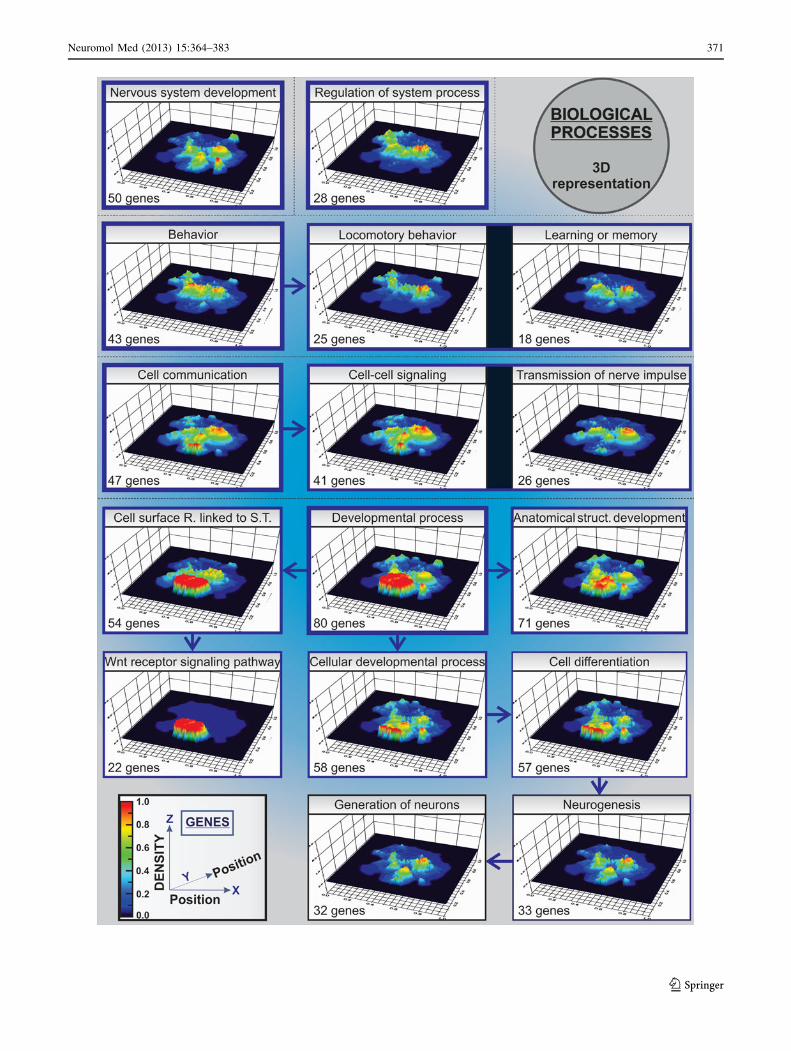
vided (Fig. 3 and supplementary table S3). In total, 16
different biological processes were shown to be affected
by the genes within GENVI network model mainly cor-
responding to both intra- and intercellular communica-
tion, Wnt signaling, nerve impulse transmission, cell
differentiation, neuronal development, neurogenesis,
behavior, and locomotion. This is shown in 3D
representation (Fig. 4) by utilizing the ViaComplex soft-
ware (http://lief.if.ufrgs.br/pub/biosoftwares/viacomplex/)
(Castro et al. 2009).
Exploration of network centralities (Supplementary
table S4 and S5) revealed that RAC1, PRKCA, GABA,
and CACNA1C were NH-Bs or nodes with high
betweenness centrality ([0.05) but lower connectivity
(\40) (Supplementary table S6, Figs. 5, 6). Addition-
ally, Ca2?, SLC6A4, sulfate, hydroxyl radicals, DRD2,
sodium ion, ATP, and magnesium were HBs or nodes
with high values of both betweenness centrality ([0.05)
and connectivity ([40) (Supplementary table S6,
Figs. 5, 6). Moreover, these bottlenecks (NH-Bs and
HBs) showed high values for stress and closeness cen-
tralities above the thresholds. This demonstrates that
such nodes are essential for the flow of information
Fig. 2 In silico network model (GENVI) integrating proposed and
novel putative gene–environment interactions in autism. The network
interconnected 122 genes/proteins and 191 factors (drugs, pollutants,
chemicals, and other compounds) through 1,461 interactions. The
model was developed by using ‘‘Databases’’ and ‘‘Experiments’’ as
input options. As explained in the inset, genetic polymorphisms and
nodes with autism-related report are represented with different shapesand colors (Color figure online)
368 Neuromol Med (2013) 15:364–383
123

through the whole network (Fig. 6a, b, d). Another
interesting aspect is related to tranylcypromine (an
irreversible inhibitor of monoamine oxidase and a potent
antidepressant) (Frieling and Bleich 2006), cholecalcif-
erol, corticosterone, and triiodothyronine. These nodes
scored the highest neighborhood connectivity values
([60) (Supplementary table S6 and Fig. 6c) and may
have a strong etiological relevance since all of them are
connected to Ca2? and/or hydroxyl radicals (HBs of
GENVI network model). Considering the clinical het-
erogeneity of autism, one could speculate that environ-
mental factors (drugs, pollutants, etc.) ‘‘targeting’’
highly connected and stressed bottlenecks in our in sil-
ico model could easily disrupt the network and affect a
higher number of biological processes. This may explain
the wide symptomatology (from mild to severe) of ASD,
because different type and number of genes/proteins
together with its associated biological processes will be
affected depending on whether the node is targeted and
its position (topologically speaking) within the network
of interactions (Fig. 2).
We shall herein discuss the role of Ca2? (HB) in autism
and the reasons why RAC1 (NH-B) may be a relevant gene
deserving further investigation in this area of research.
The Neuron-Glia Communication Mediator, Ca?2, is
a HB of GENVI Network Model: Contextualizing its
Relevance in Autism
Our data pointed to Ca2? (HB) (Fig. 7) as the most relevant
node of GENVI network model for gene–environment
interactions in the autistic context, scoring the highest
values of connectivity ([40), stress ([40,000), between-
ness ([0.05), and closeness ([0.34) centralities among 313
nodes (Supplementary table S6 and Fig. 6). Thus, abnor-
mal Ca2? influx would affect numerous interactions
between gene/proteins and compounds within the network
and, consequently, numerous biological processes. For
instance, glial cells excitability is based on variations of
cytosolic Ca2? concentrations rather than electrical chan-
ges localized in the membrane (Perea and Araque 2005).
Astrocytes express receptors for almost all neurotransmit-
ters at their membranes, giving the capability to detect any
neurotransmitter released at the synapse, activating them
through the mobilization of their intracellular Ca2?. Acti-
vated astrocytes can then modulate neuronal excitability by
releasing a number of neuroactive molecules or gliotrans-
mitters such as glutamate, ATP, D-serine, nitric oxide
(NO), atrial natriuretic factor (ANF), and prostaglandins,
Fig. 3 Hierarchical representation of cellular components, molecular functions, and biological processes of the genes belonging to the network
model (GENVI)
Neuromol Med (2013) 15:364–383 369
123

homocysteic acid, taurine, and tumor necrosis factor-a(TNF-a) (Haydon 2001; Fellin et al. 2006; Halassa et al.
2007). This efficient interrelation allows synaptic trans-
mission to continue. For instance, the expression of
plasma-membrane glutamate transporters ensures quick
clearance of the neurotransmitter from the synapse, pre-
venting the desensitization of postsynaptic receptors that
would interrupt synaptic transmission (Halassa et al. 2007).
Moreover, when presynaptic neurons are activated, astro-
cytes release ATP which is then hydrolyzed to adenosine.
If adenosine accumulates, synaptic transmission is inhib-
ited through mediation of presynaptic adenosine A1
receptors (Zhang et al. 2003; Pascual et al. 2005). The
existence of this bidirectional signaling between astrocytes
and neurons gave rise to the concept of ‘‘tripartite synapse’’
(Perea and Araque 2005).
Neurochemical studies have focused on the involvement
of neurotrophins and different neurotransmitter systems as
an important part of the neurobiological bases of autism
(Pardo and Eberhart 2007). The nerve growth factor family
is constituted by different proteins that include nerve
growth factor (NGF), neurotrophin-3, neurotrophin-4, and
brain-derived neurotrophic factor (BDNF) (Lewin and
Barde 1996; Huang and Reichardt 2003). BDNF produc-
tion was shown to be enhanced during the neonatal period
and later reduced in adult male patients of 18–26 years of
age (Nelson et al. 2001; Miyazaki et al. 2004; Connolly
et al. 2006; Hashimoto et al. 2006; Katoh-Semba et al.
2007). The presence of high levels of neurotrophin-4 and
neurotrophin-5 in neonatal blood of children, who were
later diagnosed with autism, was also reported (Nelson
et al. 2006). Abnormalities in neurotransmitter systems
including serotoninergic, dopaminergic, opioid, choliner-
gic, GABAergic, and glutamatergic may be possibly
involved in the pathology of the syndrome (Lam et al.
2006). High peripheral-blood platelet concentrations of
serotonin were shown to be associated with the disorder
(Schain and Freedman 1961; Anderson et al. 1990;
Anderson and Hoshiono 1997). Reduced serotonin trans-
porter (SERT, also known as SLC6A4) binding capacity
and low serotonin synthesis in the brain of these patients
have already been described (Makkonen et al. 2008).
Additionally, D2 dopamine receptor (DRD2) gene has
been implicated in schizophrenia, posttraumatic stress
disorder, movement disorders, and migraine (Oades et al.
2000; Noble 2003). However, whether DRD2 may play a
role in autism is not clear yet (Philippe et al. 2002).
In our analysis, both SLC6A4 and DRD2 were HBs
within GENVI model, displaying the highest connectivity
and betweenness centrality values among 122 proteins
belonging to the network (Supplementary table S6, Figs. 5,
6b), highlighting their biological relevance in silico.
SLC6A4 is targeted by multiple psychoactive drugs within
the network, such as cocaine and perinatal exposure to this
drug has already been correlated with autism and devel-
opmental abnormalities (Davis et al. 1992). Furthermore,
cocaine abuse itself has been associated with intronic
polymorphisms affecting alternative splicing of human
DRD2 (Moyer et al. 2011). This may represent an example
of how a single environmental factor may affect different
gene/proteins with potential relevance or reported associ-
ation with the neuropathology of autism.
Appreciation for cholinergic transmission in autism
steadily grew since it was reported that either its stimula-
tion or disruption could affect cognitive performance
(Andrews et al. 1994; Newhouse et al. 2004). Indeed, some
studies have claimed that, for instance, decreased nicotinic
receptor function is present in these patients (Lee et al.
2002; Martin-Ruiz et al. 2004).
Other authors have described a hyperglutaminergic
state in autism and how memantine (NMDA glutamate
receptor-antagonist) is able to improve the characteristic
symptomatology (Shinohe et al. 2006; Chez et al. 2007).
As Ghanizadeh suggested (Ghanizadeh 2010), it is
interesting that higher serum levels of neurotensin (NT)
had been detected in young patients with autistic disorder
(Angelidou et al. 2010), considering the imbalance in
glutamate-to-GABA ratios (Harada et al. 2011) and the
lower expression of GABA(A) receptors found in these
patients (Buxbaum et al. 2002; Ma et al. 2005; Fatemi
et al. 2009); since it is known that NT is also able to
promote endogenous glutamate signaling in certain brain
regions (Antonelli et al. 2007). Moreover, single-nucleo-
tide polymorphisms in both GRIN2A and GRIN2B genes
(NMDA receptor 2A and 2B, respectively) were associ-
ated with the disorder (Barnby et al. 2005; Myers et al.
2011; O’Roak et al. 2011). This all together may reflect
in a glutamate-induced brain injury as well as an
inflammatory scenario in autism.
Then, which are the direct implications that could be
expected from an abnormal Ca2? influx in the context of
autism?
(1) In general, synaptic control of glial Ca2? has been
shown in different brain areas such as cerebellum,
cortex, and hippocampus and the responsiveness of
astrocytes to the release of some neurotransmitters by
synaptic terminals such as noradrenaline, acetylcho-
line, GABA, glutamate, or NO is well described in the
literature (Perea and Araque 2005). This necessarily
means that disturbances excessively elevating intra-
cellular Ca2? would directly affect synaptic function,
neuron-astrocyte metabolic coupling, and may have
an impact in other glial cells with involvement of
Ca2? signaling like oligodendrocytes and microglia
(Agrawal et al. 2000; Schipke et al. 2002; Perea and
370 Neuromol Med (2013) 15:364–383
123
Neuromol Med (2013) 15:364–383 371
123

Araque 2005). Moreover, low sulphation or sulpho-
conjugation capacity has been noticed in ‘‘low-func-
tioning’’ autistic children, which is the mechanism that
effectively metabolize phenolic amines, including
catecholamines functioning as neurotransmitters, and
this deficiency would result in aberrant inactivation of
neurotransmitters (Alberti et al. 1999).
(2) Exposure to environmental stressors (e.g., pollutants
or drugs) may lead to overproduction of reactive
oxygen species (ROS) (e.g., hydroxyl radicals) with
ensuing neurotoxicity. In fact, xenobiotics and heavy
metals are able to inhibit metabolic pathways that
synthesize GSH and keep physiological levels of its
reduced form (Carvalho et al. 2008; Herbert 2010). In
autism, the endogenous antioxidant and detoxifier,
glutathione (GSH), may be specifically important for
the pathogenesis of the disorder since polymorphisms
of genes encoding gluthatione-dependent enzymes
were reported (e.g., glyoxalase 1 or GLO1 as well as
glutathione peroxidase 1 or GPX1) (Junaid et al. 2004;
Ming et al. 2010). Individuals with compromised
Fig. 5 Network centrality
measures and topological
analysis of the network model
(GENVI). Numeric values
corresponding to the properties
of each node (stress,
connectivity, neighborhood
connectivity, betweenness, and
closeness centralities) are
plotted in a bi-dimensional (2D)
color grading representation
(from dark blue for the lowest todark red for the highest values)
(Color figure online)
Fig. 4 Three-dimensional representation (3D) and spatial localiza-
tion of the biological processes identified in the in silico model
(GENVI). In the present figure, hierarchy of the biological processes
is additionally represented with arrows. The number of affected genes
corresponding to each biological process is shown with color gradingin the network (from dark blue for the lowest to dark red for the
highest density of genes) (Color figure online)
b
372 Neuromol Med (2013) 15:364–383
123

antioxidant defenses would be under oxidative stress
and may easily reach toxic thresholds when compared
to healthy people (Herbert 2010). It is well known that
oxidative stress levels can trigger cell death by
increasing the cytoplasmic Ca2? concentration, giving
rise to a Ca2? influx into mitochondria where it may
accelerate and disrupt the normal cellular metabolism
of these patients (Ermak and Davies 2002). Further-
more, excessive ROS production would also affect the
epigenetic regulation of gene expression due to lower
methionine synthase activity. In oxidative stress
scenarios, decreased methylation capability (in addi-
tion to compromised antioxidant/detoxification capac-
ity) would be then expected in autism (Deth et al.
2008; James et al. 2008). In other words, oxidative
stress-induced decreased DNA methylation could up-
regulate the expression of genes that are usually
controlled by methylation gene silencing, representing
one of the possible cross-talks between environmental
influences and genetic alterations in ASD.
The Rho GTPase RAC1 is a NH-B of GENVI Network
Model: A New Genetic Link to the Neurobiology
of Autism?
It is generally agreed that the characteristic fixed patterns
of behavior and lack of social interaction in ASD indicate a
disruption of critical neurodevelopmental routes and, thus,
aberrant pre- and postnatal development of important brain
structures (Muhle et al. 2004). These cerebral areas were
previously linked to social development in patients
(Adolphs 2001) and implicate parts of the frontal lobe,
superior temporal cortex, parietal cortex, and amygdala
Fig. 6 Analysis of the topological properties of the nodes belonging
to the network model (GENVI). Dashed lines are indicating the
threshold value for each property. In the graphs, genes/proteins are
represented by red circles and factors (drugs, pollutants, chemicals,
and other compounds) are plotted as black squares. Note that NH-Bs
and HBs distinguish non-hub-bottlenecks from hub-bottlenecks,
respectively (Color figure online)
Neuromol Med (2013) 15:364–383 373
123

(Amaral et al. 2008). Postmortem studies revealed a spe-
cific cytoarchitecture in both cortical and subcortical
structures of autistic patients. Several disturbances were
demonstrated, including the decreased number of neurons
and reduced dendritic arborisation in different parts of the
limbic system (amygdala, hippocampus, septum, and
anterior cingulated cortex) (Baron-Cohen et al. 2000;
Kemper and Bauman 2002; Sweeten et al. 2002; Volkmar
Fig. 7 Ca2? subnetwork analysis. Landscape of interactions of Ca2?-
gene/protein and Ca2?-factor (drugs, pollutants, chemicals, and other
compounds) through either ‘‘binding,’’ ‘‘reaction,’’ ‘‘phenotype,’’
‘‘catalysis,’’ ‘‘inhibition,’’ or ‘‘not specified’’ type of interconnection.
Reported genetic polymorphisms are marked with blue circles, as
indicated in the inset (Color figure online)
374 Neuromol Med (2013) 15:364–383
123

and Pauls 2003; Schumann and Amaral 2006). Similar
aberrations were reported in the cerebellum where a
decreased number of Purkinje and granule cells were
observed (Ritvo et al. 1986). Furthermore, both number
and structure of neocortical ‘‘minicolumns’’ (or ‘‘micro-
columns’’) seem to be altered. In the brains of ASD
patients, Casanova and colleagues found significantly
higher numbers of cortical minicolumns when compared to
the healthy controls (Casanova et al. 2002, 2006; Casanova
and Trippe 2009). They additionally observed a decreased
intercolumnar width (smaller space between cell body-
defined minicolumnar structures) and reduced neuronal
size.
In physiological conditions, during development and
adulthood, cellular migration is a crucial biological process
for neurons and glial cells to occupy characteristic posi-
tions in the central (CNS) and peripheral nervous system
(PNS) (Torrence 1991). Cytoskeleton reorganization is
required for axonal outgrowth and neuronal migration
(Dent and Gertler 2003). Interestingly, several studies have
implicated Rho GTPases in the development of axons and
dendrites (Gualdoni et al. 2007). In particular, normal
levels of RAC1 seem to be essential for early dendritic
development of mouse hippocampal neurons (Gualdoni
et al. 2007). Moreover, migration of differentiated hippo-
campal neurons in a RAC and PI3-kinases-dependent
manner has been observed (Leemhuis et al. 2004).
Our in silico analysis showed RAC1 to be a gene with
high betweenness centrality ([0.05) in our network model
(GENVI) (Supplementary table S6 and Fig. 6). It is also
remarkable that RAC1 was identified in nine of the sixteen
different biological processes representing the whole net-
work model for gene–environment interactions in the
autistic context (GENVI), such as behavior, developmental
process, nervous system development, anatomical structure
development, cell differentiation, cellular developmental
process, locomotory behavior, neurogenesis, and genera-
tion of neurons (Supplementary table S3; Figs. 3, 4).
Despite extensive research, there are no studies directly
linking RAC1 to ASD. It is an opened question whether
mutations in RAC1 gene or impaired functionality of the
protein could have a role in the neuropathology of this
disorder. On the other hand, there are some circumstantial
facts suggesting RAC1 as an interesting candidate gene/
protein to pay attention to:
(1) RAC1 is an ubiquitously expressed member of the
RHO monomeric GTPase family that participates in
the regulation of gene expression, cytoskeletal rear-
rangement, cell activation, and cell motility (Chatter-
jee et al. 2010). RAC1 has been suggested to have an
important role in toll-like receptors (TLR) function-
ing in immune cells (Ruse and Knaus 2006).
Additionally, it has been reported the existence of
differential monocyte responses to TLR ligands in
children with ASD (Enstrom et al. 2010). Therefore,
alterations of innate immunity due to TLR-mediated
neuroimmune interactions, sustained inflammatory
response, or even infection- or vaccine-induced
imbalance between innate and adaptive immunity in
autistic patients may have an interpretation through
the altered RAC1-associated signaling pathways
(Jyonouchi et al. 2005, 2008; Hagberg et al. 2012).
(2) It has been noted that a significant percentage of
autistic patients were undergoing extensive antimi-
crobial treatment and suffering from chronic diarrhea
before subsequent gradual evolution of their autistic
symptoms (Bolte 1998; Sandler et al. 2000). Some
reports suggest that disruption of protective intestinal
microbiota after several rounds of treatment with
broad-spectrum antibiotics may create a favorable
environment for opportunistic neurotoxin-producing
pathogens such as Clostridium tetani, which might
become a pathological element to consider in the
context of the idiopathic increase of autism rates as
well as when multiple cases were reported in the same
family (Sandler et al. 2000; Finegold 2008). As a
matter of fact, both qualitative and quantitative
differences between gastrointestinal microflora of
autistic children and healthy controls have been
reported as well, especially associated with Clostrid-
ium spp. (Finegold et al. 2002; Song et al. 2004;
Parracho et al. 2005). Furthermore, autistic children
have showed some improvement after oral vancomy-
cin treatment (which is not absorbed in the gut)
(Sandler et al. 2000). Even though the connection
between intestinal anaerobic bacteria and autism is
still under debate (Martirosian et al. 2009), it is
noteworthy that a number of different Clostridium
species can produce large molecular mass cytotoxins,
able to trigger effects on the actin cytoskeleton and to
disrupt actin stress fibers (Popoff et al. 1996). RAC,
RAP, and RAS small GTP-binding proteins are
indeed targets for Clostridium toxins (Just et al.
1996; Popoff et al. 1996; Leemhuis et al. 2004; Geny
et al. 2010).
(3) An additional circumstantial fact is that biological
activities of RHO family of GTPases (like CDC42,
RHOA, and RAC1) are controlled by their guanine
nucleotide binding states in cells. RhoGAPs (regu-
latory molecules of RHO GTPases) use magnesium
(Fig. 8) as a cofactor to reach catalytic efficiency
and specificity in GTP hydrolysis (Zhang et al.
2000). A significant decrease in the concentration of
magnesium and selenium in the hair and nail
samples of autistic patients, correlating with their
Neuromol Med (2013) 15:364–383 375
123

degrees of severity, has already been reported
(Lakshmi Priya and Geetha 2011). Thus, if physi-
ological levels of magnesium are reduced in autistic
patients, one could predict that both catalytic
efficiency and specificity of RhoGAPs would be
also compromised.
(4) An elegant study by Adams et al. (2011) reporting
significantly low levels of ATP in children with
autism may also support RAC1 as an interesting
candidate gene/protein in the autistic scenario since
RAC1 is known to be sensitive to ATP (Fig. 8) and
GTP levels in vitro, displaying a short-term moderate
decrease in activity after nucleotide triphosphate
depletion (Hallett et al. 2003).
In other words, based on the in silico evidences pre-
sented in our analysis together with the facts mentioned
above, we believe that RAC1, in particular, and the RHO
family of GTPases, in general, may have a role in the
neuropathological events associated with autism, presum-
ably, due to altered neuroimmune interactions.
Analysis of the Genes Belonging to the Ca2?-RHO
Family of GTPases Interactome Network Reveals
a Differential Gene Expression in the Cerebellum
of Autistic Brains
It is well described that several small GTPases collaborate
with Ca2? signaling in regulating different cellular pro-
cesses, such as cell adhesion, cell migration, and exocytosis
(Aspenstrom 2004). Our in silico results demonstrated a
topological relevance of Ca2? and RAC1 in the proposed
model for gene–environment interactions in autism
(GENVI) (Figs. 2, 6). Consequently, a constant informa-
tion flow in the interaction network is being trafficked
through these nodes. Thus, to further characterized the
interplay between Ca2?, RAC1, and other RHO family
GTPases and related proteins, we elaborated a list genes/
proteins (Supplementary table S7), and by utilizing
STITCH 3.0, the Ca2?-RHO family of GTPases interac-
tome network was developed (Fig. 9). All the genes/pro-
teins from the original list (43 nodes) were interconnected,
and 5 of them (CHP, ITPR1, PLCG1, PRKCA, and RAC3)
were directly linked to Ca2? in this newly developed net-
work (Supplementary table S7 and Fig. 9).
Synaptic control of glial Ca2? is well described in different
brain areas such as cerebellum, cortex, and hippocampus
(Perea and Araque 2005). Diverse lines of evidence suggest
the involvement of apoptosis in the cerebellum of autism
subjects, including loss and atrophy of Purkinje cells (Kern
2003; Whitney et al. 2008). Furthermore, it has been reported
brain region-specific deficits in expression levels of mito-
chondrial electron transport chain complexes in the cerebel-
lum and the frontal and temporal cortices of children with
autism (Chauhan et al. 2011). Therefore, we checked whether
the genes belonging to the Ca2?-RHO family of GTPases
interactome network could be differentially expressed in the
cerebellum of these patients. Very interestingly, we found that
15 from the 43 genes belonging to the Ca2?-RHO family of
GTPases interactome network (Table 1) were differentially
Fig. 8 RAC1 subnetwork
analysis. Landscape of
interactions of RAC1-gene/
protein and RAC1-factor (drugs,
pollutants, chemicals, and other
compounds) through either
‘‘binding,’’ ‘‘reaction,’’
‘‘phenotype,’’ ‘‘catalysis,’’
‘‘inhibition,’’ or ‘‘not specified’’
type of interconnection.
Reported genetic
polymorphisms are marked with
blue circles, as indicated in the
inset (Color figure online)
376 Neuromol Med (2013) 15:364–383
123

expressed (corrected p value \0.05) in the cerebellum of
autistic patients when compared to control samples. Three
from the 5 genes that interconnected to Ca2? in the interac-
tome network were in this group of either sub- or overex-
pressed genes (PRKCA, RAC3, and CHP). In addition, RAC1
directly connected with a number of differentially expressed
genes (e.g., CDC42, PRKCA, and RHOB) (Table 1; Fig. 9),
which is consistent with our in silico results where RAC1 was
shown to be a bottleneck of the GENVI network model, or an
essential key connector node critically relevant for the flow of
information through the entire network (Yu et al. 2007).
In our array analysis, CDC42 was strongly subexpressed
(corrected p value = 0.00200014), while RHOB displayed a
significant overexpression (corrected p value = 0.01534273)
in the cerebellum of autistic patients (Table 1). Even though
RAC1, CDC42, and RHOA/RHOB GTPases are known to be
all expressed by migrating precerebellar neurons (PCN), they
seem to have opposed roles on neuritogenesis. For instance,
inhibition of RAC and CDC42 subfamilies impairs neurite
outgrowth of PCN without affecting migration, whereas
pharmacological inhibition of RHO enhances axon outgrowth
of PCN and prevents nuclei migration (Causeret et al. 2004).
Likely, our results may reflect a compromised neurite out-
growth in cerebellar neurons of autistic patients. Besides,
RAC1 and RAC3 are important for the development of the
nervous system, wherein they play complementary roles
during late stages of brain development (Corbetta et al. 2009).
Both RAC1 and RAC3 GTPases synergistically control the
development of cortical and hippocampal GABAergic inter-
neurons (Vaghi et al. 2012), areas of the CNS where decreased
number of neurons and reduced dendritic arborisation have
been described in autism (Baron-Cohen et al. 2000; Kemper
and Bauman 2002; Sweeten et al. 2002; Volkmar and Pauls
2003; Schumann and Amaral 2006). In an elegant study by
Bolis et al. (2003), RAC3 was implicated in Purkinje cell
development, at times of neuronal differentiation and syna-
ptogenesis. Our own results showed subexpression of RAC3
in the cerebellum of autistic brains (corrected p value =
0.01470489) (Table 1). Considering that RAC3-knockout
mice are viable and recent studies started to indicate a relevant
role of RAC3 in cognitive development (Corbetta et al. 2008),
the specific role of these GTPases in the context of autism may
deserve further research.
Concluding Remarks
A general tendency is emerging toward the combination of
different etiologies to explain the heterogeneity of autism.
Casanova (2007) proposed a ‘‘Triple Hit Hypothesis of
Autism,’’ with three different degrees of etiological factors
such as precise time window of brain development, genetic
susceptibility, and different environmental stressors. Wil-
liams and Casanova (2010) further suggested that ultrasound
Fig. 9 Ca2?-RHO family of GTPases interactome network. The network interconnected Ca2? with 43 GTPases from the RHO family and
related genes/proteins (e.g., targets). The model was developed by using ‘‘Databases’’ and ‘‘Experiments’’ as input options
Neuromol Med (2013) 15:364–383 377
123

examination, commonly used in obstetrics, could be one of
these environmental stressors by exerting teratogenic/toxic
effects on the CNS. Another hypothesis proposed that
accumulative doses of mercury from different sources (e.g.,
pollution, maternal fish consumption, dental amalgams, and
vaccinations) during infant development, together with a
decreased ability to remove mercury from the body, could
increase the probability of developing and/or aggravating
autism (Zeidan-Chulia et al. 2011).
In this study, after systematic review of the literature,
we aimed to develop a model for gene–environment
interactions in ASD able to integrate the current knowledge
and findings in the topic. Thereafter, we characterized it in
detail by using systems biology tools (Fig. 1), and finally,
we further discussed the most relevant genes and/or factors
according to the output information from this approach and
the results from the microarray analysis of samples from
autistic patients. Two key conclusions can be summarized
according to our data:
(1) Ca2?- signaling molecule was the most central node of
our model of gene–environment interactions in the
autistic context (GENVI). Communication of Ca2?
signals between brain cells is a typical feature of glial
cells. In fact, astroglia-regulated neurogenesis, neuronal
structural plasticity, and synaptic rearrangement may
have an important role in the dynamics of social
functioning (Mercadante et al. 2008). Normal
development of nervous system would require well-
organized neuronal migration, axon guidance, target
selection, dendritic growth, synapse formation, and
elimination (Bolton and Eroglu 2009). But under
stressful conditions (e.g., sensory deprivation, separa-
tion stress, toxic action of xenobiotics), reactive struc-
tural plasticity can occur in an immature developing
brain which is accompanied by alterations of glial cell
activation (Musholt et al. 2009). Neuroanatomical
abnormalities observed in autism, such as reduced brain
size at birth and a sudden and excessive brain over-
growth in the areas of cerebellum, frontal lobe, and
limbic structures, with increased cerebral white matter
and decrease in cerebral cortex and hippocampal/
amygdala volumes, are changes associated with delays
in neuronal maturation and increased astrogliosis (Pardo
et al. 2005). These evidences together with studies
reporting that astroglial responses in autism, manifested
by increased astrocyte/neuron ratio in brain cortex,
alterations in intercellular communication within the
astroglial syncytium, and reactive astrogliosis (Lau-
rence and Fatemi 2005; Pardo et al. 2005; Fatemi et al.
2008) point toward a dysregulation of neuron–glia
interactions and Ca2?-mediated signaling in the devel-
oping brain of autistic children.
(2) RAC1, in particular, and the RHO family of GTPases,
in general, are highlighted as potentially relevant
genes/proteins the context of autism. Certainly, the
Table 1 Differentialy expressed genes from the Ca2?-RHO family of GTPases interactome network in the cerebellum of autistic brains versus
control samples (corrected p values \0.05 were considered significant)
Gene
symbol
Alias and/or description Ensembl ID
(ENSP)
Differential gene
expression
Corrected
p value
Interact. with
calcium
CDC42 Cell division cycle 42 (GTP-binding protein, 25 kDa) ENSP00000314458 Down 0.00200014 NO
PRKCA PKC-a; protein kinase C, a ENSP00000284384 Up 0.00353275 YES
RALA v-ral simian leukemia viral oncogene homolog A (ras
related)
ENSP00000005257 Up 0.00628922 NO
RALB v-ral simian leukemia viral oncogene homolog B (ras
related)
ENSP00000272519 Down 0.00803662 NO
RHOQ Ras homolog gene family member Q; Plasma membrane-
associated small GTPase
ENSP00000238738 Up 0.01193934 NO
RAC3 Ras-related C3 botulinum toxin substrate 3 (rho family,
small GTP-binding protein Rac3)
ENSP00000304283 Down 0.01470489 YES
RHOB Ras homolog gene family member B ENSP00000272233 Up 0.01534273 NO
RHOU Ras homolog gene family member U ENSP00000355652 Up 0.02181148 NO
ROCK1 Rho-associated coiled-coil containing protein kinase 1 ENSP00000382697 Up 0.02843342 NO
PLD1 Phospholipase D1 ENSP00000342793 Up 0.03381597 NO
PKN2 Protein kinase N2 ENSP00000359552 Up 0.034955 NO
GIT1 G protein-coupled receptor kinase interacting ArfGAP 1 ENSP00000378338 Up 0.03796387 NO
RHOG Ras homolog gene family member G (rho G) ENSP00000339467 Up 0.03837408 NO
PXN Paxillin ENSP00000228307 Up 0.04056128 NO
CHP Calcium-binding protein p22 ENSP00000335632 Down 0.04455404 YES
378 Neuromol Med (2013) 15:364–383
123

maintenance of dendritic arbor complexity during
development and into adulthood is critical for the
preservation of functional circuitry and connectivity
critical for learning and complex behaviors (Srivast-
ava et al. 2012). The Rho family of small GTPases
(especially, RAC, CDC42, and RHOA) is indeed a
key regulator of the actin cytoskeleton in response to
extracellular cues, and disturbances in their activities
often give rise to dramatic effects in dendritic
morphogenesis (Scott et al. 2003).
Finally, we believe that our approach (combination of
systematic review of the literature with systems biology
and microarray analyses) offers an attractive option to aid
researchers in the field of PDD for finding potentially rel-
evant genes, factors, or gene–environment interactions,
which could be later confirmed by clinical and/or basic
science studies.
Acknowledgments First of all, we apologize to all our colleagues
whose studies were not cited due to lack of space. We thank the
Brazilian research funding agencies FAPERGS (PqG 1008860, PqG
1008857, ARD11/1893-7, PRONEX 1000274), CAPES (PROCAD
066/2007), CNPq, PROPESQ-UFRGS, and IBN-Net #01.06.0842-00
for supporting this work. A.B.S is supported by the grant from the
Federal Program of the Russian Federation (N 8061, 2012–2013). We
are very grateful to Prof. Alexei Verkhratsky (University of Man-
chester, Manchester, UK) and Dr. Marcio L. Acencio (Universidade
Estadual Paulista, Sao Paulo, Brasil) for reading the manuscript.
Conflict of interest The authors declare that there are no conflicts
of interest.
References
Adams, J. B., Audhya, T., McDonough-Means, S., Rubin, R. A.,
Quig, D., Geis, E., et al. (2011). Nutritional and metabolic status
of children with autism vs. neurotypical children, and the
association with autism severity. Nutrition & Metabolism, 8(1),
34.
Adolphs, R. (2001). The neurobiology of social cognition. CurrentOpinion in Neurobiology, 11(2), 231–239.
Agrawal, S. K., Nashmi, R., & Fehlings, M. G. (2000). Role of L- and
N-type calcium channels in the pathophysiology of traumatic
spinal cord white matter injury. Neuroscience, 99(1), 179–188.
Alberti, A., Pirrone, P., Elia, M., Waring, R. H., & Romano, C.
(1999). Sulphation deficit in ‘‘low-functioning’’ autistic children:
A pilot study. Biological Psychiatry, 46(3), 420–424.
Amaral, D. G., Schumann, C. M., & Nordahl, C. W. (2008).
Neuroanatomy of autism. Trends in Neurosciences, 31(3),
137–145.
Anderson, G. M., Horne, W. C., Chatterjee, D., & Cohen, D. J.
(1990). The hyperserotonemia of autism. Annals of the New YorkAcademy of Sciences, 600, 331–340.
Anderson, G. M., & Hoshiono, Y. (1997). Neurochemical studies of
autism. In D. J. Cohen & F. R. Volkmar (Eds.), Handbook ofautism and pervasive developmental disorders (pp. 325–343).
New York: Wiley.
Andrews, J. S., Jansen, J. H., Linders, S., & Princen, A. (1994).
Effects of disrupting the cholinergic system on short-term spatial
memory in rats. Psychopharmacology (Berl), 115(4), 485–494.
Angelidou, A., Francis, K., Vasiadi, M., Alysandratos, K. D., Zhang,
B., Theoharides, A., et al. (2010). Neurotensin is increased in
serum of young children with autistic disorder. Journal ofNeuroinflammation, 7, 48.
Antonelli, T., Fuxe, K., Tomasini, M. C., Mazzoni, E., Agnati, L. F.,
Tanganelli, S., et al. (2007). Neurotensin receptor mechanisms
and its modulation of glutamate transmission in the brain:
Relevance for neurodegenerative diseases and their treatment.
Progress in Neurobiology, 83(2), 92–109.
Aspenstrom, P. (2004). Integration of signalling pathways regulated
by small GTPases and calcium. Biochimica et Biophysica Acta,1742(1–3), 51–58.
Barnby, G., Abbott, A., Sykes, N., Morris, A., Weeks, D. E., Mott, R.,
et al. (2005). Candidate-gene screening and association analysis
at the autism-susceptibility locus on chromosome 16p: Evidence
of association at GRIN2A and ABAT. American Journal ofHuman Genetics, 76(6), 950–966.
Baron-Cohen, S., Ring, H. A., Bullmore, E. T., Wheelwright, S., Ashwin,
C., & Williams, S. C. (2000). The amygdala theory of autism.
Neuroscience and Biobehavioral Reviews, 24(3), 355–364.
Baron-Cohen, S., Ring, H. A., Wheelwright, S., Bullmore, E. T.,
Brammer, M. J., Simmons, A., et al. (1999). Social intelligence
in the normal and autistic brain: An fMRI study. The EuropeanJournal of Neuroscience, 11(6), 1891–1898.
Bennett, T., Szatmari, P., Bryson, S., Volden, J., Zwaigenbaum, L.,
Vaccarella, L., et al. (2007). Differentiating autism and Asperger
syndrome on the basis of language delay or impairment. Journalof Autism and Developmental Disorders, 38(4), 616–625.
Bolis, A., Corbetta, S., Cioce, A., & de Curtis, I. (2003). Differential
distribution of Rac1 and Rac3 GTPases in the developing mouse
brain: Implications for a role of Rac3 in Purkinje cell differentiation.
The European Journal of Neuroscience, 18(9), 2417–2424.
Bolte, E. R. (1998). Autism and Clostridium tetani. MedicalHypotheses, 51(2), 133–144.
Bolton, M. M., & Eroglu, C. (2009). Look who is weaving the neural
web: Glial control of synapse formation. Current Opinion inNeurobiology, 19(5), 491–497.
Buxbaum, J. D., Silverman, J. M., Smith, C. J., Greenberg, D. A.,
Kilifarski, M., Reichert, J., et al. (2002). Association between a
GABRB3 polymorphism and autism. Molecular Psychiatry,7(3), 311–316.
Carvalho, C. M., Chew, E. H., Hashemy, S. I., Lu, J., & Holmgren, A.
(2008). Inhibition of the human thioredoxin system. A molecular
mechanism of mercury toxicity. The Journal of BiologicalChemistry, 283(18), 11913–11923.
Casanova, M. F. (2007). The neuropathology of autism. BrainPathology, 17(4), 422–433.
Casanova, M. F., Buxhoeveden, D. P., Switala, A. E., & Roy, E.
(2002). Minicolumnar pathology in autism. Neurology, 58(3),
428–432.
Casanova, M., & Trippe, J. (2009). Radial cytoarchitecture and
patterns of cortical connectivity in autism. Philosophical Trans-actions of the Royal Society of London. Series B, Biologicalsciences, 364, 1433–1436.
Casanova, M. F., van Kooten, I. A., Switala, A. E., van Engeland, H.,
Heinsen, H., Steinbusch, H. W., et al. (2006). Minicolumnar
abnormalities in autism. Acta Neuropathologica, 112(3), 287–303.
Castro, M. A., Filho, J. L., Dalmolin, R. J., Sinigaglia, M., Moreira, J.
C., Mombach, J. C., et al. (2009). ViaComplex: Software for
landscape analysis of gene expression networks in genomic
context. Bioinformatics, 25(11), 1468–1469.
Causeret, F., Hidalgo-Sanchez, M., Fort, P., Backer, S., Popoff, M. R.,
Gauthier-Rouviere, C., et al. (2004). Distinct roles of Rac1/
Neuromol Med (2013) 15:364–383 379
123

Cdc42 and Rho/Rock for axon outgrowth and nucleokinesis of
precerebellar neurons toward netrin 1. Development, 131(12),
2841–2852.
Chatterjee, A., Wang, L., Armstrong, D. L., & Rossie, S. (2010).
Activated Rac1 GTPase translocates protein phosphatase 5 to the
cell membrane and stimulates phosphatase activity in vitro. TheJournal of Biological Chemistry, 285(6), 3872–3882.
Chauhan, A., Gu, F., Essa, M. M., Wegiel, J., Kaur, K., Brown, W. T.,
et al. (2011). Brain region-specific deficit in mitochondrial
electron transport chain complexes in children with autism.
Journal of Neurochemistry, 117(2), 209–220.
Chez, M. G., Burton, Q., Dowling, T., Chang, M., Khanna, P., &
Kramer, C. (2007). Memantine as adjunctive therapy in children
diagnosed with autistic spectrum disorders: An observation of
initial clinical response and maintenance tolerability. Journal ofChild Neurology, 22(5), 574–579.
Connolly, A. M., Chez, M., Streif, E. M., Keeling, R. M., Golumbek,
P. T., Kwon, J. M., et al. (2006). Brain-derived neurotrophic
factor and autoantibodies to neural antigens in sera of children
with autistic spectrum disorders, Landau-Kleffner syndrome, and
epilepsy. Biological Psychiatry, 59(4), 354–363.
Corbetta, S., D’Adamo, P., Gualdoni, S., Braschi, C., Berardi, N., &
de Curtis, I. (2008). Hyperactivity and novelty-induced hyper-
reactivity in mice lacking Rac3. Behavioural Brain Research,186(2), 246–255.
Corbetta, S., Gualdoni, S., Ciceri, G., Monari, M., Zuccaro, E.,
Tybulewicz, V. L., et al. (2009). Essential role of Rac1 and Rac3
GTPases in neuronal development. FASEB Journal, 23(5),
1347–1357.
Davis, E., Fennoy, I., Laraque, D., Kanem, N., Brown, G., & Mitchell,
J. (1992). Autism and developmental abnormalities in children
with perinatal cocaine exposure. Journal of the National MedicalAssociation, 84(4), 315–319.
De Franceschi, L., Scardoni, G., Tomelleri, C., Danek, A., Walker, R.
H., Jung, H. H., et al. (2012). Computational identification of
phospho-tyrosine sub-networks related to acanthocyte generation
in neuroacanthocytosis. PLoS ONE, 7(2), e31015.
del Rio, G., Koschutzki, D., & Coello, G. (2009). How to identify
essential genes from molecular networks? BMC Systems Biol-ogy, 3, 102.
Dent, E. W., & Gertler, F. B. (2003). Cytoskeletal dynamics and
transport in growth cone motility and axon guidance. Neuron,40(2), 209–227.
Deth, R., Muratore, C., Benzecry, J., Power-Charnitsky, V. A., &
Waly, M. (2008). How environmental and genetic factors
combine to cause autism: A redox/methylation hypothesis.
Neurotoxicology, 29(1), 190–201.
Enstrom, A. M., Onore, C. E., Van de Water, J. A., & Ashwood, P.
(2010). Differential monocyte responses to TLR ligands in
children with autism spectrum disorders. Brain, Behavior, andImmunity, 24(1), 64–71.
Ermak, G., & Davies, K. J. (2002). Calcium and oxidative stress:
From cell signaling to cell death. Molecular Immunology,38(10), 713–721.
Estrada, E. (2006). Virtual identification of essential proteins within
the protein interaction network of yeast. Proteomics, 6(1),
35–40.
Fatemi, S. H., Folsom, T. D., Reutiman, T. J., & Lee, S. (2008).
Expression of astrocytic markers aquaporin 4 and connexin 43 is
altered in brains of subjects with autism. Synapse (New York, N.Y.), 62(7), 501–507.
Fatemi, S. H., Reutiman, T. J., Folsom, T. D., & Thuras, P. D. (2009).
GABA(A) receptor downregulation in brains of subjects with
autism. Journal of Autism and Developmental Disorders, 39(2),
223–230.
Fellin, T., Pascual, O., & Haydon, P. G. (2006). Astrocytes coordinate
synaptic networks: Balanced excitation and inhibition. Physiol-ogy (Bethesda), 21, 208–215.
Finegold, S. M. (2008). Therapy and epidemiology of autism–
clostridial spores as key elements. Medical Hypotheses, 70(3),
508–511.
Finegold, S. M., Molitoris, D., Song, Y., Liu, C., Vaisanen, M. L.,
Bolte, E., et al. (2002). Gastrointestinal microflora studies in
late-onset autism. Clinical Infectious Diseases, 35(Suppl 1),
S6–S16.
Fombonne, E. (2003). The prevalence of autism. JAMA, 289(1),
87–89.
Frieling, H., & Bleich, S. (2006). Tranylcypromine: New perspectives
on an ‘‘old’’ drug. European Archives of Psychiatry and ClinicalNeuroscience, 256(5), 268–273.
Gentleman, R. C., Carey, V. J., Bates, D. M., Bolstad, B., Dettling,
M., Dudoit, S., et al. (2004). Bioconductor: Open software
development for computational biology and bioinformatics.
Genome Biology, 5(10), R80.
Geny, B., Grassart, A., Manich, M., Chicanne, G., Payrastre, B.,
Sauvonnet, N., et al. (2010). Rac1 inactivation by lethal toxin
from Clostridium sordellii modifies focal adhesions upstream of
actin depolymerization. Cellular Microbiology, 12(2), 217–232.
Ghanizadeh, A. (2010). Targeting neurotensin as a potential novel
approach for the treatment of autism. Journal of Neuroinflam-mation, 7, 58.
Ginsberg, M. R., Rubin, R. A., Falcone, T., Ting, A. H., & Natowicz,
M. R. (2012). Brain transcriptional and epigenetic associations
with autism. PLoS ONE, 7(9), e44736.
Grossman, J. B., Carter, A., & Volkmar, F. R. (1997). Social behavior
in autism. Annals of the New York Academy of Sciences, 807,
440–454.
Gualdoni, S., Albertinazzi, C., Corbetta, S., Valtorta, F., & de Curtis,
I. (2007). Normal levels of Rac1 are important for dendritic but
not axonal development in hippocampal neurons. Biology of theCell, 99(8), 455–464.
Hagberg, H., Gressens, P., & Mallard, C. (2012). Inflammation during
fetal and neonatal life: Implications for neurologic and neuro-
psychiatric disease in children and adults. Annals of Neurology,71(4), 444–457.
Halassa, M. M., Fellin, T., & Haydon, P. G. (2007). The tripartite
synapse: Roles for gliotransmission in health and disease. Trendsin Molecular Medicine, 13(2), 54–63.
Hallett, M. A., Dagher, P. C., & Atkinson, S. J. (2003). Rho GTPases
show differential sensitivity to nucleotide triphosphate depletion
in a model of ischemic cell injury. American Journal ofPhysiology. Cell Physiology, 285(1), C129–C138.
Harada, M., Taki, M. M., Nose, A., Kubo, H., Mori, K., Nishitani, H.,
et al. (2011). Non-Invasive evaluation of the GABAergic/
glutamatergic system in autistic patients observed by MEGA-
editing proton MR spectroscopy using a clinical 3 tesla
instrument. Journal of Autism and Developmental Disorders,41(4), 447–454.
Hashimoto, K., Iwata, Y., Nakamura, K., Tsujii, M., Tsuchiya, K. J.,
Sekine, Y., et al. (2006). Reduced serum levels of brain-derived
neurotrophic factor in adult male patients with autism. Progressin Neuro-Psychopharmacology and Biological Psychiatry, 30(8),
1529–1531.
Haydon, P. G. (2001). GLIA: Listening and talking to the synapse.
Nature Reviews Neuroscience, 2(3), 185–193.
Herbert, M. R. (2010). Contributions of the environment and
environmentally vulnerable physiology to autism spectrum
disorders. Current Opinion in Neurology, 23(2), 103–110.
Hernandez, P., Huerta-Cepas, J., Montaner, D., Al-Shahrour, F.,
Valls, J., Gomez, L., et al. (2007). Evidence for systems-level
380 Neuromol Med (2013) 15:364–383
123

molecular mechanisms of tumorigenesis. BMC Genomics, 8,
185.
Hooper, S. D., & Bork, P. (2005). Medusa: A simple tool for
interaction graph analysis. Bioinformatics, 21(24), 4432–4433.
Huang, E. J., & Reichardt, L. F. (2003). Trk receptors: Roles in
neuronal signal transduction. Annual Review of Biochemistry,72, 609–642.
James, S. J., Melnyk, S., Jernigan, S., Hubanks, A., Rose, S., &
Gaylor, D. W. (2008). Abnormal transmethylation/transsulfura-
tion metabolism and DNA hypomethylation among parents of
children with autism. Journal of Autism and DevelopmentalDisorders, 38(10), 1976.
Junaid, M. A., Kowal, D., Barua, M., Pullarkat, P. S., Skolower
Brooks, S., & Pullarkat, R. K. (2004). Proteomic studies
identified a single nucleotide polymorphism in glyoxalase I as
autism susceptibility factor. American Journal of MedicalGenetics. Part A, 131(1), 11–17.
Just, I., Selzer, J., Hofmann, F., Green, G. A., & Aktories, K. (1996).
Inactivation of Ras by Clostridium sordellii lethal toxin-
catalyzed glucosylation. The Journal of Biological Chemistry,271(17), 10149–10153.
Jyonouchi, H., Geng, L., Cushing-Ruby, A., & Quraishi, H. (2008).
Impact of innate immunity in a subset of children with autism
spectrum disorders: A case control study. Journal of Neuroin-flammation, 5, 52.
Jyonouchi, H., Geng, L., Ruby, A., & Zimmerman-Bier, B. (2005).
Dysregulated innate immune responses in young children with
autism spectrum disorders: Their relationship to gastrointestinal
symptoms and dietary intervention. Neuropsychobiology, 51(2),
77–85.
Kanner, L. (1943). Autistic disturbances of affective contact. NervousChild, 2, 217–250.
Katoh-Semba, R., Wakako, R., Komori, T., Shigemi, H., Miyazaki,
N., Ito, H., et al. (2007). Age-related changes in BDNF protein
levels in human serum: Differences between autism cases and
normal controls. International Journal of Developmental Neu-roscience, 25(6), 367–372.
Kemper, T. L., & Bauman, M. L. (2002). Neuropathology of infantile
autism. Molecular Psychiatry, 7(Suppl 2), S12–S13.
Kern, J. K. (2003). Purkinje cell vulnerability and autism: A possible
etiological connection. Brain & Development, 25(6), 377–382.
Kogan, M. D., Blumberg, S. J., Schieve, L. A., Boyle, C. A., Perrin, J.
M., Ghandour, R. M., et al. (2007). Prevalence of parent-reported
diagnosis of autism spectrum disorder among children in the US.
Pediatrics, 124(5), 1395–1403.
Kuhn, M., von Mering, C., Campillos, M., Jensen, L. J., & Bork, P.
(2008). STITCH: Interaction networks of chemicals and pro-
teins. Nucleic Acids Research, 36(Database issue), D684–D688.
Lakshmi Priya, M. D., & Geetha, A. (2011). Level of trace elements
(copper, zinc, magnesium and selenium) and toxic elements
(lead and mercury) in the hair and nail of children with autism.
Biological Trace Element Research, 142(2), 148–158.
Lam, K. S., Aman, M. G., & Arnold, L. E. (2006). Neurochemical
correlates of autistic disorder: A review of the literature.
Research in Developmental Disabilities, 27(3), 254–289.
Landa, R. J., Holman, K. C., & Garrett-Mayer, E. (2007). Social and
communication development in toddlerswith early and later
diagnosis of autism spectrum disorders. Archives of GeneralPsychiatry, 64(7), 853–864.
Laurence, J. A., & Fatemi, S. H. (2005). Glial fibrillary acidic protein
is elevated in superior frontal, parietal and cerebellar cortices of
autistic subjects. Cerebellum, 4(3), 206–210.
Lecavalier, L. (2006). Behavioral and emotional problems in young
people with pervasive developmental disorders: Relative prev-
alence, effects of subject characteristics, and empirical
classification. Journal of Autism and Developmental Disorders,36(8), 1101–1114.
Lee, M., Martin-Ruiz, C., Graham, A., Court, J., Jaros, E., Perry, R.,
et al. (2002). Nicotinic receptor abnormalities in the cerebellar
cortex in autism. Brain, 125(Pt 7), 1483–1495.
Leemhuis, J., Mayer, U., Barth, H., Schmidt, G., & Meyer, D. K.
(2004). The small GTPase Rac is involved in clustering of
hippocampal neurons and fasciculation of their neurites. Naunyn-
Schmiedeberg’s. Archives of Pharmacology, 370(3), 211–222.
Lewin, G. R., & Barde, Y. A. (1996). Physiology of the neurotro-
phins. Annual Review of Neuroscience, 19, 289–317.
Liu, W., Li, D., Zhang, J., Zhu, Y., & He, F. (2006). SigFlux: A novel
network feature to evaluate the importance of proteins in signal
transduction networks. BMC Bioinformatics, 7, 515.
Liu, F., Wang, H., & Li, J. (2011). An integrated bioinformatics
analysis of mouse testis protein profiles with new understanding.
BMP Reports, 44(5), 347–351.
Lord, C., & Volkmar, F. (2002). Genetics of childhood disorders:
XLII. Autism, part 1: Diagnosis and assessment in autistic
spectrum disorders. Journal of the American Academy of Childand Adolescent Psychiatry, 41(9), 1134–1136.
Ma, D. Q., Whitehead, P. L., Menold, M. M., Martin, E. R., Ashley-Koch,
A. E., Mei, H., et al. (2005). Identification of significant association
and gene-gene interaction of GABA receptor subunit genes in
autism. American Journal of Human Genetics, 77(3), 377–388.
Makkonen, I., Riikonen, R., Kokki, H., Airaksinen, M. M., & Kuikka,
J. T. (2008). Serotonin and dopamine transporter binding in
children with autism determined by SPECT. DevelopmentalMedicine and Child Neurology, 50(8), 593–597.
Martin-Ruiz, C. M., Lee, M., Perry, R. H., Baumann, M., Court, J. A.,
& Perry, E. K. (2004). Molecular analysis of nicotinic receptor
expression in autism. Brain Research. Molecular BrainResearch, 123(1–2), 81–90.
Martirosian, G., Ekiel, A., Aptekorz, M., Kazek, B., Marszał, E., &
Jankowska-Steifer, E., et al. (2009). Intestinal anaerobic bacteria
and autistic mind: Is there some relations? Comment to: The
autistic mind: A case study. Katarzyna Markiewicz, Bruce
Duncan MacQueen. Medical Science Monitor, 15(1), CS5–
CS13. Medical Science Monitor, 15(3), LE2–LE3.
Mercadante, M. T., Cysneiros, R. M., Schwartzman, J. S., Arida, R.
M., Cavalheiro, E. A., & Scorza, F. A. (2008). Neurogenesis in
the amygdala: A new etiologic hypothesis of autism? MedicalHypotheses, 70(2), 352–357.
Ming, X., Johnson, W. G., Stenroos, E. S., Mars, A., Lambert, G. H.,
& Buyske, S. (2010). Genetic variant of glutathione peroxidase 1
in autism. Brain & Development, 32(2), 105–109.
Miyazaki, K., Narita, N., Sakuta, R., Miyahara, T., Naruse, H.,
Okado, N., et al. (2004). Serum neurotrophin concentrations in
autism and mental retardation: A pilot study. Brain & Devel-opment, 26(5), 292–295.
Moyer, R. A., Wang, D., Papp, A. C., Smith, R. M., Duque, L., Mash,
D. C., et al. (2011). Intronic polymorphisms affecting alternative
splicing of human dopamine D2 receptor are associated with
cocaine abuse. Neuropsychopharmacology, 36(4), 753–762.
Muhle, R., Trentacoste, S. V., & Rapin, I. (2004). The genetics of
autism. Pediatrics, 113(5), e472–e486.
Musholt, K., Cirillo, G., Cavaliere, C., Rosaria Bianco, M., Bock, J.,
Helmeke, C., et al. (2009). Neonatal separation stress reduces
glial fibrillary acidic protein and S100beta-immunoreactive
astrocytes in the rat medial precentral cortex. DevelopmentalNeurobiology, 69(4), 203–211.
Myers, R. A., Casals, F., Gauthier, J., Hamdan, F. F., Keebler, J.,
Boyko, A. R., et al. (2011). A population genetic approach to
mapping neurological disorder genes using deep resequencing.
PLoS Genetics, 7(2), e1001318.
Neuromol Med (2013) 15:364–383 381
123

Nelson, K. B., Grether, J. K., Croen, L. A., Dambrosia, J. M.,
Dickens, B. F., Jelliffe, L. L., et al. (2001). Neuropeptides and
neurotrophins in neonatal blood of children with autism or
mental retardation. Annals of Neurology, 49(5), 597–606.
Nelson, P. G., Kuddo, T., Song, E. Y., Dambrosia, J. M., Kohler, S.,
Satyanarayana, G., et al. (2006). Selected neurotrophins, neuro-
peptides, and cytokines: Developmental trajectory and concen-
trations in neonatal blood of children with autism or Down
syndrome. International Journal of Developmental Neurosci-ence, 24(1), 73–80.
Newhouse, P. A., Potter, A., & Singh, A. (2004). Effects of nicotinic
stimulation on cognitive performance. Current Opinion inPharmacology, 4(1), 36–46.
Noble, E. P. (2003). D2 dopamine receptor gene in psychiatric and
neurologic disorders and its phenotypes. American Journal ofMedical Genetics. Part B, Neuropsychiatric Genetics, 116B(1),
103–125.
O0Roak, B. J., Deriziotis, P., Lee, C., Vives, L., Schwartz, J. J.,
Girirajan, S., et al. (2011). Exome sequencing in sporadic autism
spectrum disorders identifies severe de novo mutations. NatureGenetics, 43(6), 585–589.
Oades, R. D., Rao, M. L., Bender, S., Sartory, G., & Muller, B. W. (2000).
Neuropsychological and conditioned blocking performance in
patients with schizophrenia: Assessment of the contribution of
neuroleptic dose, serum levels and dopamine D2-receptor occu-
pancy. Behavioural Pharmacology, 11(3–4), 317–330.
Pardo, C. A., & Eberhart, C. G. (2007). The neurobiology of autism.
Brain Pathology, 17(4), 434–447.
Pardo, C. A., Vargas, D. L., & Zimmerman, A. W. (2005). Immunity,
neuroglia and neuroinflammation in autism. InternationalReview of Psychiatry, 17(6), 485–495.
Parracho, H. M., Bingham, M. O., Gibson, G. R., & McCartney, A. L.
(2005). Differences between the gut microflora of children with
autistic spectrum disorders and that of healthy children. Journalof Medical Microbiology, 54(Pt 10), 987–991.
Pascual, O., Casper, K. B., Kubera, C., Zhang, J., Revilla-Sanchez, R.,
Sul, J. Y., et al. (2005). Astrocytic purinergic signaling
coordinates synaptic networks. Science, 310(5745), 113–116.
Paula, C. S., Fombonne, E., Gadia, C., Tuchman, R., & Rosanoff, M.
(2011). Autism in Brazil: Perspectives from science and society.
Revista da Associacao Medica Brasileira, 57(1), 2–5.
Pawitan, Y., Michiels, S., Koscielny, S., Gusnanto, A., & Ploner, A.
(2005). False discovery rate, sensitivity and sample size for
microarray studies. Bioinformatics, 21(13), 3017–3024.
Perea, G., & Araque, A. (2005). Glial calcium signaling and neuron-
glia communication. Cell Calcium, 38(3–4), 375–382.
Philippe, A., Guilloud-Bataille, M., Martinez, M., Gillberg, C.,
Rastam, M., Sponheim, E., et al. (2002). Analysis of ten
candidate genes in autism by association and linkage. AmericanJournal of Medical Genetics, 114(2), 125–128.
Popoff, M. R., Chaves-Olarte, E., Lemichez, E., von Eichel-Streiber,
C., Thelestam, M., Chardin, P., et al. (1996). Ras, Rap, and Rac
small GTP-binding proteins are targets for Clostridium sordellii
lethal toxin glucosylation. The Journal of Biological Chemistry,271(17), 10217–10224.
Ritvo, E. R., Freeman, B. J., Scheibel, A. B., Duong, T., Robinson, H.,
Guthrie, D., et al. (1986). Lower Purkinje cell counts in the
cerebella of four autistic subjects: Initial findings of the UCLA-
NSAC Autopsy Research Report. The American Journal ofPsychiatry, 143(7), 862–866.
Rosado, J. O., Henriques, J. P., & Bonatto, D. (2011). A systems
pharmacology analysis of major chemotherapy combination
regimens used in gastric cancer treatment: Predicting potential
new protein targets and drugs. Current Cancer Drug Targets,11(7), 849–869.
Ruse, M., & Knaus, U. G. (2006). New players in TLR-mediated
innate immunity: PI3 K and small Rho GTPases. ImmunologicResearch, 34(1), 33–48.
Rybarczyk-Filho, J. L., Castro, M. A., Dalmolin, R. J., Moreira, J. C.,
Brunnet, L. G., & de Almeida, R. M. (2011). Towards a genome-
wide transcriptogram: The Saccharomyces cerevisiae case.
Nucleic Acids Research, 39(8), 3005–3016.
Sandler, R. H., Finegold, S. M., Bolte, E. R., Buchanan, C. P.,
Maxwell, A. P., Vaisanen, M. L., et al. (2000). Short-term benefit
from oral vancomycin treatment of regressive-onset autism.
Journal of Child Neurology, 15(7), 429–435.
Scardoni, G., Petterlini, M., & Laudanna, C. (2009). Analyzing
biological network parameters with CentiScaPe. Bioinformatics,25(21), 2857–2859.
Schain, R. J., & Freedman, D. X. (1961). Studies on 5-hydroxyindole
metabolism in autistic and other mentally retarded children. TheJournal of Pediatrics, 58, 315–320.
Schipke, C. G., Boucsein, C., Ohlemeyer, C., Kirchhoff, F., &
Kettenmann, H. (2002). Astrocyte Ca2 ? waves trigger
responses in microglial cells in brain slices. FASEB Journal,16(2), 255–257.
Schumann, C. M., & Amaral, D. G. (2006). Stereological analysis of
amygdala neuron number in autism. The Journal of Neurosci-ence, 26(29), 7674–7679.
Scott, E. K., Reuter, J. E., & Luo, L. (2003). Small GTPase Cdc42 is
required for multiple aspects of dendritic morphogenesis. TheJournal of Neuroscience, 23(8), 3118–3123.
Shinohe, A., Hashimoto, K., Nakamura, K., Tsujii, M., Iwata, Y.,
Tsuchiya, K. J., et al. (2006). Increased serum levels of glutamate in
adult patients with autism. Progress in Neuro-Psychopharmacol-ogy and Biological Psychiatry, 30(8), 1472–1477.
Smoot, M. E., Ono, K., Ruscheinski, J., Wang, P. L., & Ideker, T.
(2011). Cytoscape 2.8: New features for data integration and
network visualization. Bioinformatics, 27(3), 431–432.
Song, Y., Liu, C., & Finegold, S. M. (2004). Real-time PCR
quantitation of clostridia in feces of autistic children. Appliedand Environmental Microbiology, 70(11), 6459–6465.
Srivastava, D. P., Woolfrey, K. M., Jones, K. A., Anderson, C. T., Smith,
K. R., Russell, T. A., et al. (2012). An autism-associated variant of
Epac2 reveals a role for Ras/Epac2 signaling in controlling basal
dendrite maintenance in mice. PLoS Biology, 10(6), e1001350.
Sweeten, T. L., Posey, D. J., Shekhar, A., & McDougle, C. J. (2002). The
amygdala and related structures in the pathophysiology of autism.
Pharmacology, Biochemistry and Behavior, 71(3), 449–455.
Szklarczyk, D., Franceschini, A., Kuhn, M., Simonovic, M., Roth, A.,
& Minguez, P., et al. (2011). The STRING database in 2011:
Functional interaction networks of proteins, globally integrated
and scored. Nucleic Acids Research, 39(Database issue), D561–
D568.
Tager-Flusberg, H., & Caronna, E. (2007). Language disorders:
Autism and other pervasive developmental disorders. PediatricClinics of North America, 54(3), 469–481.
Tager-Flusberg, H., & Joseph, R. M. (2003). Identifying neurocog-
nitive phenotypes in autism. Philosophical Transactions of theRoyal Society of London. Series B, Biological sciences,358(1430), 303–314.
Torrence, S. A. (1991). Positional cues governing cell migration in
leech neurogenesis. Development, 111(4), 993–1005.
Tuchman, R., & Rapin, I. (2002). Epilepsy in autism. LancetNeurology, 1(6), 352–358.
Vaghi, V., Pennucci, R., Talpo, F., Corbetta, S., Montinaro, V., &
Barone, C., et al. (2012). Rac1 and Rac3 GTPases control
synergistically the development of cortical and hippocampal
GABAergic interneurons. Cerebral Cortex. doi:10.1093/cercor/
bhs402.
382 Neuromol Med (2013) 15:364–383
123

Volkmar, F., Klin, A., & Cohen, D. (1997). Diagnosis and classifi-
cation of autism and related conditions. In D. Cohen & F.
Volkmar (Eds.), Handbook of autism and pervasive develop-mental disorders (pp. 5–40). New York: Wiley.
Volkmar, F. R., & Pauls, D. (2003). Autism. Lancet, 362(9390),
1133–1141.
Volkmar, F. R., & Rutter, M. (1995). Childhood disintegrative
disorder: Results of the DSM-IV autism field trial. Journal of theAmerican Academy of Child and Adolescent Psychiatry, 34(8),
1092–1095.
Whitney, E. R., Kemper, T. L., Bauman, M. L., Rosene, D. L., &
Blatt, G. J. (2008). Cerebellar Purkinje cells are reduced in a
subpopulation of autistic brains: A stereological experiment
using calbindin-D28 k. Cerebellum, 7(3), 406–416.
Williams, E. L., & Casanova, M. F. (2010). Potential teratogenic
effects of ultrasound on corticogenesis: Implications for autism.
Medical Hypotheses, 75(1), 53–58.
Wuchty, S., & Stadler, P. F. (2003). Centers of complex networks.
Journal of Theoretical Biology, 223(1), 45–53.
Yu, H., Kim, P. M., Sprecher, E., Trifonov, V., & Gerstein, M.
(2007). The importance of bottlenecks in protein networks:
Correlation with gene essentiality and expression dynamics.
PLoS Computational Biology, 3(4), e59.
Zeidan-Chulia, F., Gursoy, U. K., Kononen, E., & Gottfried, C.
(2011). A dental look at the autistic patient through orofacial
pain. Acta Odontologica Scandinavica, 69(4), 193–200.
Zeidan-Chulia, F., Rybarczyk-Filho, J. L., Gursoy, M., Kononen, E.,
Uitto, V. J., Gursoy, O. V., et al. (2012). Bioinformatical and
in vitro approaches to essential oil-induced matrix metallopro-
teinase inhibition. Pharmaceutical Biology, 50(6), 675–686.
Zhang, J. M., Wang, H. K., Ye, C. Q., Ge, W., Chen, Y., Jiang, Z. L.,
et al. (2003). ATP released by astrocytes mediates glutamatergic
activity-dependent heterosynaptic suppression. Neuron, 40(5),
971–982.
Zhang, B., Zhang, Y., Wang, Z., & Zheng, Y. (2000). The role of
Mg2 ? cofactor in the guanine nucleotide exchange and GTP
hydrolysis reactions of Rho family GTP-binding proteins. TheJournal of Biological Chemistry, 275(33), 25299–25307.
Neuromol Med (2013) 15:364–383 383
123