
Phosphatidylinositol 3-kinase interacts with the glucocorticoid receptor upon TLR2 activation
Upload
independentCategory
view
3download
0
1
MOLECULAR BASIS OF PHOSPHATIDYLINOSITOL 4-PHOSPHATE AND ARF1 RECOGNITION BY THE FAPP1 PH DOMAIN
Ju He1, Jordan L. Scott2, Annie Heroux3, Siddhartha Roy1, Marc Lenoir4, Michael
Overduin4, Robert V. Stahelin2,5 and Tatiana G. Kutateladze1
1Department of Pharmacology, University of Colorado School of Medicine, Aurora, CO 80045
2Department of Chemistry and Biochemistry and The Walther Center for Cancer Research, University of Notre Dame, Notre Dame, IN 46556, and 5Department of Biochemistry and Molecular Biology, Indiana University School of Medicine-South Bend, South Bend, IN
46617 3Department of Biology, Brookhaven National Laboratory, Upton, NY 11973
4School of Cancer Sciences, University of Birmingham, Birmingham, UK B15 2TT Running Title: Molecular mechanism of signaling by FAPP1 PH.
*Address correspondence to: Tatiana G. Kutateladze, e-mail:
[email protected] or Robert V. Stahelin, e-mail: [email protected]
Four-phosphate-adaptor protein 1
(FAPP1) regulates secretory transport from the trans-Golgi network (TGN) to the plasma membrane. FAPP1 is recruited to the Golgi through binding of its pleckstrin homology (PH) domain to phosphatidylinositol 4-phosphate (PtdIns(4)P) and a small GTPase ADP-ribosylation factor 1 (ARF1). Despite the critical role of FAPP1 in membrane trafficking, the molecular basis of its dual function remains unclear. Here, we report a 1.9 Å-resolution crystal structure of the FAPP1 PH domain and detail the molecular mechanisms of the PtdIns(4)P and ARF1 recognition. The FAPP1 PH domain folds into a seven-stranded β-barrel capped by an α helix at one edge, whereas the opposite edge is flanked by three loops and the β4 and β7 strands that form a lipid-binding pocket within the β-barrel. The ARF1-binding site is located on the outer side of the β-barrel as determined by NMR resonance perturbation analysis, mutagenesis and
measurements of binding affinities. The two binding sites have little overlap allowing FAPP1 PH to associate with both ligands simultaneously and independently. Binding to PtdIns(4)P is enhanced in an acidic environment and is required for membrane penetration and tubulation activity of FAPP1, whereas the GTP-bound conformation of the GTPase is necessary for the interaction with ARF1. Together, these findings provide structural and biochemical insight into the multivalent membrane anchoring by the PH domain that may augment affinity and selectivity of FAPP1 toward the TGN membranes enriched in both PtdIns(4)P and GTP-bound ARF1.
Four-phosphate-adaptor protein 1 (FAPP1) controls the formation and fission of post-Golgi vesicles and regulates secretory transport from the trans-Golgi network (TGN) to the plasma membrane (1). Displacement or knockdown of FAPP1 inhibits the cargo transfer, whereas FAPP1 overexpression impairs carrier fission (1,2).
http://www.jbc.org/cgi/doi/10.1074/jbc.M111.233015The latest version is at JBC Papers in Press. Published on March 22, 2011 as Manuscript M111.233015
Copyright 2011 by The American Society for Biochemistry and Molecular Biology, Inc.
by guest on May 15, 2016
http://ww
w.jbc.org/
Dow
nloaded from
by guest on May 15, 2016
http://ww
w.jbc.org/
Dow
nloaded from
by guest on May 15, 2016
http://ww
w.jbc.org/
Dow
nloaded from
by guest on May 15, 2016
http://ww
w.jbc.org/
Dow
nloaded from
by guest on May 15, 2016
http://ww
w.jbc.org/
Dow
nloaded from
by guest on May 15, 2016
http://ww
w.jbc.org/
Dow
nloaded from
2
Recruitment of FAPP1 to the TGN membranes, particularly to the exit sites where transport carriers are formed, requires association of its pleckstrin homology (PH) domain with phosphatidylinositol 4-phosphate (PtdIns(4)P), a primary phosphoinositide (PI) lipid in the Golgi complex, and with a small GTPase ARF1 (ADP-ribosylation factor 1) (1). Despite the vital role of FAPP1 in membrane budding and trafficking, the molecular mechanisms by which this protein exerts its functions remain unclear.
Human FAPP1 is a 300-residue protein that contains an N-terminal PH domain followed by a short proline-rich motif (Fig. 1). The FAPP1 PH domain represents a distinct subset of PH modules capable of interacting with two ligands in contrast to a typical PH domain that has a single binding partner. Approximately 20% of PH domain-containing proteins have been found to recognize PIs, with PtdIns(4,5)P2 and PtdIns(3,4,5)P3 being the most common targets (3-5). Several PH domains, including those of PLCβ2 , PLCγ2 and ARHGAP21, are involved in protein-protein interactions and association with Rac1, Rac2 and ARF1 GTPases, respectively, but these PH domains do not bind PIs (6-9). More recently, the PH domains of FAPP1, GRP1 and oxysterol binding protein were shown to recognize both PI lipids and ARF proteins (1,10-13), however the structural and biochemical basis of this dual recognition has not been determined.
ARF1 belongs to the Ras family of small GTPases that control various fundamental signaling processes (14-16). ARF1 regulates the assembly and disassembly of the vesicle coat machinery at TGN and is involved in the reorganization of actin cytoskeleton and activation of lipid-modifying enzymes (14,17). Like other GTPases, ARF1 functions as a molecular switch by way of cycling between the
inactive GDP-bound state and the active GTP-bound state. The GDP-to-GTP exchange and subsequent ARF1 activation is mediated by guanine nucleotide exchange factors (GEFs), whereas ARF1 inactivation and GTP hydrolysis is catalyzed by ARF-GAP1 (GTPase-activating protein 1). Human ARF1 is comprised of 181 residues folded into an amphipathic α-helix followed by a catalytic guanine nucleotide-binding module (18). ARF1 is myristoylated at the N-termini, and this modification is essential for localization to membranes. It has recently been shown that activated ARF1 recruits PI4-kinaseIIIβ to the Golgi membrane, elevating the local concentration of PtdIns(4)P and stimulating the rapid accumulation of FAPP1 (1). On the other hand, the ARF1 level in the TGN membranes can be increased by overexpression of FAPP1 PH that may inhibit activity of ARF-GAP1 and protect PtdIns(4)P from degradation (1). Clearly the crosstalk between ARF1, FAPP1 PH and PtdIns(4)P – the key components of the TGN membrane budding machinery – plays an important role, yet how these components are assembled is unknown.
The mechanism of the dual anchoring to membranes is central for understanding the role of FAPP1 in mediating secretory transport, however it remains undefined. In this study, we elucidate the molecular basis of PtdIns(4)P and ARF1 recognition using a combination of X-ray crystallographic and NMR analyses, measurements of binding affinities, mutagenesis, and monolayer penetration and membrane tubulation assays. Our findings provide insight into the mechanistic details of the FAPP1-PtdIns(4)P-ARF1 assembly demonstrating that the two interactions can occur simultaneously and independently.
Experimental Procedures
by guest on May 15, 2016
http://ww
w.jbc.org/
Dow
nloaded from
3
Materials 1-Palmitoyl-2-oleoyl-sn-glycero-3-phosphocholine (POPC) and 1-palmitoyl-2-oleoyl-sn-glycero-3-phosphoethanolamine (POPE) were purchased from Avanti Polar Lipids. Di-butanoyl (C4)-PtdIns(4)P and Ins(1,4)P2 were from Echelon, and di-palmitoyl (C16)-PtdIns(4)P was from Cayman Chemical Company. 15NH4Cl and 13C6-glucose were purchased from Isotech. DL-seleno-methionine, CHAPS and Thrombin were from Sigma. Talon-resin and Glutathione Sepharose columns were from Clontech Laboratories and Amersham, respectively. Subcloning, expression and purification of FAPP1 PH, ARF1 and ARF1 Q71L DNA fragments encoding residues 1-99, 1-115, 1-121, 1-125, 1-138 and 1-180 of human FAPP1 were cloned into a pET-28a vector (Invitrogen) using NcoI/XhoI restriction sites. A thrombin cleavage site was engineered between the C-terminus of FAPP1 and a 6xHis tag. The 1-99 construct of the Fapp1 PH domain carrying C94S mutation was chosen for biochemical and structural analysis. For simplicity in this study we refer to the C94S mutant as the FAPP1 PH domain. The unlabeled, 15N-labeled, 15N/13C-labeled and Se-Met labeled proteins were expressed in E.coli Rosetta (DE3) pLysS in LB or minimal media supplemented with 15NH4Cl, 13C6-glucose and DL-seleno-methionine. Bacteria were harvested by centrifugation after induction with isopropyl-1-thio-β-D-galactopyranoside (IPTG) (0.1 mM) at room temperature for 6 hours. The cells were lysed by sonication in lysis buffer (50 mM HEPES, pH 7.6, 300 mM NaCl, 5 mM β-mercaptoethanol, 10% glycerol, and a protease inhibitors cocktail). The 6xHis fusion proteins were purified on a Talon-resin column. The His tag was cleaved with
Thrombin. The proteins were further purified by size exclusion chromatography on SuperdexTM 75 column in either Bis-Tris or HEPES and concentrated in Millipore concentrators. The same protocol was used for expression and purification of K7A, R18A and K45A mutants of FAPP1 PH. The E50A, H54A and I64E mutants of GST-FAPP1 PH were expressed in E. Coli BL21(DE3) cells and purified as described (19).
The full length wild type human ARF1 cloned in pGEX-3X vector (a gift from R. Prekeris) and constitutively active mouse ARF1 Q71L lacking the N-terminal residues 1-17 (hereafter called ARF1 Q71L) cloned in pProEX HT vector (a gift from S. Wakatsuki) were expressed and purified as described above. The unlabeled and 15N-labeled proteins were expressed in E. Coli BL21(DE3) cells in LB or minimal media supplemented with 15NH4Cl. The glutathione S-transferase (GST) fusion wild type human ARF1 was purified on a Glutathione Sepharose column. The 6xHis tag fusion mouse ARF1 Q71L was purified on a Talon-resin column. The GST and His tags were cleaved with Factor Xa and Tobacco Etch Virus (TEV) protease (Invitrogen), respectively. The ARF1 proteins were further purified by FPLC on SuperdexTM 75 column and concentrated in Millipore concentrators. X-ray crystallography The crystals of FAPP1 PH domain were obtained at 18°C using a hanging drop vapor diffusion method and the precipitant solution containing 0.1 M NaAc, pH 4.6, 50 mM (NH4)2SO4 and 15% PEG1000. The crystals were soaked in mother liquor supplemented with 20% glycerol and flash frozen in liquid nitrogen. The X-ray crystallographic data were collected at the National Synchrotron Light Source at the Brookhaven National Laboratory. Data were
by guest on May 15, 2016
http://ww
w.jbc.org/
Dow
nloaded from
4
processed with D*TREK (20). The structure was determined using the SeMet SAD method and the program hkl2map. The initial models were built using COOT (21) and refined with the program Phenix (22). Statistics are shown in Supplementary Table 1. The coordinates have been deposited in the Protein Data Bank under accession number (XXX). PCR Mutagenesis Site-directed mutagenesis of the FAPP1 PH domain was performed using a QuikChange kit (Stratagene). The sequences of the C94S, K7A, K7E, R18A, R18L, K45A, E50A, H54A, and I64E constructs of FAPP1 PH were confirmed by DNA sequencing. NMR spectroscopy and sequence specific assignments Multidimensional heteronuclear NMR spectra were recorded at 25°C on a Varian INOVA 600 MHz spectrometer. The amino acid sequential assignments of the 15N/13C-labeled FAPP1 PH domain were obtained by collecting and analyzing a set of triple-resonance experiments, including HNCACB, CBCA(CO)NH, HNCO, C(CO)NH and H(CCO)NH as described in (23). Spectra were processed with NMRPipe (24) and analyzed using CCPN (25), nmrDraw and in-house software programs. NMR titrations of PtdIns(4)P, Ins(1,4)P2, ARF1 and ARF1 Q71L The 1H,15N heteronuclear single quantum coherence (HSQC) spectra of 0.1-0.5 mM 15N-labeled FAPP1 PH domain were recorded on Varian INOVA 500, 600 and 900 MHz spectrometers. Lipid and Ins(1,4)P2 binding was characterized by monitoring chemical shift changes in the 1H,15N HSQC spectra of the FAPP1 PH domain as 20 mM C4-PtdIns(4)P or Ins(1,4)P2 were added stepwise. The normalized chemical shift changes were
calculated using the equation [(ΔδH)2+(ΔδN/5)2]0.5, where ΔδH and ΔδN are 1H and 15N chemical shift changes in parts per million (ppm). Significant changes in the resonances were judged to be greater than the average plus 0.65 standard deviation for the titration of C4-PtdIns(4)P. The NMR titration of unlabeled ARF1 Q71L into the 15N-labeled FAPP1 PH domain (in the presence and absence of PtdIns(4)P) and the reverse titration of unlabeled FAPP1 PH into 15N-labeled ARF1 Q71L were carried out on a Varian INOVA 900 MHz spectrometer. Transverse Relaxation Optimized Spectroscopy (TROSY) spectra of the 15N-labeled protein (0.15 mM) were recorded as the unlabeled ligand-protein was gradually added to a 1:10 ratio. Significant changes in the resonances were judged to be greater than the average plus 0.6 (no lipid) and 0.7 (with the lipid) standard deviation. Protein-lipid overlay Protein-lipid overlay assays were performed as described in (26) using PIP-strip membranes (Echelon, Inc.) spotted with 100 pmol of various lipids. Membranes were pre-blocked with 3% BSA in buffer (10 mM Tris-HCl, pH 7.4, 150 mM NaCl and 0.1% Tween-20), incubated with GST-fusion FAPP1 PH at 4°C for 2 hr and washed 5 times with buffer. The bound protein was detected by chemiluminescence using anti-GST monoclonal antibodies and an anti-mouse horseradish peroxidase conjugate. SPR measurements The surface plasmon resonance (SPR) experiments were carried out at 25°C in 10 mM HEPES, pH 7.4 or 10 mM KH2PO4, pH 6.5 buffers containing 160 mM KCl as described in (19,27). POPC/POPE/PI4P (75:20:5) and POPC/POPE (80:20) vesicles were spread onto the active and control surfaces until 5000 RU (resonance unit) response was achieved. Equilibrium SPR
by guest on May 15, 2016
http://ww
w.jbc.org/
Dow
nloaded from
5
measurements were performed at a flow rate of 3 µl/min to allow sufficient time to reach the equilibrium response (Req). Sensorgrams were obtained using seven or more different concentrations of proteins (within a 10-fold range of Kds) and corrected for the refractive index change by subtracting the control surface response. After plotting Req versus the total protein concentration (P0), the Kd value was determined by a nonlinear least-squares analysis of binding isotherms using the equation: Req = Rmax/(1 + Kd/P0) (28). Protein-protein interactions were investigated using a CM5 chip. ARF1 or ARF1 Q71L was amine coupled to the CM5 chip by injecting the protein (50 ng/ml) at the rate of 5 µl/min over a flow channel 2. Flow channel 1 was used as a control and subjected to the amine coupling reagents but no protein was added. FAPP1 PH was injected at a rate of 10 µl/min into both flow channels. Equilibrium SPR analysis was done as described above. The koff for the interaction of FAPP1 PH with ARF1 was analyzed by fitting the dissociation phase of the binding curves and determining the t1/2 of each dissociation phase (koff = 0.69302/t1/2). Membrane tubulation assays Glass coverslips (22 mm x 40 mm) were cleaned by sonication in 1% 7x (MP Biomed). After vigorous rinses and sonication in distilled water to remove any trace of detergent, coverslips were washed with 100% ethanol and dried under N2. To generate membrane sheets, 1 µl lipid solution (POPC:POPE (80:20) or POPC:POPE:PI4P (75:20:5)) in chloroform (10 mg/ml) was spotted on each coverslip and dried under N2 for 30 min to remove traces of chloroform. Lipids were pre-hydrated for 20-30 min in a small chamber and then fully rehydrated by adding 15-20 µl of buffer (10 mM HEPES, pH 7.4, 160 mM KCl, 10 mM FM 2-10). With the chamber
mounted on a Zeiss LSM710 microscope stage, 1 µl of protein solution (2.5 mg/ml) was injected into the chamber. The deformation of membrane sheets into vesicles and tubules was monitored using laser excitation at 488 nm and recording emission above 510 nm. The formation of the narrow tubules was monitored by DIC microscopy as described in (29). Liposome pelleting assays Liposome assays were carried out as described previously (19). Briefly, FAPP1 PH (0, 1, 2, 5 and 10 µM) and DOPC:DOPE (80:20) or DOPC:DOPE:PI4P (80:18:2) liposomes were incubated for 15 minutes at room temperature in a 20 mM Tris buffer, 100 mM NaCl, 2 mM DTT. The pellets were collected by centrifugation at 100 000g for 15 minutes at 4°C. The pelleted fraction and supernatant were examined by SDS-PAGE gel electrophoresis using Coomassie blue staining. The amount of the bound protein was determined using a Ingenus light box and the GeneTools software (Syngene). Monolayer measurements The insertion of FAPP1 PH into a phospholipid monolayer was investigated by measuring the change in surface pressure (π) of the invariable surface area upon addition of the protein (23,30). POPC:POPE (80:20) or POPC:POPE:PI4P (75:20:5) monolayers were spread onto the subphase composed of 10 mM HEPES, 160 mM KCl (pH 7.4) until the desired initial surface pressure (π0) was reached. After stabilization of the signal (~5 min), 10 µg of FAPP1 PH was injected into the subphase. The surface pressure change Δπ was monitored for 45 min. GST-pulldown 10 µg His-ARF1 Q71L was incubated with 40 µg wild type or mutated GST-FAPP1 PH at 4°C for 2 hours in binding buffer (10 mM HEPES pH 7.4, 150 mM NaCl, 1 mM DTT)
by guest on May 15, 2016
http://ww
w.jbc.org/
Dow
nloaded from
6
prior to the addition of 10 µl Glutathione Sepharose beads. The beads fraction was collected by centrifugation and washed three times. His-ARF1 Q71L, precipitated with GST-FAPP1 bound to the beads, was detected by western blotting using anti-His antibodies. The experiments were performed in triplicate.
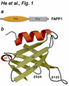
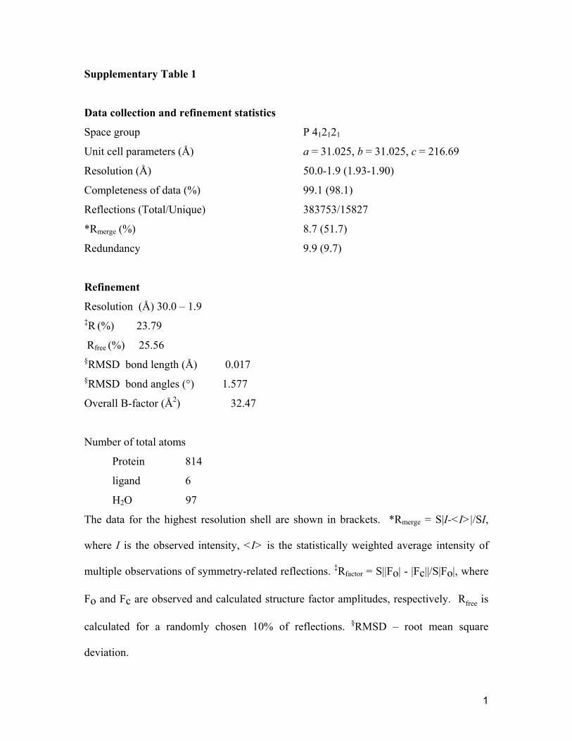
RESULTS AND DISSCUSSION Overall structure of the FAPP1 PH domain To elucidate the molecular basis of the PtdIns(4)P and ARF1 recognition, we obtained the three-dimensional structure of the human FAPP1 PH domain (residues 1-99, C94S mutant, hereafter called FAPP1 PH). The structure was determined at a 1.9 Å resolution by X-ray crystallography (Fig. 1). The PH domain crystallized as a dimer of two identical molecules linked through a C-terminal extension. The single FAPP1 PH domain folds into a seven-stranded β-barrel capped by an α helix at one edge. The opposite edge of the β-barrel is flanked by the β4 and β7 strands and three loops: a long β1-β2 loop, a β3-β4 loop that is partially invisible in the structure and a short β6-β7 loop. An additional one-turn α helix is formed between the β4 and β5 strands. The crystal structure of the FAPP1 PH domain and the solution structure of this protein (PDB 2KCJ) (31) superimposed with an rmsd of 2.1 Å over Cα atoms, with the differences seen in the position of the β5-β6 loop and the β4 and β7 strands that comprise the lipid binding site (see below). The secondary structure elements in FAPP1 PH aligned well with that of a canonical PH module, such as human GRP1 PH (PDB 1FGZ) (32) (rmsd of 1.9 Å over all Cα atoms and rmsd of 1.1 Å over Cα atoms in β-strands/α-helix), indicating that the overall fold of the β-barrel and the main α helix is conserved within the PH domain family. The diffraction data and refinement statistics
for the crystal structure are shown in Supplementary Table 1. PtdIns(4)P-binding site To define the mechanism of lipid binding, we investigated the association of the FAPP1 PH domain with PtdIns(4)P by NMR, SPR and mutagenesis (Fig. 2). The 1H,15N HSQC spectra of uniformly 15N-labeled protein were collected while a water-soluble di-butanoyl (C4) form of PtdIns(4)P was titrated in the NMR sample (Fig. 2a). As C4-PtdIns(4)P was added stepwise, a number of the FAPP1 PH amide resonances underwent changes indicating a direct interaction between the protein and the lipid, which was in agreement with the previous reports (1,26,31). Similar in directions but smaller in the magnitude chemical shift changes in the PH domain spectra were induced by Ins(1,4)P2, an isolated head group of PtdIns(4)P (Fig. 2a, b). These results suggested that both ligands occupy the same binding pocket with the inositol ring being involved in most significant contacts, however presence of the diacylglycerol moiety of the lipid is essential for the strong interaction. We note that the fast exchange regime on the NMR time scale indicated that even the short chain C4-PtdIns(4)P form was bound weakly, in the low mM range. The binding became considerably stronger when a long chain di-palmitoyl (C16)-PtdIns(4)P form embedded in POPC/POPE vesicles was used. The dissociation constant (Kd) for the interaction with C16-PtdIns(4)P, measured by SPR, was found to be 460 ± 80 nM (Fig. 2d, e).
The PtdIns(4)P-binding site was mapped based on chemical shift changes observed in FAPP1 PH upon binding of C4-PtdIns(4)P (Fig. 2b). The largest resonance perturbations occurred in the β4 and β7 strands and the β1-β2 loop of the protein suggesting that the open edge of the β-barrel comprises the binding site for the lipid.
by guest on May 15, 2016
http://ww
w.jbc.org/
Dow
nloaded from
7
Compared to the resonance perturbations previously reported for binding of the C6-form of the lipid (31), C4-PtdIns(4)P induced larger changes in the β1-β2 loop but smaller in the β4 strand. Mapping the changes onto the PH domain surface revealed a well-defined, deep pocket in the barrel’s interior where PtdIns(4)P is bound (Fig. 2c). A high positive potential of the pocket suggested that electrostatic and hydrogen-bonding interactions play an essential role in the recognition of the lipid head group.
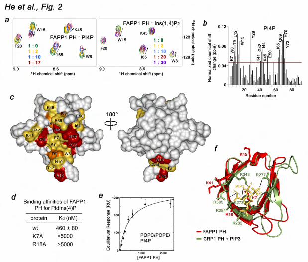
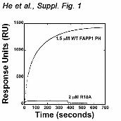
The side chains of the basic amino acids K7, R18, K41 and K45 are well positioned to coordinate the negatively charged phosphate groups of the inositol ring. These residues clearly overlay with the basic residues of the GRP1 PH domain that make major contacts with PtdIns(3,4,5)P3 (32,33) (Fig. 2f). Particularly, residues K273 and K343 form hydrogen bonds to the 4-phosphate group in the GRP1 PH-PtdIns(3,4,5)P3 complex (32,33), and similarly positioned K7 and K45 of the FAPP1 PH domain may coordinate the 4-phosphate group of PtdIns(4)P. To assess the importance of the basic residues in the PtdIns(4)P-binding pocket, we generated the K7A and R18A mutants and tested them by SPR. The data shown in Figure 2d and Supplementary Figure 1 demonstrate that substitution of K7 or R18 abrogates interaction of the FAPP1 PH domain with C16-PtdIns(4)P-containing vesicles, revealing the critical role of the basic residues in lipid binding. Interaction with PtdIns(4)P is pH dependent To gain mechanistic insight into the lipid recognition, we analyzed specificity and examined factors that may influence binding of FAPP1. The specificity of the FAPP1 PH domain was tested by a protein-lipid overlay assay. Nitrocellulose membranes were spotted with 100 pmol of PIs and other
lipids and incubated with GST-fused FAPP1 PH. The bound protein was detected by Western blot analysis using anti-GST antibodies. As shown in Figure 3a, GST-FAPP1 PH recognized PtdIns(4)P but did not bind other lipids, corroborating the finding of the Alessi group (26). Superimposition of the GRP1 and FAPP1 PH domains provide a possible explanation of the preference for PtdIns(4)P (Fig. 2f). The FAPP1 PH domain lacks of the basic residue necessary for coordination of the 5-phosphate group in the GRP1 PH-PtdIns(3,4,5)P3 complex (R277) and therefore cannot accommodate the tris-phosphorylated PI. The fact that R18 and K41 of the FAPP1 PH domain are positioned in the same way as the 3-phosphate-coordinating R284 and R305 residues of GRP1 suggests that the FAPP1 PH domain could bind bis-phosphorylated species, which is also supported by previous observations (31).
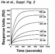
The FAPP1 PH-PtdIns(4)P interaction was facilitated by lowering pH of the media. We performed 1H,15N HSQC titration experiments at pH 6.5 and 7.4 (Fig. 3b). While at pH 7.4 addition of C4-PtdIns(4)P caused small chemical shift changes in the FAPP1 PH domain, substantially larger changes were seen at pH 6.5, implying that binding was stronger under the acidic conditions. The binding affinities measured by SPR revealed that the FAPP1 PH domain binds POPC/POPE/C16-PtdIns(4)P vesicles with a Kd of 200 ± 10 nM at pH 6.5, however it associates 2.3-fold weaker at pH 7.4 (Fig. 3c and Suppl. Fig. 2). The pH dependence is most likely due to the presence of a histidine residue in the binding site, i.e., His70 in the β7 strand that undergoes protonation in an acidic environment increasing a positive net charge of the binding pocket. Alignment of the amino acid sequences of several PH domains reveals that this histidine residue is
by guest on May 15, 2016
http://ww
w.jbc.org/
Dow
nloaded from
8
conserved in a set of proteins, including GRP1. The corresponding His355 of GRP1 forms a hydrogen bond with the 4-phosphate group of PtdIns(3,4,5)P3 (32,33) and mediates pH sensitivity (23).
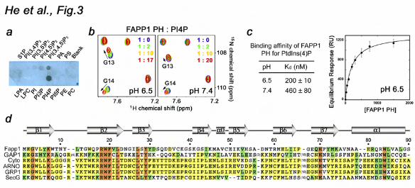
It is well established that FAPP1 localizes exclusively to the TGN membranes where intravesicular pH is 6.4. However it does not localize to the plasma membrane at the cytosolic pH of 7.4, despite the fact that the plasma membrane contains a small pool of PtdIns(4)P. Furthermore, translocation of cytosolic Arf1 to membranes depends on intravesicular acidification (34). Although further studies are required to determine whether the FAPP1 PH domain can sense the pH gradient across the Golgi membrane, it has recently been shown that another PI-binding module, the FYVE domain, displays similar pH-dependent intracellular distribution (19,35). It associates with PtdIns(3)P-enriched membranes of early endosomes that have a low pH lumen but not with other PtdIns(3)P-containing membranes. Additionally, protonation/deprotonation of the His residue in the PI binding site can modulate affinity of the FAPP1 PH domain as cytosolic pH fluctuations occur (36-39). Taken together, our data indicate that the direct interaction of the FAPP1 PH domain with PtdIns(4)P is pH dependent: it is weak at the cytosolic pH of 7.4 but becomes stronger at pH 6.5. PtdIns(4)P binding of the FAPP1 PH domain is required for the membrane insertion and tubulation It has recently been demonstrated that the β1-β2 loop of FAPP1 PH inserts into membrane-mimicking micelles causing tubulation (31). To determine whether PtdIns(4)P binding is necessary for the FAPP1 function, the wild type protein and the K7A, K7E, R18L and K45A mutants impaired in PtdIns(4)P binding were examined in membrane tubulation, pelleting
and monolayer penetration assays (Fig. 4). POPC/POPE (80:20) or POPC/POPE/PI4P (75:20:5) membrane sheets were treated with the lipophilic dye FM 2-10 to obtain high resolution imaging by a confocal microscope. While addition of the FAPP1 PH domain to POPC/POPE sheets did not cause detectable changes in the morphology of the membrane sheets up to 10 min, addition of the protein to PtdIns(4)P-containing membrane sheets rapidly induced tubulation (Fig. 4a). In contrast, when K45A mutant of FAPP1 PH defective in PtdIns(4)P binding was incubated with PtdIns(4)P-containing membrane sheets, the membrane deformation was abrogated completely. The K7A, K7E and R18L mutants of FAPP1 PH were also unable to induce the formation of narrow tubules and pelleting function of these mutants was significantly compromised (Fig. 4c, d). We concluded that interaction with PtdIns(4)P is critical for FAPP1 to induce membrane deformation.
Likewise insertion of the FAPP1 PH domain into POPC/POPE monolayers required the presence of PtdIns(4)P (Fig. 4b). The FAPP1 PH domain exhibited low intrinsic membrane penetrating ability. The POPC/POPE (80:20) monolayer surface pressure (πc) was found to be ~24 mN/m, however incorporation of 5 mol% PtdIns(4)P into the lipid monolayer greatly enhanced penetration, raising the πc value to ~33 mN/m. These results underscored the essential role of the PtdIns(4)P recognition for biological activities of FAPP1 PH. ARF1-bnding site of the FAPP1 PH domain To determine the molecular mechanism of ARF1 recognition, the ARF1-binding site of FAPP1 PH was identified by NMR, SPR, mutagenesis and pulldown assays. 1H,15N TROSY spectra of 15N-labeled FAPP1 PH were recorded as the unlabeled constitutively active GTP-locked ARF1 Q71L mutant lacking the N-terminal
by guest on May 15, 2016
http://ww
w.jbc.org/
Dow
nloaded from
9
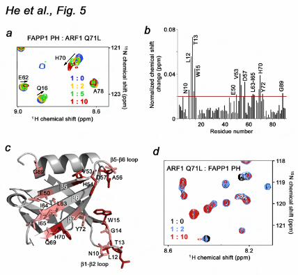
residues 1-17 (ARF1 Q71L) was added stepwise to a 1:10 ratio (Fig. 5a). Chemical shift perturbations and disappearance of the signals upon addition of ARF1 Q71L pointed to a direct interaction between the two proteins. The most pronounced chemical shift changes were observed for residues located in the long β1-β2 loop (N10, L12, T13, G14 and W15), the β5-β6 loop (A56 and D57), the β5, β6 and β7 strands (E50, V53, H54, L63, I64, I65, Q69, H70 and Y72) and the far C-terminus of the α-helix (G89) (Fig. 5b), indicating that these residues are directly or indirectly involved in the interaction. Mapping these residues on the structure of the FAPP1 PH domain revealed an extended site that spreads across the β5-β7 sheet of the β-barrel and is flanked by the β1-β2 and β5-β6 loops.
The direct interaction between ARF1 Q71L and the FAPP1 PH domain was substantiated by a reverse titration of unlabeled FAPP1 PH into the 15N-labeled ARF1 Q71L protein (Fig. 5d). As in the experiments described above, a number of amide cross peaks in 1H,15N TROSY spectra of ARF1 Q71L decreased in intensity and changed their positions upon addition of FAPP1 PH, indicating binding.
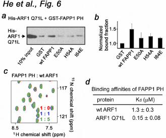
To define the role of the most perturbed residues of the FAPP1 PH domain, the E50A, H54A and I64E mutants were generated and tested by pulldown assays. His-tagged ARF1 Q71L was first incubated with the wild type or mutated GST-FAPP1 PH domain and then with Glutathione Sepharose beads. After collecting the beads by centrifugation, bound ARF1 Q71L was detected by Western blot using antibodies against the His tag. As shown in Figure 6a, b, wild type GST-FAPP1 PH precipitated ARF1 Q71L, whereas the E50A, H54A and I64E mutants of FAPP1 PH lost their ability to associate with ARF1 Q71L. Thus, the FAPP1 residues located in the β5 and β6 strands are
necessary for the interaction.
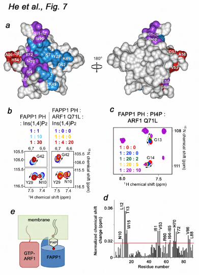
The GTP-bound form of ARF1 is essential Because binding of GTP activates ARF1, we next examined whether the ARF1 conformation plays a role in association with FAPP1 PH. A lack of significant changes in 1H,15N TROSY spectra of the 15N-labeled FAPP1 PH domain upon addition of unlabeled wild type ARF1 indicated that the GTP-free form of ARF1 does not recognize FAPP1 PH (Fig. 6c). To quantitatively characterize the effect of GTP, we measured binding affinity of the FAPP1 PH domain for wild type ARF1 and GTP-locked ARF1 Q71L by SPR. As shown in Figure 6d, FAPP1 PH binds ARF1 Q71L ~9-fold stronger than it binds wild type ARF1. Furthermore, the off-rate for the interaction with wild type ARF1 was ~10-fold faster as compared to the off-rate for the interaction with ARF1 Q71L. These data demonstrate that the GTP-bound form of ARF1 is required for the strong interaction with FAPP1 PH. FAPP1 binds PtdIns(4)P lipid and ARF1 simultaneously and independently We explored a crosstalk between the two ligands using NMR and SPR analyses (Fig. 7). The head group of the lipid, Ins(1,4)P2, was titrated into the FAPP1 PH domain alone or FAPP1 PH pre-bound to ARF1 Q71L (Fig. 7b). Almost identical in the magnitude and directions chemical shift changes were observed in both NMR experiments indicating that the head group of PtdIns(4)P was bound similarly and the formation of the FAPP1-ARF1 Q71L complex did not affect lipid binding. In agreement, when ARF1 Q71L was gradually added to the ligand-free FAPP1 PH domain or FAPP1 PH pre-bound to PtdIns(4)P, a similar pattern of resonance perturbations was seen (Figs. 5b and 7d). These results suggested that the ARF1 Q71L-binding site
by guest on May 15, 2016
http://ww
w.jbc.org/
Dow
nloaded from
10
and the strength of the FAPP1 PH-ARF1 interaction were not altered by PtdIns(4)P binding. Furthermore, the K7A mutant of FAPP1 impaired in lipid binding associated with ARF1 Q71L as strong as the wild type FAPP1 PH domain and exhibited comparable dissociation kinetics. Although the PtdIns(4)P- and ARF1-binding sites are located in close proximity and residues in the β5-β7 sheet and the β1-β2 loop were perturbed upon binding of either ligand, the sites have little overlap as the lipid-binding pocket is positioned within the β-barrel, whereas the ARF1-binding site is located on the external side of the β-barrel (Fig. 7e). Taken together, our findings suggest that FAPP1 PH is able to bind PtdIns(4)P and ARF1 simultaneously and independently.
In conclusion, this study details the molecular mechanism of the dual recognition of PtdIns(4)P and ARF1 by the FAPP1 PH domain and demonstrate that the two ligands can associate with the PH domain independently. Our results indicate that the PtdIns(4)P- and ARF1-binding sites are located on the β-barrel’s internal and external sides and that binding to PtdIns(4)P is enhanced in an acidic environment, whereas interaction with ARF1 requires the GTP-bound conformation of the GTPase. The binary association of the FAPP1 PH domain may increase binding affinity and selectivity toward the TGN membranes enriched in both PtdIns(4)P lipid and GTP-bound ARF1 anchored to membranes through a myristoyl moiety (Fig. 7e).
REFERENCES
1. Godi, A., Di Campli, A., Konstantakopoulos, A., Di Tullio, G., Alessi, D. R., Kular, G. S., Daniele, T., Marra, P., Lucocq, J. M., and De Matteis, M. A. (2004) Nat Cell Biol 6, 393-404
2. D'Angelo, G., Vicinanza, M., Di Campli, A., and De Matteis, M. A. (2008) J Cell Sci 121, 1955-1963
3. DiNitto, J. P., and Lambright, D. G. (2006) Biochim Biophys Acta 1761, 850-867 4. Lemmon, M. A. (2008) Nat Rev Mol Cell Biol 9, 99-111 5. Kutateladze, T. G. (2010) Nat Chem Biol 6, 507-513 6. Jezyk, M. R., Snyder, J. T., Gershberg, S., Worthylake, D. K., Harden, T. K., and
Sondek, J. (2006) Nat Struct Mol Biol 13, 1135-1140 7. Menetrey, J., Perderiset, M., Cicolari, J., Dubois, T., Elkhatib, N., El Khadali, F.,
Franco, M., Chavrier, P., and Houdusse, A. (2007) Embo J 26, 1953-1962 8. Bunney, T. D., Opaleye, O., Roe, S. M., Vatter, P., Baxendale, R. W., Walliser, C.,
Everett, K. L., Josephs, M. B., Christow, C., Rodrigues-Lima, F., Gierschik, P., Pearl, L. H., and Katan, M. (2009) Mol Cell 34, 223-233
9. Snyder, J. T., Singer, A. U., Wing, M. R., Harden, T. K., and Sondek, J. (2003) J Biol Chem 278, 21099-21104
10. Levine, T. P., and Munro, S. (2002) Curr Biol 12, 695-704 11. Cohen, L. A., Honda, A., Varnai, P., Brown, F. D., Balla, T., and Donaldson, J. G.
(2007) Mol Biol Cell 18, 2244-2253 12. Lemmon, M. A. (2004) Biochemical Society transactions 32, 707-711 13. Balla, A., Tuymetova, G., Tsiomenko, A., Varnai, P., and Balla, T. (2005) Mol Biol
Cell 16, 1282-1295
by guest on May 15, 2016
http://ww
w.jbc.org/
Dow
nloaded from
11
14. D'Souza-Schorey, C., and Chavrier, P. (2006) Nat Rev Mol Cell Biol 7, 347-358 15. Kahn, R. A., Cherfils, J., Elias, M., Lovering, R. C., Munro, S., and Schurmann, A.
(2006) J Cell Biol 172, 645-650 16. Gillingham, A. K., and Munro, S. (2007) Annu Rev Cell Dev Biol 23, 579-611 17. Donaldson, J. G. (2008) Biochem J 414, e1-2 18. Liu, Y., Kahn, R. A., and Prestegard, J. H. (2009) Structure 17, 79-87 19. He, J., Vora, M., Haney, R. M., Filonov, G. S., Musselman, C. A., Burd, C. G.,
Kutateladze, A. G., Verkhusha, V. V., Stahelin, R. V., and Kutateladze, T. G. (2009) Proteins 76, 852-860
20. Pflugrath, J. W. (1999) Acta Crystallogr D Biol Crystallogr 55, 1718-1725 21. Emsley, P., and Cowtan, K. (2004) Acta Crystallogr D Biol Crystallogr 60, 2126-
2132 22. Adams, P. D., Grosse-Kunstleve, R. W., Hung, L. W., Ioerger, T. R., McCoy, A. J.,
Moriarty, N. W., Read, R. J., Sacchettini, J. C., Sauter, N. K., and Terwilliger, T. C. (2002) Acta Crystallogr D Biol Crystallogr 58, 1948-1954
23. He, J., Haney, R. M., Vora, M., Verkhusha, V. V., Stahelin, R. V., and Kutateladze, T. G. (2008) J Lipid Res 49, 1807-1815
24. Delaglio, F., Grzesiek, S., Vuister, G. W., Zhu, G., Pfeifer, J., and Bax, A. (1995) J. Biomol. NMR 6, 277-293
25. Vranken, W. F., Boucher, W., Stevens, T. J., Fogh, R. H., Pajon, A., Llinas, M., Ulrich, E. L., Markley, J. L., Ionides, J., and Laue, E. D. (2005) Proteins 59, 687-696
26. Dowler, S., Currie, R. A., Campbell, D. G., Deak, M., Kular, G., Downes, C. P., and Alessi, D. R. (2000) Biochemical Journal 351, 19-31
27. Hom, R. A., Vora, M., Regner, M., Subach, O. M., Cho, W., Verkhusha, V. V., Stahelin, R. V., and Kutateladze, T. G. (2007) J Mol Biol 373, 412-423
28. Cho, W., Bittova, L., and Stahelin, R. V. (2001) Anal Biochem 296, 153-161 29. Cao, X., Coskun, U., Rossle, M., Buschhorn, S. B., Grzybek, M., Dafforn, T. R.,
Lenoir, M., Overduin, M., and Simons, K. (2009) Proc Natl Acad Sci U S A 106, 21121-21125
30. Lee, S. A., Kovacs, J., Stahelin, R. V., Cheever, M. L., Overduin, M., Setty, T. G., Burd, C. G., Cho, W., and Kutateladze, T. G. (2006) J Biol Chem 281, 37091-37101
31. Lenoir, M., Coskun, U., Grzybek, M., Cao, X., Buschhorn, S. B., James, J., Simons, K., and Overduin, M. (2010) EMBO Rep 11, 279-284
32. Lietzke, S. E., Bose, S., Cronin, T., Klarlund, J., Chawla, A., Czech, M. P., and Lambright, D. G. (2000) Mol Cell 6, 385-394
33. Ferguson, K. M., Kavran, J. M., Sankaran, V. G., Fournier, E., Isakoff, S. J., Skolnik, E. Y., and Lemmon, M. A. (2000) Mol Cell 6, 373-384
34. Zeuzem, S., Feick, P., Zimmermann, P., Haase, W., Kahn, R. A., and Schulz, I. (1992) Proc. Nat. Acad. Sci. USA 89, 6619-6623
35. Lee, S. A., Eyeson, R., Cheever, M. L., Geng, J., Verkhusha, V. V., Burd, C., Overduin, M., and Kutateladze, T. G. (2005) Proc Natl Acad Sci U S A 102, 13052-13057
36. Gottlieb, R. A., Giesing, H. A., Zhu, J. Y., Engler, R. L., and Babior, B. M. (1995) Proc Natl Acad Sci U S A 92, 5965-5968
37. Moolenaar, W. H., Tsien, R. Y., van der Saag, P. T., and de Laat, S. W. (1983) Nature 304, 645-648
by guest on May 15, 2016
http://ww
w.jbc.org/
Dow
nloaded from
12
38. Schuldiner, S., and Rozengurt, E. (1982) Proc Natl Acad Sci U S A 79, 7778-7782 39. Thangaraju, M., Sharma, K., Leber, B., Andrews, D. W., Shen, S. H., and Srikant, C.
B. (1999) J Biol Chem 274, 29549-29557
FOOTNOTES
We thank Y. Gedle, M. Lemmon, C. Musselman, E. Odintsova and R. Zhao for discussions and helping with experiments, and R. Prekeris and S. Wakatsuki for providing cDNAs of wild type and ARF1 Q71L. This research was supported by grants from the National Institutes of Health, GM071424 and CA95144 (T.G.K.), Indiana University Biomedical Research and the American Cancer Society (IRG-84-002-22) (R.V.S.) and Cancer Research UK (MO). X-ray crystallographic data were collected at beamline X25 of the NSLS. Financial support for NSLS comes from the Offices of Biological and Environmental Research and of Basic Energy Sciences of the US Department of Energy, and from the National Center for Research Resources of the NIH (P41RR012408). T.G.K. is an Independent NARSAD Investigator. Abbreviations: FAPP1, four-phosphate-adaptor protein 1; PI, phosphoinositide; PtdIns(4)P, phosphatidylinositol 4-phosphate; PH, pleckstrin homology; ARF1, ADP-ribosylation factor 1; NMR, nuclear magnetic resonance; TGN, trans-Golgi network; SPR, surface plasmon resonance; HSQC, heteronuclear single quantum coherence; Ins(1,4)P2, inositol 1,4-bisphosphate; POPC, 1-palmitoyl-2-oleoyl-sn-glycero-3-phosphocholine; POPE, 1-palmitoyl-2-oleoyl-sn-glycero-3-phosphoethanolamine; PtdIns, phosphatidylinositol.
FIGURE LEGENDS
Figure 1. The crystal structure of the PH domain of FAPP1 (C94S) determined at 1.9 Å resolution. (a) Architecture of FAPP1: the amino-terminal PH domain and a proline-rich motif. (b) Ribbon diagram of the FAPP1 PH structure. Figure 2. The PtdIns(4)P-binding site of the FAPP1 PH domain. (a) Superimposed 1H,15N HSQC spectra of 15N-labeled FAPP1 PH collected during titration with C4-PtdIns(4)P (PI4P) or the lipid head group, Ins(1,4)P2. The spectra are color coded according to the concentration of the ligands. (b) The histogram shows normalized chemical shift changes induced in the backbone amides of the PH domain by PtdIns(4)P. (c) Residues that display significant chemical shift change in (b) are labeled on the FAPP1 PH domain surface and colored red, orange and yellow for large, medium and small changes, respectively. (d) Binding affinities of the wild type and mutant FAPP1 PH domain for POPC/POPE/PI4P (75:20:5) vesicles were measured by SPR. (e) Representative binding isotherms generated from saturation response values at respective FAPP1 PH concentrations were used to calculate Kds. (f) Overlay of the β-barrels of the FAPP1 PH domain (red) and the PtdIns(3,4,5)P3-bound GRP1 PH domain (1FGY). PtdIns(3,4,5)P3 is shown as a stick model and colored yellow. Selected hydrogen bonds in the GRP1 complex are depicted as gray lines.
by guest on May 15, 2016
http://ww
w.jbc.org/
Dow
nloaded from
13
Figure 3. PtdIns(4)P binding is pH sensitive. (a) The specificity of FAPP1 PH was assessed by a protein-lipid overlay assay. (b) Superimposed 1H,15N HSQC spectra of 15N-labeled FAPP1 PH collected during gradual addition of C4-PtdIns(4)P at pH 6.5 and 7.4. (c) Binding affinities of the wild type FAPP1 PH domain for POPC/POPE/PI4P (75:20:5) vesicles as measured by SPR. Representative binding isotherm at pH 6.5 used to calculate Kds. (d) Alignment of the PH domain sequences: absolutely, moderately and weakly conserved residues are colored orange, yellow and green, respectively. The secondary structure of FAPP1 PH is shown above the sequences. Figure 4. Binding of the FAPP1 PH domain to PtdIns(4)P is necessary for membrane tubulation, pelleting and insertion. (a) (top panels) POPC/POPE (80:20) and POPC/POPE/PI4P (75:20:5) membrane sheets labeled with FM 2-10 dye. (middle panels) 2.5 mg/ml FAPP1 PH was injected. Images shown after 5 min (left) and 2 min (right) incubation with FAPP1 PH. (bottom panel) POPC/POPE/PI4P membrane sheets after 5 min incubation with 2.5 mg/ml K45A FAPP1 PH. (b) Insertion of the FAPP1 PH domain into a POPC/POPE (80:20) monolayer (open circles) and a POPC/POPE/PtdIns(4)P (75:20:5) monolayer (filled circles). (c) The formation of narrow membrane tubules caused by wt or mutated FAPP1 PH was examined by DIC microscopy. (d) Graphs showing normalized pelleted fraction of wt and mutant FAPP1 PH plotted as a function of the initial protein concentration. Figure 5. The ARF1-binding site of the FAPP1 PH domain. (a) Superimposed 1H,15N TROSY spectra of 15N-labeled FAPP1 PH collected as unlabeled ARF1 Q71L was titrated in. The spectra are color coded according to the concentration of ARF1 Q71L. (b) The histogram shows normalized chemical shift changes induced in the backbone amides of the PH domain by ARF1 Q71L. (c) Residues that display significant chemical shift change in (b) are labeled on the ribbon diagram of FAPP1 PH and colored red and pink for large and medium changes, respectively. (d) Superimposed 1H,15N TROSY spectra of 15N-labeled ARF1 Q71L recorded while unlabeled FAPP1 PH was added stepwise. Figure 6. The GTP-bound conformation of ARF1 is essential. (a, b) Mutations of the ARF1-binding site residues disrupt binding. Pulldown assays and the histogram show His-ARF1 Q71L is precipitated by wild type or mutant GST-FAPP1 PH bound to the Glutathione Sepharose beads. (c) Superimposed 1H,15N TROSY spectra of 15N-labeled FAPP1 PH collected as wild type ARF1 was titrated in. (d) Binding affinities of the FAPP1 PH domain for ARF1 as measured by SPR. Kinetic curves were used to calculate Kd and koff. Figure 7. Molecular mechanism of PtdIns(4)P and ARF1 recognition. (a) The residues of FAPP1 PH most perturbed upon interaction with either PtdIns(4)P, ARF1 Q71L, or both ligands are mapped on the surface of the PH domain and colored blue, red and purple, respectively. (b) Superimposed 1H,15N HSQC spectra of FAPP1 PH (left) and 1H,15N TROSY spectra of FAPP1 PH pre-bound to ARF1 Q71L collected during titration with Ins(1,4)P2. (c) Superimposed 1H,15N TROSY spectra of FAPP1 PH recorded as first C4-PtdIns(4)P and then ARF1 Q71L were titrated in. (d) The histogram shows normalized chemical shift changes induced in the backbone amides of the C4-PtdIns(4)P-bound FAPP1
by guest on May 15, 2016
http://ww
w.jbc.org/
Dow
nloaded from
14
PH domain by ARF1 Q71L. (e) A model of anchoring of FAPP1 PH to the Golgi membranes via the double interaction with PtdIns(4)P lipid and myristoylated GTP-bound ARF1.
by guest on May 15, 2016
http://ww
w.jbc.org/
Dow
nloaded from
He et al., Fig. 1
FAPP1
β1-β2β3-β4
a
b
β7
by guest on May 15, 2016
http://ww
w.jbc.org/
Dow
nloaded from
1
Supplementary Table 1
Data collection and refinement statistics
Space group P 412121
Unit cell parameters (Å) a = 31.025, b = 31.025, c = 216.69
Resolution (Å) 50.0-1.9 (1.93-1.90)
Completeness of data (%) 99.1 (98.1)
Reflections (Total/Unique) 383753/15827
*Rmerge (%) 8.7 (51.7)
Redundancy 9.9 (9.7)
Refinement
Resolution (Å) 30.0 – 1.9 ‡R (%) 23.79
Rfree (%) 25.56
§RMSD bond length (Å) 0.017 §RMSD bond angles (°) 1.577
Overall B-factor (Å2) 32.47
Number of total atoms
Protein 814
ligand 6
H2O 97
The data for the highest resolution shell are shown in brackets. *Rmerge = S|I-<I>|/SI,
where I is the observed intensity, <I> is the statistically weighted average intensity of
multiple observations of symmetry-related reflections. ‡Rfactor = S||Fo| - |Fc||/S|Fo|, where
Fo and Fc are observed and calculated structure factor amplitudes, respectively. Rfree is
calculated for a randomly chosen 10% of reflections. §RMSD – root mean square
deviation.
2
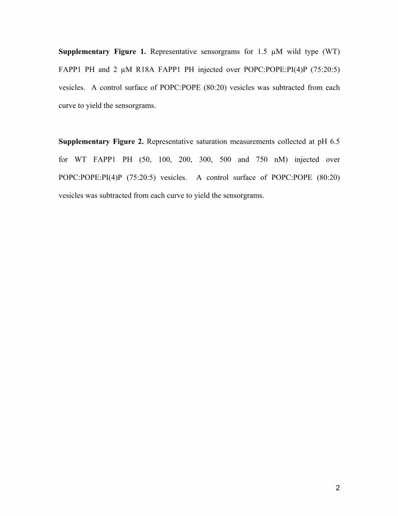
Supplementary Figure 1. Representative sensorgrams for 1.5 µM wild type (WT)
FAPP1 PH and 2 µM R18A FAPP1 PH injected over POPC:POPE:PI(4)P (75:20:5)
vesicles. A control surface of POPC:POPE (80:20) vesicles was subtracted from each
curve to yield the sensorgrams.
Supplementary Figure 2. Representative saturation measurements collected at pH 6.5
for WT FAPP1 PH (50, 100, 200, 300, 500 and 750 nM) injected over
POPC:POPE:PI(4)P (75:20:5) vesicles. A control surface of POPC:POPE (80:20)
vesicles was subtracted from each curve to yield the sensorgrams.
Robert V. Stahelin and Tatiana G. KutateladzeJu He, Jordan L. Scott, Annie Heroux, Siddhartha Roy, Marc Lenoir, Michael Overduin,
FAPP1 PH domainMolecular basis of phosphatidylinositol 4-phosphate and ARF1 recognition by the
published online March 22, 2011J. Biol. Chem.
10.1074/jbc.M111.233015Access the most updated version of this article at doi:
Alerts:
When a correction for this article is posted•
When this article is cited•
to choose from all of JBC's e-mail alertsClick here
Supplemental material:
http://www.jbc.org/content/suppl/2011/03/22/M111.233015.DC1.html
http://www.jbc.org/content/early/2011/03/22/jbc.M111.233015.full.html#ref-list-1
This article cites 0 references, 0 of which can be accessed free at
by guest on May 15, 2016
http://ww
w.jbc.org/
Dow
nloaded from
Copyright © 2022 FDOKUMEN