PKB Binding Proteins
11
Cell, Vol. 111, 293–303, November 1, 2002, Copyright 2002 by Cell Press Review PKB Binding Proteins: Getting in on the Akt to play a modulatory role in PKB regulation. It is the aim of this review to focus on proteins that have been reported to bind or interact with PKB, rather than pro- Derek P. Brazil, 1 Jongsun Park, and Brian A. Hemmings Friedrich Miescher Institute for Biomedical Research teins that serve as substrates. The reader is directed to other comprehensive reviews for additional details of Maulbeerstrasse 66 CH-4058 Basel PKB substrates (Datta et al., 1999; Kandel and Hay, 1999; Lawlor and Alessi, 2001). We will focus on the role Switzerland of nonsubstrate PKB binding proteins in cell function, and limit the discussion to a few examples of PKB sub- strates that have been shown to interact with PKB. We will then touch on future directions for this work and how Protein kinase B (PKB) has emerged as the focal point for many signal transduction pathways, regulating this large volume of data can enhance our knowledge of PKB signaling networks in a physiological setting. multiple cellular processes such as glucose metabo- lism, transcription, apoptosis, cell proliferation, angio- genesis, and cell motility. In addition to acting as a Protein Ligands for the PH Domain of PKB kinase toward many substrates involved in these pro- All three isoforms of PKB (PKB, , ) contain an amino- cesses, PKB forms complexes with other proteins that terminal pleckstrin homology (PH) domain that is highly are not substrates, but rather act as modulators of conserved in all three members. The PH domain of PKB PKB activity and function. In this review, we discuss is crucial for kinase activation, as it binds the membrane the implications of these data in understanding the phospholipid PtdIns-3,4,5-P 3 and mediates recruitment multitude of functions predicted for PKB in cells. to the plasma membrane (see Figure 2). Recent data have suggested that the role of the PH domain in PKB regulation may not be limited to lipid binding, but may Introduction also be used as a protein interaction domain. These Since its discovery just over ten years ago, protein ki- results are discussed below. nase B (PKB, also known as Akt) has been the subject of One protein that interacts with the PH domain of PKB close to 3000 papers currently available in the PubMed is TCL1. Many human T cell malignancies involve trans- database. Many excellent reviews have covered the locations at 14q32.1 or Xq28, chromosomal regions con- molecules and biological processes controlled by and taining genes for a family of proto-oncogenes typified involving PKB (Datta et al., 1999; Galetic et al., 1999; by TCL1 (Virgilio et al., 1994). TCL1 is a 14 kDa protein Kandel and Hay, 1999; Scheid and Woodgett, 2001). whose expression is limited to a specific subset of T The activation of PKB by numerous receptor tyrosine and B cells during development (Virgilio et al., 1994). In kinases controls a range of diverse cellular functions 2000, two research teams identified TCL1 as a binding that are important both physiologically and pathophysi- protein for PKB using a yeast two-hybrid screen (Laine ologically (Brazil and Hemmings, 2001). Recent develop- et al., 2000) or coimmunoprecipitation (Pekarsky et al., ments in the field such as the generation of mice with 2000). Both these groups clearly demonstrated that the targeted deletion of PKB isoforms provide the possibility PH domain of PKB was required for TCL1 binding, which to determine the physiological role of these kinases in increases kinase activity. These data suggest that bind- mammalian systems and also to verify the bona fide ing of TCL1 to PKB may mimic the conformation change nature of the multitude of reported PKB substrates. Ad- induced when PtdIns-3,4,5-P 3 engages the kinase, thus ditionally, the determination of the crystal structure of triggering kinase activation. PKB (Figure 1) has provided essential information into Significant differences exist between these two pa- the mechanism of PKB activation and regulation by pers that are worth noting. Laine et al. report that TCL1 multisite phosphorylation (Yang et al., 2002a, 2002b). can interact with both PKB and PKB in a PH domain- Within the plethora of papers containing data on PKB dependent manner. In contrast, Pekarsky and cowork- are a number of reports detailing the interaction of PKB ers suggest that TCL1 selectively interacts with PKB. with other cellular proteins. By definition, PKB phos- Another divergent conclusion involves the requirement phorylates proteins containing the consensus motif R-x- of PKB kinase activity and phosphorylation for TCL1 R-x-x-S/T-F/L (Alessi et al., 1996). Protein kinases do interaction. Pekarsky et al. state that the interaction of not generally form stable complexes with their sub- TCL1 with PKB is independent of kinase activation or strates, as rapid on/off rates for phosphorylation are phosphorylation status and do not observe increased essential for signaling cascades to function effectively PKB phosphorylation on Ser473. In direct contrast, as “gate keepers” in cells. However, many researchers Laine et al. show that TCL1 binding to PKB increases have demonstrated that PKB can exist in a stable com- the phosphorylation of Thr308 in the kinase domain, and plex with several of its substrate proteins (e.g., Mdm2, also Ser473 in the hydrophobic motif. These data are TSC2, EDG-1) and can also interact with many proteins supported by the author’s demonstration of increased that do not serve as PKB substrates, but rather seem PKB-mediated phosphorylation of GSK3 and BAD, two key substrates of this kinase. These biochemical obser- vations were further extended by this group, which 1 Correspondence: [email protected]
Transcript of PKB Binding Proteins

Cell, Vol. 111, 293–303, November 1, 2002, Copyright 2002 by Cell Press
ReviewPKB Binding Proteins:Getting in on the Akt
to play a modulatory role in PKB regulation. It is theaim of this review to focus on proteins that have beenreported to bind or interact with PKB, rather than pro-
Derek P. Brazil,1 Jongsun Park,and Brian A. HemmingsFriedrich Miescher Institute for
Biomedical Research teins that serve as substrates. The reader is directed toother comprehensive reviews for additional details ofMaulbeerstrasse 66
CH-4058 Basel PKB substrates (Datta et al., 1999; Kandel and Hay,1999; Lawlor and Alessi, 2001). We will focus on the roleSwitzerlandof nonsubstrate PKB binding proteins in cell function,and limit the discussion to a few examples of PKB sub-strates that have been shown to interact with PKB. Wewill then touch on future directions for this work and howProtein kinase B (PKB) has emerged as the focal point
for many signal transduction pathways, regulating this large volume of data can enhance our knowledge ofPKB signaling networks in a physiological setting.multiple cellular processes such as glucose metabo-
lism, transcription, apoptosis, cell proliferation, angio-genesis, and cell motility. In addition to acting as a Protein Ligands for the PH Domain of PKBkinase toward many substrates involved in these pro- All three isoforms of PKB (PKB�, �, �) contain an amino-cesses, PKB forms complexes with other proteins that terminal pleckstrin homology (PH) domain that is highlyare not substrates, but rather act as modulators of conserved in all three members. The PH domain of PKBPKB activity and function. In this review, we discuss is crucial for kinase activation, as it binds the membranethe implications of these data in understanding the phospholipid PtdIns-3,4,5-P3 and mediates recruitmentmultitude of functions predicted for PKB in cells. to the plasma membrane (see Figure 2). Recent data
have suggested that the role of the PH domain in PKBregulation may not be limited to lipid binding, but may
Introduction also be used as a protein interaction domain. TheseSince its discovery just over ten years ago, protein ki- results are discussed below.nase B (PKB, also known as Akt) has been the subject of One protein that interacts with the PH domain of PKBclose to 3000 papers currently available in the PubMed is TCL1. Many human T cell malignancies involve trans-database. Many excellent reviews have covered the locations at 14q32.1 or Xq28, chromosomal regions con-molecules and biological processes controlled by and taining genes for a family of proto-oncogenes typifiedinvolving PKB (Datta et al., 1999; Galetic et al., 1999; by TCL1 (Virgilio et al., 1994). TCL1 is a 14 kDa proteinKandel and Hay, 1999; Scheid and Woodgett, 2001). whose expression is limited to a specific subset of TThe activation of PKB by numerous receptor tyrosine and B cells during development (Virgilio et al., 1994). Inkinases controls a range of diverse cellular functions 2000, two research teams identified TCL1 as a bindingthat are important both physiologically and pathophysi- protein for PKB using a yeast two-hybrid screen (Laineologically (Brazil and Hemmings, 2001). Recent develop- et al., 2000) or coimmunoprecipitation (Pekarsky et al.,ments in the field such as the generation of mice with 2000). Both these groups clearly demonstrated that thetargeted deletion of PKB isoforms provide the possibility PH domain of PKB was required for TCL1 binding, whichto determine the physiological role of these kinases in increases kinase activity. These data suggest that bind-mammalian systems and also to verify the bona fide ing of TCL1 to PKB may mimic the conformation changenature of the multitude of reported PKB substrates. Ad- induced when PtdIns-3,4,5-P3 engages the kinase, thusditionally, the determination of the crystal structure of triggering kinase activation.PKB (Figure 1) has provided essential information into Significant differences exist between these two pa-the mechanism of PKB activation and regulation by pers that are worth noting. Laine et al. report that TCL1multisite phosphorylation (Yang et al., 2002a, 2002b). can interact with both PKB� and PKB� in a PH domain-Within the plethora of papers containing data on PKB dependent manner. In contrast, Pekarsky and cowork-are a number of reports detailing the interaction of PKB ers suggest that TCL1 selectively interacts with PKB�.with other cellular proteins. By definition, PKB phos- Another divergent conclusion involves the requirementphorylates proteins containing the consensus motif R-x- of PKB kinase activity and phosphorylation for TCL1R-x-x-S/T-F/L (Alessi et al., 1996). Protein kinases do interaction. Pekarsky et al. state that the interaction ofnot generally form stable complexes with their sub- TCL1 with PKB is independent of kinase activation orstrates, as rapid on/off rates for phosphorylation are phosphorylation status and do not observe increasedessential for signaling cascades to function effectively PKB phosphorylation on Ser473. In direct contrast,as “gate keepers” in cells. However, many researchers Laine et al. show that TCL1 binding to PKB increaseshave demonstrated that PKB can exist in a stable com- the phosphorylation of Thr308 in the kinase domain, andplex with several of its substrate proteins (e.g., Mdm2, also Ser473 in the hydrophobic motif. These data areTSC2, EDG-1) and can also interact with many proteins supported by the author’s demonstration of increasedthat do not serve as PKB substrates, but rather seem PKB-mediated phosphorylation of GSK3� and BAD, two
key substrates of this kinase. These biochemical obser-vations were further extended by this group, which1Correspondence: [email protected]

Cell294
Figure 1. Crystal Structure of PKB and Sites of Interaction of Partner Proteins
The PH domain of PKB� (reproduced with kind permission from Thomas et al. [2002]) and catalytic and regulatory domain of PKB� (reproducedfrom Yang et al. [2002a]) are shown. The regions that are important for the interaction of several proteins are indicated. Proteins requiringthe complete PH domain (TCL1 and IMPDH) are also listed.
showed that cellular responses regulated by PKB activ- been used to explain the difficulty in identifying the so-called PDK2 or Ser473 kinase. The recently solved crys-ity such as cell proliferation and cell survival were en-
hanced by TCL1. Overall, the more comprehensive char- tal structure of PKB� demonstrates the unique natureof PKB activation by multisite phosphorylation andacterization of TCL1 binding to PKB performed by Laine
et al. suggests that the effect of this binding is to in- strongly argues that autophosphorylation cannot be thephysiological mechanism of Ser473 phosphorylationcrease the activity of PKB.
An interesting consequence of TCL1 binding appears (Yang et al., 2002a). Recent data from our laboratoryhas identified a kinase activity that can phosphorylateto be the oligomerization of PKB. Laine et al. showed
that TCL1 forms trimers in cells and suggested that this PKB on Ser473, independent of PDK1 and PKB activity(Hill et al., 2002). Since the expression of TCL1 and itstrimeric complex can bind to three molecules of PKB.
These data were further extended by a second report relatives is limited to the lymphoid tissue, this suggeststhat PKB activity may be primed specifically in thesefrom this group, which showed that TCL1 could induce
heterooligomerization of PKB� and PKB� and sug- cells, based on the ability of TCL1 to boost its activation.As Laine et al. point out, however, the presence of an-gested that this facilitated transphosphorylation of PKB
molecules (Laine et al., 2002). Of note, this paper also other PH domain-containing kinase, e.g., PDK1 orSer473 kinase, in the signaling complex that may bedemonstrated a degree of specificity of the TCL1 family
in its binding to PKB isoforms. Both PKB� and PKB� responsible for increasing PKB phosphorylation and ac-tivation cannot be ruled out. Experiments utilizing T andcould interact with all three TCL1 family members,
whereas PKB� specifically interacted with TCL1. Recent B cells lacking TCL1 proteins will be essential in de-termining the physiological role of TCL1 in PKB acti-data have identified key amino acids in TCL1 that are
involved in PKB binding (French et al., 2002; Kunstle et vation.In addition to TCL1 family members, the PH domainal., 2002). The model proposed by these authors in-
volved transphosphorylation of PKB molecules on of PKB has been reported to bind to other proteins.Interaction of inosine-5� monophosphate dehydroge-Ser473 in the hydrophobic motif. Autophosphorylation
on this site has been suggested as the physiological nase (IMPDH) with the PH domain of PKB� led to anincrease in IMPDH enzymatic activity (Ingley and Hem-mechanism of Ser473 phosphorylation (Toker and New-
ton, 2000), but this topic remains controversial and has mings, 2000). Tanaka et al. showed that myosin II could

Review295
Figure 2. Mechanism of PKB Activation
Growth factor-mediated activation of PI3Kleads to the generation of PtdIns-3,4,5-P3,which recruits inactive PKB from the cytosolto the plasma membrane via PH domain bind-ing. Residue Thr308 in the catalytic site isthen phosphorylated by PDK1, and Ser473phosphorylation by an as yet unidentifiedSer473 kinase. The crystal structure of PKBdemonstrates the requirement for Ser473phosphorylation, as this induces an order-to-disorder transition in PKB, stabilizing the �Chelix and allowing ATP binding (Yang et al.,2002a). Activation of PKB is inhibited by theaction of PTEN, a lipid phosphatase, andCTMP, which inhibits phosphorylation onSer473. Activated PKB can then phosphory-late substrates at the plasma membrane, inthe cytosol, and in the nucleus. The kinase isdeactivated by phosphatases such as PP2A(not pictured).
also bind to the PH domain of PKB and that this binding Heat shock protein 90 (Hsp90) was originally identifiedin a kinase complex with v-Src and a cochaperone calledcould be competed with increasing concentrations of
PtdIns-4,5-P2 (Tanaka et al., 1999). The physiological Cdc37 (reviewed in Hunter and Poon, 1997). Two groupspreviously demonstrated that PKB could form com-relevance of IMPDH and myosin II binding to PKB re-
mains to be established. However, the ability of PtdIns- plexes with Hsp27 and 90 (Konishi et al., 1997; Sato etal., 2000). Sato and colleagues identified amino acids4,5-P2 to compete with myosin II binding to the PH do-
main raises an interesting dilemma for the field: what 229–309 of PKB as a crucial region for complex forma-tion with Hsp90. The crystal structure of PKB revealseffect does protein binding to the PH domain of PKB
have on lipid binding to the same domain? Activation that this region corresponds to �-D and �-E helices ofthe C lobe and � sheets 6–8 exactly proximal to theof PI3K via growth factor stimulation leads to the genera-
tion of PtdIns-3,4,5-P3, which is essential for PKB activa- Thr308 residue (Yang et al., 2002a). Disruption of theinteraction of endogenous PKB with Hsp90 using a trun-tion, as translocation of PKB to the plasma membrane
is dependent on this lipid. Furthermore, membrane cated mutant form of PKB (amino acids 1–309) de-creased both kinase activation and phosphorylation andtranslocation of PDK1, the kinase responsible for phos-
phorylation of PKB on Thr308, is also PtdIns-3,4,5-P3 ultimately led to cellular apoptosis as measured byPARP cleavage (Sato et al., 2000). These authors con-dependent. Indeed, the ligands for most PH domains
studied to date are membrane phospholipids, although cluded that inhibition of the PKB-Hsp90 complex forma-tion promotes PP2A-mediated dephosphorylation ofother PH domain proteins such as �-adrenergic receptor
kinase (�ARK) can also bind protein ligands (G protein PKB and kinase inactivation, without a major change inthe level of PKB protein, and suggested that heat shock-�� subunits) as well as lipids (Touhara et al., 1994). We
currently favor the model of “mutually exclusive” lipid induced PKB activation requires Hsp90.Basso et al. used ansamycin antibiotics (Hsp90 inhibi-and protein binding to the PH domain of PKB. A more
likely scenario is that after initial membrane transloca- tors) to disrupt the PKB-Hsp90 complex. This group hadpreviously shown that ansamycins induced an inhibitiontion and activation via PtdIns-3,4,5-P3 binding to its PH
domain, PKB dissociates from the plasma membrane, of PKB activation and cyclin D expression in breastcancer cells overexpressing HER2 (Basso et al., 2002a,thus freeing its PH domain to interact with protein li-
gands such as TCL1. The recent elegant description of 2002b). Similar to results obtained by Sato et al., treat-ment of cells with geldanamycin induced a dephosphor-the crystal structure of the PH domain of PKB� com-
plexed to PtdIns-3,4,5-P3 should improve our ability to ylation and inactivation of PKB that was especially evi-dent in SKBr-3 breast cancer cells. In addition, theseaddress the question of lipid versus protein binding to
this module (Thomas et al., 2002). authors observed a proteosome-mediated decrease inPKB protein levels and showed that coincubation ofcells with ansamycins and a proteosome inhibitor pre-Regulation of PKB by Heat Shock Proteins
Heat shock proteins are molecular chaperones that are vented PKB degradation, which facilitated an apparentsubcellular relocalization of PKB to the perinuclear vesi-involved in protecting proteins against degradation
when cells are exposed to environmental stresses cles (Basso et al., 2002a, 2002b). Importantly, the inter-action of PKB with Hsp90 was suggested to be indirect(Parsell and Lindquist, 1993). Heat shock has been re-
ported to increase the activation of PKB (Shaw et al., and dependent on the presence of the Cdc37 cochaper-one. PKB present in protein complexes containing1998; Konishi et al., 1999), which was maximal at 10 min
post heat shock, and was sensitive to PI3K inhibition Hsp90 and Cdc37 was catalytically active and phos-phorylated GSK-3� in vitro. Hsp90 complex formation(Shaw et al., 1998). Recent publications have sought to
decipher the mechanism of heat shock-induced PKB with PKB may therefore facilitate kinase activation bypreventing both dephosphorylation and PKB degrada-activation.
Cell296
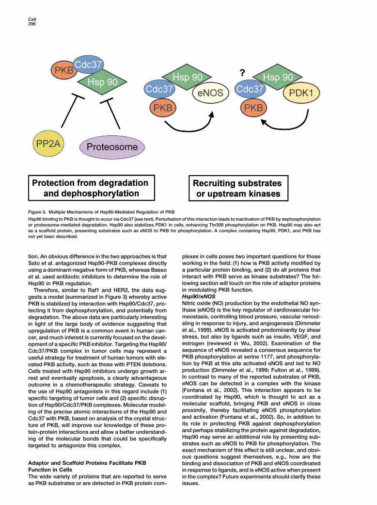
Figure 3. Multiple Mechanisms of Hsp90-Mediated Regulation of PKB
Hsp90 binding to PKB is thought to occur via Cdc37 (see text). Perturbation of this interaction leads to inactivation of PKB by dephosphorylationor proteosome-mediated degradation. Hsp90 also stabilizes PDK1 in cells, enhancing Thr308 phosphorylation on PKB. Hsp90 may also actas a scaffold protein, presenting substrates such as eNOS to PKB for phosphorylation. A complex containing Hsp90, PDK1, and PKB hasnot yet been described.
tion. An obvious difference in the two approaches is that plexes in cells poses two important questions for thoseworking in the field: (1) how is PKB activity modified bySato et al. antagonized Hsp90-PKB complexes directlya particular protein binding, and (2) do all proteins thatusing a dominant-negative form of PKB, whereas Bassointeract with PKB serve as kinase substrates? The fol-et al. used antibiotic inhibitors to determine the role oflowing section will touch on the role of adaptor proteinsHsp90 in PKB regulation.in modulating PKB function.Therefore, similar to Raf1 and HER2, the data sug-Hsp90/eNOSgests a model (summarized in Figure 3) whereby activeNitric oxide (NO) production by the endothelial NO syn-PKB is stabilized by interaction with Hsp90/Cdc37, pro-thase (eNOS) is the key regulator of cardiovascular ho-tecting it from dephosphorylation, and potentially frommeostasis, controlling blood pressure, vascular remod-degradation. The above data are particularly interestingeling in response to injury, and angiogenesis (Dimmelerin light of the large body of evidence suggesting thatet al., 1999). eNOS is activated predominantly by shearupregulation of PKB is a common event in human can-stress, but also by ligands such as insulin, VEGF, andcer, and much interest is currently focused on the devel-estrogen (reviewed in Wu, 2002). Examination of theopment of a specific PKB inhibitor. Targeting the Hsp90/sequence of eNOS revealed a consensus sequence forCdc37/PKB complex in tumor cells may represent aPKB phosphorylation at serine 1177, and phosphoryla-useful strategy for treatment of human tumors with ele-tion by PKB at this site activated eNOS and led to NOvated PKB activity, such as those with PTEN deletions.production (Dimmeler et al., 1999; Fulton et al., 1999).Cells treated with Hsp90 inhibitors undergo growth ar-In contrast to many of the reported substrates of PKB,rest and eventually apoptosis, a clearly advantageouseNOS can be detected in a complex with the kinaseoutcome in a chemotherapeutic strategy. Caveats to(Fontana et al., 2002). This interaction appears to bethe use of Hsp90 antagonists in this regard include (1)coordinated by Hsp90, which is thought to act as aspecific targeting of tumor cells and (2) specific disrup-molecular scaffold, bringing PKB and eNOS in closetion of Hsp90/Cdc37/PKB complexes. Molecular model-proximity, thereby facilitating eNOS phosphorylationing of the precise atomic interactions of the Hsp90 andand activation (Fontana et al., 2002). So, in addition toCdc37 with PKB, based on analysis of the crystal struc-its role in protecting PKB against dephosphorylationture of PKB, will improve our knowledge of these pro-and perhaps stabilizing the protein against degradation,tein-protein interactions and allow a better understand-Hsp90 may serve an additional role by presenting sub-ing of the molecular bonds that could be specificallystrates such as eNOS to PKB for phosphorylation. Thetargeted to antagonize this complex.exact mechanism of this effect is still unclear, and obvi-ous questions suggest themselves, e.g., how are the
Adaptor and Scaffold Proteins Facilitate PKB binding and dissociation of PKB and eNOS coordinatedFunction in Cells in response to ligands, and is eNOS active when presentThe wide variety of proteins that are reported to serve in the complex? Future experiments should clarify these
issues.as PKB substrates or are detected in PKB protein com-

Review297
Axin/GSK-3� to axin-conductin is reported to participate in Wnt sig-naling (Yuan et al., 1999).The activity of glycogen synthase kinase 3� (GSK-3�)
How can these two sets of conclusions be consoli-is regulated by diverse ligands such as insulin and Wnts,dated? One explanation proposed by Fukumoto et al.controlling cellular processes such as glucose metabo-is that prolonged exposure of cells to Wnt is required forlism and development (Weston and Davis, 2001). De-the recruitment of PKB to the GSK-3�/Axin/Dishevelledfects in GSK-3� signaling are implicated in diabetes,complex, and suggested that the role of PKB is to sus-developmental disorders, and cancer. GSK-3� was thetain rather than initiate the signaling process in responsefirst PKB substrate identified (Cross et al., 1995), andto Wnt (Fukumoto et al., 2001). An alternative explana-insulin-stimulated phosphorylation of GSK-3� by PKBtion provided by the recently solved crystal structure ofinactivates the kinase, leading to increased glycogenGSK-3� provides a significant insight into this problemstorage in muscle. Many groups have reported that insu-(Weston and Davis, 2001). For substrates to be phos-lin-mediated GSK-3� inactivation causes increased gly-phorylated by GSK-3�, a residue at position P � 4 mustcogen synthase activity without a concomitant changefirst be phosphorylated in a “priming” mechanism. Phos-in �-catenin levels. In contrast, inactivation of GSK-3�phorylation of GSK-3� on Ser9 by PKB allows the aminoby the Wnt pathway is reported to be independent ofterminus of GSK-3� to act as a pseudosubstrate, occu-Ser9 phosphorylation and leads to the stabilization ofpying the same site used by primed substrates, thus�-catenin levels in cells, without changing the activitypreventing kinase activation (Dajani et al., 2001; Frameof glycogen synthase. Therefore, a major question re-et al., 2001; ter Haar et al., 2001). The reported inabilitygarding GSK-3� signaling is: how are the insulin andof Wnt to phosphorylate Ser9 on GSK-3� suggests thatWnt signaling pathways leading to GSK-3� inhibitionthis site is protected when GSK-3� is complexed withinsulated from each other (Dajani et al., 2001)? The fol-other proteins such as axin, but protein complexes con-lowing section will discuss the data both for and againsttaining Dishevelled may perturb the overall structure ofthis “insulated signaling” hypothesis.GSK3�, thus reexposing the Ser9 site for phosphoryla-The “insulated” nature of GSK-3� signaling events intion by PKB. This provocative hypothesis still needs toresponse to different ligands was challenged by databe tested experimentally.from Fukumoto et al. who, in contrast to other groupsCarboxy-Terminal Modulator Protein (CTMP)(see below), reported that PKB played a role in the WntCTMP is a 27 kDa protein that binds to the carboxy-signaling pathway, and have shown that expression ofterminal end of PKB�, encompassing the hydrophobicWnt or Dishevelled increased PKB kinase activity inmotif and Ser473 residue. The protein was identified inPC12 cells (Fukumoto et al., 2001). These authors alsoa yeast two-hybrid screen and interacts with PKB� indemonstrated that endogenous PKB is recruited into avitro and in vivo (Maira et al., 2001). Colocalization ofcomplex with axin, Dishevelled, and GSK-3� when cellsCTMP and PKB� was observed at the plasma membraneare stimulated with Wnt, with a concomitant increase inof serum-starved cells, and an endogenous complex ofPKB activity toward GSK3-�. Finally, dominant-negativethese two proteins was observed in the same cell frac-PKB reversed Wnt-induced changes in neuronal celltion. CTMP binding inhibits phosphorylation of PKB�morphology. Fukumoto and colleagues suggest that theon Ser473, and to a lesser extent on Thr308, with aconcerted activation of PKB and Dishevelled down-corresponding decrease in in vitro PKB kinase activitystream of Wnt is required for �-catenin stabilization, andand phosphorylation of downstream effectors such aspropose the hypothesis that the inability of insulin toGSK-3� (Maira et al., 2001). Stable expression of CTMPmediate stabilization of �-catenin is due to its inabilityin AKT8 cells (CCL-64 mink lung epithelial cells trans-to activate Dishevelled in this complex.formed with v-Akt) induced phenotypic regression ofThese data appear at odds with several reports thatthese cells to that of the wild-type. AKT8 cells express-have also explored downstream GSK-3� signaling ining CTMP were also compromised in their ability to formresponse to insulin and Wnt. Using a range of differenttumors in nude mice.cell lines (HEK293, C57MG mouse mammary gland epi-
CTMP therefore seems to represent a new class ofthelial cells, CH0-IR, and NIH3T3 cells), these groupsproteins that can interact with PKB� at the plasma mem-
failed to observe phosphorylation of GSK-3� on Ser9 inbrane and maintain the kinase in an inactive state. CTMP
response to Wnt and concluded that the decreases in dissociation from PKB may be provoked by phosphory-GSK-3� induced by insulin and Wnt occur via distinct lation, by an as yet unidentified kinase, to allow a poten-downstream events (Yuan et al., 1999). Initially reports tial Ser473 kinase to access the hydrophobic motif anddemonstrated that activated PKB phosphorylated and thus facilitate PKB� activation.inhibited GSK-3� but did not induce accumulation of JNK Interacting Protein-1 (JIP1)cytosolic �-catenin or activate LEF-1-mediated tran- The role of PKB as a mediator of cell survival and apo-scription (Yuan et al., 1999). These data were supported ptosis has been well described. Downstream targets ofby two reports from the group of McCormick, who PKB such as BAD, Forkhead family members, and IKK�showed that decreases in GSK-3� activity induced by are reported to mediate this antiapoptotic affect. A re-insulin and Wnt were distinct cellular events, and sug- cent report from Kim et al. has uncovered a novel mech-gested a role for PKCs in the Wnt signaling response anism for PKB-mediated protection from apoptosis in(Chen et al., 2000; Ding et al., 2000). These authors neurons exposed to excitotoxins (Kim et al., 2002).suggest that the specificity in signaling could be These data highlight the importance of protein-proteinachieved by GSK-3� forming unique protein complexes interactions for PKB function and further cement thein response to insulin and Wnt. GSK-3� has been shown role of PKB as a crucial protagonist of cell survival into form complexes with axin-conductin, APC, and Di- response to environmental stresses.
The JNK pathway is coordinated by the scaffold pro-shevelled. Of note, only the pool of GSK-3� complexed

Cell298
tein JIP1 (Whitmarsh et al., 2001). Treatment of neurons targeting of PKB to the plasma membrane induceswith kainate, an excitotoxin, increases the activity of the constitutive activation of the kinase (Andjelkovic et al.,JNK pathway and induces apoptosis both in vitro and 1997; Barthel et al., 1997). Therefore, these data suggestin vivo (Kim et al., 2002). PKB� and JIP1 colocalize in a that Grb10 serves as a coactivator of PKB downstreamneuronal plasma membrane compartment. This com- of PI3K.plex was disrupted by kainate treatment, without a par-allel change in PKB phosphorylation, but was accompa- PKB Complex Formation with Other Protein Kinasesnied by an increase in JNK-JIP1 complex formation (Kim So far, the PKB binding proteins discussed in this reviewet al., 2002). Binding of JIP1 appeared to be direct and mainly serve a modulatory role in terms of regulatinginvolved the PH domain of PKB�. Interestingly, this inter- PKB function. The canonical pathway for PKB activationaction was selective for PKB� over PKB�. The authors involves upstream kinases such as PI3K and PDK1, par-then elegantly demonstrated that expression of PKB� allel effectors such as p70S6K and PKC�, and down-inhibits the ability of JIP1 to potentiate JNK1 activation stream target kinases such as GSK-3� and IKK� (Dattain cells, and that this inhibition was independent of PKB et al., 1999). Of note, a complex of PDK1 and PKB haskinase activity. The mechanism of this effect is sug- recently been demonstrated (Filippa et al., 2000; Conusgested to be an inhibition of JIP1’s ability to assemble et al., 2002).the signaling scaffold (involving JNK, MKK7, and MLK3) Early PKB cloning papers from the group of Kikkawarequired for JNK activation and induction of apoptosis. reported that all three isoforms of PKB could interactIn agreement with this hypothesis, neurons from mice with PKC� and that these interactions were mediatedlacking PKB� were more susceptible to kainate-induced by the PH domains of PKB (Konishi et al., 1994a, 1994b,apoptosis (Kim et al., 2002). 1995). Investigations into the role of keratin K10 as a
These data suggest a new and fascinating role for negative modulator of keratinocyte cell cycle prolifera-PKB in preventing neuronal apoptosis. Rather than tion showed that overexpression of keratin K10 inducedphosphorylation of downstream targets such as BAD cell cycle arrest, and loss of this protein was observedand FKHRL1, PKB may act as a “steric hindrance” to during tumor development (Paramio et al., 2001). Rolesprevent assembly of the JNK signaling cassette around for PKB and PKC� in this effect were delineated bythe scaffold JIP1. This effect involves the PH domain of Paramio and colleagues, who showed that cell cyclePKB and raises the question why a protein kinase is arrest induced by keratin K10 could be reversed by ex-required for a cellular function where the kinase activity pression of PKB or PKC�. Keratin K10 colocalized PKBis surplus to requirements. Also, given the high degree and PKC� to the keratin cytoskeleton and maintainedof identity between the PH domains of PKB� and PKB�, these kinases in an inactive state. These data suggestit is perhaps surprising that JIP1 selectively interacts that in keratinocytes, both PKB and PKC� are importantwith PKB�, suggesting that a small number of PH do- mediators of cell proliferation, and sequestration ofmain amino acids may mediate this selectivity. Clearly, these kinases by keratin K10 inhibits their activity andprotein-protein interactions of PKB may be a novel induces cell cycle arrest. Thus, a multikinase complexmechanism of apoptosis regulation by this kinase.
containing PKB, PKC�, and possibly other kinases mayGrb10
be nucleated by keratin K10 in a specific subcellularThe Grb10 family of proteins are adaptor molecules that
compartment. The exact function of this complex re-transduce signals from receptors such as EGFR, Ret,
mains to be determined.and IGF-R (Jahn et al., 2002). Grb10 interacts with aExtracellular signals that trigger PKB can also activatenumber of protein kinases, including MEK1, Src, and
pathways that exert opposing effects on the cell. Formitochondrial-associated Raf. Jahn et al. identifiedexample, the PI3K/PKB pathway in muscle cells exertsGrb10 as a component of the c-kit receptor signalingan opposing effect to that of the Raf-MEK-ERK pathwaycascade, and showed that Grb10 was phosphorylated(Rommel et al., 1999). Two groups have explored thison Ser/Thr residues in a wortmannin-sensitive mannerphenomenon and have shown that PKB and Raf associ-when the c-kit receptor is activated. This interactionate in C2C12 myotubes. Furthermore, it has been dem-between c-kit and Grb10 is dependent on both tyrosineonstrated that phosphorylation of Raf on Ser259 by PKBphosphorylation of the receptor and the SH2 domain ofinhibits Raf activity, facilitating 14-3-3 binding (MuslinGrb10. Interestingly, endogenous PKB� forms a com-et al., 1996; Rommel et al., 1999; Zimmermann and Moel-plex with Grb10, and complex formation is dependentling, 1999). Therefore, interaction of PKB with Raf repre-on neither the PH domain nor the SH2 domain of Grb10,sents a means of crosstalk between the PI3K and ERKsuggesting that this interaction is not direct. This associ-pathways in cells. The interaction between PKB and Rafation is not perturbed by PI3K inhibition, and overex-only occurs in differentiated myotubes and not in thepression of Grb10 in the presence of activated c-kitmyoblast precursors (Rommel et al., 1999). PKB-medi-induced PKB activity. Interestingly, while neither the PHated inhibition of Raf may only occur in specific celldomain nor the SH2 domain of Grb10 was required fortypes and may also be dependent on the stage of cellbinding to PKB, both were required for the activation ofdifferentiation. Another possibility is that adaptor pro-the kinase. The SH2 domain was clearly required toteins specific to different cells or to a particular stageengage the phospho-tyrosine residue of the c-kit recep-of cell differentiation are required for this interaction.tor, and Jahn and colleagues suggest that the PH do-Hsp90 and Grb10 have been reported to bind both PKBmain mutant of Grb10 may act in a “dominant-negative”-and Raf and would therefore represent two candidateslike manner, preventing the release of PKB from thethat could mediate the interaction of these two kinases.complex and thereby preventing full activation. Work
from many groups, including our own, has shown that Recent data from Kane and colleagues have identified

Review299
the oncogenic serine/threonine kinase Cot1 (also known p70S6K (reviewed in McManus and Alessi, 2002). Muchdiscussion in the insulin-signaling field has existed as toas Tpl2) as both a PKB binding protein and a PKB sub-
strate (Kane et al., 2002). PKB and Cot1 are detectable how PKB controls p70S6K activation. The breakthroughcame when the group of Cantley showed that TSC2in a protein complex in Jurkat T cells, and cotransfection
of these kinases increases Cot1-mediated transcription served as a substrate for PKB and that this phosphoryla-tion decreases the ability of TSC2 to inhibit p70S6Kin Jurkat T cells. PKB phosphorylates Cot1 on two sites,
Ser400 and Ser413, and mutation of these sites does (Manning et al., 2002). Several reports then followed,showing that PKB could phosphorylate TSC2 in vitronot affect the interaction of these proteins, but does
affect the ability of Cot1 to activate NF�B-dependent and in vivo (listed in McManus and Alessi, 2002). Theconsequence of TSC2 phosphorylation appears to be atranscription. The consequence of PKB-mediated phos-
phorylation of Cot1 may also involve the recruitment of disruption of the TSC1/2 complex, and some groupshave further reported a ubiquitination-mediated degra-Cot1 substrates or modifiers of Cot1 function.dation of TSC2 when not complexed to TSC1 (Inoki etal., 2002). Therefore, PKB-mediated phosphorylation ofPKB Targeting to GLUT4 VesiclesTSC2 may relieve the TSC1/2-mediated inhibition ofActivation of PKB is a key component of the insulinp70S6K and thus facilitate activation of this kinase (Fig-response, mediating many different aspects of glucoseure 2; McManus and Alessi, 2002).metabolism. Different PKB isoforms appear to play dif-
Of specific interest to this review, Dan et al. reportferent roles in the response of cellular responses tothat PKB� can associate with the TSC1/2 complex (Daninsulin, with PKB� predominantly involved in this meta-et al., 2002). This association promotes the phosphoryla-bolic pathway. Initial clues to the differing roles of PKBtion of TSC2 by PKB, with a concomitant reduction inisoforms came from Calera et al., who showed that PKB�the levels of p27KIP and increased cell proliferation.had a much wider subcellular distribution than PKB�These authors suggest that PKB binding to the TSC1/2(Calera et al., 1998). This group then clearly demon-complex is mediated by TSC2, although the relevantstrated insulin-stimulated recruitment of PKB� todomains on either protein were not identified. DespiteGLUT4-containing vesicles, an observation supportedclear evidence that TSC2 serves as a PKB substrate,by numerous subsequent publications (for example, Ku-some confusion exists as to the exact consequencespriyanova and Kandror, 1999). Expression of PKB� isof TSC2 phosphorylation. For example, several groupsinduced upon differentiation of preadipocytes (Hill et al.,observe that PKB-mediated phosphorylation of TSC21999), and this kinase can phosphorylate componentdisrupts the TSC1/2 complex and mediates TSC2 degra-proteins of the GLUT4 vesicles (Kupriyanova and Kan-dation (Dan et al., 2002; Potter et al., 2002). In contrast,dror, 1999). The importance of PKB� activity for thisManning et al. do not observe any significant changesresponse is highlighted by the inhibition of glucose up-in TSC2 levels in tumor cells lacking PTEN that displaytake and GLUT4 translocation by PTEN, a negative regu-high levels of PKB activation and TSC2 phosphorylationlator of PKB activity (Nakashima et al., 2000). The exact(Manning et al., 2002). The reason for these differencesnature of the recruitment of PKB� to these vesicles isis not clear, and more experiments are required to estab-still unclear. Recent data from Saltiel and colleagueslish the consequences of PKB-mediated phosphoryla-has described several proteins present in lipid rafts (e.g.,tion of TSC2, as well as the role of PKB association withCbl, Crk, flotillin, and TC10) that are important in GLUT4this complex. Phosphorylation of TSC2 by other kinasestranslocation (reviewed in Saltiel and Kahn, 2001). Themay also be important and, in this regard, Li et al. reportinteraction of PKB� with proteins in these lipid raftsthat 14-3-3 binding to phospho-Ser1210 of TSC2, a non-remains to be investigated.PKB phosphorylation site, inhibits TSC2 function (Li etConfirmation of the importance of PKB� in the physio-al., 2002). Future experiments will focus on the extentlogical response to insulin came from Birnbaum andof TSC2 phosphorylation (and indeed the overall proteincolleagues, who showed that mice lacking PKB� pre-levels) in cells lacking PKB and other kinases, which willsented with insulin resistance and a diabetes-like syn-hopefully help us to determine whether phosphoryla-drome (Cho et al., 2001). The inability of other PKBtion-dependent inhibition of TSC1/2 represents a novelisoforms to compensate for the lack of PKB� in insulin-effector pathway for PKB in cells.sensitive tissues clearly suggests a tissue-specific role
for PKB�, which may be mediated by its ability to interactwith specific proteins contained in the GLUT4 vesicles PKB Controls p53 Levels in Cellsor lipid rafts. via Mdm2 Phosphorylation
The pathways linking PKB and p53 have recently beenreviewed (Mayo and Donner, 2002). Mdm2 is a ubiquitinPKB-Mediated Phosphorylation of TSC2
Tuberous sclerosis (TSC) is an autosomal-dominant ligase that catalyzes the addition of ubiquitin moieties top53, leading to proteosome-mediated p53 degradation.condition characterized by hamartoma formation in or-
gans such as brain, skin, heart, lung, and kidney Mdm2 is also an oncogene that is amplified in high-grade, aggressive, metastatic tumors of the breast and(Cheadle et al., 2000). Both the familial and sporadic
forms of TSC are caused by mutations in either the TSC1 prostate (Zhou et al., 2001a). In contrast, p53 is perhapsthe best-characterized tumor suppressor protein stud-or TSC2 tumor suppressor gene (Inoki et al., 2002). The
proteins encoded by these genes (also called hamartin ied to date. A link between the Mdm2/p53 pathway andPKB was suggested by the prediction of several PKBand tuberin) form a physical complex in vivo. Many re-
cent reports have shown that TSC1/2 act to antagonize phosphorylation sites on Mdm2 (http://scansite.mit.edu,M. Yaffe and J. Obenauer). Phosphorylation of Mdm2the insulin-signaling pathway through inhibition of
Cell300
Table 1. Examples of PKB Interacting Proteins
EndogenousInteraction
Interacting Protein Biological Effect Demonstrated? Ref
NonsubstratesTCL1 Increases PKB kinase activity and nuclear translocation � 1–4CTMP Inhibits S473 phosphorylation on PKB � 5Hsp90/Cdc37 Protection from dephosphorylation, degradation of PKB, and recruiting � 6–8
its substratesHsp27 Activation of kinase activity � 9GLUT4 vesicles Exact role unclear, important for insulin-stimulated glucose uptake � 10, 11JIP1 Negative regulation of the JNK pathway � 12, 13Axin-GSK3�-Dvl Mediates GSK3�/� regulation in Wnt signaling pathways � 14PKC �,, � (Functional relevance remains to be determined) � 15–17Keratin 10 Inhibits intracellular translocation of PKB � 18, 19Grb 10 Translocation of PKB to the plasma membrane � 20Myosin II (Functional relevance remains to be determined) � 21IMPDH Regulation of GTP biosynthesis � 22APPL (Functional relevance remains to be determined) � 23G protein �� subunit (Functional relevance remains to be determined) � 15Btk Positively activates PKB in H2O2 signaling in B cells � 24
SubstratesMdm2/Hdm2 Nuclear translocation of Mdm2, decreases p53 levels � 25–28p21Cip/WAF1 Enhances protein stability of p21 and promotes cell survival � 29EDG-1 Activation of GPCRs in G(i)-independent manner � 30BAD Phosphorylated BAD binds to 14-3-3 and is sequestered in the cytoplasm � 31, 32TSC2 Destabilizes TSC2 and disrupts its interaction with TSC1, leading to � 33–35
p70S6K activationRaf1 Inhibition of Raf1 signaling � 36–3714-3-3� (Functional relevance remains to be determined) � 38Cot1/TPL2 Increase in NF-�B-dependent transcription � 39IRS-1 Positive regulation of IRS-1 function � 40Nur77 Inhibits the transactivation activity of Nur77 in a nuclear receptor pathway � 41Gab2 Negative regulation of mitogenic signaling � 42
1 Laine, et al., 2002; 2French et al., 2002; 3Kunstle et al., 2002; 4Laine et al., 2000; 5Maira et al., 2001; 6Fontana et al., 2002; 7Basso et al., 2002a;8Sato et al., 2000; 9Konishi et al., 1997; 10Kupriyanova and Kandror, 1999; 11Calera et al., 1998; 12Whitmarsh et al., 2001; 13Kim et al., 2002;14Fukumoto et al., 2001; 15Konishi et al., 1995; 16Konishi et al., 1994b; 17Konishi et al., 1994a; 18Paramio et al., 2001; 19Paramio et al., 1999;20Jahn et al., 2002; 21Tanaka et al., 1999; 22Ingley and Hemmings, 2000; 23Mitsuuchi et al., 1999; 24Lindvall and Islam, 2002; 25Mayo and Donner,2001; 26Lin et al., 2002; 27Ogawara et al., 2002; 28Zhou et al., 2001a; 29Zhou et al., 2001b; 30Lee et al., 2001; 31Galarneau et al., 2002; 32Datta etal., 1997; 33Inoki et al., 2002; 34Potter et al., 2002; 35Dan et al., 2002; 36Rommel et al., 1999; 37Zimmermann and Moelling, 1999; 38Powell et al.,2002; 39Kane et al., 2002; 40Paz et al., 1999; 41Pekarsky et al., 2001; 42Lynch and Daly, 2002.
by PKB occurs on several residues and is reported to affect the other. New data and experiments will clarifysome cloudy areas in this field and hopefully allow im-mediate translocation of Mdm2 to the nucleus, where it
can modify p53 (Mayo and Donner, 2001). Zhou and proved therapeutic intervention into diseases such ascancer.colleagues have also demonstrated that PKB can form
a complex with Mdm2, and this association is indepen-dent of phosphorylation of Mdm2 by PKB. Two primary Perspectives
If the data discussed in this review demonstrate onesites of PKB phosphorylation have been identified onMdm2: Ser166 and Ser186. In support of Mdm2 and thing, it is that the cellular regulation of PKB signaling
is a highly complex process. The caveats about makingPKB association in cells, Lin and colleagues report thatMdm2 forms a protein complex with PKB and the andro- firm conclusions based on overexpression systems im-
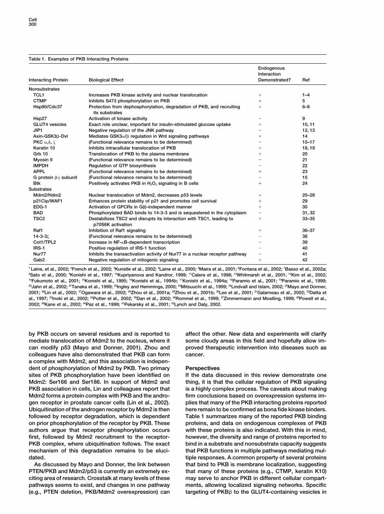
plies that many of the PKB interacting proteins reportedgen receptor in prostate cancer cells (Lin et al., 2002).Ubiquitination of the androgen receptor by Mdm2 is then here remain to be confirmed as bona fide kinase binders.
Table 1 summarizes many of the reported PKB bindingfollowed by receptor degradation, which is dependenton prior phosphorylation of the receptor by PKB. These proteins, and data on endogenous complexes of PKB
with these proteins is also indicated. With this in mind,authors argue that receptor phosphorylation occursfirst, followed by Mdm2 recruitment to the receptor- however, the diversity and range of proteins reported to
bind in a substrate and nonsubstrate capacity suggestsPKB complex, where ubiquitination follows. The exactmechanism of this degradation remains to be eluci- that PKB functions in multiple pathways mediating mul-
tiple responses. A common property of several proteinsdated.As discussed by Mayo and Donner, the link between that bind to PKB is membrane localization, suggesting
that many of these proteins (e.g., CTMP, keratin K10)PTEN/PKB and Mdm2/p53 is currently an extremely ex-citing area of research. Crosstalk at many levels of these may serve to anchor PKB in different cellular compart-
ments, allowing localized signaling networks. Specificpathways seems to exist, and changes in one pathway(e.g., PTEN deletion, PKB/Mdm2 overexpression) can targeting of PKB� to the GLUT4-containing vesicles in

Review301
directly due to space limitations. Special thanks to Dr. Daan M.F.adipocytes supports this hypothesis and clearly illus-van Aalten (University of Dundee, Scotland, UK) and Dr. David Bar-trates the importance of subcellular localization for PKBford (ICR, London, UK) for permission to adapt Figure 1, and tosignaling in insulin action. The interaction of other pro-Thomas Radimerski (FMI) for helpful discussions regarding TSC1/2.
teins (e.g., TCL1, Raf1) with PKB may be cell specific. We gratefully acknowledge funding from Oncosuisse (D.P.B) andHow should we interpret this in the context of individual the BBW/EU (J.P.).PKB effectors? Or, to put the question another way,
Referenceshow are cellular responses that involve activation ofPKB differentiated from each other? For example, in
Alessi, D.R., Caudwell, F.B., Andjelkovic, M., Hemmings, B.A., andendothelial cells, when PKB is activated and phosphory-Cohen, P. (1996). Molecular basis for the substrate specificity of
lates eNOS, are all other substrates of PKB being phos- protein kinase B; comparison with MAPKAP kinase-1 and p70 S6phorylated at the same time? Or when insulin induces kinase. FEBS Lett. 399, 333–338.PKB-dependent inhibition of GSK-3� in insulin-sensitive Andjelkovic, M., Alessi, D.R., Meier, R., Fernandez, A., Lamb, N.J.,tissues, are other proteins also being modified? The Frech, M., Cron, P., Cohen, P., Lucocq, J.M., and Hemmings, B.A.
(1997). Role of translocation in the activation and function of proteinanswer to these questions, based on the multitude ofkinase B. J. Biol. Chem. 272, 31515–31524.proteins that modify PKB activity in cells, is probablyBarthel, A., Kohn, A.D., Luo, Y., and Roth, R.A. (1997). A constitutivelyno. The number of candidate proteins that interact withactive version of the Ser/Thr kinase Akt induces production of thePKB suggests that these interactions may mediate dis-ob gene product, leptin, in 3T3–L1 adipocytes. Endocrinology 138,tinct effects on PKB function. These proteins may be3559–3562.
specific for multiple tissues or cell types, different sub-Basso, A.D., Solit, D.B., Chiosis, G., Giri, B., Tsichlis, P., and Rosen,
cellular compartments, or may depend on various post- N. (2002a). Akt forms an intracellular complex with Hsp90 and Cdc37translational modifications to interact with PKB. All of and is destabilized by inhibitors of Hsp90 function. J. Biol. Chem.the above could contribute to achieving specificity via 277, 39858–39866.
a common intermediate protein such as PKB in different Basso, A.D., Solit, D.B., Munster, P.N., and Rosen, N. (2002b). Ansa-mycin antibiotics inhibit Akt activation and cyclin D expression inpathways (Ding et al., 2000).breast cancer cells that overexpress HER2. Oncogene 21, 1159–These data allow us to speculate that, at any one time1166.in any given cell, PKB may be regulated by multipleBrazil, D.P., and Hemmings, B.A. (2001). Ten years of protein kinaseprotein-protein interactions. These interactions may in-B signalling: a hard Akt to follow. Trends Biochem. Sci. 26, 657–664.volve many different regulatory proteins and/or sub-Calera, M.R., Martinez, C., Liu, H., Jack, A.K., Birnbaum, M.J., andstrates, and distinct proteins would therefore be pre-Pilch, P.F. (1998). Insulin increases the association of Akt-2 with
dicted to interact with PKB in different tissues, and in Glut4-containing vesicles. J. Biol. Chem. 273, 7201–7204.cells under different conditions such as cellular stress or Cheadle, J.P., Reeve, M.P., Sampson, J.R., and Kwiatkowski, D.J.insulin resistance. The existence of three PKB isoforms (2000). Molecular genetic advances in tuberous sclerosis. Hum.suggests an additional level of complexity, and the iso- Genet. 107, 97–114.form-specific interaction of proteins such as TCL1 and Chen, R.H., Ding, W.V., and McCormick, F. (2000). Wnt signaling to
beta-catenin involves two interactive components. Glycogen syn-JIP1 suggests that different PKB proteins may exist inthase kinase-3beta inhibition and activation of protein kinase C. J.distinct protein complexes in cells. The roles of individ-Biol. Chem. 275, 17894–17899.ual PKB family members in various human cancers (e.g.,Cho, H., Mu, J., Kim, J.K., Thorvaldsen, J.L., Chu, Q., Crenshaw,PKB� in stomach cancer, PKB� in ovarian cancer) fur-E.B., 3rd, Kaestner, K.H., Bartolomei, M.S., Shulman, G.I., and Birn-ther suggests subtleties in signaling specificity basedbaum, M.J. (2001). Insulin resistance and a diabetes mellitus-like
on unique protein-protein interactions. The recent gen- syndrome in mice lacking the protein kinase Akt2 (PKB beta). Sci-eration of mice lacking individual isoforms of PKB will ence 292, 1728–1731.help enormously in deciphering the physiological basis Conus, N.M., Hannan, K.M., Cristiano, B.E., Hemmings, B.A., andof protein binding in PKB function in cells. Proteomic Pearson, R.B. (2002). Direct identification of tyrosine 474 as a regula-
tory phosphorylation site for the akt protein kinase. J. Biol. Chem.and mass spectrometry analysis of PKB protein com-277, 38021–38028.plexes from these mice will allow us to determineCross, D.A., Alessi, D.R., Cohen, P., Andjelkovich, M., and Hem-whether protein modules are nucleated around eachmings, B.A. (1995). Inhibition of glycogen synthase kinase-3 by insu-PKB isoform, and also whether individual isoforms canlin mediated by protein kinase B. Nature 378, 785–789.compensate for the absence of one form of PKB byDajani, R., Fraser, E., Roe, S.M., Young, N., Good, V., Dale, T.C.,recruiting proteins or substrates specific for the deletedand Pearl, L.H. (2001). Crystal structure of glycogen synthase kinase
isoform. This approach will also allow more precise anal- 3 beta: structural basis for phosphate-primed substrate specificityysis of PKB protein-protein interactions and will en- and autoinhibition. Cell 105, 721–732.hance our knowledge about the physiological interac- Dan, H.C., Sun, M., Yang, L., Feldman, R.I., Sui, X.M., Ou, C.C.,tions of PKB with other proteins in the cell. The Nellist, M., Yeung, R.S., Halley, D.J., Nicosia, S.V., et al. (2002).
Phosphatidylinositol 3-kinase/Akt pathway regulates tuberous scle-description of these interactions will then hopefully allowrosis tumor suppressor complex by phosphorylation of tuberin. J.specific targeted strategies of pharmacological inhibi-Biol. Chem. 277, 35364–35370.tion in diseases such as cancer, diabetes, and neurode-Datta, S.R., Dudek, H., Tao, X., Masters, S., Fu, H., Gotoh, Y., andgeneration. We eagerly anticipate the mapping of theGreenberg, M.E. (1997). Akt phosphorylation of BAD couples sur-entire complement of PKB binding proteins in the cellvival signals to the cell-intrinsic death machinery. Cell 91, 231–241.
and the elucidation of their function in signaling.Datta, S.R., Brunet, A., and Greenberg, M.E. (1999). Cellular survival:a play in three Akts. Genes Dev. 13, 2905–2927.
AcknowledgmentsDimmeler, S., Fleming, I., Fisslthaler, B., Hermann, C., Busse, R., andZeiher, A.M. (1999). Activation of nitric oxide synthase in endothelialThe Friedrich Miescher Institute for Biomedical Research is part ofcells by Akt-dependent phosphorylation. Nature 399, 601–605.the Novartis Research Foundation. We offer our sincere apologies
to all colleagues whose original publications could not be cited Ding, V.W., Chen, R.H., and McCormick, F. (2000). Differential regula-

Cell302
tion of glycogen synthase kinase 3beta by insulin and Wnt signaling. and their association with protein kinase C zeta. Biochem. Biophys.Res. Commun. 205, 817–825.J. Biol. Chem. 275, 32475–32481.
Filippa, N., Sable, C.L., Hemmings, B.A., and Van Obberghen, E. Konishi, H., Kuroda, S., Tanaka, M., Matsuzaki, H., Ono, Y., Kame-yama, K., Haga, T., and Kikkawa, U. (1995). Molecular cloning and(2000). Effect of phosphoinositide-dependent kinase 1 on protein
kinase B translocation and its subsequent activation. Mol. Cell. Biol. characterization of a new member of the RAC protein kinase family:association of the pleckstrin homology domain of three types of RAC20, 5712–5721.protein kinase with protein kinase C subspecies and beta gammaFontana, J., Fulton, D., Chen, Y., Fairchild, T.A., McCabe, T.J., Fujita,subunits of G proteins. Biochem. Biophys. Res. Commun. 216,N., Tsuruo, T., and Sessa, W.C. (2002). Domain mapping studies526–534.reveal that the M domain of hsp90 serves as a molecular scaffold to
regulate Akt-dependent phosphorylation of endothelial nitric oxide Konishi, H., Matsuzaki, H., Tanaka, M., Takemura, Y., Kuroda, S.,Ono, Y., and Kikkawa, U. (1997). Activation of protein kinase B (Akt/synthase and NO release. Circ. Res. 90, 866–873.RAC-protein kinase) by cellular stress and its association with heatFrame, S., Cohen, P., and Biondi, R.M. (2001). A common phosphateshock protein Hsp27. FEBS Lett. 410, 493–498.binding site explains the unique substrate specificity of GSK3 and
its inactivation by phosphorylation. Mol. Cell 7, 1321–1327. Konishi, H., Fujiyoshi, T., Fukui, Y., Matsuzaki, H., Yamamoto, T.,Ono, Y., Andjelkovic, M., Hemmings, B.A., and Kikkawa, U. (1999).French, S.W., Shen, R.R., Koh, P.J., Malone, C.S., Mallick, P., andActivation of protein kinase B induced by H(2)O(2) and heat shockTeitell, M.A. (2002). A modeled hydrophobic domain on the TCL1through distinct mechanisms dependent and independent of phos-oncoprotein mediates association with AKT at the cytoplasmicphatidylinositol 3-kinase. J. Biochem. (Tokyo) 126, 1136–1143.membrane. Biochemistry 41, 6376–6382.Kunstle, G., Laine, J., Pierron, G., Kagami Si, S., Nakajima, H., Hoh,Fukumoto, S., Hsieh, C.M., Maemura, K., Layne, M.D., Yet, S.F., Lee,F., Roumestand, C., Stern, M.H., and Noguchi, M. (2002). Identifica-K.H., Matsui, T., Rosenzweig, A., Taylor, W.G., Rubin, J.S., et al.tion of Akt association and oligomerization domains of the Akt kinase(2001). Akt participation in the Wnt signaling pathway through Di-coactivator TCL1. Mol. Cell. Biol. 22, 1513–1525.shevelled. J. Biol. Chem. 276, 17479–17483.Kupriyanova, T.A., and Kandror, K.V. (1999). Akt-2 binds to Glut4-Fulton, D., Gratton, J.P., McCabe, T.J., Fontana, J., Fujio, Y., Walsh,containing vesicles and phosphorylates their component proteinsK., Franke, T.F., Papapetropoulos, A., and Sessa, W.C. (1999). Regu-in response to insulin. J. Biol. Chem. 274, 1458–1464.lation of endothelium-derived nitric oxide production by the protein
kinase Akt. Nature 399, 597–601. Laine, J., Kunstle, G., Obata, T., Sha, M., and Noguchi, M. (2000).The protooncogene TCL1 is an Akt kinase coactivator. Mol. Cell 6,Galarneau, A., Primeau, T., Trudeau, L.E., and Michnick, S.W. (2002).395–407.beta-Lactamase protein fragment complementation assays as in
vivo and in vitro sensors of protein protein interactions. Nat. Biotech- Laine, J., Kunstle, G., Obata, T., and Noguchi, M. (2002). Differentialregulation of Akt kinase isoforms by the members of the TCL1 onco-nol. 20, 619–622.gene family. J. Biol. Chem. 277, 3743–3751.Galetic, I., Andjelkovic, M., Meier, R., Brodbeck, D., Park, J., and
Hemmings, B.A. (1999). Mechanism of protein kinase B activation Lawlor, M.A., and Alessi, D.R. (2001). PKB/Akt: a key mediator ofcell proliferation, survival and insulin responses? J. Cell Sci. 114,by insulin/insulin-like growth factor-1 revealed by specific inhibitors
of phosphoinositide 3-kinase—significance for diabetes and cancer. 2903–2910.Pharmacol. Ther. 82, 409–425. Lee, M.J., Thangada, S., Paik, J.H., Sapkota, G.P., Ancellin, N., Chae,
S.S., Wu, M., Morales-Ruiz, M., Sessa, W.C., Alessi, D.R., and Hla,Hill, M.M., Clark, S.F., Tucker, D.F., Birnbaum, M.J., James, D.E.,and Macaulay, S.L. (1999). A role for protein kinase Bbeta/Akt2 in T. (2001). Akt-mediated phosphorylation of the G protein-coupled
receptor EDG-1 is required for endothelial cell chemotaxis. Mol. Cellinsulin-stimulated GLUT4 translocation in adipocytes. Mol. Cell.Biol. 19, 7771–7781. 8, 693–704.
Li, Y., Inoki, K., Yeung, R., and Guan, K.L. (2002). Regulation of TSC2Hill, M.M., Feng, J., and Hemmings, B. (2002). Identification of aplasma membrane raft-associated PKB Ser473 kinase activity that by 14–3-3 binding. J. Biol. Chem., in press. Published online October
2, 2002. 10.1074/jbc.C200510200.is distinct from ILK and PDK1. Curr. Biol. 12, 1251–1255.
Hunter, T., and Poon, R.Y. (1997). Cdc37: a protein kinase chaper- Lin, H.K., Wang, L., Hu, Y.C., Altuwaijri, S., and Chang, C. (2002).Phosphorylation-dependent ubiquitylation and degradation of an-one? Trends Cell Biol. 7, 157–161.drogen receptor by Akt require Mdm2 E3 ligase. EMBO J. 21, 4037–Ingley, E., and Hemmings, B.A. (2000). PKB/Akt interacts with ino-4048.sine-5� monophosphate dehydrogenase through its pleckstrin ho-
mology domain. FEBS Lett. 478, 253–259. Lindvall, J., and Islam, T.C. (2002). Interactions of Btk and Akt in Bcell signaling. Biochem. Biophys. Res. Commun. 293, 1319–1326.Inoki, K., Li, Y., Zhu, T., Wu, J., and Guan, K.L. (2002). TSC2 is
phosphorylated and inhibited by Akt and suppresses mTOR signal- Lynch, D.K., and Daly, R.J. (2002). PKB-mediated negative feedbacktightly regulates mitogenic signalling via Gab2. EMBO J. 21, 72–82.ling. Nat. Cell Biol. 4, 648–657.
Jahn, T., Seipel, P., Urschel, S., Peschel, C., and Duyster, J. (2002). Maira, S.M., Galetic, I., Brazil, D.P., Kaech, S., Ingley, E., Thelen,M., and Hemmings, B.A. (2001). Carboxyl-terminal modulator proteinRole for the adaptor protein Grb10 in the activation of Akt. Mol. Cell.
Biol. 22, 979–991. (CTMP), a negative regulator of PKB/Akt and v-Akt at the plasmamembrane. Science 294, 374–380.Kandel, E.S., and Hay, N. (1999). The regulation and activities of the
multifunctional serine/threonine kinase Akt/PKB. Exp. Cell Res. 253, Manning, B.D., Tee, A.R., Logsdon, M.N., Blenis, J., and Cantley,L.C. (2002). Identification of the tuberous sclerosis complex-2 tumor210–229.suppressor gene product tuberin as a target of the phosphoinositideKane, L.P., Mollenauer, M.N., Xu, Z., Turck, C.W., and Weiss, A.3-kinase/akt pathway. Mol. Cell 10, 151–162.(2002). Akt-dependent phosphorylation specifically regulates Cot
induction of NF-kappa B-dependent transcription. Mol. Cell. Biol. Mayo, L.D., and Donner, D.B. (2001). A phosphatidylinositol3-kinase/Akt pathway promotes translocation of Mdm2 from the22, 5962–5974.cytoplasm to the nucleus. Proc. Natl. Acad. Sci. USA 98, 11598–Kim, A.H., Yano, H., Cho, H., Meyer, D., Monks, B., Margolis, B.,11603.Birnbaum, M.J., and Chao, M.V. (2002). Akt1 regulates a JNK scaf-
fold during excitotoxic apoptosis. Neuron 35, 697–709. Mayo, L., and Donner, D. (2002). The PTEN, Mdm2, p53 tumor sup-pressor-oncoprotein network. Trends Biochem. Sci. 27, 462–467.Konishi, H., Kuroda, S., and Kikkawa, U. (1994a). The pleckstrin
homology domain of RAC protein kinase associates with the regula- McManus, E.J., and Alessi, D.R. (2002). TSC1–TSC2: a complex taleof PKB-mediated S6K regulation. Nat. Cell Biol. 4, E214–E216.tory domain of protein kinase C zeta. Biochem. Biophys. Res. Com-
mun. 205, 1770–1775. Mitsuuchi, Y., Johnson, S.W., Sonoda, G., Tanno, S., Golemis, E.A.,and Testa, J.R. (1999). Identification of a chromosome 3p 14.3-21.1Konishi, H., Shinomura, T., Kuroda, S., Ono, Y., and Kikkawa, U.
(1994b). Molecular cloning of rat RAC protein kinase alpha and beta gene, APPL, encoding an adaptor molecule that interacts with the

Review303
oncoprotein-serine/threonine kinase AKT2. Oncogene 18, 4891– (1994). Binding of G protein beta gamma-subunits to pleckstrin ho-mology domains. J. Biol. Chem. 269, 10217–10220.4898.
Muslin, A.J., Tanner, J.W., Allen, P.M., and Shaw, A.S. (1996). Inter- Virgilio, L., Narducci, M.G., Isobe, M., Billips, L.G., Cooper, M.D.,Croce, C.M., and Russo, G. (1994). Identification of the TCL1 geneaction of 14–3-3 with signaling proteins is mediated by the recogni-
tion of phosphoserine. Cell 84, 889–897. involved in T-cell malignancies. Proc. Natl. Acad. Sci. USA 91,12530–12534.Nakashima, N., Sharma, P.M., Imamura, T., Bookstein, R., and Olef-
sky, J.M. (2000). The tumor suppressor PTEN negatively regulates Weston, C.R., and Davis, R.J. (2001). Signal transduction: signalingspecificity—a complex affair. Science 292, 2439–2440.insulin signaling in 3T3-L1 adipocytes. J. Biol. Chem. 275, 12889–
12895. Whitmarsh, A.J., Kuan, C.Y., Kennedy, N.J., Kelkar, N., Haydar, T.F.,Mordes, J.P., Appel, M., Rossini, A.A., Jones, S.N., Flavell, R.A., etOgawara, Y., Kishishita, S., Obata, T., Isazawa, Y., Suzuki, T., Ta-
naka, K., Masuyama, N., and Gotoh, Y. (2002). Akt enhances Mdm2- al. (2001). Requirement of the JIP1 scaffold protein for stress-induced JNK activation. Genes Dev. 15, 2421–2432.mediated ubiquitination and degradation of p53. J. Biol. Chem. 277,
21843–21850. Wu, K.K. (2002). Regulation of endothelial nitric oxide synthase activ-ity and gene expression. Ann. N Y Acad. Sci. 962, 122–130.Paramio, J.M., Casanova, M.L., Segrelles, C., Mittnacht, S., Lane,
E.B., and Jorcano, J.L. (1999). Modulation of cell proliferation by Yang, J., Cron, P., Thompson, V., Good, V.M., Hess, D., Hemmings,cytokeratins K10 and K16. Mol. Cell. Biol. 19, 3086–3094. B.A., and Barford, D. (2002a). Molecular mechanism for the regula-
tion of protein kinase B/Akt by hydrophobic motif phosphorylation.Paramio, J.M., Segrelles, C., Ruiz, S., and Jorcano, J.L. (2001). Inhibi-tion of protein kinase B (PKB) and PKCzeta mediates keratin K10- Mol. Cell 9, 1227–1240.induced cell cycle arrest. Mol. Cell. Biol. 21, 7449–7459. Yang, J., Cron, P., Good, V., Thompson, V., Hemmings, B., and
Barford, D. (2002b). Crystal structure of an activated Akt/ProteinParsell, D.A., and Lindquist, S. (1993). The function of heat-shockproteins in stress tolerance: degradation and reactivation of dam- Kinase B ternary complex with GSK3-peptide and AMP-PNP. Nat.
Struct. Biol., in press.aged proteins. Annu. Rev. Genet. 27, 437–496.
Paz, K., Liu, Y.F., Shorer, H., Hemi, R., LeRoith, D., Quan, M., Kanety, Yuan, H., Mao, J., Li, L., and Wu, D. (1999). Suppression of glycogensynthase kinase activity is not sufficient for leukemia enhancer fac-H., Seger, R., and Zick, Y. (1999). Phosphorylation of insulin receptor
substrate-1 (IRS-1) by protein kinase B positively regulates IRS-1 tor-1 activation. J. Biol. Chem. 274, 30419–30423.function. J. Biol. Chem. 274, 28816–28822. Zhou, B.P., Liao, Y., Xia, W., Zou, Y., Spohn, B., and Hung, M.C.
(2001a). HER-2/neu induces p53 ubiquitination via Akt-mediatedPekarsky, Y., Koval, A., Hallas, C., Bichi, R., Tresini, M., Malstrom,S., Russo, G., Tsichlis, P., and Croce, C.M. (2000). Tcl1 enhances MDM2 phosphorylation. Nat. Cell Biol. 3, 973–982.Akt kinase activity and mediates its nuclear translocation. Proc. Zhou, B.P., Liao, Y., Xia, W., Spohn, B., Lee, M.N., and Hung, M.C.Natl. Acad. Sci. USA 97, 3028–3033. (2001b). Cytoplasmic localization of p21Cip1/WAF1 by Akt-induced
phosphorylation in HER-2/neu-overexpressing cells. Nat. Cell Biol.Pekarsky, Y., Hallas, C., Palamarchuk, A., Koval, A., Bullrich, F.,Hirata, Y., Bichi, R., Letofsky, J., and Croce, C.M. (2001). Akt phos- 3, 245–252.phorylates and regulates the orphan nuclear receptor Nur77. Proc. Zimmermann, S., and Moelling, K. (1999). Phosphorylation and regu-Natl. Acad. Sci. USA 98, 3690–3694. lation of Raf by Akt (protein kinase B). Science 286, 1741–1744.Potter, C.J., Pedraza, L.G., and Xu, T. (2002). Akt regulates growthby directly phosphorylating Tsc2. Nat. Cell Biol. 4, 658–665.
Powell, D.W., Rane, M.J., Chen, Q., Singh, S., and McLeish, K.R.(2002). Identification of 14-3-3zeta as a protein kinase B/Akt sub-strate. J. Biol. Chem. 277, 21639–21642.
Rommel, C., Clarke, B.A., Zimmermann, S., Nunez, L., Rossman, R.,Reid, K., Moelling, K., Yancopoulos, G.D., and Glass, D.J. (1999).Differentiation stage-specific inhibition of the Raf-MEK-ERK path-way by Akt. Science 286, 1738–1741.
Saltiel, A.R., and Kahn, C.R. (2001). Insulin signalling and the regula-tion of glucose and lipid metabolism. Nature 414, 799–806.
Sato, S., Fujita, N., and Tsuruo, T. (2000). Modulation of Akt kinaseactivity by binding to Hsp90. Proc. Natl. Acad. Sci. USA 97, 10832–10837.
Scheid, M.P., and Woodgett, J.R. (2001). PKB/AKT: functional in-sights from genetic models. Nat. Rev. Mol. Cell Biol. 2, 760–768.
Shaw, M., Cohen, P., and Alessi, D.R. (1998). The activation of pro-tein kinase B by H2O2 or heat shock is mediated by phosphoinosi-tide 3-kinase and not by mitogen-activated protein kinase-activatedprotein kinase-2. Biochem. J. 336, 241–246.
Tanaka, M., Konishi, H., Touhara, K., Sakane, F., Hirata, M., Ono,Y., and Kikkawa, U. (1999). Identification of myosin II as a bindingprotein to the PH domain of protein kinase B. Biochem. Biophys.Res. Commun. 255, 169–174.
ter Haar, E., Coll, J.T., Austen, D.A., Hsiao, H.M., Swenson, L., andJain, J. (2001). Structure of GSK3beta reveals a primed phosphoryla-tion mechanism. Nat. Struct. Biol. 8, 593–596.
Thomas, C., Deak, M., Alessi, D., and van Aalten, D. (2002). High-resolution structure of the pleckstrin homology domain of proteinkinase b/akt bound to phosphatidylinositol (3,4,5)-trisphosphate.Curr. Biol. 12, 1256–1262.
Toker, A., and Newton, A.C. (2000). Akt/protein kinase B is regulatedby autophosphorylation at the hypothetical PDK-2 site. J. Biol.Chem. 275, 8271–8274.
Touhara, K., Inglese, J., Pitcher, J.A., Shaw, G., and Lefkowitz, R.J.