GAS2-like proteins mediate communication between microtubules and actin through interaction with...
16
Journal of Cell Science RESEARCH ARTICLE GAS2-like proteins mediate communication between microtubules and actin through interactions with end-binding proteins Matthew J. Stroud 1, * ,` , Alicja Nazgiewicz 1,` , Edward A. McKenzie 2 , Yisu Wang 1 , Richard A. Kammerer 3 and Christoph Ballestrem 1,§ ABSTRACT Crosstalk between the microtubule (MT) and actin cytoskeletons is fundamental to many cellular processes including cell polarisation and cell motility. Previous work has shown that members of the growth-arrest-specific 2 (GAS2) family mediate the crosstalk between filamentous actin (F-actin) and MTs, but the molecular basis of this process remained unclear. By using fluorescence microscopy, we demonstrate that three members of this family, GAS2-like 1, GAS2-like 2 and GAS2-like 3 (G2L1, G2L2 and G2L3, also known as GAS2L1, GAS2L2 and GAS2L3, respectively) are differentially involved in mediating the crosstalk between F-actin and MTs. Although all localise to actin and MTs, only the exogenous expression of G2L1 and G2L2 influenced MT stability, dynamics and guidance along actin stress fibres. Biochemical analysis and live- cell imaging revealed that their functions are largely due to the association of these proteins with MT plus-end-binding proteins that bind to SxIP or SxLP motifs located at G2L C-termini. Our findings lead to a model in which end-binding (EB) proteins play a key role in mediating actin–MT crosstalk. KEY WORDS: GAS2 family, GAS2-like 1, GAS2-like 2, GAS2-like 3, End-binding protein, Microtubule, Actin, MT-tip localising signal, MtLS INTRODUCTION Crosstalk between microtubules (MTs) and actin is essential for normal cellular function. The most prominent candidates for this role are the spectraplakins, proteins that crosslink MTs and actin (Leung et al., 1999; Kodama et al., 2003; Wu et al., 2008; Prokop et al., 2013). Various medical conditions and developmental defects arise as a result of mutations in genes encoding spectraplakins, including mental retardation, cancer and chronic skin blistering (Sonnenberg and Liem, 2007). Spectraplakins are multi-domain proteins with binding sites for filamentous actin (F- actin) in their calponin homology (CH) domains, and for MTs in their C-terminal regions. Mice lacking MT-actin crosslinking factor 1 (MACF1), a spectraplakin family member, show embryonic lethality, and dystonin mutant patients or mice suffer from sensory neuropathy, which has been correlated with instability and aberrant organisation of MTs (Bernier et al., 1995; Brown et al., 1995; Guo et al., 1995; Goryunov et al., 2007). Cells without MACF1 display defects in the guidance of MTs along actin stress fibres and in cellular polarisation (Kodama et al., 2003). Expression of a mini-version of MACF1, consisting of the actin- and MT-binding domains only, was sufficient to rescue these perturbations (Kodama et al., 2003), implying that these are key functional domains of spectraplakins. Members of the growth-arrest-specific 2 (GAS2) family resemble spectraplakins as they possess both actin- and MT- binding regions, but they lack plakin domains. The GAS2 family consists of four members: GAS2, GAS2-like 1, GAS2-like 2 and GAS2-like 3 (G2L1, G2L2 and G2L3, also known as GAS2L1, GAS2L2 and GAS2L3, respectively) (Schneider et al., 1988; Goriounov et al., 2003; Stroud et al., 2011). GAS2 is widely expressed in human tissues (Collavin et al., 1998). It has been found to play a role in apoptosis (Brancolini et al., 1995; Lee et al., 1999; Sgorbissa et al., 1999) and to inhibit cell division in Xenopus embryos (Zhang et al., 2011). G2L1 is expressed in testis and brain and is involved in inhibiting the growth of red blood cells downstream of thyroid receptor signalling (Goriounov et al., 2003; Gamper et al., 2009). G2L2 is exclusively expressed in skeletal muscle, but little is known about its function (Goriounov et al., 2003). G2L3 is found in many cell types and we have previously demonstrated that it binds to actin and MTs (Stroud et al., 2011). It is also specifically upregulated during mitosis and contributes to cell cycle regulation (Wolter et al., 2012). Knockdown of G2L3 in human BJ fibroblasts and HCT116 cells resulted in aneuploidy, implying that deregulation of G2L3 might play a role in tumorigenesis (Wolter et al., 2012). Although a potential function of the GAS2 family in the crosstalk between actin and MTs has been proposed, little is known about how it is mediated (Goriounov et al., 2003). All GAS2 family members contain a CH domain (a putative active- binding site) and a GAS2-related (GAR) domain (a putative MT- binding domain), but only the GAS2-like proteins contain a larger unstructured C-terminus. Further examination of the C-termini of G2L1, G2L2 and G2L3 proteins has revealed that, like spectraplakins, they contain evolutionarily-conserved MT-tip localisation signals (MtLSs) comprising the amino acid sequence Ser/Thr-Xaa-Ile/Leu-P (or SxIP motifs), necessary to interact with MT plus-end-binding (EB) proteins (Honnappa et al., 2009). G2L1 and G2L2 have recently been identified in a proteome-wide screen for EB-binding proteins (Jiang et al., 1 Wellcome Trust Centre for Cell-Matrix Research, Faculty of Life Sciences, University of Manchester, Manchester M13 9PT, UK. 2 Manchester Institute of Biotechnology, Faculty of Life Sciences, 131 Princess Street, Manchester M1 7DN, UK. 3 Laboratory of Biomolecular Research, OFLC 106, Paul Scherrer Institut, 5232 Villigen PSI, Switzerland. *Present address: Department of Medicine, University of California San Diego, 9500 Gilman Drive, La Jolla. CA 92093, USA. ` These authors contributed equally to this work § Author for correspondence ([email protected]) This is an Open Access article distributed under the terms of the Creative Commons Attribution License (http://creativecommons.org/licenses/by/3.0), which permits unrestricted use, distribution and reproduction in any medium provided that the original work is properly attributed. Received 12 August 2013; Accepted 12 March 2014 2672 ß 2014. Published by The Company of Biologists Ltd | Journal of Cell Science (2014) 127, 2672–2682 doi:10.1242/jcs.140558
-
Upload
manchester-us -
Category
Documents
-
view
0 -
download
0
Transcript of GAS2-like proteins mediate communication between microtubules and actin through interaction with...

Jour
nal o
f Cel
l Sci
ence
RESEARCH ARTICLE
GAS2-like proteins mediate communication between microtubulesand actin through interactions with end-binding proteins
Matthew J. Stroud1,*,`, Alicja Nazgiewicz1,`, Edward A. McKenzie2, Yisu Wang1, Richard A. Kammerer3 andChristoph Ballestrem1,§
ABSTRACT
Crosstalk between the microtubule (MT) and actin cytoskeletons is
fundamental to many cellular processes including cell polarisation
and cell motility. Previous work has shown that members of the
growth-arrest-specific 2 (GAS2) family mediate the crosstalk
between filamentous actin (F-actin) and MTs, but the molecular
basis of this process remained unclear. By using fluorescence
microscopy, we demonstrate that three members of this family,
GAS2-like 1, GAS2-like 2 and GAS2-like 3 (G2L1, G2L2 and G2L3,
also known as GAS2L1, GAS2L2 and GAS2L3, respectively) are
differentially involved in mediating the crosstalk between F-actin
and MTs. Although all localise to actin and MTs, only the exogenous
expression of G2L1 and G2L2 influenced MTstability, dynamics and
guidance along actin stress fibres. Biochemical analysis and live-
cell imaging revealed that their functions are largely due to the
association of these proteins with MT plus-end-binding proteins that
bind to SxIP or SxLP motifs located at G2L C-termini. Our findings
lead to a model in which end-binding (EB) proteins play a key role in
mediating actin–MT crosstalk.
KEY WORDS: GAS2 family, GAS2-like 1, GAS2-like 2, GAS2-like 3,
End-binding protein, Microtubule, Actin, MT-tip localising signal,
MtLS
INTRODUCTIONCrosstalk between microtubules (MTs) and actin is essential for
normal cellular function. The most prominent candidates for this
role are the spectraplakins, proteins that crosslink MTs and actin
(Leung et al., 1999; Kodama et al., 2003; Wu et al., 2008; Prokop
et al., 2013). Various medical conditions and developmental
defects arise as a result of mutations in genes encoding
spectraplakins, including mental retardation, cancer and chronic
skin blistering (Sonnenberg and Liem, 2007). Spectraplakins are
multi-domain proteins with binding sites for filamentous actin (F-
actin) in their calponin homology (CH) domains, and for MTs in
their C-terminal regions. Mice lacking MT-actin crosslinking
factor 1 (MACF1), a spectraplakin family member, show
embryonic lethality, and dystonin mutant patients or mice
suffer from sensory neuropathy, which has been correlated with
instability and aberrant organisation of MTs (Bernier et al., 1995;
Brown et al., 1995; Guo et al., 1995; Goryunov et al., 2007). Cells
without MACF1 display defects in the guidance of MTs along
actin stress fibres and in cellular polarisation (Kodama et al.,
2003). Expression of a mini-version of MACF1, consisting of the
actin- and MT-binding domains only, was sufficient to rescue
these perturbations (Kodama et al., 2003), implying that these are
key functional domains of spectraplakins.
Members of the growth-arrest-specific 2 (GAS2) family
resemble spectraplakins as they possess both actin- and MT-
binding regions, but they lack plakin domains. The GAS2 family
consists of four members: GAS2, GAS2-like 1, GAS2-like 2 and
GAS2-like 3 (G2L1, G2L2 and G2L3, also known as GAS2L1,
GAS2L2 and GAS2L3, respectively) (Schneider et al., 1988;
Goriounov et al., 2003; Stroud et al., 2011).
GAS2 is widely expressed in human tissues (Collavin et al.,
1998). It has been found to play a role in apoptosis (Brancolini
et al., 1995; Lee et al., 1999; Sgorbissa et al., 1999) and to inhibit
cell division in Xenopus embryos (Zhang et al., 2011). G2L1 is
expressed in testis and brain and is involved in inhibiting the
growth of red blood cells downstream of thyroid receptor
signalling (Goriounov et al., 2003; Gamper et al., 2009). G2L2
is exclusively expressed in skeletal muscle, but little is known
about its function (Goriounov et al., 2003). G2L3 is found in
many cell types and we have previously demonstrated that
it binds to actin and MTs (Stroud et al., 2011). It is also
specifically upregulated during mitosis and contributes to cell
cycle regulation (Wolter et al., 2012). Knockdown of G2L3 in
human BJ fibroblasts and HCT116 cells resulted in aneuploidy,
implying that deregulation of G2L3 might play a role in
tumorigenesis (Wolter et al., 2012).
Although a potential function of the GAS2 family in the
crosstalk between actin and MTs has been proposed, little is
known about how it is mediated (Goriounov et al., 2003). All
GAS2 family members contain a CH domain (a putative active-
binding site) and a GAS2-related (GAR) domain (a putative MT-
binding domain), but only the GAS2-like proteins contain a larger
unstructured C-terminus. Further examination of the C-termini of
G2L1, G2L2 and G2L3 proteins has revealed that, like
spectraplakins, they contain evolutionarily-conserved MT-tip
localisation signals (MtLSs) comprising the amino acid
sequence Ser/Thr-Xaa-Ile/Leu-P (or SxIP motifs), necessary to
interact with MT plus-end-binding (EB) proteins (Honnappa
et al., 2009). G2L1 and G2L2 have recently been identified in a
proteome-wide screen for EB-binding proteins (Jiang et al.,
1Wellcome Trust Centre for Cell-Matrix Research, Faculty of Life Sciences,University of Manchester, Manchester M13 9PT, UK. 2Manchester Institute ofBiotechnology, Faculty of Life Sciences, 131 Princess Street, Manchester M17DN, UK. 3Laboratory of Biomolecular Research, OFLC 106, Paul ScherrerInstitut, 5232 Villigen PSI, Switzerland.*Present address: Department of Medicine, University of California San Diego,9500 Gilman Drive, La Jolla. CA 92093, USA.`These authors contributed equally to this work
§Author for correspondence ([email protected])
This is an Open Access article distributed under the terms of the Creative Commons AttributionLicense (http://creativecommons.org/licenses/by/3.0), which permits unrestricted use, distributionand reproduction in any medium provided that the original work is properly attributed.
Received 12 August 2013; Accepted 12 March 2014
2672
� 2014. Published by The Company of Biologists Ltd | Journal of Cell Science (2014) 127, 2672–2682 doi:10.1242/jcs.140558

Jour
nal o
f Cel
l Sci
ence
2012), but it was not clear whether these sites are functionallyrelevant or what role they might have.
In the present study, we aimed to gain mechanistic insight intothe role of GAS2 family members in cells. We found that whereasfull-length GAS2 localised exclusively to actin stress fibres,G2L1, G2L2 and G2L3 colocalised with both actin stress fibres
and MTs, and contributed to different levels of actin–MT co-alignment. The identification of EB-binding motifs in the C-termini of G2L proteins led to our hypothesis that EB binding
might play an important role in the cytoskeletal crosstalk. Thiswas indeed the case for G2L1 and G2L2, which influenced notonly MT guidance along actin stress fibres, but also MT dynamics
and stability.
RESULTSExpression of G2L1 and G2L2 induce actin–microtubuleco-alignmentTo compare the subcellular localisation of the GAS2 family ofproteins (Fig. 1A) we transiently expressed them in NIH3T3
fibroblasts. GAS2, G2L1 and G2L2 localised predominantly toactin stress fibres. In the case of GAS2, MTs seemed to localiseindependently of actin, whereas for G2L1 and G2L2 they showed
high incidence of co-alignment with stress fibres, suggesting arole for these two proteins in MT-actin crosslinking. Despite thelocalisation of G2L3 to actin and MTs we found little co-
alignment of the two (Fig. 1B).
MtLSs in G2L proteins are essential for microtubule plus-endlocalisationPrevious studies have suggested that the C-termini of G2Lproteins are crucial for MT binding (Goriounov et al., 2003;Stroud et al., 2011; Jiang et al., 2012). This was supported by our
previous observations that GAS2, the only member of the familywithout an extended C-terminus, localises only to actin stress
fibres, and that the other members lacking the C-terminal taillocalise exclusively to stress fibres (Goriounov et al., 2003;
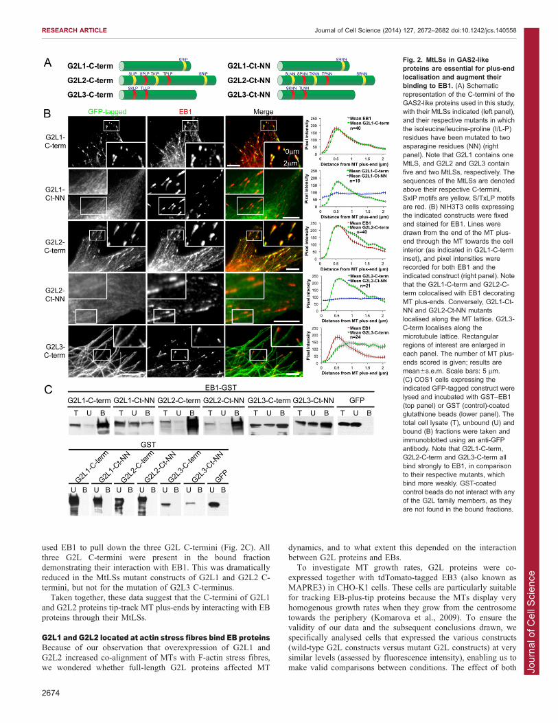
Stroud et al., 2011). To provide more detailed understanding ofG2L-protein–MT interactions, we analysed the sequences of theirC-termini and revealed that all of them contained putative bindingsites for EB proteins. G2L1 had one potential MtLS, G2L2 had
five and G2L3 had two (Fig. 2A). The single MtLS in G2L1, andthe last MtLS in G2L2 are well conserved in both mice andzebrafish.
To investigate the role of the C-termini of G2L proteins andtheir functional significance to EB proteins, we expressed theisolated C-termini fused to GFP in NIH3T3 cells, fixed the cells
and then stained for EB1 (also known as MAPRE1). The resultsshowed that both G2L1-C-term and G2L2-C-term (Fig. 2A)decorated the MT plus-ends, where endogenous EBs reside. Line-
tracing analysis of the MT plus-end revealed that the C-termini ofboth G2L1 and G2L2 colocalised with EB1, consistent with theidea that they interact with EB1 (Fig. 2B). Time-lapse recordingsdemonstrated that G2L1 and G2L2 C-termini robustly track the
tip of (tip-track) MT plus-ends in a similar manner to the EBproteins (supplementary material Movie 1). Interestingly, G2L3-C-term did not tip track MTs but decorated the whole MT shaft
(Fig. 2B).To examine the function of the potential EB-binding sites in
G2L proteins we mutated the IP or LP residues in the MtLSs
to two asparagine residues (NN) (Fig. 2A). These mutationsabolished MT-end localisation and MT-plus-end tracking of theC-termini of G2L1 and G2L2. The C-terminus of G2L3 remained
at the MT shaft despite mutating its potential EB-binding sites(Fig. 2B), suggesting that there are other motifs in its C-terminusthat mediate MT binding.
To confirm the interaction of EBs with the C-termini of the
G2L proteins, we performed GST-pulldown assays. For theseexperiments, we expressed G2L C-termini in COS1 cells and
Fig. 1. Subcellular localisation ofthe GAS2 family members.(A) Schematic representation ofmembers of the GAS2 family. Thecalponin homology (CH) and GAS2-related (GAR) domains are depictedin red and yellow, respectively, andthe number of amino acids for eachfamily member is noted above the C-termini. The sequences of the MtLSsare denoted above their respective C-termini, SxIP motifs are yellow, S/TxLP motifs are red. Note that the b-isoforms of G2L1 and G2L2 aredepicted. (B) NIH3T3 cells expressingthe indicated constructs were fixedand stained for MTs and actin. Dottedred lines indicate where the actincytoskeleton is in respect to the MTs.Note that GAS2 (yellow arrows)localised exclusively to the actincytoskeleton (black arrows), whereasG2L1, G2L2 and G2L3 (yellowarrowheads) localised to both MTs(black arrowheads) and actin-richstructures (white arrowheads). Scalebars: 10 mm.
RESEARCH ARTICLE Journal of Cell Science (2014) 127, 2672–2682 doi:10.1242/jcs.140558
2673
Jour
nal o
f Cel
l Sci
ence
used EB1 to pull down the three G2L C-termini (Fig. 2C). Allthree G2L C-termini were present in the bound fractiondemonstrating their interaction with EB1. This was dramatically
reduced in the MtLSs mutant constructs of G2L1 and G2L2 C-termini, but not for the mutation of G2L3 C-terminus.
Taken together, these data suggest that the C-termini of G2L1
and G2L2 proteins tip-track MT plus-ends by interacting with EBproteins through their MtLSs.
G2L1 and G2L2 located at actin stress fibres bind EB proteinsBecause of our observation that overexpression of G2L1 andG2L2 increased co-alignment of MTs with F-actin stress fibres,we wondered whether full-length G2L proteins affected MT
dynamics, and to what extent this depended on the interactionbetween G2L proteins and EBs.
To investigate MT growth rates, G2L proteins were co-
expressed together with tdTomato-tagged EB3 (also known asMAPRE3) in CHO-K1 cells. These cells are particularly suitablefor tracking EB-plus-tip proteins because the MTs display very
homogenous growth rates when they grow from the centrosometowards the periphery (Komarova et al., 2009). To ensure thevalidity of our data and the subsequent conclusions drawn, wespecifically analysed cells that expressed the various constructs
(wild-type G2L constructs versus mutant G2L constructs) at verysimilar levels (assessed by fluorescence intensity), enabling us tomake valid comparisons between conditions. The effect of both
Fig. 2. MtLSs in GAS2-likeproteins are essential for plus-endlocalisation and augment theirbinding to EB1. (A) Schematicrepresentation of the C-termini of theGAS2-like proteins used in this study,with their MtLSs indicated (left panel),and their respective mutants in whichthe isoleucine/leucine-proline (I/L-P)residues have been mutated to twoasparagine residues (NN) (rightpanel). Note that G2L1 contains oneMtLS, and G2L2 and G2L3 containfive and two MtLSs, respectively. Thesequences of the MtLSs are denotedabove their respective C-termini,SxIP motifs are yellow, S/TxLP motifsare red. (B) NIH3T3 cells expressingthe indicated constructs were fixedand stained for EB1. Lines weredrawn from the end of the MT plus-end through the MT towards the cellinterior (as indicated in G2L1-C-terminset), and pixel intensities wererecorded for both EB1 and theindicated construct (right panel). Notethat the G2L1-C-term and G2L2-C-term colocalised with EB1 decoratingMT plus-ends. Conversely, G2L1-Ct-NN and G2L2-Ct-NN mutantslocalised along the MT lattice. G2L3-C-term localises along themicrotubule lattice. Rectangularregions of interest are enlarged ineach panel. The number of MT plus-ends scored is given; results aremean6s.e.m. Scale bars: 5 mm.(C) COS1 cells expressing theindicated GFP-tagged construct werelysed and incubated with GST–EB1(top panel) or GST (control)-coatedglutathione beads (lower panel). Thetotal cell lysate (T), unbound (U) andbound (B) fractions were taken andimmunoblotted using an anti-GFPantibody. Note that G2L1-C-term,G2L2-C-term and G2L3-C-term allbind strongly to EB1, in comparisonto their respective mutants, whichbind more weakly. GST-coatedcontrol beads do not interact with anyof the G2L family members, as theyare not found in the bound fractions.
2674
RESEARCH ARTICLE Journal of Cell Science (2014) 127, 2672–2682 doi:10.1242/jcs.140558

Jour
nal o
f Cel
l Sci
ence
G2L1 and G2L2 expression was dramatic. In cells expressingintermediate to high levels of these proteins, EB3–tdTomato
appeared immobilised on actin stress fibres (supplementarymaterial Fig. 1A). Even in cells expressing barely detectablelevels of G2L1– or G2L2–GFP (as assessed by fluorescenceintensity levels), the dramatic impact on EB protein localisation
was apparent. In control cells expressing GFP with EB3–tdTomato,recordings showed that MT plus-ends grew in a random orientationin cells, whereas MT plus-ends grew more slowly and often
appeared to tether along actin stress fibres in cells expressing lowlevels of G2L1 (supplementary material Movie 2). Interestingly,although G2L1 and G2L2 appeared to dramatically affect EB
protein localisation and MT growth rates, G2L3 expression hadlittle, if any, effect even when expressed at higher levels (data notshown).
Because of the dramatic extent of EB3–tdTomatocolocalisation with G2L1 and G2L2, we wondered whetherthe overexpression of EB proteins would give similar results. Wetherefore assessed endogenous EB protein localisation using
antibodies in cells expressing G2L proteins. In agreement withthe above data, both full-length G2L1 and G2L2 recruitedendogenous EB1 to actin stress fibres, whereas G2L3 and GAS2
had no effect on EB1 recruitment (Fig. 3; supplementary materialFig. S1B). In order to verify that this effect was mediated throughdirect interactions between EB proteins and G2L1 or G2L2,
we expressed either the EB-protein-binding defective mutants(G2L1-FL-NN and G2L2-FL-NN, respectively) or versions ofG2L1 and G2L2 lacking C-termini (G2L1-DC-term and G2L2-
DC-term, respectively). We found that these mutants fullyblocked the G2L-mediated EB recruitment and their effect onMT dynamics was lost (Fig. 3; supplementary material Fig.S1A,B).
Previous observations have shown that the expression of G2LC-termini alone are able to induce MT bundling (Goriounovet al., 2003; Stroud et al., 2011), which suggests that their
expression alone could affect MT dynamics. However, only the
expression of G2L1-C-term led to a very small but significantreduction of MT growth rate as measured by tracking of co-
expressed EB3–tdTomato; this was fully rescued upon mutationof its EB-protein-binding site. None of the other C-terminal G2Lconstructs showed any significant effect on the MT plus-endgrowth rate (supplementary material Fig. S1C).
Taken together, these data indicate that G2L1 and G2L2 areable to recruit EB proteins to actin stress fibres, where they mightguide MT growth along actin. The observations that G2L3
expression had little impact on both EB recruitment and MTdynamics, suggests that this family member has a differentfunction.
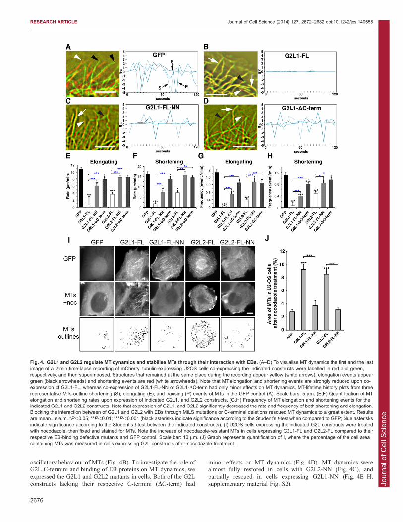
G2L1 and G2L2 regulate microtubule dynamics through theirinteraction with EBsIn the previous experiments we measured MT behaviourindirectly by analysing localisation and dynamics of EBproteins. To measure the direct impact of G2L1 and G2L2expression on MT dynamics we expressed G2L proteins
together with mCherry–tubulin in U2OS cells. These cellsdisplayed a less dense MT network than other cells andtherefore were ideal for the analysis of MT dynamics at the
cell periphery. A hallmark of MT dynamics is their dynamicoscillation, with phases of growth and shrinkage (Kirschner andMitchison, 1986). Such dynamic instability is essential for
normal cellular function and parameters commonly measuredinclude the rates and frequencies of elongation (E) andshortening (S) (Fig. 4A) (Kirschner and Mitchison, 1986;
Desai and Mitchison, 1997).We observed that expression of GAS2 (supplementary material
Fig. S2D) and G2L3 (data not shown) had no effect; however,expression of G2L1 and G2L2 dramatically decreased MT
dynamics by significantly increasing the times MTs spentpausing, and decreasing the rates of elongation and shortening(Fig. 4B,E,F; supplementary material Fig. S2). Overall, G2L1
had the most pronounced effect by strongly reducing the
Fig. 3. G2L1 recruits EB proteinsalong the actin cytoskeleton.NIH3T3 cells expressing GAS2,G2L3 or G2L1 were fixed and stainedfor EB1 and MTs. Note the G2L1colocalisation with EB1 at the actincytoskeleton (black arrowheads).MtLS mutations (FL-NN) or deletionof the C-terminus (DC-term) restoreEB1 localisation to MT plus-ends(white arrowheads). GAS2 and G2L3do not affect the localisation of EB1(white arrowheads). Scale bars:5 mm.
RESEARCH ARTICLE Journal of Cell Science (2014) 127, 2672–2682 doi:10.1242/jcs.140558
2675
Jour
nal o
f Cel
l Sci
ence
oscillatory behaviour of MTs (Fig. 4B). To investigate the role ofG2L C-termini and binding of EB proteins on MT dynamics, we
expressed the G2L1 and G2L2 mutants in cells. Both of the G2Lconstructs lacking their respective C-termini (DC-term) had
minor effects on MT dynamics (Fig. 4D). MT dynamics werealmost fully restored in cells with G2L2-NN (Fig. 4C), and
partially rescued in cells expressing G2L1-NN (Fig. 4E–H;supplementary material Fig. S2).
Fig. 4. G2L1 and G2L2 regulate MT dynamics and stabilise MTs through their interaction with EBs. (A–D) To visualise MT dynamics the first and the lastimage of a 2-min time-lapse recording of mCherry–tubulin-expressing U2OS cells co-expressing the indicated constructs were labelled in red and green,respectively, and then superimposed. Structures that remained at the same place during the recording appear yellow (white arrows); elongation events appeargreen (black arrowheads) and shortening events are red (white arrowheads). Note that MT elongation and shortening events are strongly reduced upon co-expression of G2L1-FL, whereas co-expression of G2L1-FL-NN or G2L1-DC-term had only minor effects on MT dynamics. MT-lifetime history plots from threerepresentative MTs outline shortening (S), elongating (E), and pausing (P) events of MTs in the GFP control (A). Scale bars: 5 mm. (E,F) Quantification of MTelongation and shortening rates upon expression of indicated G2L1, and G2L2 constructs. (G,H) Frequency of MT elongation and shortening events for theindicated G2L1 and G2L2 constructs. Note that expression of G2L1, and G2L2 significantly decreased the rate and frequency of both shortening and elongation.Blocking the interaction between of G2L1 and G2L2 with EBs through MtLS mutations or C-terminal deletions rescued MT dynamics to a great extent. Resultsare mean6s.e.m. *P,0.05; **P,0.01; ***P,0.001 (black asterisks indicate significance according to the Student’s t-test when compared to GFP; blue asterisksindicate significance according to the Student’s t-test between the indicated constructs). (I) U2OS cells expressing the indicated G2L constructs were treatedwith nocodazole, then fixed and stained for MTs. Note the increase of nocodazole-resistant MTs in cells expressing G2L1-FL and G2L2-FL compared to theirrespective EB-binding defective mutants and GFP control. Scale bar: 10 mm. (J) Graph represents quantification of I, where the percentage of the cell areacontaining MTs was measured in cells expressing G2L constructs after nocodazole treatment.
RESEARCH ARTICLE Journal of Cell Science (2014) 127, 2672–2682 doi:10.1242/jcs.140558
2676
Jour
nal o
f Cel
l Sci
ence
A previous study has shown that the expression of G2L1 andG2L2 increased the number of stable MTs that are resistant to the
MT depolymerising reagent nocodazole (Goriounov et al., 2003).To test whether this stability was conferred by their interactionswith EB proteins, we treated cells expressing the wild-type andmutant G2L proteins with nocodazole for 10, 20, or 30 min prior
to fixation, and subsequently stained for MTs. No MTs remainedin cells treated for 20 or 30 min with nocodazole (data notshown). However, when cells were treated for 10 min, we
observed a more than twofold increase in the amount of stabilisedMTs in cells expressing G2L1 and G2L2, compared totheir respective EB-binding-defective mutants or GFP alone
(Fig. 4I,J). Overall, these data demonstrate that EB binding to theC-termini of G2L1 and G2L2 plays an important role in theirability to regulate MT dynamics and contributes to MT stability.
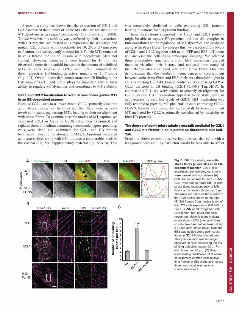
G2L1 and G2L2 localisation to actin stress fibres guides MTsin an EB-dependent mannerBecause G2L1, and to a lesser extent G2L2, primarily decorate
actin stress fibres, we hypothesised that they were activelyinvolved in capturing passing MTs, leading to their co-alignmentwith stress fibres. To examine possible modes of MT capture, we
expressed G2L1 or G2L2 in U2OS cells, then trypsinised andreplated them in medium containing nocodazole. Upon spreading,cells were fixed and examined for G2L- and EB protein
localisation. Despite the absence of MTs, EB proteins decoratedactin stress fibres along with G2L proteins at comparable levels tothe control (Fig. 5A; supplementary material Fig. S3A,B). This
was completely abolished in cells expressing G2L proteinsbearing mutations for EB protein binding.
These observations suggested that G2L1 and G2L2 proteinsmight be able to capture EB proteins, and that this complex inturn contributes to the regulation of MT dynamics and guidancealong actin stress fibres. To address this, we expressed low levels
of G2L1 and G2L2 together with actin–CFP and EB3–tdTomatoand analysed the cells using time-lapse imaging. We selectedthree consecutive time points from EB3 recordings, merged
them to visualise their history, and analysed how many ofthe EB-triplicates co-aligned with actin stress fibres. Our datademonstrated that the number of coincidences of co-alignment
between actin stress fibres and EB3 tracks was threefold higher incells expressing G2L1-FL than in control cells expressing GFP orG2L1 deficient in EB binding (G2L1-FL-NN) (Fig. 5B,C). In
contrast to G2L1, we were unable to quantify co-alignment forG2L2 because EB3 localisation appeared to be static, even incells expressing very low levels of G2L2. EB3 localisation wasfully restored to growing MT plus-ends in cells expressing G2L2-
FL-NN, thereby confirming that the crosstalk between actin andMT mediated by G2L2 is primarily coordinated by its ability tobind EB proteins.
The degree of actin–microtubule crosstalk mediated by G2L1and G2L2 is different in cells plated on fibronectin and Cell-TakFrom the above observations, we hypothesised that cells with aless-pronounced actin cytoskeleton would be less able to affect
Fig. 5. G2L1 localising on actinstress fibres guides MTs in an EB-dependent manner. U2OS cellsexpressing the indicated constructswere treated with nocodazole (A).Note that in contrast to G2L1-FL-NN,G2L1 was able to retain EB1 at actinstress fibres independently of MTs(black arrowheads). Scale bar: 5 mm.The white line indicates the position ofthe RGB profile shown to the right.(B) Still frames from movies taken ofNIH 3T3 cells expressing G2L1-FL orG2L1-FL-NN or GFP together withEB3 (green, red, blue) and actin(magenta). Magnifications indicatelocalisation of EB3 (merge of threeconsecutive time frames taken every5 s) and actin stress fibres. Note thatEB3 was guided along actin stressfibres in G2L1-FL-transfected cells.This phenomenon was no longerobserved in cells expressing the EB-binding-defective mutant G2L1-FL-NN. Scale bar: 10 mm. (C) Graphrepresents quantification of B whereco-alignment of three consecutivetime frames of EB3 along actin stressfibres was considered as onecoincidence event.
RESEARCH ARTICLE Journal of Cell Science (2014) 127, 2672–2682 doi:10.1242/jcs.140558
2677

Jour
nal o
f Cel
l Sci
ence
MT growth rates. To test this we plated cells on Cell-Tak, areagent that promotes cell spreading without engaging integrins.Cells on Cell-Tak lack focal adhesions, and consequently have
vastly reduced numbers of actin stress fibres (Fig. 6A). Underthese conditions, G2L1 and G2L2 still localised to the fine actinmeshwork, but showed a more pronounced colocalisation withEB-enriched MT tips (Fig. 6B). The presence of EB1 itself at the
tip of MTs indicated that these MTs are dynamic. To visualiseMT growth under these conditions we expressed EB3–tdTomatotogether with G2L1 or G2L2 in CHO-K1 cells and plated them on
fibronectin or Cell-Tak. In agreement with our data in Fig. 4,cells expressing intermediate levels of G2L1 or G2L2 that wereplated on fibronectin displayed significantly attenuated MT plus-
end growth rates compared to the GFP control. In contrast, MTgrowth rates of cells on Cell-Tak were unaffected, despitethem expressing similar levels of G2L proteins (as assessed by
fluorescence intensity), compared to GFP-expressing control cells(Fig. 6C,D; supplementary material Movies 3, 4).
These experiments demonstrate that G2L1 and G2L2 supportefficient crosstalk between actin and MTs, and that the impact of
this crosstalk on MT dynamics greatly varies depending on thepresence of extracellular matrix.
DISCUSSIONPrevious publications have outlined the potential of GAS2 familymembers being involved in crosslinking between actin and MTs
(Goriounov et al., 2003; Stroud et al., 2011). From thesepublications, it seemed clear that the binding of these proteinsto actin stress fibres was mediated through their N-terminalCH domains, and the binding to MTs was mediated by their C-
termini, which vary between the different members. In this study,we first compared four GAS2 proteins and analysed their roles inregulating the crosstalk between MTs and F-actin. We identified
classical EB-binding motifs in the C-termini of two out of thefour members, and demonstrated that the ability of the differentfamily members to mediate actin–MT crosstalk strongly
depended on the presence of these binding motifs.
Role of G2L association with EBs and relationship withspectraplakinsAlthough the mode and role of EB protein binding to G2L3 needsfurther investigation, our mutational analysis showed that EBprotein binding is the main mechanism by which G2L1 and G2L2
regulate communication with MTs. The interaction with EBproteins was not only responsible for MT guidance along actin
Fig. 6. The effect of G2L proteins on MT dynamics is dependent on the extracellular matrix. (A) CHO-K1 cells expressing GFP were plated on eitherfibronectin (left panel) or Cell-Tak (right panel), and actin was visualised with either phalloidin or Lifeact–RFP. Note the absence of prominent actin stress fibres incells plated on Cell-Tak in comparison to fibronectin (black arrowheads). (B) U2OS cells expressing the indicated constructs plated on Cell-Tak. Note that G2L1was present on fine actin structures, but predominantly localised to EB1-positive MT plus-ends (black arrowheads), whereas the EB-binding-defective mutant(G2L1-FL-NN) predominantly localised to actin. White lines indicate regions taken for intensity profile measurements shown to the right. (C) CHO-K1 cellsexpressing EB3–tdTomato were plated either on fibronectin or Cell-Tak and time-lapse images were recorded in 5-s intervals. Consecutive frames of EB3displayed in red, green and blue, were superimposed to visualise MT growth over time. The decrease in distance between red, green and blue MT tips in cellsplated on fibronectin versus cells on Cell-Tak outlines the impact of actin organisation on MT dynamics (white arrowheads). (D) Quantification of MT growthspeed measured by plus-end-tip displacement of cells expressing indicated constructs (n5165 MT plus-ends from .5 cells per condition). Note the reduction ofMT growth rates on fibronectin (FN) and the failure of G2L1 and G2L2 to stabilise MTwhen plated on Cell-Tak. ***P,0.001; ns, not significant (P.0.1), accordingto the Student’s t-test between indicated constructs. Scale bars: 5 mm.
RESEARCH ARTICLE Journal of Cell Science (2014) 127, 2672–2682 doi:10.1242/jcs.140558
2678

Jour
nal o
f Cel
l Sci
ence
fibres, but also determined the state of MT dynamics andtheir stability. This interaction with EBs and their role inMT guidance places G2L1 and G2L2 in a close functional
relationship with spectraplakins (Alves-Silva et al., 2012). Towhat extent the function of mammalian spectraplakins, MACF1,dystonin (human) and ACF7 (mouse), in guiding MTs along actinstress fibres is dependent on their C-terminal EB-binding sites has
not been explored in detail (Leung et al., 1999; Kodama et al.,2003; Slep et al., 2005; Honnappa et al., 2009). However, it islikely that they have similarly important roles to those of G2L1 or
G2L2, because it has been shown that the MtLS in the Drosophila
spectraplakin homologue Short stop (Shot) is important for itslocalisation to growing MT tips (Applewhite et al., 2010) and
essential for its physiological function in axonal outgrowth(Alves-Silva et al., 2012).
Like spectraplakins, EB binding seems not to be the sole link
between G2L proteins and MTs (Leung et al., 1999; Goriounovet al., 2003), especially because isolated C-terminal ends of G2L1and G2L2 with mutated MtLS still localise (albeit weakly) tothe MT lattice. We speculate that such interactions might be
mediated through the positive charges in the G2L C-termini,which have been shown previously to enable electrostatic peptideinteractions with MTs [which have an overall negative charge
(Wolff, 1998)]. In support of this, positively charged residues inthe C-terminus of Shot were found to be important for both MTshaft and plus-end localisation (Alves-Silva et al., 2012). What
precise role such interactions might have for the function of G2Lproteins remains to be determined, but they might be important tostabilise MTs.
G2L proteins and the regulation of MT dynamics versusstabilityOur data clearly show that members of the G2L proteins regulate
MT dynamics, which is dependent on the presence of EB-bindingsites, and that G2L proteins promote MT stability (measured bynocodazole resistance). Increased MT stability was limited to
short-term drug treatment (10 min) and was almost completelyabolished in MtLS mutants, demonstrating that EB binding is animportant modulator of G2L-induced MT stability. EB binding,
however, is not likely to be the only stabilising factor for G2L-mediated MT stability, because Goriounov and colleagues havefound that constructs containing the GAR domain and C-terminusalso contribute to nocodazole resistance (Goriounov et al., 2003).
Similar experiments with the Drosophila spectraplakin Shot haveshown a role for the GAR domain in MT stabilisation (Alves-Silva et al., 2012) suggesting contributions from both the C-
terminal tail and the GAR domain.
Cellular and physiological roles of GAS2 family membersThe physiological role of GAS2 family members in mammals is
unclear, but clues about their function have been revealed inDrosophila, which express only a single member of the GAS2family, named ‘Pigs’. Pigs-null Drosophila are semi-viable,
and display muscle degeneration and defects in follicle celldifferentiation (Pines et al., 2010). The C-terminus of Pigs has
Table 1. A summary of the characteristics and behaviour of the GAS2 protein family
Properties or protein GAS2 G2L1 G2L2 G2L3
Localisation Actin a Actin and MTs b Actin and MTs b Actin and MTsc
Actin–MT crosstalk 2 +b + b +/2d
MT plus-tip behaviour of C-terminus n.a. +e +e 2
Canonical MtLS 2 +e +e 2
Endogenous EB relocalisation 2 + + 2
EB-regulated MT dynamics 2 + + 2
EB-dependent MT stability n.d. + + n.d.EB-dependent MT guidance along actin n.d. + + n.d.Substrate dependent actin–MT crosstalk n.d. + + n.d.
+, strong crosstalk; +/2, weak crosstalk; 2, no crosstalk; n.d., not determined; n.a., not applicable.aData from Brancolini et al., 1992, Brancolini and Schneider, 1994, Goriounov et al., 2003 and Sgorbissa et al., 1999.bData from Goriounov et al., 2003 and Jiang et al., 2012.cData from Stroud et al., 2011 and Wolter et al., 2012.dData from Stroud et al., 2011.eData from Jiang et al., 2012.
Fig. 7. Model of G2L-mediated crosstalk. (A) G2L1 and G2L2 act assensors and detect actin structures that require crosstalk with MTs.(B) Changes in requirements of crosstalk in the cell might be regulated eitherby modulating their endogenous expression levels or by changing thebinding strength to actin or EBs.
RESEARCH ARTICLE Journal of Cell Science (2014) 127, 2672–2682 doi:10.1242/jcs.140558
2679

Jour
nal o
f Cel
l Sci
ence
been found to be important for its correct localisation inDrosophila cells, but the role of the four putative MtLS motifs
in its C-terminus has yet to be determined.In mammals, it appears that GAS2 family members have
diverse cellular functions despite their highly similar domainorganisation. Although GAS2 seems to be involved in the
regulation of apoptosis (Schneider et al., 1988; Brancolini et al.,1995), G2L3 seems to be involved in mitosis (Wolter et al.,2012). Cellular and physiological functions of G2L1 and G2L2
are less established (Gamper et al., 2009), but our data suggestthat their functions are based on their strong ability to mediatecrosstalk between the actin and MTs through EB proteins. This
could be important in growth arrest, when their otherwise verylow expression is upregulated (Brancolini et al., 1995), or undercircumstances that promote G2L binding to actin. The latter
scenario is supported by our observation that the crosstalkbetween actin and MTs mediated by G2L1 and G2L2 is mostpronounced when cells develop stable actin stress fibres (Fig. 1).Table 1 summarises the characteristics and behaviour of the
GAS2 family proteins. From these data, we propose a modelwhereby G2L1 and G2L2, which have a high affinity to actin,bind to cytoplasmic EB proteins that might, in turn, associate with
MTs. Alternatively, G2L1 and G2L2 might capture MT tipsenriched in EB proteins, thus guiding them along actin stressfibres (Fig. 7A). Hence, G2L1 and G2L2 sense the state of actin
structures that can change under different environmentalconditions. A further possibility to modulate the crosstalk in thecell might occur through the regulation of endogenous G2L
protein expression levels (Fig. 7B).
MATERIALS AND METHODSCell culture and transfectionsAll cells were grown in DMEM (Sigma Aldrich, Dorset, UK)
supplemented with 10% FBS, and 1% glutamine in a 5% CO2
humidified incubator. For transfections of CHO-K1, NIH3T3 and COS-
1, cells were plated in six-well dishes (for immunofluorescence) or 10-cm
Petri dishes (for EB1–GST pull-down experiments) using 1–1.5 mg or
8 mg of DNA, respectively, and Lipofectamine Plus (Invitrogen, Paisley,
UK) in accordance with the manufacturer’s instructions. U2OS cells were
transfected with Lipofectamine 2000 (Invitrogen, Paisley, UK) with 1–
1.5 mg of DNA. For immunofluorescence, cells were replated after 3–4 h
in glass-bottomed dishes (MatTek Corporation, Ashland, USA) coated
with 10 mg/ml bovine fibronectin (pFN; Sigma) or 3.5 mg/cm2 Cell-
Tak (Becton, Dickinson Biosciences, Oxford, UK). For pulldown
experiments, cells were washed three times in PBS (Lonza, Verviers,
Belgium), and once in complete DMEM, and left overnight before lysis
the following day.
Fixed immunofluorescence imagingCells were fixed and permeabilised with 3% paraformaldehyde (Sigma)
containing 0.25% Triton X-100 (Sigma) and 0.05% glutaraldehyde (Sigma)
for 15 min, before being washed in PBS (Lonza). Autofluorescence was
quenched using 0.01% sodium tetrahydroborate (Sigma) in PBS. Cells were
incubated with anti-tubulin (DM1A, Sigma) antibody, at 1:500 dilution,
followed by secondary antibodies conjugated to DyLight 488, 594 or 649
(Jackson ImmunoResearch Laboratories, Suffolk, UK). For actin, Texas-
Red-, FITC- or Alexa-Fluor-633-labelled phalloidin (Invitrogen) was added
together with the secondary antibody. For the EB1 antibody (1A11/4, Santa
Cruz Biotechnology), cells were fixed in 220 C methanol for 5 min, before
being rehydrated in PBS and stained using anti-tubulin antibodies (as
above). For the experiments involving nocodazole (Sigma) and Cell-Tak,
cells were spread on glass-bottomed dishes in the presence of 10 or 4 mM
nocodazole or DMSO (control) and Cell-Tak, respectively, for 1 h prior to
fixation. Cells were then imaged using an oil-immersed 1006 objective,
with 1.35 numerical aperture, on an inverted microscope (IX71; Olympus)
controlled by a Deltavision system (Applied Precision, Washington, USA).
Images were captured using a Coolsnap HQ CCD camera (Princeton
Instruments, Lurgan, UK).
Live-cell imagingFor MT dynamics and EB-tracking experiments, cells were plated on
glass-bottomed dishes coated with fibronectin or Cell-Tak and spread for
1 h prior to live imaging. Cells were imaged in Ham’s F12 medium
supplemented with 25 mM HEPES, 1% L-glutamine, 1% penicillin/
streptomycin and the pH was adjusted to 7.3 with NaOH (as described in
Carisey et al., 2011) either using an oil-immersed 606 objective, with
1.35 numerical aperture, on a Yokogawa spinning disk microscope
(Intelligent Imaging Innovations, Colorado, US) equipped with an Evolve
EMCCD camera (Photometrics, AZ, USA) or an oil-immersed 1006objective, with 1.35 numerical aperture, on an inverted microscope
(IX71; Olympus) controlled by a Deltavision system (Applied Precision).
Images were captured using a Coolsnap HQ CCD camera (Princeton
Instruments).
Image processingTo measure MT plus-end growth rates, automated tracking software was
used (Matov et al., 2010), Briefly, cells co-expressing EB3–tdTomato
and G2L C-termini were imaged every 2 s and the default settings
were used for analysis. Analysis of MT dynamics were performed as
described previously (Dhamodharan and Wadsworth, 1995), with some
modifications. Briefly, to assess rates and frequencies of MT elongation,
shortening and pausing, cells were imaged every 5 s. A pause phase was
determined when MT movement was not greater than 60.5 mm, the
shortening phase as MT movement greater than 20.5 mm, and elongation
as MT movement greater than +0.5 mm. MT dynamics were measured
manually by using the Fiji manual tracking plugin (Schindelin et al.,
2012). To measure the percentage of the cell area containing MTs in cells
treated with nocodazole, images were processed and analysed with Fiji
software. To detect MTs, the image background was subtracted, a region
of the cell surface was selected and an FFT bandpass filter and threshold
was applied. Coinciding events were measured manually. Briefly, at least
30 cells were imaged every 5 s, RGB merges of three consecutive time
frames of EB3–tdTomato and Actin–CFP were prepared using Fiji
software. Each co-alignment of three consecutive time frames of EB3
along actin stress fibres was considered as one coincidence event. To
measure colocalisation of GAS2 family members and EB proteins, RGB
merges were prepared by using Fiji software. MATLAB was used to
create fluorescence intensity line profiles over merged images and graphs
representing normalised fluorescence intensity over the drawn line.
Adobe Photoshop CS5 and Adobe Illustrator CS4 were used in the
preparation of figures for this manuscript.
Constructs and sub-cloningFor immunofluorescence, G2L3 and G2L3-C-T were cloned into pEGFP-
N1 or mCherry-N1 vectors (Clontech) using the restriction endonucleases
(Fermentas) NheI and HindIII at the amino acids indicated in Fig. 1A.
G2L3-C-T was also cloned into the pEGFP-C1 vector (Clontech) using
the restriction endonuclease BamHI. GAS2 was cloned into pEGFP-N1
with XhoI and KpnI, and pEGFP-C1 with EcoRI and BamHI. G2L1-FL
and G2L1-C-term were cloned into pEGFP-C1 with EcoRI and BamHI,
G2L1-C-term was cloned into pEGFP-N1 with BglII and HindIII. G2L2-
FL and G2L2-C-term were cloned in to pEGFP-C1 with BglII and EcoRI,
G2L2-C-term was cloned in to pEGFP-N1 with NheI and HindIII.
For recombinant expression in E. coli, GST–EB1 was cloned into
the pHisGST vector (Kammerer et al., 1998) using the restriction
endonucleases EcoRI and BamHI. All recombinant insert DNA was
verified by DNA sequencing (GATC Biotech). EB1–GFP was a kind gift
from the laboratory of Shoichiro Tsukita. EB3–tdTomato construct
was a kind gift from the laboratory of Philip Woodman (University of
Manchester, UK). The mCherry–tubulin construct was a kind gift from
the laboratory of Viki Allan (University of Manchester, UK). Lifeact-
RFP construct was a kind gift from the laboratory of Roland Wedlich-
Soldner’s lab (MPI Munich, Germany).
RESEARCH ARTICLE Journal of Cell Science (2014) 127, 2672–2682 doi:10.1242/jcs.140558
2680

Jour
nal o
f Cel
l Sci
ence
Protein expression and purificationThe recombinant GST-EB1 was expressed in E. coli JM109 (DE3)
(Merck Biosciences) and BL21 CodonPlus (DE3) (Stratagene, UK), as
previously described (Kammerer et al., 1995) using an autoinduction
protocol (as described by Studier, 2005). GST–EB1-C-T was purified
using glutathione-coated Sepharose beads (GE Healthcare). Purification
was performed under denaturing condition using 8 M urea as described in
the pET system manual (Merck Biosciences). Removal of the GST tag by
thrombin was performed as previously described (Kammerer et al.,
1995). Determination of protein concentration was achieved by
measuring the absorbance of tryptophan and tyrosine residues at
280 nm (Edelhoch, 1967).
GST pull-down assay from cell lysatesCOS cells were washed with PBS, trypsinised, centrifuged and lysed in
500 ml ice-cold lysis buffer (1% NP40, 50 mM Tris-HCl pH 7.4,
120 mM NaCl, 2.5 mM EGTA 10 mM MgCl, supplemented with 26protease inhibitor cocktail solution). Lysates were passed through a 27G
needle ten times, centrifuged in a pre-cooled centrifuge at 4 C for 15 min
at 800 g. The supernatant was pre-cleared with glutathione-coated
Sepharose beads (GE Healthcare) for 1 h at 4 C, followed by
centrifugation at 2000 g and aspiration of the supernatant. Glutathione-
coated Sepharose beads were incubated with either GST–EB1 or GST
alone (control). After pre-clearing, the GST–EB1 or GST-coated beads
were washed three times in PBS containing 0.01% Tween 20 (PBS-T),
and were resuspended in the pre-cleared lysate and rotated for an hour at
4 C. The samples were centrifuged at 2000 g. The beads were washed
four times in PBS-T and the bound proteins were eluted from the beads in
26 SDS-loading buffer. The individual fractions were then separated
by SDS-PAGE and analysed by immunoblotting, using an anti-GFP
antibody (1:1000, Roche).
Statistical analysisStudent’s t-tests were performed using Microsoft Excel.
AcknowledgementsWe thank the following colleagues at the University of Manchester (Manchester,UK): Sanjai Patel, Aishwariya Papilly and the Protein Expression Facility forexcellent technical assistance; Alexandre Carisey for his kind support with theMATLAB script used for line profile analysis; David Garrod, Janet Askari andAndreas Prokop for critical reading of the manuscript; and Peter March and themembers of the Faculty of Life Sciences Bioimaging core facility for the help withvarious microscopes.
Competing interestsThe authors declare no competing interests.
Author contributionsM.J.S., A.N., E.A.M. and Y.W. performed experiments. M.J.S., A.N., R.A.K. andC.B. designed experiments, analysed data, and contributed to writing. M.J.S. andC.B. wrote the manuscript.
FundingThis work was supported by a Wellcome Trust Ph.D. studentship to M.J.S.; aWellcome Trust Senior Research Fellowship [grant number 080878/Z/06/Z toR.A.K.]; and the Biotechnology and Biological Sciences Research Council [grantnumber BB/G004552/1 to C.B.]. Deposited in PMC for immediate release.
Supplementary materialSupplementary material available online athttp://jcs.biologists.org/lookup/suppl/doi:10.1242/jcs.140558/-/DC1
ReferencesAlves-Silva, J., Sanchez-Soriano, N., Beaven, R., Klein, M., Parkin, J., Millard,T. H., Bellen, H. J., Venken, K. J., Ballestrem, C., Kammerer, R. A. et al.(2012). Spectraplakins promote microtubule-mediated axonal growth byfunctioning as structural microtubule-associated proteins and EB1-dependent+TIPs (tip interacting proteins). J. Neurosci. 32, 9143-9158.
Applewhite, D. A., Grode, K. D., Keller, D., Zadeh, A., Slep, K. C. and Rogers,S. L. (2010). The spectraplakin Short stop is an actin-microtubule cross-linkerthat contributes to organization of the microtubule network. Mol. Biol. Cell 21,1714-1724.
Bernier, G., Brown, A., Dalpe, G., De Repentigny, Y., Mathieu, M. and Kothary,R. (1995). Dystonin expression in the developing nervous system predominatesin the neurons that degenerate in dystonia musculorum mutant mice. Mol. Cell.Neurosci. 6, 509-520.
Brancolini, C. and Schneider, C. (1994). Phosphorylation of the growth arrest-specific protein Gas2 is coupled to actin rearrangements during GoRG1transition in NIH 3T3 cells. J. Cell Biol. 124, 743-756.
Brancolini, C., Bottega, S. and Schneider, C. (1992). Gas2, a growth arrest-specific protein, is a component of the microfilament network system. J. CellBiol. 117, 1251-1261.
Brancolini, C., Benedetti, M. and Schneider, C. (1995). Microfilamentreorganization during apoptosis: the role of Gas2, a possible substrate forICE-like proteases. EMBO J. 14, 5179-5190.
Brown, A., Bernier, G., Mathieu, M., Rossant, J. and Kothary, R. (1995). Themouse dystonia musculorum gene is a neural isoform of bullous pemphigoidantigen 1. Nat. Genet. 10, 301-306.
Carisey, A., Stroud, M., Tsang, R. and Ballestrem, C. (2011). Fluorescencerecovery after photobleaching. Methods Mol. Biol. 769, 387-402.
Collavin, L., Buzzai, M., Saccone, S., Bernard, L., Federico, C., DellaValle,G., Brancolini, C. and Schneider, C. (1998). cDNA characterizationand chromosome mapping of the human GAS2 gene. Genomics 48, 265-269.
Desai, A. and Mitchison, T. J. (1997). Microtubule polymerization dynamics.Annu. Rev. Cell Dev. Biol. 13, 83-117.
Dhamodharan, R. and Wadsworth, P. (1995). Modulation of microtubule dynamicinstability in vivo by brain microtubule associated proteins. J. Cell Sci. 108,1679-1689.
Edelhoch, H. (1967). Spectroscopic determination of tryptophan and tyrosine inproteins. Biochemistry 6, 1948-1954.
Gamper, I., Koh, K. R., Ruau, D., Ullrich, K., Bartunkova, J., Piroth, D., Hacker,C., Bartunek, P. and Zenke, M. (2009). GAR22: a novel target gene of thyroidhormone receptor causes growth inhibition in human erythroid cells. Exp.Hematol. 37, 539-548, e534.
Goriounov, D., Leung, C. L. and Liem, R. K. (2003). Protein products of humanGas2-related genes on chromosomes 17 and 22 (hGAR17 and hGAR22)associate with both microfilaments and microtubules. J. Cell Sci. 116, 1045-1058.
Goryunov, D., Adebola, A., Jefferson, J. J., Leung, C. L., Messer, A. and Liem,R. K. (2007). Molecular characterization of the genetic lesion in Dystoniamusculorum (dt-Alb) mice. Brain Res. 1140, 179-187.
Guo, L., Degenstein, L., Dowling, J., Yu, Q. C., Wollmann, R., Perman, B. andFuchs, E. (1995). Gene targeting of BPAG1: abnormalities in mechanicalstrength and cell migration in stratified epithelia and neurologic degeneration.Cell 81, 233-243.
Honnappa, S., Gouveia, S. M., Weisbrich, A., Damberger, F. F., Bhavesh, N. S.,Jawhari, H., Grigoriev, I., van Rijssel, F. J., Buey, R. M., Lawera, A. et al.(2009). An EB1-binding motif acts as a microtubule tip localization signal. Cell138, 366-376.
Jiang, K., Toedt, G., Montenegro Gouveia, S., Davey, N. E., Hua, S., van derVaart, B., Grigoriev, I., Larsen, J., Pedersen, L. B., Bezstarosti, K. et al.(2012). A proteome-wide screen for mammalian sxip motif-containingmicrotubule plus-end tracking proteins. Curr. Biol. 22, 1800-1807.
Kammerer, R. A., Antonsson, P., Schulthess, T., Fauser, C. and Engel, J.(1995). Selective chain recognition in the C-terminal a-helical coiled-coil regionof laminin. J. Mol. Biol. 250, 64-73.
Kammerer, R. A., Schulthess, T., Landwehr, R., Lustig, A., Fischer, D. andEngel, J. (1998). Tenascin-C hexabrachion assembly is a sequential two-step process initiated by coiled-coil alpha-helices. J. Biol. Chem. 273, 10602-10608.
Kirschner, M. W. and Mitchison, T. (1986). Microtubule dynamics. Nature 324,621.
Kodama, A., Karakesisoglou, I., Wong, E., Vaezi, A. and Fuchs, E.(2003). ACF7: an essential integrator of microtubule dynamics. Cell 115, 343-354.
Komarova, Y., De Groot, C. O., Grigoriev, I., Gouveia, S. M., Munteanu, E. L.,Schober, J. M., Honnappa, S., Buey, R. M., Hoogenraad, C. C., Dogterom,M. et al. (2009). Mammalian end binding proteins control persistent microtubulegrowth. J. Cell Biol. 184, 691-706.
Lee, K. K., Tang, M. K., Yew, D. T., Chow, P. H., Yee, S. P., Schneider, C. andBrancolini, C. (1999). gas2 is a multifunctional gene involved in the regulationof apoptosis and chondrogenesis in the developing mouse limb. Dev. Biol. 207,14-25.
Leung, C. L., Sun, D., Zheng, M., Knowles, D. R. and Liem, R. K. (1999).Microtubule actin cross-linking factor (MACF): a hybrid of dystonin anddystrophin that can interact with the actin and microtubule cytoskeletons.J. Cell Biol. 147, 1275-1286.
Matov, A., Applegate, K., Kumar, P., Thoma, C., Krek, W., Danuser, G. andWittmann, T. (2010). Analysis of microtubule dynamic instability using a plus-end growth marker. Nat. Methods 7, 761-768.
Pines, M. K., Housden, B. E., Bernard, F., Bray, S. J. and Roper, K. (2010). Thecytolinker Pigs is a direct target and a negative regulator of Notch signalling.Development 137, 913-922.
Prokop, A., Beaven, R., Qu, Y. and Sanchez-Soriano, N. (2013). Using flygenetics to dissect the cytoskeletal machinery of neurons during axonal growthand maintenance. J. Cell Sci. 126, 2331-2341.
RESEARCH ARTICLE Journal of Cell Science (2014) 127, 2672–2682 doi:10.1242/jcs.140558
2681

Jour
nal o
f Cel
l Sci
ence
Schindelin, J., Arganda-Carreras, I., Frise, E., Kaynig, V., Longair, M.,Pietzsch, T., Preibisch, S., Rueden, C., Saalfeld, S., Schmid, B. et al.(2012). Fiji: an open-source platform for biological-image analysis. Nat. Methods9, 676-682.
Schneider, C., King, R. M. and Philipson, L. (1988). Genes specificallyexpressed at growth arrest of mammalian cells. Cell 54, 787-793.
Sgorbissa, A., Benetti, R., Marzinotto, S., Schneider, C. and Brancolini, C.(1999). Caspase-3 and caspase-7 but not caspase-6 cleave Gas2 in vitro:implications for microfilament reorganization during apoptosis. J. Cell Sci. 112,4475-4482.
Slep, K. C., Rogers, S. L., Elliott, S. L., Ohkura, H., Kolodziej, P. A. andVale, R. D. (2005). Structural determinants for EB1-mediated recruitment ofAPC and spectraplakins to the microtubule plus end. J. Cell Biol. 168, 587-598.
Sonnenberg, A. and Liem, R. K. (2007). Plakins in development and disease.Exp. Cell Res. 313, 2189-2203.
Stroud, M. J., Kammerer, R. A. and Ballestrem, C. (2011). Characterization ofG2L3 (GAS2-like 3), a new microtubule- and actin-binding protein related tospectraplakins. J. Biol. Chem. 286, 24987-24995.
Studier, F. W. (2005). Protein production by auto-induction in high density shakingcultures. Protein Expr. Purif. 41, 207-234.
Wolff, J. (1998). Promotion of microtubule assembly by oligocations: cooperativitybetween charged groups. Biochemistry 37, 10722-10729.
Wolter, P., Schmitt, K., Fackler, M., Kremling, H., Probst, L., Hauser, S., Gruss,O. J. and Gaubatz, S. (2012). GAS2L3, a target gene of the DREAM complex,is required for proper cytokinesis and genomic stability. J. Cell Sci. 125, 2393-2406.
Wu, X., Kodama, A. and Fuchs, E. (2008). ACF7 regulates cytoskeletal-focaladhesion dynamics and migration and has ATPase activity. Cell 135, 137-148.
Zhang, T., Dayanandan, B., Rouiller, I., Lawrence, E. J. and Mandato, C. A.(2011). Growth-arrest-specific protein 2 inhibits cell division in Xenopusembryos. PLoS ONE 6, e24698.
RESEARCH ARTICLE Journal of Cell Science (2014) 127, 2672–2682 doi:10.1242/jcs.140558
2682

Fig. S1. (A) CHO-K1 cells co-expressing EB3 and full-length G2L1 and G2L2 and their respective NN and ∆C-term mutants were imaged live at 5 seconds intervals. Consecutive frames of EB3 were imaged in red and green and superimposed on one another. Note the large overlap between red and green channels from consecutive frames in the wild-type (G2L1-FL and G2L2-FL) panel, outlining the static nature of EB3 in these cells. Conversely, preventing EB3 binding to G2L1 and G2L2 by MtLS mutations or by deletion of C-termini, rescued EB3 MT plus-end tip-tracking. (B) NIH 3T3 cells expressing the full-length G2L2 were fixed and stained for EB1 and MTs. Note G2L2 colocalisation with EB1 at the actin cytoskeleton (black arrowheads). MtLSs mutations (FL-NN) or removal of the C-terminus (ΔC-term) restored EB1 localisation to growing MT plus-ends (white arrowheads). (C) CHO-K1 cells co-expressing EB3 and GAS2-like C-termini and their respective NN mutants were recorded live at 5 seconds intervals and imaged as in (A). Average speeds of MT plus-ends are indicated in the histogram. More than 4000 MT plus-ends were tracked for each condition. Error bars indi-cate standard deviations from the mean. Black asterisks indicates p<0.005 according to the Student’s T-test. Scale bars, 5mm.
Journal of Cell Science | Supplementary Material

Fig. S2. Micrographs from panels A-D. Overlays of the first frame (MTs in red) and the last frame at 2 min (MTs in green) from time-lapse imaging of MTs in U2OS cells expressing the indicated constructs of G2L2 (A-C), or GFP (D) (control). White arrowheads outline MTs that have undergone shortening; black arrowheads outline newly polymerised MTs; and white arrows indicate paused MTs. Note that the expression of G2L2-FL dramatically attenuates MT dynamics. MT lifetime-history plots of three representative MTs from their respective micrographs to the left. The shortening (S), elongating (E) and pausing (P) phases of MT dynamic instability are denoted in the GFP control (D). Note that MTs in cells expressing G2L2-FL exhibit very little dynamic instability in comparison to the GFP-ex-pressing control. (E) Percentage time spent by MTs in each phase of dynamic instability for the indicated G2L1 and G2L2 constructs, respectively. Note the amount of time MTs spent in pause is significantly increased in cells expressing G2L1 and G2L2. White asterisks indicate p<0.05 according to the Student’s T-test compared to GFP, blue and red asterisks indicate p<0.05 according to the Student’s T-test between the indicated constructs and G2L1-FL and G2L2-FL, respectively. Scale bars, 5 μm.
Journal of Cell Science | Supplementary Material

Fig. S3. U2OS cells expressing the indicated constructs were treated with DMSO (A). Note that in contrast to G2L1-FL and G2L2-FL (black arrowheads), GAS2, G2L1-FL-NN and G2L2-FL-NN are unable to recruit EB1 to actin structures. (B) U2OS cells expressing the indicated constructs were treated with nocodazole. Note that G2L2-FL was able to retain EB1 at actin stress fibres independently of MTs (black arrowheads), whereas EB1 localises diffusely in cells expressing GAS2 or G2L1-FL-NN. (C) Cells expressing the indicated constructs were plated on Cell-Tak. Note that G2L2-FL was present on fine actin structures, but predominantly localised to EB1-positive MT plus-ends (black arrowheads), whereas EB-binding defective mutant G2L2-FL-NN and GAS2 predominantly localised to actin. White lines indicate regions taken for intensity profile measurements to the right. Scale bars, 5 μm.
Journal of Cell Science | Supplementary Material

Movie 1. G2L1- and G2L2- C-termini tip track microtubule plus-ends. CHO-K1 cells expressing GFP-conjugated C-termini of either G2L1 or G2L2 were recorded in 5 second intervals for the duration of 60 seconds. Movies are played back at 5 frames per second.
Movie 2. G2L1 requires its C-terminus to guide MTs along actin. Left panels: CHO-K1 cells co-expressing GFP-conjugated G2L1-FL or G2L1-∆C-term (G2L1-D-C-term) (red) with EB3-tdTomato (green). Right panels show EB3 track overlays for their respective movies in the left panels. Note that for G2L1-FL, EB3 often grows slowly along the G2L1-decorated actin cytoskeleton or dramatically slows down when it contacts actin stress fibres. Conversely, in cells expressing G2L1-∆C-term, EB3 speeds are unaffected when passing through actin and EB3 is apparently not guided along actin. Cells were imaged live at 2 second intervals for 60 seconds. Movies are played back at 7 frames per second.
Journal of Cell Science | Supplementary Material

Movie 4. The crosstalk between actin and MTs mediated by G2L2 and EBs is matrix-dependent. CHO-K1 cells co-expressing full-length G2L2 and EB3 plated on fibronectin show high levels of crosstalk between MTs and actin as indicated by the reduced lev-els of EB3 plus-end tracking. Conversely, very little crosstalk is observed in cells plated on Cell-Tak, which have less prominent actin structures. Selected cells have similar expression levels of both proteins. Cells were recorded every 5 seconds for 120 seconds. Movies are played back at 20 frames per second.
Movie 3. The crosstalk between actin and MTs mediated by G2L1 and EBs is matrix-dependent. CHO-K1 cells co-expressing full-length G2L1 and EB3 plated on FN show high levels of crosstalk between MTs and actin as indicated by the reduced levels of EB3 plus-end tracking. Conversely, very little crosstalk is observed in cells plated on Cell-Tak, which have less prominent actin structures. Selected cells have similar expression levels of both proteins. Cells were recorded every 5 seconds for 120 seconds. Movies are played back at 20 frames per second.
Journal of Cell Science | Supplementary Material