Interaction of high molecular weight kininogen binding proteins on endothelial cells
17
Interaction of high-molecular-weight kininogen with endothelial cell binding proteins suPAR, gC1qR and cytokeratin 1 determined by Surface Plasmon Resonance (BiaCore) Robin A. Pixley 1,* , Ricardo G. Espinola 1,* , Berhane Ghebrehiwet 2 , Kusumam Joseph 3 , Alice Kao 4 , Khalil Bdeir 4 , Douglas B. Cines 4 , and Robert W. Colman 1 1 The Sol Sherry Thrombosis Research Center, Temple University School of Medicine, Philadelphia, Pennsylvania, USA 2 ((Affiliation??)) 3 Medical University of South Carolina, Charleston, South Carolina, USA 4 University of Pennsylvania, Philadelphia, Pennsylvania, USA Summary The physiologic activation of the plasma kallikrein-kinin system requires the assembly of its constituents on a cell membrane. High-molecular-weight kininogen (HK) and cleaved HK (HKa) both interact with at least three endothelial cell binding proteins: urokinase plasminogen activator receptor (uPAR), globular C1q receptor (gC1qR,) and cytokeratin 1 (CK1). The affinity of HK and HKa for endothelial cells are K D =7–52 nM. The contribution of each protein is unknown. We examined the direct binding of HK and HKa to the soluble extracellular form of uPAR (suPAR), gC1qR and CK1 using surface plasmon resonance. Each binding protein linked to a CM-5 chip and the association, dissociation and K D (equilibrium constant) were measured. The interaction of HK and HKa with each binding protein was zinc-dependent. The affinity for HK and HKa was gC1qR>CK1>suPAR, indicating that gC1qR is dominant for binding. The affinity for HKa compared to HK was the same for gC1qR, 2.6-fold tighter for CK1 but 53-fold tighter for suPAR. Complex between binding proteins was only observed between gC1qR and CK1 indicating that a binary CK1-gC1qR complex can form independently of kininogen. Although suPAR has the weakest affinity of the three binding proteins, it is the only one that distinguished between HK and HKa. This finding indicates that uPAR may be a key membrane binding protein for differential binding and signalling between the cleaved and uncleaved forms of kininogen. The role of CK1 and gC1qR may be to initially bind HK to the membrane surface before productive cleavage to HKa. Keywords Kininogen; surface plasmon resonance; suPAR; gC1qR; cytokeratin 1 © Schattauer 2011 Correspondence to: Robert W. Colman, MD, The Sol Sherry Thrombosis Research Center, Temple University School of Medicine, 3400 N. Broad Street, Room 418 OMS, Philadelphia, PA 19140, USA, Tel.: +1 215 707 2779, Fax: +1 215 707 2783 [email protected]. * Equal first authors. Conflict of interest None declared. NIH Public Access Author Manuscript Thromb Haemost. Author manuscript; available in PMC 2011 August 4. Published in final edited form as: Thromb Haemost. 2011 June 6; 105(6): 1053–1059. doi:10.1160/TH10-09-0591. NIH-PA Author Manuscript NIH-PA Author Manuscript NIH-PA Author Manuscript
-
Upload
independent -
Category
Documents
-
view
6 -
download
0
Transcript of Interaction of high molecular weight kininogen binding proteins on endothelial cells

Interaction of high-molecular-weight kininogen with endothelialcell binding proteins suPAR, gC1qR and cytokeratin 1determined by Surface Plasmon Resonance (BiaCore)
Robin A. Pixley1,*, Ricardo G. Espinola1,*, Berhane Ghebrehiwet2, Kusumam Joseph3, AliceKao4, Khalil Bdeir4, Douglas B. Cines4, and Robert W. Colman1
1The Sol Sherry Thrombosis Research Center, Temple University School of Medicine,Philadelphia, Pennsylvania, USA2((Affiliation??))3Medical University of South Carolina, Charleston, South Carolina, USA4University of Pennsylvania, Philadelphia, Pennsylvania, USA
SummaryThe physiologic activation of the plasma kallikrein-kinin system requires the assembly of itsconstituents on a cell membrane. High-molecular-weight kininogen (HK) and cleaved HK (HKa)both interact with at least three endothelial cell binding proteins: urokinase plasminogen activatorreceptor (uPAR), globular C1q receptor (gC1qR,) and cytokeratin 1 (CK1). The affinity of HKand HKa for endothelial cells are KD=7–52 nM. The contribution of each protein is unknown. Weexamined the direct binding of HK and HKa to the soluble extracellular form of uPAR (suPAR),gC1qR and CK1 using surface plasmon resonance. Each binding protein linked to a CM-5 chipand the association, dissociation and KD (equilibrium constant) were measured. The interaction ofHK and HKa with each binding protein was zinc-dependent. The affinity for HK and HKa wasgC1qR>CK1>suPAR, indicating that gC1qR is dominant for binding. The affinity for HKacompared to HK was the same for gC1qR, 2.6-fold tighter for CK1 but 53-fold tighter for suPAR.Complex between binding proteins was only observed between gC1qR and CK1 indicating that abinary CK1-gC1qR complex can form independently of kininogen. Although suPAR has theweakest affinity of the three binding proteins, it is the only one that distinguished between HK andHKa. This finding indicates that uPAR may be a key membrane binding protein for differentialbinding and signalling between the cleaved and uncleaved forms of kininogen. The role of CK1and gC1qR may be to initially bind HK to the membrane surface before productive cleavage toHKa.
KeywordsKininogen; surface plasmon resonance; suPAR; gC1qR; cytokeratin 1
© Schattauer 2011Correspondence to: Robert W. Colman, MD, The Sol Sherry Thrombosis Research Center, Temple University School of Medicine,3400 N. Broad Street, Room 418 OMS, Philadelphia, PA 19140, USA, Tel.: +1 215 707 2779, Fax: +1 215 707 [email protected].*Equal first authors.Conflict of interestNone declared.
NIH Public AccessAuthor ManuscriptThromb Haemost. Author manuscript; available in PMC 2011 August 4.
Published in final edited form as:Thromb Haemost. 2011 June 6; 105(6): 1053–1059. doi:10.1160/TH10-09-0591.
NIH
-PA Author Manuscript
NIH
-PA Author Manuscript
NIH
-PA Author Manuscript

IntroductionHigh-molecular-weight kininogen (HK) is the substrate and cofactor of the plasmakallikrein-kinin system (KKS) (1). HK is present on plasma at a concentration of 660 nM (1)binds to platelet GPI-V-IX complex, and inhibits binding of thrombin to platelets (2) servingas an antithrombotic protein in vivo (3). Domain 2 of HK exhibits antiplatelet activity byinhibiting platelet calpain, a cysteine protease which is a signalling molecule in platelets (4).HK inhibits adhesion of neutrophils to blood compatible surfaces under flow conditions (5)and enhances cellular fibinolysis (6). The proenzyme, prekallikrein, after activation forms anactive enzyme plasma kallikrein which can cleave HK to two chain HKa and bradykinin (7),a potent bioactive peptide which stimulates endothelial cell prostacyclin synthesis and nitricoxide formation (8) resulting in vasodilation. The formation of HKa occurs on platelet orendothelial cell surfaces where kallikrein is activated even in the presence of plasmaprotease inhibitors (9, 10).
The transformation of HK to HKa is accompanied by dramatic conformational changesdetected by circular dichroism (11) and electron microscopy (12). These physical alterationsresult in new functional activities. HKa stimulates monocytes to synthesise and secretecytokines and chemokines (13) and thus is anti-inflammatory in experimental inflammatoryarthritis (14,15). The action of HKa on monocytes is mediated by integrin receptors such asαvβ3 and α5β1 as well as the adaptor protein uPAR (16). HKa is also antiangiogenic byvirtue of inhibiting proliferation and migration of stimulated endothelial cells (17) as well asinterfering with tube formation in vitro (18) and angiogenesis in vivo (19).
On the surface of human umbilical vein endothelial cells (HU-VECs) both HK and HKaassociate with binding proteins of uPAR (20), gC1qR (21) and CK1(22). Each of these threeproteins are required for binding of HKa to endothelial cells because antibodies to any oneprotein block binding(18, 23)and a peptide from the light chain of HK, isolated by anaffinity column inhibits both binding and contact activation (24). The sub-domains by whichHK and HKa binds to each binding protein are known. For example, a peptide derived fromdomain 5 of HK, G486-K502 (25) inhibits cell adhesion to vitronectin. In this paper, weconfine ourselves to cell surface binding proteins which are intrinsic to endothelial cells. Tobetter understand the function of each binding protein, we now determine the relativeaffinity of each soluble purified binding protein for both HK and HKa using surfaceplasmon resonance. This method also allows evaluation of multiple interactions includingthe possible occurrence of binary binding protein complexes. The differential binding of HKand HKa to each binding protein was assessed in a quantitative approach in the presence ofzinc ion.
Materials and methodsMaterials
Purified proteins were made or obtained from participants in this study. Cytokeratin 1 was aspurified and characterised in Joseph et al.(23). Soluble expressed urokinase plasminogenactivator receptor (suPAR) was produced as described previously (26). Single-chainurokinase-type plasminogen activator (scuPA or PUK) was purchased from AmericanDiagnostica Inc. (Stamford, CT, USA). gC1qR (also known as p32, p33, C1QBP, TAP) waspurified as described(27). Single-chain high-molecular-weight kininogen (HK) and two-chain high-molecular-weight kininogen (HKa) was purchased from Enzyme ResearchLaboratories (South Bend, IN, USA). Low-molecular-weight kininogen (LK) was purchasedfrom Calbiochem (Darmstadt, Germany). Ovalbumin was purchased from Sigma-AldrichChemical Co. (St Louis, MO, USA).
Pixley et al. Page 2
Thromb Haemost. Author manuscript; available in PMC 2011 August 4.
NIH
-PA Author Manuscript
NIH
-PA Author Manuscript
NIH
-PA Author Manuscript
MethodsPurified proteins were examined for purity by Sodium Dodecyl Sulfate (SDS)polyacrylamide gel electrophoresis (PAGE). The samples 3–5 μg (reduced and non-reduced), were applied to a 4–20% NuPage gel using MOPS buffer (Invitrogen, Carlsbad,CA, USA). Invitrogen-SeeBlue Plus2 was used as standard and Simply blue Safe stain usedto observe the position of the proteins.
Real-time biomolecular interaction analysis was performed using a BiaCore 2000 instrument(GE Heathcare, Piscataway, NJ, USA). Purified proteins were covalently linked at pH 5.0 in10 mM sodium acetate buffer to a CM5 Chip using EDC/NHS amine coupling of theprimary amine of the protein to a carboxyl group of a chip linked carboxymethylateddextran using HBS-P running buffer (10 mM HEPES, 0.15 M NaCl, 0.005% polysorbate 20,pH 7.4)at 25°C. Ethanolamine was used to block unwanted carboxy groups after linkage.The base values of suPAR(950 RU), gC1qR (600 RU), cytokeratin 1 (750 RU), HK (924),HKa (1400 RU) and LK (1190 RU) were used as the specific ligands where 1 RU representsthe mass of 1 pg of linked protein. Each RU represents 1 pg of mass giving the number ofmolecules linked to the chip. The mass of gC1qR, suPAR, cytokeratin 1, HK, HKa and LK;when used for calculations were 33 kDa (as a non-covalent trimer),40 kDa(20),54 kDa, 120kDa, 110 kDa and 64 kDa respectively. An ethanolamine blocked Fc1 was used as blank forbackground subtraction of any non-specific response to the derivatised dextran chip. Inkinetic determinations, concentrations of different proteins were produced by programmeddilution from a stock solution using the BiaCore software starting from low concentrationsto higher concentrations. Kinject was used for injection. Flow rate was 30 μgl/minute(min)for suPAR immobilised competition studies (▸ Fig. 2) and for HKa/PUK competitionstudies on suPAR. Other studies were at 60 μl/min. Temperature for all studies were set at25°C. The running buffer used was BiaCore HBS-P (10 mM HEPES, 0.15 M NaCl, 0.005%polysorbate 20, pH = 7.4) containing 5 μg/ml (111 nM) chicken egg albumin (SigmaChemical Co.) as well as either 10 μM ZnCl2 or 3 mM EDTA (absence of Zinc runs). Aftereach analyte injection, the chip surfaces were regenerated by two injections of 100 mMEDTA + 2 M NaCl. In the PUK/HKa competitive study, regeneration was performed byshort injection series of 0.1 M acetic acid, 2 M NaCl, and 3 mM EDTA. For affinity constantdeterminations, merging of triplicate injections of each concentration was performed toobtain a single sensorgram line at each concentration. In determining the affinity constants,to stay well below Rmax values, only the lower three or four concentrations were used to fitcurves for kinetic determinations shown in ▸ Table 1. The remaining lines are for illustrativepurposes. A Lang-muir binding model with local fit(stoichiometry of 1:1) was used toanalyse kon (association rate constant), koff (dissociation rate constant) and KD (equilibriumdissociation constant). Chi2 for the fits were usually <1 or some studies or <4.
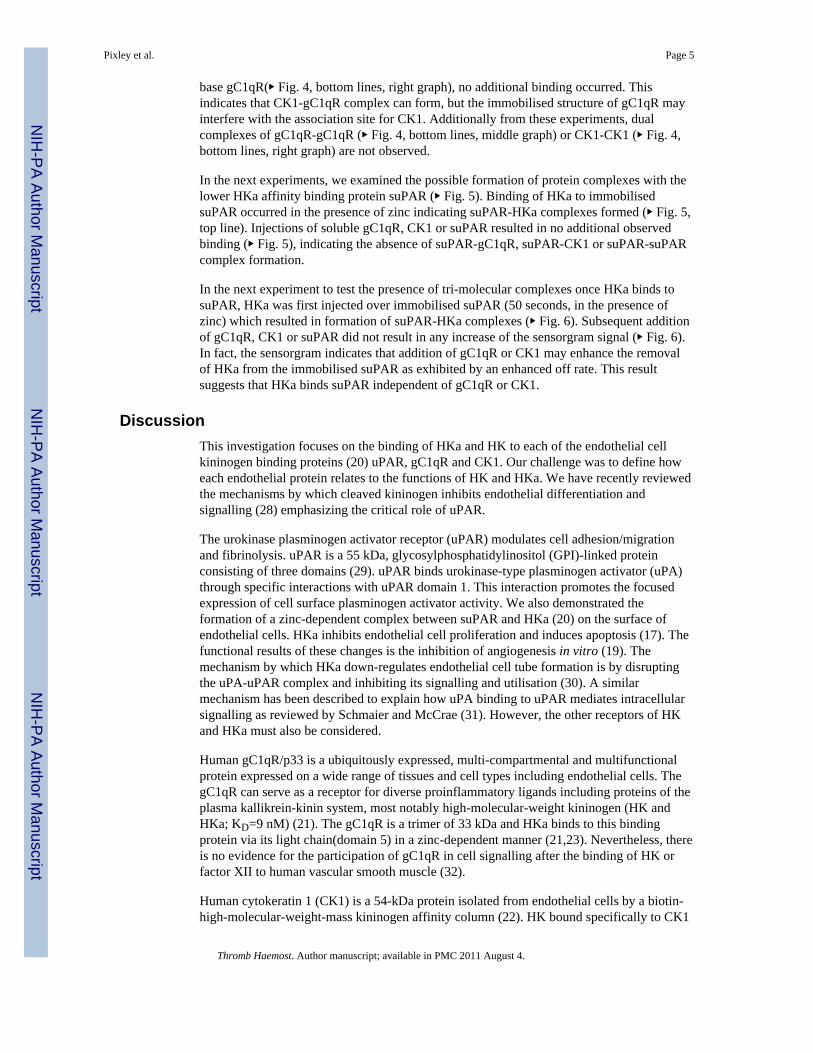
ResultsThe purity and functionality of each of the proteins were tested by gel electrophoresispatterns and by competitive tests. ▸ Figure 1 shows SDS gel electrophoretic patterns for HK,HKa, suPAR and gC1qR at the expected locations under non-reduced and reducedconditions. Each binding protein was tested for interaction with HKa using 1:1 ratio mixingstudies of HKa with each binding protein. Lowering of the ΔRU of HKa sensorgram bindingto immobilised suPAR in the presence of the soluble binding protein indicates there iscompetition between HKa binding to the immobilised suPAR and the tested soluble purifiedbinding protein (▸ Fig. 2). Immobilised suPAR was also tested for functional activity. ThesuPAR is a specific receptor for single-chain urokinase plasminogen activator (scuPA orprourokinase), and was found to bind to suPAR at a high affinity as previously described(KD =0.1–1 nM, data not shown) (26) indicating its functionality. The results indicate that
Pixley et al. Page 3
Thromb Haemost. Author manuscript; available in PMC 2011 August 4.
NIH
-PA Author Manuscript
NIH
-PA Author Manuscript
NIH
-PA Author Manuscript

after complex formation with HKa in solution, suPAR, gC1qR and CK1 competed with HKabinding to the immobilised suPAR.
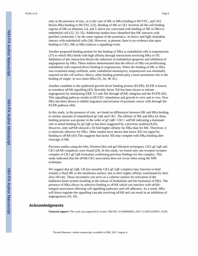
▸ Figure 3 shows the sensorgrams for the BiaCore method determinations of the kineticbinding of HK and HKa to each binding protein gC1qR (▸ Fig. 3A), suPAR (▸ Fig. 3B) andCK1 (▸ Fig. 3C) in the presence of 10 μM ZnCl2 or in the presence of 2 mM EDTA (toassure the absence of bivalent cations). Each concentration used were diluted and injected intriplicate and the three curves were averaged to one curve per concentration (as shown) andanalysed using the Biaevaluation program as described in Methods. In the presence ofEDTA, there is no apparent binding of HK nor HKa in the range of 25–400 nM examined.In the presence of 10 μM ZnCl2, there is significant binding of both HK and HKa with allthe linked binding proteins with HKa having a greater ΔRU binding peak than HK using thesame concentrations. These results confirm the requirement for Zn++ for both HK and HKabinding to endothelial cells.
The sensorgram is displayed for the binding of HK and HKa to immobilised gC1qR (▸ Fig.3A) and the data in the presence of 10 μM zinc is displayed in ▸ Table 1. The kon is twice asfast for HKa as for HK and as for all three binding proteins is absolutely dependent on thepresence of Zn2+. The koff is very slow almost flat which accounts for the very tight bindingKd = 0.7–0.8 nM for both HK and HKa (▸ Table 1). There is little selectivity with gC1qRfor either form of kininogen. The sensorgram for CK1 is dominated by the slow kon, but dueto the low koff the affinity is 6–15 nM (▸ Fig. 3C, ▸ Table 1). There is little preference forHKa over HK.
The sensorgram for immobilised suPAR is strikingly different (▸ Table 1 and Fig. 3B). Thelower kon and higher koff of HK over HKa accounts for the lower Kd (53-fold higheraffinity) of HKa binding to suPAR.
We probed whether binding to the cell binding proteins involved the heavy chain of HK. LKcontains only the heavy chain (domains 1–3) and does not contain the light chaincomponents domain 5 and 6. We compared the sensorgram binding of LK and HKa toimmobilised CK1, suPAR and gC1qR in the presence of 10 μM ZnCl2. No binding of LKwas observed in the presence or absence of zinc ions to any of these immobilised bindingproteins (data not shown). Therefore it is likely that the light chain domain of HK or HKa isrequired for the binding of HKa binding to these membrane proteins.
Up to this point we have only considered the interaction of kininogens with each of the threebinding proteins separately. In the next set of experiments, the binding proteins were studiedas soluble proteins and as immobilised proteins. We first examined the higher affinityproteins gC1qR and CK1. First it is shown that HKa (arrow indicates injection) binds toboth immobilised binding proteins (▸ Fig. 4, left side of graph, base CK1 and gC1qR). Withbase gC1qR, when additional soluble gC1qR or CK1 (arrow indicated) was injected to thebound gC1qR-HKa complex (▸ Fig. 4, top line, middle and right graph), no additional tri-molecular gC1qR-HKa-gC1qR (A) or gC1qR-HKa-CK1 complex formation are observed.This finding indicates that once HKa is bound to gC1qR, the binding sites can no longerbind to additional CK1 and gC1qR. In contrast, when base CK1 with HKa bound is injectedwith gC1qR (▸ Fig. 4, middle line, middle graph), additional binding is observed forming aCK1-HKa-gC1qR complex (▸ Fig. 4B). Additional injection of CK1 does not add nordisplace the complex (▸ Fig. 4, middle line, right graph). This raises the question as towhether CK1 and gC1qR can associate with each other independent of HKa. Using baseCK1 with addition of gC1qR (▸ Fig. 4, bottom lines, mid graph) additional binding ofgC1qR to immobilised CK1 was observed (▸ Fig. 4C) and the RU displacement was similarto that observed with base CK1-HKa-gC1qR complex (▸ Fig. 4B). When CK1 was added to
Pixley et al. Page 4
Thromb Haemost. Author manuscript; available in PMC 2011 August 4.
NIH
-PA Author Manuscript
NIH
-PA Author Manuscript
NIH
-PA Author Manuscript
base gC1qR(▸ Fig. 4, bottom lines, right graph), no additional binding occurred. Thisindicates that CK1-gC1qR complex can form, but the immobilised structure of gC1qR mayinterfere with the association site for CK1. Additionally from these experiments, dualcomplexes of gC1qR-gC1qR (▸ Fig. 4, bottom lines, middle graph) or CK1-CK1 (▸ Fig. 4,bottom lines, right graph) are not observed.
In the next experiments, we examined the possible formation of protein complexes with thelower HKa affinity binding protein suPAR (▸ Fig. 5). Binding of HKa to immobilisedsuPAR occurred in the presence of zinc indicating suPAR-HKa complexes formed (▸ Fig. 5,top line). Injections of soluble gC1qR, CK1 or suPAR resulted in no additional observedbinding (▸ Fig. 5), indicating the absence of suPAR-gC1qR, suPAR-CK1 or suPAR-suPARcomplex formation.
In the next experiment to test the presence of tri-molecular complexes once HKa binds tosuPAR, HKa was first injected over immobilised suPAR (50 seconds, in the presence ofzinc) which resulted in formation of suPAR-HKa complexes (▸ Fig. 6). Subsequent additionof gC1qR, CK1 or suPAR did not result in any increase of the sensorgram signal (▸ Fig. 6).In fact, the sensorgram indicates that addition of gC1qR or CK1 may enhance the removalof HKa from the immobilised suPAR as exhibited by an enhanced off rate. This resultsuggests that HKa binds suPAR independent of gC1qR or CK1.
DiscussionThis investigation focuses on the binding of HKa and HK to each of the endothelial cellkininogen binding proteins (20) uPAR, gC1qR and CK1. Our challenge was to define howeach endothelial protein relates to the functions of HK and HKa. We have recently reviewedthe mechanisms by which cleaved kininogen inhibits endothelial differentiation andsignalling (28) emphasizing the critical role of uPAR.
The urokinase plasminogen activator receptor (uPAR) modulates cell adhesion/migrationand fibrinolysis. uPAR is a 55 kDa, glycosylphosphatidylinositol (GPI)-linked proteinconsisting of three domains (29). uPAR binds urokinase-type plasminogen activator (uPA)through specific interactions with uPAR domain 1. This interaction promotes the focusedexpression of cell surface plasminogen activator activity. We also demonstrated theformation of a zinc-dependent complex between suPAR and HKa (20) on the surface ofendothelial cells. HKa inhibits endothelial cell proliferation and induces apoptosis (17). Thefunctional results of these changes is the inhibition of angiogenesis in vitro (19). Themechanism by which HKa down-regulates endothelial cell tube formation is by disruptingthe uPA-uPAR complex and inhibiting its signalling and utilisation (30). A similarmechanism has been described to explain how uPA binding to uPAR mediates intracellularsignalling as reviewed by Schmaier and McCrae (31). However, the other receptors of HKand HKa must also be considered.
Human gC1qR/p33 is a ubiquitously expressed, multi-compartmental and multifunctionalprotein expressed on a wide range of tissues and cell types including endothelial cells. ThegC1qR can serve as a receptor for diverse proinflammatory ligands including proteins of theplasma kallikrein-kinin system, most notably high-molecular-weight kininogen (HK andHKa; KD=9 nM) (21). The gC1qR is a trimer of 33 kDa and HKa binds to this bindingprotein via its light chain(domain 5) in a zinc-dependent manner (21,23). Nevertheless, thereis no evidence for the participation of gC1qR in cell signalling after the binding of HK orfactor XII to human vascular smooth muscle (32).
Human cytokeratin 1 (CK1) is a 54-kDa protein isolated from endothelial cells by a biotin-high-molecular-weight-mass kininogen affinity column (22). HK bound specifically to CK1
Pixley et al. Page 5
Thromb Haemost. Author manuscript; available in PMC 2011 August 4.
NIH
-PA Author Manuscript
NIH
-PA Author Manuscript
NIH
-PA Author Manuscript
only in the presence of zinc, as is the case of HK or HKa binding to HUVEC, and CK1blocks HKa binding to HUVEC (22). Binding of HK to CK1 involves all the cell bindingregions of HK on domains 3,4, and 5 which are concerned with binding of HK or HKa toendothelial cell (22, 33–35). Additional studies have identified that HK interacts withpurified cytokeratin 1 by the same regions of the protein(i.e. its heavy and light chain)thatinteract with endothelial cells (36). However, at present, there is no evidence that uponbinding to CK1, HK or HKa induces a signalling event.
Another proposed binding protein for the binding of HKa to endothelial cells is tropomyosin(37) to which HKa binds with high affinity through interactions involving HKa or D5.Inhibition of this interaction blocks the induction of endothelial apoptosis and inhibition ofangiogenesis by HKa. These authors demonstrated that the effects of HKa on proliferatingendothelial cells required direct binding to tropomyosin. When the binding of HK or HKawas examined using confluent, static endothelial monolayers, tropomyosin was minimallyexposed on the cell surface. Hence, other binding proteins play a more prominent role in thebinding of single- or two-chain HKa (33, 34, 38–41).
Another candidate is the epidermal growth factor binding protein (EGFR). EGFR is knownto transduce uPAR signalling (42). Recently factor XII has been shown to initiateangiogenesis by stimulating ERK 1/2 and Akt through uPAR, integrins and the EGFR (43).This signalling pathway results in HUVEC stimulation and growth in vitro and in vivo. NowHKa has been shown to inhibit migration and invasion of prostrate cancer cells through theEGFR pathway (44).
In this study, in the presence of zinc, we found no differences between HK and HKa bindingto similar amounts of immobilised gC1qR and CK1. The affinity of HK and HKa for thesebinding proteins was greater in the order of gC1qR> CK1> suPAR indicating a dominantrole in initial binding for gC1qR as has been suggested by a previous studies(24,45).However, only suPAR showed a 50-fold higher affinity for HKa than for HK. Therefore, itis relatively selective for HKa. Other studies have shown that factor XII can signal bybinding to uPAR (43) This suggests that factor XII may compete with HKa binding aftercleavage of HK.
Previous studies using dot blot, Western blot and gel filtration techniques, CK1-gC1qR andCK1-uPAR complexes were found (24). In this study, we found only one receptor-receptorcomplex of CK1-gC1qR formation confirming previous findings for this complex. Thisstudy indicated that the uPAR-CK1 association does not occur when using the SPRtechnique.
We suggest that gC1qR, CK1(or possibly CK1-gC1qR complex) may function to bindinitially a fluid HK to the membrane surface, due to their higher affinity contributed by theirslow off-rate. These encounters can serve as a cofactor surface for activation of thekallikrein-kinin system resulting in the release of bradykinin and the formation of HKa. Thepresence of HKa allows its selective binding to uPAR which can interfere with uPAR-integrin association affecting cell signalling pathways and cell adhesion. As a result, HKawill down regulate the signalling cascade involving uPAR and can result in an inhibition ofangeogenesis (18, 19).
AcknowledgmentsFinancial support: This work was supported by Grants: NIH R01 AI-060866(BG), R01 CA-083121(RWC, RAP).
Pixley et al. Page 6
Thromb Haemost. Author manuscript; available in PMC 2011 August 4.
NIH
-PA Author Manuscript
NIH
-PA Author Manuscript
NIH
-PA Author Manuscript

References1. Scott CF, Shull B, Muller-Esterl W, et al. Rapid direct determination of low and high molecular
weight kininogen in human plasma by Particle Concentration Fluorescence Immunoassay (PCFIA).Thromb Haemost. 1997; 77:109–118. [PubMed: 9031459]
2. Bradford HN, Dela Cadena RA, Kunapuli SP, et al. Human kininogens regulate thrombin binding toplatelets through the glycoprotein Ib-IX-V complex. Blood. 1997; 90:1508–1515. [PubMed:9269768]
3. Hassan S, Sainz IM, Khan MM, et al. Antithrombotic activity of kininogen is mediated by inhibitoryeffects of domain 3 during arterial injury in vivo. Am J Physiol Heart Circ Physiol. 2007;292:H2959–2965. [PubMed: 17293494]
4. Bradford HN, Jameson BA, Adam AA, et al. Contiguous binding and inhibitory sites on kininogensrequired for the inhibition of platelet calpain. J Biol Chem. 1993; 268:26546–26551. [PubMed:8253784]
5. Yung LY, Lim F, Khan MM, et al. Neutrophil adhesion on surfaces preadsorbed with highmolecular weight kininogen under well-defined flow conditions. Immunopharmacology. 1996;32:19–23. [PubMed: 8796260]
6. Lin Y, Harris RB, Yan W, et al. High molecular weight kininogen peptides inhibit the formation ofkallikrein on endothelial cell surfaces and subsequent urokinase-dependent plasmin formation.Blood. 1997; 90:690–697. [PubMed: 9226169]
7. Jacobsen S, Kriz M. Some data on two purified kininogens from human plasma. Br J PharmacolChemother. 1967; 29:25–36. [PubMed: 19108236]
8. Palmer RM, Ferrige AG, Moncada S. Nitric oxide release accounts for the biological activity ofendothelium-derived relaxing factor. Nature. 1987; 327:524–526. [PubMed: 3495737]
9. Schapira M, Scott CF, Colman RW. Contribution of plasma protease inhibitors to the inactivation ofkallikrein in plasma. J Clin Invest. 1982; 69:462–468. [PubMed: 6173399]
10. Silverberg M, Longo J, Kaplan AP. Study of the effect of high molecular weight kininogen uponthe fluid-phase inactivation of kallikrein by C1 inhibitor. J Biol Chem. 1986; 261:14965–14968.[PubMed: 3639874]
11. Villanueva GB, Leung L, Bradford H, et al. Conformation of high molecular weight kininogen:effects of kallikrein and factor XIa cleavage. Biochem Biophys Res Commun. 1989; 158:72–79.[PubMed: 2783551]
12. Weisel JW, Nagaswami C, Woodhead JL, et al. The shape of high molecular weight kininogen.Organization into structural domains, changes with activation, and interactions with prekallikrein,as determined by electron microscopy. J Biol Chem. 1994; 269:10100–10106. [PubMed: 8144509]
13. Khan MM, Bradford HN, Isordia-Salas I, et al. High-molecular-weight kininogen fragmentsstimulate the secretion of cytokines and chemokines through uPAR, Mac-1, and gC1qR inmonocytes. Arterioscler Thromb Vasc Biol. 2006; 26:2260–2266. [PubMed: 16902163]
14. Keith JC Jr, Sainz IM, Isordia-Salas I, et al. A monoclonal antibody against kininogen reducesinflammation in the HLA-B27 transgenic rat. Arthritis Res Ther. 2005; 7:R769–776. [PubMed:15987478]
15. Espinola RG, Uknis A, Sainz IM, et al. A monoclonal antibody to high-molecular weightkininogen is therapeutic in a rodent model of reactive arthritis. Am J Pathol. 2004; 165:969–976.[PubMed: 15331420]
16. Colman RW. The contact system: a proinflammatory pathway with antithrombotic activity. NatMed. 1998; 4:277–278. [PubMed: 9500598]
17. Guo YL, Wang S, Colman RW. Kininostatin, anangiogenic inhibitor, inhibits proliferation andinduces apoptosis of human endothelial cells. Arterioscler Thromb Vasc Biol. 2001; 21:1427–1433. [PubMed: 11557667]
18. Cao DJ, Guo YL, Colman RW. Urokinase-type plasminogen activator receptor is involved inmediating the apoptotic effect of cleaved high molecular weight kininogen in human endothelialcells. Circ Res. 2004; 94:1227–1234. [PubMed: 15044324]
Pixley et al. Page 7
Thromb Haemost. Author manuscript; available in PMC 2011 August 4.
NIH
-PA Author Manuscript
NIH
-PA Author Manuscript
NIH
-PA Author Manuscript

19. Colman RW, Jameson BA, Lin Y, et al. Domain 5 of high molecular weight kininogen(kininostatin)down-regulates endothelial cell proliferation and migration and inhibits angiogenesis.Blood. 2000; 95:543–550. [PubMed: 10627460]
20. Colman RW, Pixley RA, Najamunnisa S, et al. Binding of high molecular weight kininogen tohuman endothelial cells is mediated via a site within domains 2 and 3 of the urokinase receptor. JClin Invest. 1997; 100:1481–1487. [PubMed: 9294114]
21. Joseph K, Ghebrehiwet B, Peerschke EI, et al. Identification of the zinc-dependent endothelial cellbinding protein for high molecular weight kininogen and factor XII: identity with there ceptor thatbinds to the globular heads“ of C1q(gC1q-R). Proc Natl Acad Sci USA. 1996; 93:8552–8557.[PubMed: 8710908]
22. Hasan AA, Zisman T, Schmaier AH. Identification of cytokeratin 1 as a binding protein andpresentation receptor for kininogens on endothelial cells. Proc Natl Acad Sci USA. 1998;95:3615–3620. [PubMed: 9520414]
23. Joseph K, Ghebrehiwet B, Kaplan AP. Cytokeratin 1 and gC1qR mediate high molecular weightkininogen binding to endothelial cells. Clin Immunol. 1999; 92:246–255. [PubMed: 10479529]
24. Joseph K, Tholanikunnel BG, Ghebrehiwet B, et al. Interaction of high molecular weightkininogen binding proteins on endothelial cells. Thromb Haemost. 2004; 91:61–70. [PubMed:14691569]
25. Chavakis T, Kanse SM, Pixley RA, et al. Regulation of leukocyte recruitment by polypeptidesderived from high molecular weight kininogen. Faseb J. 2001; 15:2365–2376. [PubMed:11689462]
26. Bdeir K, Kuo A, Sachais BS, et al. The kringle stabilizes urokinase binding to the urokinasereceptor. Blood. 2003; 102:3600–3608. [PubMed: 12881310]
27. Ghebrehiwet B, Peerschke EI. Structure and function of gC1q-R: a multiligand binding cellularprotein. Immunobiology. 1998; 199:225–238. [PubMed: 9777408]
28. Colman RW, Wu Y, Liu Y. Mechanisms by which cleaved kininogen inhibits endothelial celldifferentiation and signalling. Thromb Haemost. 2010; 104:875–885. [PubMed: 20886177]
29. Ploug M, Ronne E, Behrendt N, et al. Cellular receptor for urokinase plasminogen activator.Carboxyl-terminal processing and membrane anchoring by glycosylphosphatidylinositol. J BiolChem. 1991; 266:1926–1933. [PubMed: 1846368]
30. Liu Y, Cao DJ, Sainz IM, et al. The inhibitory effect of HKa in endothelial cell tube formation ismediated by disrupting the uPA-uPAR complex and inhibiting its signaling and internalization.Am J Physiol Cell Physiol. 2008; 295:C257–267. [PubMed: 18495808]
31. Schmaier AH, McCrae KR. The plasma kallikrein-kinin system: its evolution from contactactivation. J Thromb Haemost. 2007; 5:2323–2329. [PubMed: 17883591]
32. Fernando AN, Fernando LP, Fukuda Y, et al. Assembly, activation, and signaling by kinin-formingproteins on human vascular smooth muscle cells. Am J Physiol Heart Circ Physiol. 2005;289:H251–257. [PubMed: 15961376]
33. Herwald H, Hasan AA, Godovac-Zimmermann J, et al. Identification of an endothelial cell bindingsite on kininogen domain D3. J Biol Chem. 1995; 270:14634–14642. [PubMed: 7540175]
34. Hasan AA, Cines DB, Herwald H, et al. Mapping the cell binding site on high molecular weightkininogen domain 5. J Biol Chem. 1995; 270:19256–19261. [PubMed: 7642598]
35. Zini JM, Schmaier AH, Cines DB. Bradykinin regulates the expression of kininogen binding siteson endothelial cells. Blood. 1993; 81:2936–2946. [PubMed: 8388750]
36. Shariat-Madar Z, Mahdi F, Schmaier AH. Mapping binding domains of kininogens on endothelialcell cytokeratin 1. J Biol Chem. 1999; 274:7137–7145. [PubMed: 10066772]
37. Zhang JC, Donate F, Qi X, et al. The antiangiogenic activity of cleaved high molecular weightkininogen is mediated through binding to endothelial cell tropomyosin. Proc Natl Acad Sci USA.2002; 99:12224–12229. [PubMed: 12196635]
38. Herwald H, Renne T, Meijers JC, et al. Mapping of the discontinuous kininogen binding site ofprekallikrein. A distal binding segment is located in the heavy chain domain A4. J Biol Chem.1996; 271:13061–13067. [PubMed: 8662705]
Pixley et al. Page 8
Thromb Haemost. Author manuscript; available in PMC 2011 August 4.
NIH
-PA Author Manuscript
NIH
-PA Author Manuscript
NIH
-PA Author Manuscript

39. Hasan AA, Cines DB, Zhang J, et al. The carboxyl terminus of bradykinin and amino terminus ofthe light chain of kininogens comprise an endothelial cell binding domain. J Biol Chem. 1994;269:31822–31830. [PubMed: 7989355]
40. Dedio J, Muller-Esterl W. Kininogen binding protein p33/gC1qR is localized in the vesicularfraction of endothelial cells. FEBS Lett. 1996; 399:255–258. [PubMed: 8985157]
41. Dedio J, Jahnen-Dechent W, Bachmann M, et al. The multiligand-binding protein gC1qR, putativeC1q receptor, is a mitochondrial protein. J Immunol. 1998; 160:3534–3542. [PubMed: 9531316]
42. Jo M, Thomas KS, O’Donnell DM, et al. Epidermal growth factor receptor-dependent and -independent cell-signaling pathways originating from the urokinase receptor. J Biol Chem. 2003;278:1642–1646. [PubMed: 12426305]
43. LaRusch GA, Mahdi F, Shariat-Madar Z, et al. Factor XII stimulates ERK1/2 and Akt throughuPAR, integrins, and the EGFR to initiate angiogenesis. Blood. 2010; 115:5111–5120. [PubMed:20228268]
44. Liu Y, Pixley R, Fusaro M, et al. Cleaved high-molecular-weight kininogen and its domain 5inhibit migration and invasion of human prostate cancer cells through the epidermal growth factorreceptor pathway. Oncogene. 2009; 28:2756–2765. [PubMed: 19483730]
45. Nakazawa, YJK.; Kaplan, AP. Kinin 2002; 2002. Charleston, South Carolina, USA: InternationalImmunopharmacology; 2002. Inhibition of contact activation by a kininogen peptide (HK20)derived from domain 5; p. 18-1885.
Pixley et al. Page 9
Thromb Haemost. Author manuscript; available in PMC 2011 August 4.
NIH
-PA Author Manuscript
NIH
-PA Author Manuscript
NIH
-PA Author Manuscript

What is known about this topic?
• The activation of the plasma kallikrein-kinin system on a cell membranerequires the binding of factor XII, plasma prekallikrein, and high-molecular-weight kininogen (HK).
• It is known that both HK and cleaved HK (HKa) bind to three different bindingproteins, uPAR, gC1qR and cytokeratin 1(CK1) on endothelial cells.
• HK by releasing bradykinin initiates angiogenesis, while HKa terminates theangiogenic process by inhibiting endothelial cell proliferation, migration, andtube formation.
What does this paper add?
• Using surface plasmon resonance, the affinity for HK and HKa is gC1qR >CK1> suPAR due to a very slow koff (dissociation rate constant).
• Complex formation between the binding proteins was only observed betweengC1qR and immobilised CK1 and was independent of HK.
• Of the three membrane binding proteins, only suPAR has significantly tighterbinding to HKa than HK which may explain their differences in regulatingangiogenesis.
Pixley et al. Page 10
Thromb Haemost. Author manuscript; available in PMC 2011 August 4.
NIH
-PA Author Manuscript
NIH
-PA Author Manuscript
NIH
-PA Author Manuscript

Figure 1. SDS PAGE of purified suPAR, gC1qR, HK and HKaSDS PAGE was performed on purified HK, HKa, suPAR and gC1qR under reduced (left)and non-reduced (right) conditions on using a 4–20% Bis/Tris gel using MOPS buffer. Tothe left of the gel are the positions of molecular standard markers.
Pixley et al. Page 11
Thromb Haemost. Author manuscript; available in PMC 2011 August 4.
NIH
-PA Author Manuscript
NIH
-PA Author Manuscript
NIH
-PA Author Manuscript
Figure 2. Binding protein integrity sensorgramIn the presence of 10 μM ZnCl2, HKa (100 nM) alone or preincubated with gC1qR (100nM), cytokeratin-1 (100 nM) or suPAR (100 nM) was injected over a suPAR immobilisedCM5 chip. All reagents were found to be functionally active by HKa competing with thesoluble binding protein to prevent binding to the chip immobilised suPAR. HKa alone (solidline), HKa + gC1qR (dotted line), HKa + cytokeratin 1 (short dashed line), HKa + suPAR(long dashed line). Each line represents a mean of two similar injections. Analyte injectionwas terminated at 50 seconds.
Pixley et al. Page 12
Thromb Haemost. Author manuscript; available in PMC 2011 August 4.
NIH
-PA Author Manuscript
NIH
-PA Author Manuscript
NIH
-PA Author Manuscript
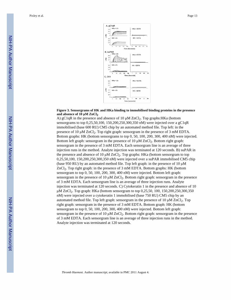
Figure 3. Sensorgrams of HK and HKa binding to immobilised binding proteins in the presenceand absence of 10 μM ZnCl2A) gC1qR in the presence and absence of 10 μM ZnCl2. Top graphs:HKa (bottomsensorgrams to top 0,25,50,100, 150,200,250,300,350 nM) were injected over a gC1qRimmobilised (base 600 RU) CM5 chip by an automated method file. Top left: in thepresence of 10 μM ZnCl2. Top right graph: sensorgram in the presence of 3 mM EDTA.Bottom graphs: HK (bottom sensorgrams to top 0, 50, 100, 200, 300, 400 nM) were injected.Bottom left graph: sensorgram in the presence of 10 μM ZnCl2. Bottom right graph:sensorgram in the presence of 3 mM EDTA. Each sensorgram line is an average of threeinjection runs in the method. Analyte injection was terminated at 120 seconds. B) suPAR inthe presence and absence of 10 μM ZnCl2. Top graphs: HKa (bottom sensorgram to top0,25,50,100, 150,200,250,300,350 nM) were injected over a suPAR immobilised CM5 chip(base 950 RU) by an automated method file. Top left graph: in the presence of 10 μMZnCl2. Top right graph: in the presence of 3 mM EDTA. Bottom graphs: HK (bottomsensorgram to top 0, 50, 100, 200, 300, 400 nM) were injected. Bottom left graph:sensorgram in the presence of 10 μM ZnCl2. Bottom right graph: sensorgram in the presenceof 3 mM EDTA. Each sensorgram line is an average of three injection runs. Analyteinjection was terminated at 120 seconds. C) Cytokeratin 1 in the presence and absence of 10μM ZnCl2. Top graph: HKa (bottom sensorgram to top 0,25,50, 100, 150,200,250,300,350nM) were injected over a cytokeratin 1 immobilised (base 750 RU) CM5 chip by anautomated method file. Top left graph: sensorgram in the presence of 10 μM ZnCl2. Topright graph: sensorgram in the presence of 3 mM EDTA. Bottom graph: HK (bottomsensorgram to top 0, 50, 100, 200, 300, 400 nM) were injected. Bottom left graph:sensorgram in the presence of 10 μM ZnCl2. Bottom right graph: sensorgram in the presenceof 3 mM EDTA. Each sensorgram line is an average of three injection runs in the method.Analyte injection was terminated at 120 seconds.
Pixley et al. Page 13
Thromb Haemost. Author manuscript; available in PMC 2011 August 4.
NIH
-PA Author Manuscript
NIH
-PA Author Manuscript
NIH
-PA Author Manuscript

Figure 4. CK1-gC1qR interactionsSensorgrams show comparative binding of purified binding proteins and HKa in thepresence of 10 μM ZnCl2 to immobilised cytokeratin 1 and gC1qR. HKa, cytokeratin 1, andgC1qR (all 100 nM) were injected in the sequence indicated in the figure. Injection timeswere adjusted, but the ΔRU were not adjusted, for comparative illustrative purposes. Arrowswith vertical dotted line indicates the position where each analyte was injected. The chip-linked binding protein base is indicated on the right each line. Each analyte injection was for50 seconds. A: peak of suPAR-HKa-gC1qR complex; B: peak of CK1-HKa-gC1qRcomplex; C: peak of CK1-gC1qR complex.
Pixley et al. Page 14
Thromb Haemost. Author manuscript; available in PMC 2011 August 4.
NIH
-PA Author Manuscript
NIH
-PA Author Manuscript
NIH
-PA Author Manuscript

Figure 5. CK1, gC1qR, suPAR interactionsSensorgram indicating lack of binding of CK1, gC1qR and suPAR to immobilised suPAR inthe presence of 10μM ZnCl2 (no binding was found in the absence of Zinc). 100 nM ofHKa, gC1qR, CK1 and suPAR were each injected separately at zero time. Each plotrepresents the mean of duplicate runs. Each analyte was injected for 60 seconds.
Pixley et al. Page 15
Thromb Haemost. Author manuscript; available in PMC 2011 August 4.
NIH
-PA Author Manuscript
NIH
-PA Author Manuscript
NIH
-PA Author Manuscript

Figure 6. HKa binding protein complexes on suPARSensorgram indicating lack of binding of CK1, gC1qR and suPAR to suPAR bound HKa.HKa (100 nM) in the presence of 10 μM ZnCl2 was first injected over an immobilisedsuPAR chip. Subsequent addition of 100 nM CK1, gC1qR, or suPAR did not increase thebinding signal. Each analyte injection was for 50 seconds.
Pixley et al. Page 16
Thromb Haemost. Author manuscript; available in PMC 2011 August 4.
NIH
-PA Author Manuscript
NIH
-PA Author Manuscript
NIH
-PA Author Manuscript

NIH
-PA Author Manuscript
NIH
-PA Author Manuscript
NIH
-PA Author Manuscript
Pixley et al. Page 17
Tabl
e 1
Bin
ding
con
stan
ts o
f HK
and
HK
a to
imm
obili
sed
kini
noge
n bi
ndin
g pr
otei
ns in
the
pres
ence
of 1
0 μ
M Z
nCl 2
. Mea
n ±
SD o
f n=3
–4 o
f com
bine
d se
nsor
gram
s of e
ach
conc
entra
tion
usin
g B
iaC
ore
Lang
mui
r bin
ding
mod
el (s
ee M
ater
ials
and
met
hods
).
HK
HK
a
KD
kon
koff
KD
kon
koff
Imm
obili
sed
Prot
ein
nMM
−1 s−
1 ×10
4s−
1 ×10
4−nM
M−
1 s−
1 ×10
4s−
1 ×10
−4
gC1q
R (p
32)
0.8
± 0.
712
.3 ±
5.0
0.8
± 0.
50.
7 ±
0.5
24.4
± 1
0.9
1.4
± 0.
5
CK
115
.1 ±
1.0
3.6
± 0.
35.
4 ±
0.7
6.3
± 1.
95.
8 ±
0.3
3.6
± 0.
9
suPA
R2,
313.
5 ±
1,46
5.3
1.0
± 0.
716
4.8
± 22
.243
.6 ±
16.
621
.3 ±
11.
978
.5 ±
10.
6
Thromb Haemost. Author manuscript; available in PMC 2011 August 4.