Transdifferentiation of glioblastoma cells into vascular endothelial cells
Pajusola, Seppo Ylä-Herttuala and Kari AlitaloTuomas Tammela, Yulong He, Johannes Lyytikkä, Michael Jeltsch, Johanna Markkanen, Katri
ProteinsGrowth Factor-C/Vascular Endothelial Growth Factor Heparin-Binding Domain Fusion Distinct Architecture of Lymphatic Vessels Induced by Chimeric Vascular Endothelial
Print ISSN: 0009-7330. Online ISSN: 1524-4571 Copyright © 2007 American Heart Association, Inc. All rights reserved.is published by the American Heart Association, 7272 Greenville Avenue, Dallas, TX 75231Circulation Research
doi: 10.1161/01.RES.0000269043.51272.6d2007;100:1468-1475; originally published online May 3, 2007;Circ Res.
http://circres.ahajournals.org/content/100/10/1468World Wide Web at:
The online version of this article, along with updated information and services, is located on the
http://circres.ahajournals.org//subscriptions/
is online at: Circulation Research Information about subscribing to Subscriptions:
http://www.lww.com/reprints Information about reprints can be found online at: Reprints:
document. Permissions and Rights Question and Answer about this process is available in the
located, click Request Permissions in the middle column of the Web page under Services. Further informationEditorial Office. Once the online version of the published article for which permission is being requested is
can be obtained via RightsLink, a service of the Copyright Clearance Center, not theCirculation Researchin Requests for permissions to reproduce figures, tables, or portions of articles originally publishedPermissions:
by guest on August 23, 2014http://circres.ahajournals.org/Downloaded from by guest on August 23, 2014http://circres.ahajournals.org/Downloaded from
Distinct Architecture of Lymphatic Vessels Induced byChimeric Vascular Endothelial Growth Factor-C/Vascular
Endothelial Growth Factor Heparin-Binding DomainFusion Proteins
Tuomas Tammela,* Yulong He,* Johannes Lyytikka, Michael Jeltsch, Johanna Markkanen,Katri Pajusola, Seppo Yla-Herttuala, Kari Alitalo
Abstract—Vascular endothelial growth factor (VEGF)-C and VEGF-D are composed of the receptor-binding VEGFhomology domain and a carboxy-terminal silk homology domain that requires proteolytic cleavage for growth factoractivation. Here, we explored whether the C-terminal heparin-binding domain of the VEGF165 or VEGF189 isoform alsocontaining neuropilin-binding sequences could substitute for the silk homology domain of VEGF-C. Such VEGF-C/VEGF–heparin-binding domain chimeras were produced and shown to activate VEGF-C receptors, and, whenexpressed in tissues via adenovirus or adeno-associated virus vectors, stimulated lymphangiogenesis in vivo. However,both chimeras induced a distinctly different pattern of lymphatic vessels when compared with VEGF-C. WhereasVEGF-C–induced vessels were initially a dense network of small diameter vessels, the lymphatic vessels induced by thechimeric growth factors tended to form directly along tissue borders, along basement membranes that are rich in heparansulfate. For example, in skeletal muscle, the chimeras induced formation of lumenized lymphatic vessels moreefficiently than wild-type VEGF-C. We conclude that the matrix-binding domain of VEGF can target VEGF-C activityto heparin-rich basement membrane structures. These properties may prove useful for tissue engineering and attemptsto regenerate lymphatic vessels in lymphedema patients. (Circ Res. 2007;100:1468-1475.)
Key Words: VEGF-C � VEGF-A � heparin-binding � lymphangiogenesis
The 5 mammalian vascular endothelial growth factor(VEGF) family members identified to date, VEGF,
VEGF-B, VEGF-C, VEGF-D, and placenta growth factor, arekey effectors of physiological and pathological regulation ofvasculogenesis, hematopoiesis, angiogenesis, lymphangio-genesis, and vascular permeability.1–3 VEGF is a key growthfactor for blood vessel formation and plays an essential rolein this process via VEGF receptor (VEGFR)-1 and VEGFR-2.1 VEGF-C and VEGF-D activate primarily VEGFR-34–8
and induce lymphangiogenesis in transgenic mice and inother in vivo models.8–11
VEGF is expressed as multiple forms, including the majorforms VEGF121, VEGF145, VEGF165, VEGF189, and VEGF206,which result from alternative RNA splicing.1 An importantbiological property that distinguishes these VEGF isoformsfrom each other is their different binding affinity to heparinand other heparan sulfates. Except for VEGF121, all of theother forms described above contain a heparin-binding do-main (HBD) encoded by exon 6 and/or exon 7. The 24-aa
residues encoded by exon 6 contain the HBD and alsoelements that enable its binding to the extracellular matrix.12
VEGF molecules containing the cationic polypeptide se-quence encoded by exon 7 (44 aa) are also heparin-bindingand remain bound to the cell surface and the extracellularmatrix.13 VEGF exon 7–encoded domain also enables itsbinding to neuropilin-1 (NP-1).14 Other members of theVEGF family that contain a HBD include VEGF-B167
15 andplacenta growth factor-2.16,17 There is increasing evidencepointing to the importance of the HBD for the biologicalactivity of VEGF.18,19
VEGF-C and VEGF-D have a C-terminal domain homol-ogous to certain silk proteins, plus an amino terminal propep-tide. A proteolytic cleavage between the growth factordomain and the silk domain activates VEGF-C binding toVEGFR-3, and the N-terminally cleaved mature form(VEGF-C�N�C) can also activate VEGFR-2 in blood vesselendothelium, resulting in angiogenic activity.20–24 However,in transgenic models in which both wild-type and mutant
Original received February 5, 2007; revision received March 30, 2007; accepted April 25, 2007.From the Molecular/Cancer Biology Laboratory and Ludwig Institute for Cancer Research (T.T., Y.H., J.L., M.J., K.P., K.A.), Biomedicum Helsinki,
the Haartman Institute and Helsinki University Central Hospital, University of Helsinki; and Department of Biotechnology and Molecular Medicine (J.M.,S.Y.-H.), A.I. Virtanen Institute, University of Kuopio, Finland.
*Both authors contributed equally to this work.Correspondence to Dr Kari Alitalo, University of Helsinki, Molecular/Cancer Biology Program, Biomedicum Helsinki, POB 23 (Haartmaninkatu 8),
Helsinki 00014, Finland. E-mail [email protected]© 2007 American Heart Association, Inc.
Circulation Research is available at http://circres.ahajournals.org DOI: 10.1161/01.RES.0000269043.51272.6d
1468 by guest on August 23, 2014http://circres.ahajournals.org/Downloaded from

forms of VEGF-C induced lymphangiogenesis in the skin, noangiogenic effect was observed, suggesting that during em-bryonic development, VEGF-C may not be fully processed toa form that activates the VEGFR-2 in blood vessels.7,9
In this study, we investigated the contribution of a matrix-binding domain to the in vivo activity of VEGF-C. The exon6 to 8 or exon 7 to 8 encoded domains from VEGF were fusedto the C terminus of the fully processed VEGF-C�N�Clacking the N- and C-terminal propeptides. With these con-structs, we wanted to investigate whether the heparin- andneuropilin-binding property can alter the lymphangiogenic orangiogenic effects of VEGF-C�N�C.
Materials and MethodsCell Culture293T cells from American Type Culture Collection were maintainedin DMEM (HaartBio, Helsinki, Finland) supplemented with2 mmol/L L-glutamine, penicillin (100 U/mL), streptomycin (100�g/mL), and 10% FBS (Autogen Bioclear). Hela cells were main-tained in DMEM, and Ba/F3 cells25 were grown in DMEM as above,with the addition of Zeocin (200 �g/mL) and recombinant humanVEGF-C�N�C (corresponding to the fully processed mature shortform, 100 ng/mL).
CloningcDNAs encoding the fusion proteins comprising the VEGF-C signalsequence (amino acids 1 to 31), the VEGF-C�N�C domain (amino acids103 to 225) and the C terminus of VEGF (exon 6 to 8 encoded polypeptidefragment, named CA89; or exon 6 to 7 encoded fragment, named CA65)were constructed by PCR amplification using the following primers:VEGF-C�N�C, 5�-ACATTGGTGTGCACCTCCAAGC-3� and 5�-AATAATGGAATGAACTTGTCTGTAAAC-3�; VEGF C-terminalregions, 5�-AAATCAGTTCGAGGAAAGGGAAAG-3� or 5�-CCCTGTGGGCCTTGCTCAGAG-3� and 5�-ACCATGCTCGAGAGT-CTTTCCTGGTGAGAGATCTGG-3�. The PCR products were digestedwith HindIII (5�-HindIII/3�-blunt) or XhoI (5�-blunt-3�-XhoI) and clonedinto the pEBS7 expression vector that had been digested with the sameenzymes, resulting in constructs pEBS7/CA89 and pEBS7/CA65.
Transfection and Immunoprecipitation293T cells were transfected with pEBS7/CA89, pEBS7/CA65,pEBS7/VEGF-C�N�C, or pEBS7 vector using liposomes (FuGENE6, Roche). Transfected cells were cultured for 24 hours and werethen metabolically labeled in methionine-free and cysteine-freemodified Eagle’s medium supplemented with [35S]methionine/[35S]cysteine (Promix, GE Healthcare) at 100 �Ci/mL for 8 hours.Cells transfected with pEBS7/CA89 were cultured with or withoutheparin (20 U/mL). Conditioned medium was harvested, cleared ofparticulate material by centrifugation, depleted of VEGF by immu-noprecipitation with anti-VEGF antibody (R&D Systems), andincubated with soluble VEGFR1-Ig, VEGFR2-Ig, VEGFR3-Ig, orpolyclonal antibodies against VEGF-C.20 The formed growth factor–antibody or –receptor complexes were bound to protein A–Sepharoseor protein G–Sepharose (GE Healthcare), which were washed twicewith 0.5% BSA/0.02% Tween-20 in PBS and once with PBS andanalyzed in SDS-PAGE under reducing conditions.
For the analysis of neuropilin-binding, conditioned medium fromtransfected cells was used. Briefly, both NP-1–Ig and NP-2(a22)–Igfusion proteins26 were transiently transfected into 293T cells. Thetransfected cells were cultured in serum-free medium for 48 hours,the conditioned medium was collected, cleared by centrifugation,and used for binding analysis as described above.
Production and In Vivo Delivery of CA89 andCA65 by Viral VectorsThe adeno-associated virus (AAV) vector psub-CAG-WPRE hasbeen derived by substituting the cytomegalovirus (CMV) promoter
fragment of psub-CMV-WPRE with the CMV–chicken �-actininsert. The cDNAs encoding CA89 and CA65 were cloned asblunt-end fragments into the psub-CAG-WPRE plasmid, and therecombinant AAVs (AAV-CA89 and AAV-CA65, AAV serotype 2)were produced as previously described.26 The cDNAs encodingCA89 and CA65 were also cloned into the pAdBglII vector(AdCA89 and AdCA65), and recombinant adenoviruses were pro-duced as described previously.27 HeLa cells used for expressionanalysis were transduced with AAVs (2000 multiplicities of infec-tion) or adenoviruses (50 multiplicities of infection). Expression ofthe recombinant proteins was examined by metabolic labeling andimmunoprecipitation, followed by SDS-PAGE analysis as describedabove.
All animal experiments were approved by the Committee forAnimal Experiments of the District of Southern Finland. Approxi-mately 3�108 plaque forming units of AdCA89, AdCA65,AdVEGF-C, AdC�N�C,- or �-galactosidase-encoding AdLacZcontrol virus or AAVs (AAV-CA89, AAV-CA65, AAV-VEGF-C,AAV-C�N�C, AAV-VEGF-B167, or AAV-EGFP; approximately1�1010 viral particles) were injected subcutaneously into mouseears. Tissues were collected for histological analysis 2 weeks afteradenoviral, or 6 weeks or 2 years after AAV transduction. Semi-membranosus muscles of rabbit hindlimbs were transduced withadenoviral vectors, as previously.28
Bioassay for Growth Factor–MediatedCell SurvivalBa/F3 cells expressing the VEGFR-3/EpoR chimeric receptor25 wereseeded in 96-well plates at 15 000 cells per well in triplicate, andsupplied with conditioned medium from Hela cells transduced withAdLacZ, AdVEGF-C, AdVEGF-C�N�C, AdCA65, or AdCA89(1:80 dilution). Cell viability was quantified by a colorimetric assayafter 48 hours. Briefly, 3-(4,5-dimethylthiazol-2-yl)-2,5-diphenyltetrazolium bromide (MTT) (Sigma; 0.5 mg/mL) was added intoeach well and incubated for 4 hours at 37°C. The reaction wasterminated by adding lysis buffer (10% SDS, 10 mmol/L HCl), andthe resulting formazan products were solubilized overnight at 37°Cin a humid atmosphere. The absorbance, at 540 nm, was measuredwith a Multiscan microtiter plate reader (Labsystems).
Generation of AntiseraWe generated novel rabbit antisera to the core domain of humanVEGF-C (VEGF-C�N�C), designated VEGF-C�N�C no. 3 andVEGF-C�N�C no. 4. Recombinant, mature hexahistidine-taggedVEGF-C (amino acids 103 to 215) was produced using the FastBacsystem (Invitrogen) and purified using Ni2�–nitrilotriacetic acidaffinity chromatography, followed by dialysis against PBS. Poly-clonal rabbit antiserum was obtained using standard procedures.29
ImmunohistochemistryFor whole-mount staining, mice were fixed by intracardiac perfusionwith 1% paraformaldehyde, blocked with 3% milk in PBS, andincubated with polyclonal antibodies against the lymphatic endothe-lial–specific hyaluronan receptor LYVE-1,30 full-length VEGF-C,31
VEGF-C�N�C (#3 and #4) or VEGFR-3 (R&D Systems) ormonoclonal antibodies against platelet endothelial cell adhesionmolecule (PECAM)-1 (PharMingen) or phosphohistone H3 (Up-state) overnight at 4°C.32 Appropriate Alexa 647–, Alexa594–, orAlexa488– conjugated secondary antibodies (Molecular Probes)were used for staining. All fluorescently labeled samples weremounted with Vectashield containing DAPI (4�,6-diamidino-2-phenylindole) (VectorLabs) and analyzed with a compound fluores-cent microscope (Zeiss 2, Carl Zeiss, Gottingen, Germany; �10objective with 0.30 numerical aperture [NA]) or a confocal micro-scope (Zeiss LSM 510; 40� objective with 1.3 NA and �63objective with 1.4 NA) by using multichannel scanning in framemode. Three-dimensional projections were digitally constructedfrom confocal z-stacks. For staining of tissue sections, tissues werefixed in 4% paraformaldehyde overnight at 4°C and paraffin sections
Tammela et al Biological Activity of Heparin-Binding VEGF-C 1469
by guest on August 23, 2014http://circres.ahajournals.org/Downloaded from

(6 �m) were immunostained with anti–LYVE-1 as described previ-ously26 and were analyzed with a Leica DMLB microscope.
Statistical AnalysisThe number of LYVE-1–positive lumenized vessels was countedfrom �400 micrographs containing the highest vessel density (3micrographs/section). Proliferating blood vascular endothelial cellswere counted as phosphohistone H3 and PECAM-1 double positivecells from �400 confocal micrographs of whole mount preparationsof the AAV-tranduced mouse ears. Colocalization was assessed fromsingle confocal z-sections, as previously.32 The cross-sectionalsurface area of capillaries in rabbit semimembranosus muscle wascounted from PECAM-1–stained sections. All quantifications werecounted from three �400 microscopic fields in each ear section andat least 3 sections from each ear or muscle, and the analyticaltechnique and time point were analyzed. At least 3 animals wereused for each time point and analytical technique, and each experi-ment was repeated at least 3 times. Statistical analysis was performedusing Student’s unpaired t test. A probability value of less than 0.05was considered to be statistically significant.
Results
Fusion of the HBD of VEGF to the C Terminus ofMature VEGF-CTo investigate whether the HBD of VEGF, which is knownto play an important role in regulating VEGF activity,33,34
would alter the lymphangiogenic or angiogenic effects ofVEGF-C�N�C, we constructed plasmids encoding chi-meric proteins comprising the VEGF-C signal sequence,followed by the VEGF-C�N�C domain and VEGF exon 6to 8 or exon 7 to 8 encoded sequences. These constructs,named CA89 and CA65, are schematically shown in Figure1A. We also made constructs where both N- andC-terminal propeptides were swapped (ACA), but these
were very poorly expressed, suggesting protein foldingproblems (data not shown).
CA65 and CA89 Proteins Bind to VEGFR-2,VEGFR-3, and NeuropilinsProduction of the chimeric VEGF-C proteins into the mediaof transfected cells was tested by immunoprecipitation usingpolyclonal antibodies against VEGF-C. The results showedthat CA65 is secreted into the medium, whereas CA89 wasnot released from the cells unless heparin was included in theculture medium (Figure 1B, compare lanes �H and �H),indicating that this form was strongly bound to cell surfaceheparan sulfate similar to what has been described forVEGF189. Analysis of the receptor binding profiles of thechimeric molecules indicated that similar to VEGF-C�N�C,both CA89 and CA65 bound to VEGFR-2 and VEGFR-3, butnot to VEGFR-1 (Figure 1B). In agreement with the bindingof VEGF exon 7 containing sequences to neuropilin,14,35 bothCA89 and CA65 also bound strongly to NP-1 and moreweakly to NP-2, whereas VEGF-C�N�C showed barelydetectable binding to NP-2 but not to NP-1 (Figure 1C).
Biological Activity of CA65 and CA89 Expressedvia Adenovirus VectorTo further characterize the biological functions of the chi-meric proteins in vivo, the cDNAs encoding CA89 and CA65were used to generate recombinant adenoviruses (AdCA89and AdCA65). Recombinant AAVs (AAV-CA89 and AAV-CA65) were produced to study the effect of long-termexpression of the chimeric proteins in skeletal muscle. Shownin Figure 1D is the analysis of polypeptides produced by therecombinant adenoviruses and AAVs. The biological activity
Figure 1. Biochemical analysis of thechimeric proteins. A, Schematic illustra-tion of the VEGF-C/VEGF chimeric mole-cules comprised of the signal sequence(SS) of VEGF-C, VEGF-C�N�C, andVEGF exon 6 to 8 or exon 7 to 8encoded sequences (CA89 and CA65,respectively). The depictions are notdrawn to scale. B through D, Immuno-precipitation and polyacrylamide gelelectrophoresis of metabolically labeledproteins from the conditioned medium of293T cells transfected with pEBS7/CA89(CA89) (B), pEBS7/CA65 (CA65) (B),pEBS7/VEGF-C�N�C (�N�C) (D), or thepEBS7 vector with anti–VEGF-C serum(B), VEGFR-1–Ig (R-1) (B), VEGFR-2–Ig(R-2) (B), and VEGFR-3–Ig (R-3) (B) orNP-1–Ig (NP1) (C) and NP-2–Ig (NP2) (C).�H and �H indicate medium with orwithout heparin (20 U/mL), respectively.D, Recombinant AAV and adenoviralexpression of CA89, CA65, VEGF-C�N�C, and VEGF-C were analyzed byimmunoprecipitation of metabolicallylabeled proteins with anti–VEGF-C serumas above. E, The biological activity ofthe VEGF-C chimeric proteins assessedin a bioassay using Ba/F3 cells express-
ing a chimeric VEGFR-3/erythropoietin receptor (EpoR) that mediates Ba/F3/VEGFR-3 cell survival as detailed in Materials and Meth-ods. Data represent the mean values from 3 triplicate assays.
1470 Circulation Research May 25, 2007
by guest on August 23, 2014http://circres.ahajournals.org/Downloaded from

of the chimeric proteins was demonstrated in a bioassay usingBa/F3 cells expressing a chimeric VEGFR-3/erythropoietinreceptor (Ba/F3-VEGFR-3/EpoR).25 Conditioned mediumfrom adenovirus-transduced cells containing CA89 or CA65was shown to induce survival and proliferation of these cells(Figure 1E).
A Distinct Pattern of Lymphatic Vessels IsInduced by the CA65 and CA89 Chimeric ProteinsFor comparison of their vascular effects, adenoviruses encod-ing CA89, CA65, VEGF-C�N�C, and VEGF-C were in-jected subcutaneously into the ears of nude mice. Two weeksafter virus transduction, tissues were collected for whole-mount immunostaining of lymphatic vessels using antibodiesagainst the lymphatic endothelial–specific hyaluronan recep-tor LYVE-1. Both AdCA89 and AdCA65 were shown toinduce robust lymphangiogenesis (Figure 2A and 2B) incomparison with the AdLacZ control virus (Figure 2E).However, the lymphangiogenic responses to both CA65 andCA89 appeared different from those induced by full-lengthVEGF-C or mature VEGF-C (C�N�C) (Figure 2A through2D). The CA65- and CA89-generated vessels were thickerand formed a more sparse network, whereas a robust lym-phangiogenic response consisting of a dense network ofvery-fine lymphatic capillaries was induced with the full-length VEGF-C (Figure 2C), whereas VEGF-C�N�C in-duced a weaker widespread lymphangiogenic effect charac-terized by lymphatic sprouting and hyperplasia (Figure 2D).The average number of LYVE-1–positive vessels determinedfrom 3 microscopic fields of the highest vessel density isshown in Figure 2F. Assuming that roughly similar levels ofgrowth factor proteins were expressed in each case from theadenovirus vector, it appeared that the ability of VEGF-C�N�C, CA65, and CA89 to induce formation of lymphaticvessels was significantly weaker than that of full-lengthVEGF-C (P�0.001).
CA65 and CA89 Delivered via the AAV VectorInduce Lymphangiogenesis Along Muscle FibersConsistent with the fact that AAV mainly transduces musclecells,36 we detected expression of the growth factors only inthe thin layer of skeletal muscle of the mouse ear by stainingwith antibodies that detect the VEGF homology domain ofVEGF-C (VEGF-C/VHD) (Figure 3A through 3C). Theseantibodies did not detect skeletal muscle fibers transducedwith AAV-VEGF-B167 used as a control (Figure 3D). At 6weeks after transduction, the AAV-CA65–induced lymphaticcapillaries were organized mostly in parallel along the musclefibers (arrowheads, Figure 3A and 3E), whereas the AAV-VEGF-C and AAV-C�N�C–induced a very dense networkof thin, rather unorganized vessels (Figure 3B, 3C, 3F, and3G). Few lymphatic vessels forming along the muscle fiberswere seen in the AAV-VEGF-C–transduced ears (arrow-heads, Figure 3F). AAV-VEGF-B167 transduction did notstimulate lymphangiogenesis, and only a few quiescent lym-phatic capillaries were observed in the muscle layer (Figure1D and 1H). Even tighter association of developing lymphaticvessels with the muscle fibers was seen in samples treated
with AAV-CA89 (Figure 3I), when compared with thoseinduced in response to AAV-VEGF-C (Figure 3J).
The VEGF-C–induced lymphatic capillaries reorganizedslowly along the muscle fibers, as seen when mice injectedwith the AAV vectors were analyzed at 6 weeks and at 2years after transduction (Figure 3K and 3L). In strikingcontrast, the CA65-induced vessels were already well-oriented along the fibers at 6 weeks (Figure 3M). Forcomparison, shown are lymphatic capillaries in the overlyingskin in Figure 3N. In histological sections, the AAV-CA65–and AAV-CA89–induced LYVE-1–positive vessels wereobserved in between the myofibers (Figure 3O), whereas onlya few lymphatic vessels were found in corresponding sectionsfrom the control mice (Figure 3P). We found very fewlymphatic vessels in the skeletal muscle layer of mouse earsof mice that received AAV-VEGF-B167, whereas abundantlumenized vessels were found in AAV-CA65– or AAV-CA89–transduced ears (Figure 3Q). Importantly, more ves-sels containing a lumen were found in the muscle layer of earsexpressing the chimeric growth factors when compared with
Figure 2. In vivo effects of the chimeric growth factors afteradenoviral gene transduction. A through E, Whole-mount stain-ing for LYVE-1 (red) of the mouse ears 2 weeks after transduc-tion with AdCA65 (A), AdCA89 (B), AdVEGF-C (C), AdVEGF-C�N�C (D), or AdLacZ (E). F, Quantification of LYVE–positivelumenized vessels from sections of the ear skins transducedwith adenoviruses. *P�0.05 compared with LacZ, **P�0.05compared with all other groups. Scale bar�100 �m.
Tammela et al Biological Activity of Heparin-Binding VEGF-C 1471
by guest on August 23, 2014http://circres.ahajournals.org/Downloaded from

AAV-VEGF-C– or AAV-C�N�C–transduced ears (Figure3Q).
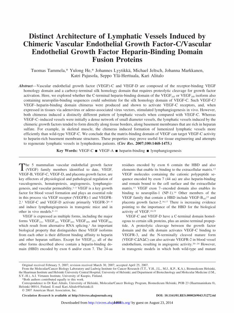
CA65 and CA89 Induce Mild Changes in theBlood VesselsA weak angiogenic response characterized by increasedarterial tortuosity was observed in PECAM-1–stained skininjected with AdCA65, AdCA89, AdVEGF-C, or AdVEGF-C�N�C (arrowheads in Figure 4A through 4E; data notshown). We did not detect statistically significant changes invessel area density in any of the growth factor–transducedears when compared with control (data not shown), although
all 4 factors also stimulated proliferation of blood vascularendothelial cells when expressed in the ear via AAV vectors,with CA89 displaying a trend toward the strongest response(P�0.119; Figure 4F). The CA65 and CA89 adenovirusesinduced a barely detectable widening of muscle capillaries inrabbit hindlimb (Figure 4G through 4I). CA65 increased thecross-sectional capillary surface area 1.9-fold (P�0.011) andCA89 1.6-fold (P�0.0072) when compared with AdLacZ.We also observed a slight trend toward an increase incapillary permeability, as measured by the Miles permeabilityassay,28 but the increase was not statistically significantcompared with the control (data not shown).
Figure 3. Induction of lymphatic vesselgrowth along muscle fibers by AAV-CA65 and AAV-CA89. A through H,Whole-mount immunostaining ofVEGFR-3 (red) and VEGF-C �N�C(green), included in all chimeric and wild-type VEGF-C constructs used in thestudy, in the ears of mice transduced 6weeks before analysis with AAV-CA65(A), AAV-VEGF-C (B), AAV-C�N�C (C),or AAV-VEGF-B167 (D). Arrowheads indi-cate lymphatic vessels forming alongmuscle fibers transduced with the vector.I and J, Very-high-resolution confocalanalysis of ears transduced with AAV-CA89 (I) or AAV-VEGF-C (J). The smallerimages at right in I and J are cross-sections of confocal z-stacks. VEGFR-3staining is in red, VEGF-C�N�C in green,and DNA in blue. K through N, Lym-phatic vessels visualized by whole-mountLYVE-1 immunostaining (red) in the ears6 weeks (K and M) or 2 years (L and N)after transduction with AAV-VEGF-C.Normal LYVE-1–positive lymphatic ves-sels (red) and AAV-EGFP expression(green) 2 years after transduction areshown in the inset in L. O and P, LYVE-1immunostaining (red) and hematoxylincounterstaining (blue) of paraffin sectionsfrom ears transduced with AAV-CA65 orAAV-EGFP. LYVE-1–positive lymphaticvessels with a lumen are indicated witharrows. Scale bars: 100 �m (A through Hand K through P); 10 �m (I and J). Q,Quantification of the number of LYVE-1–positive vessels containing a lumen inmouse ears 6 weeks after transductionwith AAV vectors. *P�0.05.
1472 Circulation Research May 25, 2007
by guest on August 23, 2014http://circres.ahajournals.org/Downloaded from
DiscussionHere, we have sought to determine the ability of a strongpericellular matrix-binding domain to influence the in vivoactivity of VEGF-C. We demonstrated that the chimericCA89 and CA65 proteins accommodating the growth factordomain of VEGF-C and the HBD of VEGF stimulateddistinct patterning of the growth factor–induced lymphaticvessels.
The altered biological activity of CA89 and CA65 incomparison with VEGF-C may result from redistribution ofthe growth factors by binding of the HBD to heparin-richpericellular matrix structures that typically are present inbasal lamina and on the surface of certain cells. It is possiblethat by fusion with the HBD the 3D diffusion of VEGF-C�N�C is largely replaced by 2D mobility in the plane of thecell surface heparan sulfate matrix, which leads to more of thegrowth factor available for the high-affinity signal–transduc-ing receptors. The importance of the exon 6 and 7 encodedsequences in VEGF165 and VEGF189 was emphasized whenCarmeliet et al observed impaired myocardial angiogenesisand ischemic cardiomyopathy in mice lacking the corre-sponding mouse VEGF isoforms.18 In fact, the lymphangio-genic response we obtained with VEGF-C�N�C is highlysimilar to the angiogenic response that has been observedwith VEGF121,18,37 as both factors potently stimulate endothe-lial cell proliferation but fail to provide the cells withsufficient guidance cues, which leads to formation of endo-thelial sheets instead of functional vessels.
Processing of heparan sulfate–bound VEGF by plasminmatrix metalloproteinases regulates its bioavailability andvascular patterning.37 Interestingly, VEGF signaling in endo-thelial cells is fully supported by heparan sulfate expressed in
trans by adjacent perivascular smooth muscle cells.38 Suchability of the HBD to regulate vessel patterning was alsoreflected in the lymphangiogenic activity of the correspond-ing chimeras CA65 and CA89. Importantly, we did notobserve any changes in lymphatic vessels after gene transferof VEGF-B167, which contains similar HBD and neuropilin-binding domains as VEGF189 and CA89,15 indicating thatheparin and neuropilin binding capacities alone are notsufficient to stimulate either lymphangiogenesis or angiogen-esis unless the factor is able to activate VEGFR-2 and/orVEGFR-3.
Some of the distinct biological activities of CA65 andCA89 may also be attributable to the observed increasedbinding to NP-1 or NP-2, which regulate vessel patterning. Ithas been shown that NP-1 enhances VEGF165 binding toVEGFR-2 by forming a ternary complex on endothelial cellsurfaces,39 whereas NP-2, a possible coreceptor for VEGF-C,is required for the normal development of lymphatic ves-sels.40,41 NP-1–signaling activity has been shown to regulatetip cell guidance and the fusion of sprouts of adjacentvessels.42 Furthermore, recent experiments have indicatedthat NP-1 is essential for VEGF-induced vascularremodeling.43
VEGF-C was shown to induce angiogenesis in mousecorneas,22 and a dose-dependent angiogenic response wasalso observed with adenoviral expression in normal mouseskin,44 as well as in wounds of diabetic mice.45 Consistentwith this, all tested adenoviral vectors induced a modestincrease in endothelial cell proliferation. The CA89 growthfactor chimera that binds very tightly to the pericellularmatrix showed a trend toward highest angiogenic activity.Furthermore, we also investigated the vascular effects in a
Figure 4. Blood vascular effects of the chi-meric growth factors. A through E, Whole-mount analysis of blood vascular changes(PECAM-1, green) in the mouse ear 2 weeksafter adenoviral gene transduction. Arrow-heads indicate tortuous vessels. F, Quantita-tive analysis of proliferating blood vascularendothelial cells (phosphohistoneH3/PECAM-1 double-positive cells) from�400 confocal micrographs 6 weeks afterAAV-mediated gene transfer. G through I,PECAM-1 immunostaining of the rabbitsemimembranosus muscle 6 days aftergene transfer. Arrowheads indicate enlargedcapillaries. Scale bars�100 �m.
Tammela et al Biological Activity of Heparin-Binding VEGF-C 1473
by guest on August 23, 2014http://circres.ahajournals.org/Downloaded from
rabbit nonischemic limb model, in which AdCA89 andAdCA65 were injected into the semimembranosus muscle,and animals were euthanized 6 days later. Consistent with themouse data, a slight increase in capillary diameter wasobserved.
In summary, the heparin-binding chimeric VEGF-C formsinduced a distinct pattern of lymphatic vessel growth longi-tudinally along the basement membranes and muscle fibers.This suggests that by using a heparin-binding growth factor,one can achieve a more defined localization of growth factorexpression in a given tissue and, therefore, minimize thedanger of obtaining aberrant side effects at other sites. Moregenerally, we envision that such growth factor domain swapcombinations should have a great potential for buildingvascular networks in tissue-engineering and other therapeuticapplications.
AcknowledgmentsWe acknowledge the Biomedicum Molecular Imaging Unit forexpertise with confocal microscopy. We thank Mari Helantera, PaulaHyvarinen, Tanja Laakkonen, Sanna Lampi, Anna Malinen, andTapio Tainola for excellent technical assistance.
Sources of FundingThis work was supported by grants from the NIH (5 R01 HL075183-02), the European Union (Lymphangiogenomics, LSHG-CT-2004-503573), the Academy of Finland (202852 and 204312), and theSigrid Juselius Foundation. T.T. was supported by grants from theEmil Aaltonen Foundation, the Biomedicum Helsinki Foundation,the Finnish Cancer Organizations, the Finnish Cultural Foundation,the Paulo Foundation, the University of Helsinki, and the OskarOflund Foundation.
DisclosuresKari Alitalo is a minority shareholder and board member ofLymphatix Ltd.
References1. Ferrara N, Gerber HP, LeCouter J. The biology of VEGF and its
receptors. Nat Med. 2003;9:669–676.2. Alitalo K, Tammela T, Petrova TV. Lymphangiogenesis in development
and human disease. Nature. 2005;438:946–953.3. Carmeliet P. Mechanisms of angiogenesis and arteriogenesis. Nat Med.
2000;6:389–395.4. Joukov V, Pajusola K, Kaipainen A, Chilov D, Lahtinen I, Kukk E,
Saksela O, Kalkkinen N, Alitalo K. A novel vascular endothelial growthfactor, VEGF-C, is a ligand for the Flt4 (VEGFR-3) and KDR(VEGFR-2) receptor tyrosine kinases. EMBO J. 1996;15:290–298.
5. Lee J, Gray A, Yuan J, Luoh S-M, Avraham H, Wood WI. Vascularendothelial growth factor-related protein: A ligand and specific activatorof the tyrosine kinase receptor Flt4. Proc Natl Acad Sci U S A. 1996;93:1988–1992.
6. Achen MG, Jeltsch M, Kukk E, Makinen T, Vitali A, Wilks AF, AlitaloK, Stacker SA. Vascular endothelial growth factor D (VEGF-D) is aligand for the tyrosine kinases VEGF receptor 2 (Flk1) and VEGFreceptor 3 (Flt4). Proc Natl Acad Sci U S A. 1998;95:548–553.
7. Veikkola T, Jussila L, Makinen T, Karpanen T, Jeltsch M, Petrova TV,Kubo H, Thurston G, McDonald DM, Achen MG, Stacker SA, Alitalo K.Signalling via vascular endothelial growth factor receptor-3 is sufficientfor lymphangiogenesis in transgenic mice. EMBO J. 2001;6:1223–1231.
8. Makinen T, Jussila L, Veikkola T, Karpanen T, Kettunen MI, PulkkanenKJ, Kauppinen R, Jackson DG, Kubo H, Nishikawa S, Yla-Herttuala S,Alitalo K. Inhibition of lymphangiogenesis with resulting lymphedema intransgenic mice expressing soluble VEGF receptor-3. Nat Med. 2001;7:199–205.
9. Jeltsch M, Kaipainen A, Joukov V, Meng X, Lakso M, Rauvala H, SwartzM, Fukumura D, Jain RK, Alitalo K. Hyperplasia of lymphatic vessels inVEGF-C transgenic mice. Science. 1997;276:1423–1425.
10. Oh S-J, Jeltsch MM, Birkenhager R, McCarthy JE, Weich HA, Christ B,Alitalo K, Wilting J. VEGF and VEGF-C: specific induction of angio-genesis and lymphangiogenesis in the differentiated avian chorioallantoicmembrane. Dev Biol. 1997;188:96–109.
11. Yoon YS, Murayama T, Gravereaux E, Tkebuchava T, Silver M, Curry C,Wecker A, Kirchmair R, Hu CS, Kearney M, Ashare A, Jackson DG,Kubo H, Isner JM, Losordo DW. VEGF-C gene therapy augmentspostnatal lymphangiogenesis and ameliorates secondary lymphedema.J Clin Invest. 2003;111:717–725.
12. Poltorak Z, Cohen T, Sivan R, Kandelis Y, Spira G, Vlodavsky I, KeshetE, Neufeld G. VEGF145, a secreted vascular endothelial growth factorisoform that binds to extracellular matrix. J Biol Chem. 1997;272:7151–7158.
13. Gitay-Goren H, Cohen T, Tessler S, Soker S, Gengrinovitch S, RockwellP, Klagsbrun M, Levi BZ, Neufeld G. Selective binding of VEGF(121) toone of the three vascular endothelial growth factor receptors of vascularendothelial cells. J Biol Chem. 1996;271:5519–5523.
14. Soker S, Fidder H, Neufeld G, Klagsbrun M. Characterization of novelvascular endothelial growth factor (VEGF) receptors on tumor cells thatbind VEGF165 via its exon 7-encoded domain. J Biol Chem. 1996;271:5761–5767.
15. Makinen T, Olofsson B, Karpanen T, Hellman U, Soker S, Klagsbrun M,Eriksson U, Alitalo K. Differential binding of vascular endothelial growthfactor B splice and proteolytic isoforms to neuropilin-1. J Biol Chem.1999;274:21217–21222.
16. Maglione D, Guerriero V, Viglietto G, Ferraro MG, Aprelikova O, AlitaloK, Del Vecchio S, Lei K-J, Chou JY, Persico MG. Two alternativemRNAs coding for the angiogenic factor, placenta growth factor (PlGF),are transcribed from a single gene of chromosome 14. Oncogene. 1993;8:925–931.
17. Hauser S, Weich HA. A heparin-binding form of placenta growth factor(PlGF-2) is expressed in human umbilical vein endothelial cells and inplacenta. Growth Factors. 1993;9:259–268.
18. Carmeliet P, Ng YS, Nuyens D, Theilmeier G, Brusselmans K, Cor-nelissen I, Ehler E, Kakkar VV, Stalmans I, Mattot V, Perriard JC,Dewerchin M, Flameng W, Nagy A, Lupu F, Moons L, Collen D,D’Amore PA, Shima DT. Impaired myocardial angiogenesis and ische-mic cardiomyopathy in mice lacking the vascular endothelial growthfactor isoforms VEGF164 and VEGF188. Nat Med. 1999;5:495–502.
19. Ruhrberg C, Gerhardt H, Golding M, Watson R, Ioannidou S, FujisawaH, Betsholtz C, Shima DT. Spatially restricted patterning cues providedby heparin-binding VEGF-A control blood vessel branching morpho-genesis. Genes Dev. 2002;16:2684–2698.
20. Joukov V, Sorsa T, Kumar V, Jeltsch M, Claesson-Welsh L, Cao Y,Saksela O, Kalkkinen N, Alitalo K. Proteolytic processing regulatesreceptor specificity and activity of VEGF-C. EMBO J. 1997;16:3898–3911.
21. Stacker SA, Stenvers K, Caesar C, Vitali A, Domagala T, Nice E, RoufailS, Simpson RJ, Moritz R, Karpanen T, Alitalo K, Achen MG. Biosyn-thesis of vascular endothelial growth factor-D involves proteolytic pro-cessing which generates non-covalent homodimers. J Biol Chem. 1999;274:32127–32136.
22. Cao Y, Linden P, Farnebo J, Cao R, Eriksson A, Kumar V, Qi J-H,Claesson-Welsh L, Alitalo K. Vascular endothelial growth factor Cinduces angiogenesis in vivo. Proc Natl Acad Sci U S A. 1998;95:14389–14394.
23. Witzenbichler B, Asahara T, Murohara T, Silver M, Spyridopoulos I,Magner M, Principe N, Kearney M, Hu J-S, Isner JM. Vascular endo-thelial growth factor-C (VEGF-C/VEGF-2) promotes angiogenesis in thesetting of tissue ischemia. Am J Pathol. 1998;153:381–394.
24. Marconcini L, Marchio S, Morbidelli L, Cartocci E, Albini A, Ziche M,Bussolino F, Oliviero S. c-fos-induced growth factor/vascular endothelialgrowth factor D induces angiogenesis in vivo and in vitro. Proc Natl AcadSci U S A. 1999;96:9671–9676.
25. Achen MG, Roufail S, Domagala T, Catimel B, Nice EC, Geleick DM,Murphy R, Scott AM, Caesar C, Makinen T, Alitalo K, Stacker SA.Monoclonal antibodies to vascular endothelial growth factor-D block itsinteractions with both VEGF receptor-2 and VEGF receptor-3. EurJ Biochem. 2000;267:2505–2515.
26. Karkkainen MJ, Saaristo A, Jussila L, Karila KA, Lawrence EC, Pajusola K,Bueler H, Eichmann A, Kauppinen R, Kettunen MI, Yla-Herttuala S,Finegold DN, Ferrell RE, Alitalo K. A model for gene therapy of humanhereditary lymphedema. Proc Natl Acad Sci U S A. 2001;98:12677–12682.
27. Laitinen M, Makinen K, Manninen H, Matsi P, Kossila M, Agrawal RS,Pakkanen T, Luoma JS, Viita H, Hartikainen J, Alhava E, Laakso M,
1474 Circulation Research May 25, 2007
by guest on August 23, 2014http://circres.ahajournals.org/Downloaded from

Yla-Herttuala S. Adenovirus-mediated gene transfer to lower limb arteryof patients with chronic critical leg ischemia. Hum Gene Ther. 1998;9:1481–1486.
28. Rissanen TT, Markkanen JE, Gruchala M, Heikura T, Puranen A,Kettunen MI, Kholova I, Kauppinen RA, Achen MG, Stacker SA, AlitaloK, Yla-Herttuala S. VEGF-D is the strongest angiogenic and lym-phangiogenic effector among VEGFs delivered into skeletal muscle viaadenoviruses. Circ Res. 2003;92:1098–1106.
29. Harlow E, Lane D. Antibodies: A Laboratory Manual. Cold SpringHarbor, NY: Cold Spring Harbor Laboratory Press; 1988.
30. Karkkainen MJ, Haiko P, Sainio K, Partanen J, Taipale J, Petrova TV,Jeltsch M, Jackson DG, Talikka M, Rauvala H, Betsholtz C, Alitalo K.Vascular endothelial growth factor C is required for sprouting of the firstlymphatic vessels from embryonic veins. Nat Immunol. 2004;5:74–80.
31. Baluk P, Tammela T, Ator E, Lyubynska N, Achen MG, Hicklin DJ,Jeltsch M, Petrova TV, Pytowski B, Stacker SA, Yla-Herttuala S, JacksonDG, Alitalo K, McDonald DM. Pathogenesis of persistent lymphaticvessel hyperplasia in chronic airway inflammation. J Clin Invest. 2005;115:247–257.
32. Tammela T, Saaristo A, Lohela M, Morisada T, Tornberg J, Norrmen C,Oike Y, Pajusola K, Thurston G, Suda T, Yla-Herttuala S, Alitalo K.Angiopoietin-1 promotes lymphatic sprouting and hyperplasia. Blood.2005;105:4642–4648.
33. Gitay-Goren H, Soker S, Vlodavsky I, Neufeld G. The binding ofvascular endothelial growth factor to its receptors is dependent on cellsurface-associated heparin-like molecules. J Biol Chem. 1992;267:6093–6098.
34. Tessler S, Rockwell P, Hicklin D, Cohen T, Levi BZ, Witte L, Lemischka IR,Neufeld G. Heparin modulates the interaction of VEGF165 with soluble andcell associated flk-1 receptors. J Biol Chem. 1994;269:12456–12461.
35. Neufeld G, Kessler O, Herzog Y. The interaction of neuropilin-1 andneuropilin-2 with tyrosine-kinase receptors for VEGF. Adv Exp Med Biol.2002;515:81–90.
36. Daly TM. Overview of adeno-associated viral vectors. Methods Mol Biol.2004;246:157–165.
37. Lee S, Jilani SM, Nikolova GV, Carpizo D, Iruela-Arispe ML. Processingof VEGF-A by matrix metalloproteinases regulates bioavailability andvascular patterning in tumors. J Cell Biol. 2005;169:681–691.
38. Jakobsson L, Kreuger J, Holmborn K, Lundin L, Eriksson I, Kjellen L,Claesson-Welsh L. Heparan sulfate in trans potentiates VEGFR-mediatedangiogenesis. Dev Cell. 2006;10:625–634.
39. Soker S, Miao HQ, Nomi M, Takashima S, Klagsbrun M. VEGF165mediates formation of complexes containing VEGFR-2 and neuropilin-1 thatenhance VEGF165-receptor binding. J Cell Biochem. 2002;85:357–368.
40. Karkkainen MJ, Jussila L, Ferrell RE, Finegold DN, Alitalo K. Molecularregulation of lymphangiogenesis and targets for tissue oedema. TrendsMol Med. 2001;7:18–22.
41. Yuan L, Moyon D, Pardanaud L, Breant C, Karkkainen MJ, Alitalo K,Eichmann A. Abnormal lymphatic vessel development in neuropilin 2mutant mice. Development. 2002;129:4797–4806.
42. Gerhardt H, Ruhrberg C, Abramsson A, Fujisawa H, Shima D, BetsholtzC. Neuropilin-1 is required for endothelial tip cell guidance in thedeveloping central nervous system. Dev Dyn. 2004;231:503–509.
43. Pan Q, Chanthery Y, Liang WC, Stawicki S, Mak J, Rathore N, Tong RK,Kowalski J, Yee SF, Pacheco G, Ross S, Cheng Z, Le Couter J, PlowmanG, Peale F, Koch AW, Wu Y, Bagri A, Tessier-Lavigne M, Watts RJ.Blocking neuropilin-1 function has an additive effect with anti-VEGF toinhibit tumor growth. Cancer Cell. 2007;11:53–67.
44. Saaristo A, Veikkola T, Enholm B, Hytonen M, Arola J, Pajusola K,Turunen P, Jeltsch M, Karkkainen MJ, Kerjaschki D, Bueler H, Yla-Herttuala S, Alitalo K. Adenoviral VEGF-C overexpression inducesblood vessel enlargement, tortuosity, and leakiness but no sproutingangiogenesis in the skin or mucous membranes. FASEB J. 2002;16:1041–1049.
45. Saaristo A, Tammela T, Farkkila A, Karkkainen M, Suominen E, Yla-Herttuala S, Alitalo K. Vascular endothelial growth factor-C acceleratesdiabetic wound healing. Am J Pathol. 2006;169:1080–1087.
Tammela et al Biological Activity of Heparin-Binding VEGF-C 1475
by guest on August 23, 2014http://circres.ahajournals.org/Downloaded from
Copyright © 2022 FDOKUMEN