Isolation and Angiogenesis by Endothelial Progenitors in the Fetal Liver

British Joumal of Cancer (1998) 77(7), 1123-1129© 1998 Cancer Research Campaign
Linomide blocks angiogenesis by breast carcinomavascular endothelial growth factor transfectants
MZiche1, S Donnini1, L MorbideIi1, A Parenti1, G Gasparini2 and F Ledda
'Department of Pharmacology, University of Florence, Viale Morgagni 65, 50134 Florence; 2Department of Oncology, St Bortolo Hospital, Vicenza, Italy
Summary The blocking of angiogenesis provides a novel therapeutic target to inhibit tumour spreading. In this study, we investigated theeffect of linomide on angiogenesis induced in vivo by highly angiogenic breast carcinoma cells. The rabbit cornea was used to assessneovascular growth in the absence of a tumour mass. MCF-7 cells stably transfected with the cDNAencoding for vascular endothelial growthfactor 121 (VEGF121) (V12 clone) were used to elicit a potent VEGF-dependent corneal angiogenesis. After tumour cell implant, albino rabbitsreceived 100 mg kg-1 day-1 linomide for 5 consecutive days. Daily observation of neovascular progression indicated that linomide blockedangiogenesis. The antiangiogenic effect of linomide was apparent within 48 h from the beginning of the treatment and was bothangiosuppressive and angiostatic. The block of neovascular growth lasted over 10 days from treatment suspension, and preformed vessels,which had regressed, remained dormant, suggesting the persistence of unfavourable conditions for capillary progression. Linomide (50-200 gg ml-1) was not cytotoxic in vitro on resting capillary endothelial cells but blocked endothelial cell replication induced by VEGF. Our dataindicate that linomide can efficiently and persistently block VEGF-dependent angiogenesis in vivo in the absence of a growing tumour mass.These data suggest that linomide could be a chemopreventive drug in breast cancer patients and a valuable tool in clinical settings in whichmetastatic spreading occurs in the absence of a detectable tumour mass.
Keywords: Linomide; angiogenesis; vascular endothelial growth factor; breast carcinoma; endothelial cells
There is compelling experimental evidence that angiogenesis isnecessary for tumour growth and spreading. Clinicopathologicalstudies have shown that this can be translated in human tumours,such as breast cancer (Horak et al, 1992; Weidner et al, 1992; Toiet al, 1993: Gasparini et al, 1994; Gasparini and Harris, 1995). Inthe metastatic cascade angiogenesis is implicated both in the stageof shedding from a primary tumour and upon arrival of metastasesat their distant site (Folkman, 1995). Based on these observations,antiangiogenic therapy has been proposed as adjuvant therapy insolid primary tumours to prevent invasion and metastasis (Fan etal, 1995; Folkman, 1995).
Vascular endothelial growth factor (VEGF) appears to play akey role in tumour angiogenesis (Brown et al, 1993; Klagsbrunand Soker, 1993; O'Brien et al, 1995). In breast cancer, the expres-sion and levels (Toi et al, 1996; Gasparini et al, 1997) of VEGFcorrelate with high microvessel density, and both features areassociated with poor prognosis. Thus, breast cancer with highmicrovessel density and/or VEGF expression may be a likelycandidate for antiangiogenic treatment.
Transfection of VEGF121 into the human breast carcinoma cellline (MCF-7 cells) has been previously shown to enhance tumourgrowth and vascular density in vivo and to promote a strong angio-genic response (Zhang et al, 1995). VEGF121 transfectants (V12cells) thus provide a useful model for monitoring the antiangio-genesis effect of specific inhibitors.
The quinoline-3-carboxamide linomide has been reported tohave immunomodulatory activity (Kalland et al, 1985; Larsson et
Received 16 May 1997Revised 15 September 1997Accepted 16 September 1997
Correspondence to: MZiche
al, 1987; Bengtsson et al, 1992) and to block angiogenesis effi-ciently in experimental tumour models (Kalland, 1986; Harning etal, 1989; Ichikawa et al, 1992; Vukanovic et al, 1993; Joseph et al,1996). Moreover, we recently demonstrated the ability of linomideto selectively inhibit VEGF-induced migration and growth ofmicrovascular endothelium (Parenti et al, 1996; Ziche et al,1997a), indicating that the drug could target the inhibition ofVEGF-dependent tumour angiogenesis in vivo.
The cornea is an ideal model for monitoring the occurrence ofangiogenesis as it allows the direct observation of vascularprogression over time. By using wild-type MCF-7 and VEGF121transfectants, the aim of this study was to assess in vivo the effectof linomide on the angiogenesis induced by breast carcinoma cellpopulations. The effect of drug treatment was assessed on angio-genesis that had already been established by the tumour cells aswell as before neovascular growth had become apparent, twoexperimental settings that mimic the clinical conditions in whichtumour angiogenesis contributes to tumour progression andspreading. The effect of linomide on tumour and endothelial cellgrowth has also been investigated.
MATERIALSANDMETHODS
Angiogenesis in vivo: rabbit cornea assay
Angiogenesis was studied in the cornea of albino rabbits, as this isan avascular and transparent tissue in which inflammatory reac-tions and growing capillaries can be easily monitored and themodification can be quantitated by stereomicroscopic examina-tion. Female NewZealand white rabbits (2.5-3 kg, Charles River,Calco, Como, Italy) were used in accordance with the guidelinesof the European Economic Community for animal care andwelfare (EEC law no. 86/609). In animals anaesthetized by sodium
1123

1124 MZiche et al
pentothal (30 mg kg-'), a micropocket (1.5x3 mm) was surgicallyproduced using a pliable iris spatula 1.5 mmwide in the lower halfof the rabbit eye. Approximately 2.5x10⁵ wild-type MCF-7 cells(WT) or VI 2 transfectants as a cell suspension in a 10-,l volumewere implanted in the micropocket following a procedurepreviously reported (Ziche and Gullino, 1982). Subsequent dailyobservations of the implants were made with a slit lamp stereo-microscope. An angiogenic response was scored positive whenbudding of vessels from the limbal plexus occurred after 48 h andcapillaries progressed to reach the implant according to thescheme previously reported (Ziche et al, 1989). The number ofpositive implants over the total implants performed was scoredduring each observation.
The potency of angiogenic activity was evaluated on the basisof the number and growth rate of newly formed capillaries, and anangiogenic score was calculated (vessel density x distance fromlimbus) (Ziche et al, 1997b). A density value of 1 corresponded to0-25 vessels, 2 from 25 to 50, 3 from 50 to 75, 4 from 75 to 100, 5from 100 to 200 and 6 for more than 200 vessels. The distance(mm) from the limbus was graded with the aid of an ocular grid.Corneas were removed at the end of the experiment as well as ata defined interval from surgery and/or treatment and fixed informalin for histological examination.
Linomide treatment
Linomide was kindly provided by Dr Beryl Hartley-Asp of KabiPharmacia Therapeutics, Lund, Sweden. The dry powder wassolubilized in water to obtain a concentration of 200 mgml. Thisvolume of solution was calculated to provide the required amountin 1-1.5 ml for i.v. injection. After solubilization, the compoundwas stored at 4°C. To assess the effect of the compound as ablocker of neovascular growth, linomide was administered by i.v.injections at the dose of 100 mg kg-1 day-1 for 5 consecutive days.The treatment was given 24 h after the surgical implant of thecells, i.e. before any angiogenesis had occurred. To assess the
20-1
cn
a)U1)
cJ cnC) co0
._ >
C)
U)ccO)
CU)
UJ)U1)
15-
10-
5-
o-II I4 6 8 10 12 14 16 18 20
Time (days)Figure 1 In vivo angiogenesis by wild-type human breast carcinoma cellline MCF-7 (WT) (O) and VEGFtransfectants (V12) (-). Data are expressedas the angiogenic score calculated by vessel density x vessel length asdescribed in Materials and methods
effect of linomide once angiogenesis had been induced by tumourcells, the drug was given to the rabbits at the same dosage andschedule as above, starting from day 7 after the implant of the cellsin the cornea.
Control animals received an equal volume (1 ml) of salinebuffer solution following the same schedule of administration.
Cell lines and culture conditions
Low passages of the human breast carcinoma cell line MCF-7(wild type, WT) and the VEGF21-transfected MCF-7 cells (cloneV12) (Zhang et al, 1995) were provided by Dr Roy Bicknell,Imperial Cancer Research Fund, University of Oxford, Oxford,UK. The cell lines were kept in culture in Dulbecco's modifiedEagle medium (DMEM) with 4500 mg of glucose per 1 supple-mented with 10% fetal calf serum (FCS). V12 transfectants,selected for neomycin resistance, were maintained in culture in thepresence of the antibiotic G418 (500 ,ug ml-').
Post-capillary endothelial cells (CVECs) were obtained by abead perfusion technique of the bovine coronary sinus (Schellinget al, 1988) and were a gift from Dr Harris J Granger, Micro-circulation Research Institute and Department of Medical Physi-ology, Texas A&MUniversity, College Station, USA. Cells weremaintained in culture in DMEMsupplemented with antibioticsand 10% FCS on gelatin-coated dishes. Subclones growing un-modified for morphological appearance and biological response tothe angiogenesis factors up to 28 passages were selected and cellsbetween passage 12 and 18 were used in these experiments.
Proliferation assay
Cell growth was quantified by the total cell number recoveredafter exposure to test substances as previously reported (Zicheet al, 1997b). Experiments were performed using DMEMsupple-mented with 10% FCS for tumour cells and 1% FCS for CVEC.Cells (3x10³ per 500p1) suspended in 5% FCS medium wereseeded in 48 multiwell plates. After incubation with testsubstances (2 days for CVECand 4 days for WTand V12 cells),cells were fixed with 100% methanol overnight at 4°C and stainedwith Diff-Quik (Mertz+Dade AG, Dudingen, Switzerland). Thenumber of cells was counted under blind conditions in sevenrandom fields of each well at a magnification of 200 with the aidof an ocular grid. Data (means±s.e.m.) are expressed as thenumber of cells counted per well.
Statistical analysisNumerical values are expressed as mean ± s.e.m. Statisticalanalysis was performed using Student's t-test for paired and/orunpaired data. A P-value < 0.05 was taken as significant.
RESULTS
Characterization of the angiogenic phenotype of MCF-7transfectants
The morphogenetic evolution of angiogenesis by MCF-7 overex-pressing or not overexpressing VEGFwas monitored and charac-terized over time after cell transplantation into the avascularcorneal tissue. The implant of wild-type MCF-7 (WT) did notinduce angiogenesis during the first 10 days of observation, but
British Journal of Cancer (1998) 77(7), 1123-1129
r-
0 Cancer Research Campaign 1998

Angiosuppressive effect of linomide on breast cancer 1125
Figure 2 Corneal angiogenesis by wild-type MCF-7 and VEGFtransfectants (V12 cells): effect of linomide. Representative pictures of wild-type MCF-7(A) and V12 cells (B) after 15 days from the surgical implant in the cornea of albino rabbits. (C) Vascular progression elicited by V12 transfectants 7 days afterimplant in the rabbit cornea. At this stage of angiogenesis, systemic linomide treatment (i.v. 100 mg kg-' day-') given to the animals for 5 consecutive daysstrongly suppressed corneal neovascularization. (D) V12 transfectants, day 15 post implant, compared with control untreated animal in B
prompted a detectable neovascular response 2 weeks aftertransplantation (Figure 1 and Figure 2A). Conversely, VEGF-expressing cells (V12) rapidly elicited the appearance of a strongangiogenic response (Figure 2B). Within 3 days from surgicalimplant, a consistent number of capillaries had grown from thelimbus and after 1 week angiogenesis had progressed over 1 mminto the avascular cornea (Figures 1 and 2C). The area of cornealvascularization induced at day 10 was between 6 and 10 mm2forV12 clone cells and between 2 and 4 mm2for WTcells.
Linomide is angiostatic and angiosuppressive onvascularization induced by VEGFtransfectants
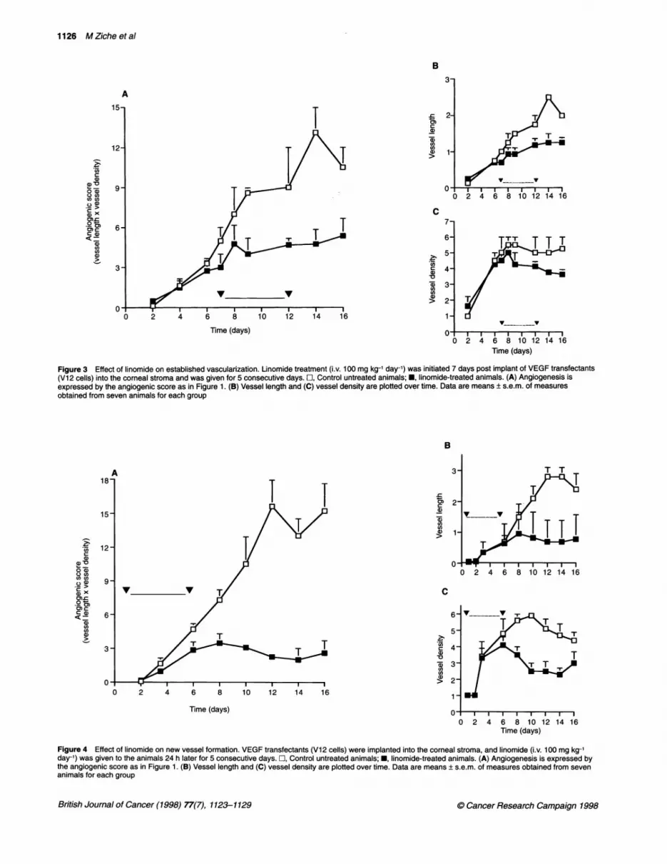
In a general clinical situation, tumours are vascularized and angio-genesis is present at the site of tumour diffusion and spreading.Thus, this type of neovascularization is presumably present at thetime of tumour detection and removal and is a likely target of theantiangiogenic treatment to prevent metastases. To mimic thiscondition, linomide was given at the time when V12 cells hadelicited a consistent number of capillaries to travel about 1 mmfrom the limbus and to actively grow into the corneal stroma(Figure 2C). Treatment started at day 7 from the cell implant intothe rabbit cornea and was given for the following 5 days. After 2days of treatment, the number and the growth rate of the newlyformed capillaries was only slightly different between the treatedanimals and the controls. In the following observations, however,
the advancement of the capillary front into the corneal stroma wasconsistently repressed in treated animals compared with thecontrols (Figure 3A). The progression of growing capillaries(length) appeared to be blocked (Figure 3B). Involution of thepreformed capillaries occurred, leading to a reduction in the densityof the vessels (Figures 2D and 3C). Despite discontinuation of thetreatment, the angiostatic/angiosuppressive effect of linomidepersisted and a consistent reduction of angiogenesis was stillpresent after 7 days from the end of treatment (data not shown).
Treatment with linomide prevents the occurrence ofneoangiogenesis
To assess the effect of linomide in a model of tumour cells in aprevascular stage, animals were treated for 5 consecutive days24 h after the corneal implant of VI 2 cells and before any angio-genesis occurred. The number and growth rate of neovessels(density) and their ability to invade and progress into the avascularcornea (length) were reduced compared with untreated rabbits(Figure 4). Four days after the implant (3 days of treatment),treated animals had only few buds sprouting from the limbalvessels, while in the control group a dense network of capillaryoutgrowth had progressed approximately 0.7 mm(23%) from thelimbus. The angiosuppressive effect was still present after 10 daysfrom the discontinuation of the treatment (Figure 4) and persistedup to the third week of observation (data not shown). In the
British Journal of Cancer (1998) 77(7), 1123-1129
n%
0 Cancer Research Campaign 1998
1126 MZicheetal
B
-C0)Ca)
7,)U)12)
a)
-oZa)
C,)(1)
16_v _ _v
0 2 4 6 8 10 12 14 16Time (days)
Figure 3 Effect of linomide on established vascularization. Linomide treatment (i.v. 100 mg kg-1 day-') was initiated 7 days post implant of VEGFtransfectants(Vl 2 cells) into the corneal stroma and was given for 5 consecutive days. O, Control untreated animals; *, linomide-treated animals. (A) Angiogenesis isexpressed by the angiogenic score as in Figure 1. (B) Vessel length and (C) vessel density are plotted over time. Data are means ± s.e.m. of measuresobtained from seven animals for each group
B
0)
a)
CO)CO)a)
V V C
6-
5-
4a)'a*0cn
: 2-
1-~0 2 4 6 8 10 12 14 16
Time (days)0 I I I I I I I I
0 2 4 6 8 10 12 14 16Time (days)
Figure 4 Effect of linomide on new vessel formation. VEGFtransfectants (Vi2 cells) were implanted into the corneal stroma, and linomide (i.v. 100 mg kg-1day-') was given to the animals 24 h later for 5 consecutive days. L, Control untreated animals; *, linomide-treated animals. (A) Angiogenesis is expressed bythe angiogenic score as in Figure 1. (B) Vessel length and (C) vessel density are plotted over time. Data are means ± s.e.m. of measures obtained from sevenanimals for each group
British Journal of Cancer (1998) 77(7), 1123-1129
A
._n
a)C1)v0D2)
.2 T0).) >
a x
U)C
o m-
a)cna1)
Time (days)
7-
6-
5-
4-
3-
2-
1-~
O0
A
U)n
a)
o a)QCu
0).C).n Th._ >0) x
Ca)U)
7U)ca
aL)
0 Cancer Research Campaign 1998

Angiosuppressive effect of linomide on breast cancer 1127
8-
06-c
IDIo0
. 1*
I
0-
0- El
r 1TI I
lbTime (days)
Figure 5 Effect of linomide on angiogenesis induced by wild-type MCF-7cells. MCF-7 cells were implanted into the corneal stroma, and linomide(i.v. 100 mg kg-' day-') was given 24 h later for 5 consecutive days. In controluntreated animals (E) angiogenesis appeared at 14 days; at this timelinomide (O) was able to reduce the neovascular growth induced by MCF-7cells. Data are expressed as the angiogenic score as in Figure 1. Data aremeans ± s.e.m. of measures obtained from five animals for each group
Table 1 Effect of linomide on the proliferation of tumour and endothelialcells
Counted cells per well
Control +Linomide +Linomide +Linomide50 jg ml-, 100 ,ug ml-' 200 ,ug ml-'
WT 245 ± 5 ND 274 ± 12 211 ± 19
V12 222±4 ND 216±5 228±10
CVECBasal 40 + 3 40 ± 1.7 46 ± 1.1 30 ± 0.5VEGF 58 ± 1.7 34 ± 2.5** 42 ± 2** 34 ± 0.5**bFGF 59±2 42±3.2* 45±2.1 42±4.2*
Proliferation was quantified by the number of cells recovered after 2 daysand 4 days of exposure to test substances for CVECand WTN12cellsrespectively. To evaluate the effect of linomide on endothelial cellproliferation, cells were pretreated with the appropriate concentration of theagent 2 h before VEGFand bFGFwere added. Data are expressed as totalcells counted per well in seven random fields at a magnification of 200. Datarepresent mean ± s.e.m. of three experiments run in triplicate. *P < 0.01 and**P < 0.001 vs bFGF and VEGFalone respectively. ND, not done.
absence of VEGF transfection, MCF-7 elicited a delayed angio-genesis, suggesting a dormant phenotype for angiogenesis (Figure1). The administration of linomide in this experimental conditionfurther delayed the occurrence of angiogenesis (Figure 5).
Effect of linomide on tumour and endothelial cellgrowth in vitro
The possibility that the antiangiogenic effect of linomide could belinked to a direct cytotoxic effect on tumour cell growth was ruledout by exposing the cells in culture to the compound. As shown inTable 1, we found that the growth pattern of WTand V12 cellsexposed for 4 days to 100 and 200 ,ug ml-1 linomide was notmodified by the agent.
Linomide was then assessed on the growth of post-capillaryendothelial cells (CVEC). Linomide at 50 and 100 ,ug ml' didnot affect endothelial cell proliferation, while at 200 ,ug ml-l itslightly reduced the spontaneous replication of serum-deprivedcells. When CVECwere stimulated to grow with either VEGF(20 ng ml') or bFGF (10 ng ml-'), linomide abolished the prolifer-ation induced by either growth factor (Table 1). Interestingly,VEGF-treated cells were more sensitive to linomide inhibition.
DISCUSSION
This study documents for the first time that linomide exerts a
specific antiangiogenic effect in vivo on breast tumour cells andefficiently blocks capillary growth elicited by VEGFbreast cancertransfectants. The results of our study demonstrate by continuousand direct monitoring of the capillary sprouting in vivo thatlinomide prevents and blocks the angiogenesis elicited by breastcarcinoma cells by modifying endothelial cell responsiveness tothe angiogenic trigger. These observations are substantiated by invitro studies showing that microvascular endothelium treated withlinomide does not proliferate in response to VEGF.
Angiogenesis and VEGF expression are correlated with poorprognosis in breast cancer patients. Among the angiogenic factorsproduced by tumours, VEGFseems to be the predominant angio-genic factor expressed in primary breast carcinoma and appears toplay a key role in pathological angiogenesis (Toi et al, 1996;Gasparini et al, 1997). Linomide has been reported to reduce thegrowth of prostatic tumours by modifying the immune response ofthe host (Ichikawa et al, 1992), by cytokine production (Vukanovicand Isaacs, 1995) and by inducing vascular alterations withinthe tumour, leading to angio-inhibition (Vukanovic et al, 1993;Vukanovic and Isaacs, 1995). Moreover, linomide has been shownto inhibit mammary carcinogenesis in rodents and to block vas-cular proliferation induced in matrigel (Joseph et al, 1996). Basedon these considerations, our aim was to assess whether linomidecould be an efficient antiangiogenic drug for breast cancer andwhether it could antagonize a specific endothelial mitogen respon-sible for tumour angiogenesis. The MCF-7 transfectants are an
appropriate model to reproduce a highly angiogenic phenotype ofa tumour cell population linked to VEGF overexpression. Thetransplant of tumour cells into the avascular cornea of albinorabbits allows the assessment of the morphogenetic evolution ofangiogenesis during time and the comparison in the same animalof cell populations with distinct angiogenic phenotypes (Ziche andGullino, 1982). More importantly, the effect of drug treatmenton angiogenesis can be evaluated in each animal during time,independently of tumour growth, thus allowing an accurate andspecific monitoring of the antiangiogenic effect. Two experimentalprotocols were designed to mimic the clinical situations that anantiangiogenic treatment should target: the neovascularizationalready established by the tumour at the time of detection and theoccurrence of angiogenesis linked to metastasis spreading andgrowth. Our results indicate that linomide is able to affect the earlysteps of new vessel formation, resulting in suppression of theangiogenesis elicited by highly angiogenic tumour cell popula-tions and further delaying the occurrence of angiogenesis oftumour cells with a dormant angiogenic profile. Linomide given tothe animals before frank angiogenesis was produced, leads to along-lasting angio-inhibition. Consistently, once florid angiogen-esis has been established by the VEGFtransfectants, the treatmentblocks the neovascular progression and results in the involution of
British Journal of Cancer (1998) 77(7), 1123-11290 Cancer Research Campaign 1998

1128 MZicheetal
the corneal vascularization. The effects of linomide are thus bothangiostatic and angiosuppressive. In keeping with the finding thata specific inhibitor of angiogenesis does not directly interfere withthe growth of tumour cells in vitro (Ichikawa et al, 1992; Yamaokaet al, 1993), we found that linomide does not affect the growthpattern of either MCF-7 or V12 cells in vitro.
In a previous study, linomide was reported to block in vitro thelong-term replication of the endothelium without cytotoxicity(Vukanovic et al, 1993). We recently reported the ability of lino-mide to selectively target VEGF-induced endothelial cell migration(Parenti et al, 1996; Ziche et al, 1997a). Consistently in the presentstudy we document that the drug efficiently impairs endothelial cellreplication promoted by VEGF, substantiating that linomide canspecifically counteract the effect of VEGFon angiogenesis.
In patients with breast carcinoma, there is evidence that angio-genesis plays a role in primary tumour progression (Brem et al,1977; Zajchowski et al, 1990) and in the development of metas-tasis (Gasparini and Harris, 1995). Breast cancer patients withhighly vascularized tumours have poor outcome even if treatedwith conventional adjuvant chemotherapy or hormone therapy(Gasparini et al, 1995). Several reports show that there exists asignificant association between intratumoral microvessel densityand the expression and levels of VEGFin primary invasive breastcarcinomas and that VEGF is the most important angiogenicpeptide in this neoplasm (Toi et al, 1996; Gasparini et al, 1997).These observations indicate that breast cancer patients are likely tobenefit from antiangiogenic treatment and emphasize that knowl-edge of the specific mechanisms of the antiangiogenic interventionare needed for a rational therapeutic approach based on themodulation of angiogenesis.
In conclusion, linomide appears to be a promising antiangio-genic inhibitor that should be tested in patients with breast canceras a new anti-cancer therapeutic strategy. Recent evidencesuggests that, in spite of the redundancy of angiogenic factorspotentially involved in pathological angiogenesis, strategies aimedat antagonizing one specific endothelial cell mitogen may form thebasis for an effective and safe treatment of cancer and metastases.Patients with primary tumours with high microvessel density andwith high VEGFexpression are likely to obtain the highest benefitfrom angiogenesis inhibition with linomide given alone or incombination with conventional adjuvant anti-cancer treatments, inaccordance with the approach of Teicher et al (1992), whichinvolves the targeting of two-cell compartments.
ABBREVIATIONSVEGF, vascular endothelial growth factor; bFGF, basic fibroblastgrowth factor; CVEC, coronary venular endothelial cells; MCF-7,human breast carcinoma cell line; WT, wild type; V12, VEGF121transfectant clone; DMEM,Dulbecco's modified Eagle medium;FCS, fetal calf serum
ACKNOWLEDGEMENTSWe are grateful to Dr Roy Bicknell, Imperial Cancer ResearchFund, Oxford, UK, and to Dr Harris J Granger, Texas A&MUniversity, College Station, USA, for the cells used in this study.We wish to thank Dr Beryl Hartley-Asp of Kabi PharmaciaTheurapeutics, for supplying linomide. This work was supportedby funds from the Italian Association for Cancer Research(AIRC) (Special Project 'Angiogenesis'), European Communities
BIOMED-2 (PL950669) 'Angiogenesis and Cancer' and theNational Research Council of Italy to MZ (project no.96.03745.14).
REFERENCES
Bengtsson M, Simonsson B, Carlsson K, Nilsson B, Smedmyr B, Termander B,Oberg Gand Totterman TH (1992) Stimulation of NKcell, T cell, andmonocyte functions by the novel immunomodulator Linomide after autologousbone marrow transplanation. Transplanation 53: 882-888
Brem SS, Gullino PMand Medina D (1977) Angiogenesis: a marker forneoplastic transformation of mammarypapillary hyperplasia. Science 195:880-881
Brown LF, Berse B, Jackman RW, Tognazzi K, Manseau EJ, Dvorak HF and SengerDR(1993) Increased expression of vascular permeability factor (vascularendothelial growth factor) and its receptors in kidney and bladder carcinomas.AmJ Pathol 143: 1255-1262
Fan T-P, Jaggar R and Bicknell R (1995) Controlling the vasculature: angiogenesis,anti-angiogenesis and vascular targeting of gene therapy. Trenlds Pharmacol Sci16: 57-66
Folkman J 1995 Angiogenesis in cancer, vascular, rheumatoid and other disease.Nature Med 1: 27-31
Gasparini Gand Harris AL (1995) Clinical importance of the determination of tumorangiogenesis in breast cancer: much more than a new prognostic tool. J ClinOncol 13: 765-782
Gasparini G, Weidner N, Bevilacqua P, Maluta S, Dalla Palma P, Caffo 0,Barbareschi M, Boracchi P, Marubini E and Pozza F (1994) Tumor microvesseldensity, p53 expression, tumor size and peritumoral lymphatic vessel invasionare relevant prognostic markers in node-negative breast carcinoma. J ClinOncol 12: 454-466
Gasparini G, Barbareschi M, Boracchi P, Verderio P, Caffo 0, Meli S, Dalla Palma P,Marubini E and Bevilacqua P (1995) Tumor angiogenesis predicts clinicaloutcome of node-positive breast cancer patients treated with adjuvant hormonetherapy or chemotherapy. Cancer J Sci Am1: 131-141
Gasparini G, Toi M, Gion M, Verderio P, DittaDi R, Hanatani M, Matsubara I,Viante 0, Bonoldi E, Boracchi P, Gatti C, Suzuki H and Tominaga T (1997)Prognostic significance of vascular endothelial growth factor protein in nodenegative breast carcinoma. J Natil Catcer Inst 89: 139-147
Haming R, Koo GCand Szalay J (1989) Regulation of the metastasis of murineocular melanoma by natural killer cells. Inivest Ophthalmol Vis Sci 30:1909-1915
Horak ER, Leek R, Klenk N, Lejeune S, Smith K, Stuart N, Greenall M,Stepniewska Mand Harris AL (1992) Angiogenesis, assessed byplatelet/endothelial cell adhesion molecule antibodies, as indicator of nodemetastases and survival in breast cancer. Lancet 340: 1120-1124
Ichikawa T, Lamb JC, Chistensson PI, Hartley-ASP B and Isaacs JT (1992) Theantitumor effects of the quinoline-3-carboxamide Linomide on dunning R-3327rat prostatic cancers. Cancer Res 52: 3022-3028
Joseph IBJK, Vukanovich J and Isaacs JT (1996) Antiangiogenic treatment withLinomide as chemoprevention for prostate, seminal vesicle, and breastcarcinogenesis in rodents. Cancer Res 56: 3404-3408
Kalland T (1986) Effects of the immunomodulator LS-26 16 on growth andmetastasis of the murine B16-Fl0 melanoma. Ca,tcer Res 46: 3018-3022
Kalland T, Alm G and Stalhandske T (1985) Augmentation of mouse natural killercell activity by LS-26 16, a new immunomodulator. J Immunol 134: 3956-3961
Klasbrun Mand Soker S (1993) VEGF/VPF: the angiogenesis factor found? ClurrBiol 3: 699-702
Larsson E-L, Joki A and Stalhandske T (1987) Mechanism of action of the newimmunomodulator LS-2616 on T cell responses. Ihtt J Immuniopharmiiaceuit 9:425-431
O'Brien T, Cranston D, Fuggle S, Bicknell R and Harris A (1995) Differentangiogenic pathways characterize superficial and invasive bladder cancer.Canicer Res 55: 5 10-513
Parenti A, Donnini S, Morbidelli L, Granger HJ and Ziche M(1996) The effectof Linomide on the migration and the proliferation of capillary endothelialcells elicited by vascular endothelial growth factor. Br J Pharmacol 119:619-621
Schelling ME, Meininger CJ, Hawker JR and Granger HJ (1988) Venular endothelialcells from bovine heart. AmJ Physiol 254: H121 1 -H 1217
Teicher BA, Sotomayor EA and Huang ZD (1992) Antiangiogenic agents potentiatecytotoxic cancer therapies against primary and metastatic disease. Cancer Res52: 6702-6704
British Journal of Cancer (1998) 77(7), 1123-1129 © Cancer Research Campaign 1998

Angiosuppressive effect of linomide on breast cancer 11 29
Toi M, Kashitani J and Tominaga T (1993) Tumor angiogenesis is an independentprognostic indicator in primary breast carcinoma. Int J Cancer 55: 371-374
Toi M, Kondo S, Suzuki H, Yamamoto Y, Inada K, Imazawa T, Taniguchi T andTominaga T (1996) Quantitative analyses of vascular endothelial growth factorin primary breast cancer. Cancer Res 77: 1101-1105
Vukanovic J and Isaacs JT (1995) Linomide inhibits angiogenesis, growth,metastasis, and macrophage infiltration within rat prostatic cancers. Cancer Res55: 1499-1504
Vukanovic J, Passaniti A, Hirata T, Traystman RJ, Hartley-Asp B and Isaacs JT(I1993) Antiangiogenic effects of the quinoline-3-carboxamide Linomide.Cancer Res 53: 1833-1837
Weidner N, Folkman J, Pozza F, Bevilacqua P, Allred EN, Moore DH, Meli S andGasparini G (1992) Tumor angiogenesis: a new significant and independentprognostic indicator in early-stage breast carcinoma. J Natl Cancer Inst 84:1875-1887
Yamaoka M, Yamamoto T, Ikeyama S, Sudo K and Fujita T (1993) Angiogenesisinhibitor TNP-470 (AGM- 1470) potently inhibits the tumor growth ofhormone-independent human breast and prostate carcinoma cell lines. CancerRes 53: 5233-5236
Zajchowski DA, Band V, Trask DK, Kling D, Connolly JL and Sager R (1990)Suppression of tumor-forming ability and related traits in MCF-7 human breastcancer cell by fusion with immortal mammary epithelial cells. Cell Biol 87:2314-2318
Zhang H-T, Craft P, Scott PAE, Ziche M, Weilch HA, Harris AL and Bicknell R(1995) Enhancement of tumor growth and vascular density by transfection ofvascular endothelial cell growth factor into MCF-7 human breast carcinomacells. J Natl Cancer Inst 87: 213-219
Ziche Mand Gullino PM(1982) Angiogenesis and neoplastic progression in vitro.J Natl Cancer Inst 69: 483-487
Ziche M, Alessandri Gand Gullino PM(1989) Gangliosides promote the angiogenicresponse. Lab Invest 61: 629-634
Ziche M, Gasparini G, Choudhuri R, Bicknell R, Morbidelli L, Parenti A and LeddaF (1997a) Antiangiogenic effect of Linomide on breast cancer VEGFtransfectants. Oncol Rep 4: 253-256
Ziche M, Morbidelli L, Choudhuri R, Zhang H-T, Donnini S, Granger HJ andBicknell R (1997b) Nitric oxide synthase lies downstream from vascularendothelial growth factor-induced but not basic fibroblast growth factor-induced angiogenesis. J Clin Invest 99: 2625-2634
C Cancer Research Campaign 1998 British Journal of Cancer (1998) 77(7), 1123-1129
Copyright © 2022 FDOKUMEN