Sorafenib blocks tumour growth, angiogenesis and metastatic potential in preclinical models of...
17
BioMed Central Page 1 of 17 (page number not for citation purposes) Molecular Cancer Open Access Research Sorafenib blocks tumour growth, angiogenesis and metastatic potential in preclinical models of osteosarcoma through a mechanism potentially involving the inhibition of ERK1/2, MCL-1 and ezrin pathways Ymera Pignochino* 1 , Giovanni Grignani 1 , Giuliana Cavalloni 1 , Manuela Motta 2 , Marta Tapparo 1 , Stefania Bruno 3 , Alessia Bottos 4 , Loretta Gammaitoni 1 , Giorgia Migliardi 1 , Giovanni Camussi 3 , Marco Alberghini 5 , Bruno Torchio 2 , Stefano Ferrari 4 , Federico Bussolino 6 , Franca Fagioli 6 , Piero Picci 4 and Massimo Aglietta 1 Address: 1 Division of Medical Oncology, University of Torino Medical School, A.O. Ordine Mauriziano, Institute for Cancer Research and Treatment Candiolo, Torino, Italy, 2 Unit of Pathology, A.O. Ordine Mauriziano, Torino, Italy, 3 Department of Internal Medicine, Center for Molecular Biotechnology and Center of Experimental Research and Medical Sciences, University of Torino, 4 Division of Molecular Angiogenesis, University of Torino Medical School, Department of oncological science, Institute for Cancer Research and Treatment Candiolo, Torino, Italy, 5 Chemotherapy Division of Department of Musculoskeletal Oncology, Istituti Ortopedici Rizzoli, Bologna, Italy and 6 Division of Pediatric Onco- Hematology, Regina Margherita Children's Hospital, Torino, Italy Email: Ymera Pignochino* - [email protected]; Giovanni Grignani - [email protected]; Giuliana Cavalloni - [email protected]; Manuela Motta - [email protected]; Marta Tapparo - [email protected]; Stefania Bruno - [email protected]; Alessia Bottos - [email protected]; Loretta Gammaitoni - [email protected]; Giorgia Migliardi - [email protected]; Giovanni Camussi - [email protected]; Marco Alberghini - [email protected]; Bruno Torchio - [email protected]; Stefano Ferrari - [email protected]; Federico Bussolino - [email protected]; Franca Fagioli - [email protected]; Piero Picci - [email protected]; Massimo Aglietta - [email protected] * Corresponding author Abstract Background: Osteosarcoma (OS) is the most common primary bone tumour in children and young adults. Despite improved prognosis, metastatic or relapsed OS remains largely incurable and no significant improvement has been observed in the last 20 years. Therefore, the search for alternative agents in OS is mandatory. Results: We investigated phospho-ERK 1/2, MCL-1, and phospho-Ezrin/Radixin/Moesin (P-ERM) as potential therapeutic targets in OS. Activation of these pathways was shown by immunohistochemistry in about 70% of cases and in all OS cell lines analyzed. Mutational analysis revealed no activating mutations in KRAS whereas BRAF gene was found to be mutated in 4/30 OS samples from patients. Based on these results we tested the multi-kinase inhibitor sorafenib (BAY 43-9006) in preclinical models of OS. Sorafenib inhibited OS cell line proliferation, induced apoptosis and downregulated P-ERK1/2, MCL-1, and P-ERM in a dose-dependent manner. The dephosphorylation of ERM was not due to ERK inhibition. The downregulation of MCL-1 led to an increase in apoptosis in OS cell lines. In chick embryo chorioallantoic membranes, OS supernatants induced angiogenesis, which was blocked by sorafenib and it was also shown that sorafenib reduced Published: 10 December 2009 Molecular Cancer 2009, 8:118 doi:10.1186/1476-4598-8-118 Received: 14 September 2009 Accepted: 10 December 2009 This article is available from: http://www.molecular-cancer.com/content/8/1/118 © 2009 Pignochino et al; licensee BioMed Central Ltd. This is an Open Access article distributed under the terms of the Creative Commons Attribution License (http://creativecommons.org/licenses/by/2.0 ), which permits unrestricted use, distribution, and reproduction in any medium, provided the original work is properly cited.
-
Upload
independent -
Category
Documents
-
view
2 -
download
0
Transcript of Sorafenib blocks tumour growth, angiogenesis and metastatic potential in preclinical models of...

BioMed CentralMolecular Cancer
ss
Open AcceResearchSorafenib blocks tumour growth, angiogenesis and metastatic potential in preclinical models of osteosarcoma through a mechanism potentially involving the inhibition of ERK1/2, MCL-1 and ezrin pathwaysYmera Pignochino*1, Giovanni Grignani1, Giuliana Cavalloni1, Manuela Motta2, Marta Tapparo1, Stefania Bruno3, Alessia Bottos4, Loretta Gammaitoni1, Giorgia Migliardi1, Giovanni Camussi3, Marco Alberghini5, Bruno Torchio2, Stefano Ferrari4, Federico Bussolino6, Franca Fagioli6, Piero Picci4 and Massimo Aglietta1Address: 1Division of Medical Oncology, University of Torino Medical School, A.O. Ordine Mauriziano, Institute for Cancer Research and Treatment Candiolo, Torino, Italy, 2Unit of Pathology, A.O. Ordine Mauriziano, Torino, Italy, 3Department of Internal Medicine, Center for Molecular Biotechnology and Center of Experimental Research and Medical Sciences, University of Torino, 4Division of Molecular Angiogenesis, University of Torino Medical School, Department of oncological science, Institute for Cancer Research and Treatment Candiolo, Torino, Italy, 5Chemotherapy Division of Department of Musculoskeletal Oncology, Istituti Ortopedici Rizzoli, Bologna, Italy and 6Division of Pediatric Onco-Hematology, Regina Margherita Children's Hospital, Torino, Italy
Email: Ymera Pignochino* - [email protected]; Giovanni Grignani - [email protected]; Giuliana Cavalloni - [email protected]; Manuela Motta - [email protected]; Marta Tapparo - [email protected]; Stefania Bruno - [email protected]; Alessia Bottos - [email protected]; Loretta Gammaitoni - [email protected]; Giorgia Migliardi - [email protected]; Giovanni Camussi - [email protected]; Marco Alberghini - [email protected]; Bruno Torchio - [email protected]; Stefano Ferrari - [email protected]; Federico Bussolino - [email protected]; Franca Fagioli - [email protected]; Piero Picci - [email protected]; Massimo Aglietta - [email protected]
* Corresponding author
AbstractBackground: Osteosarcoma (OS) is the most common primary bone tumour in children andyoung adults. Despite improved prognosis, metastatic or relapsed OS remains largely incurable andno significant improvement has been observed in the last 20 years. Therefore, the search foralternative agents in OS is mandatory.
Results: We investigated phospho-ERK 1/2, MCL-1, and phospho-Ezrin/Radixin/Moesin (P-ERM)as potential therapeutic targets in OS. Activation of these pathways was shown byimmunohistochemistry in about 70% of cases and in all OS cell lines analyzed. Mutational analysisrevealed no activating mutations in KRAS whereas BRAF gene was found to be mutated in 4/30 OSsamples from patients. Based on these results we tested the multi-kinase inhibitor sorafenib (BAY43-9006) in preclinical models of OS. Sorafenib inhibited OS cell line proliferation, inducedapoptosis and downregulated P-ERK1/2, MCL-1, and P-ERM in a dose-dependent manner. Thedephosphorylation of ERM was not due to ERK inhibition. The downregulation of MCL-1 led to anincrease in apoptosis in OS cell lines. In chick embryo chorioallantoic membranes, OS supernatantsinduced angiogenesis, which was blocked by sorafenib and it was also shown that sorafenib reduced
Published: 10 December 2009
Molecular Cancer 2009, 8:118 doi:10.1186/1476-4598-8-118
Received: 14 September 2009Accepted: 10 December 2009
This article is available from: http://www.molecular-cancer.com/content/8/1/118
© 2009 Pignochino et al; licensee BioMed Central Ltd. This is an Open Access article distributed under the terms of the Creative Commons Attribution License (http://creativecommons.org/licenses/by/2.0), which permits unrestricted use, distribution, and reproduction in any medium, provided the original work is properly cited.
Page 1 of 17(page number not for citation purposes)

Molecular Cancer 2009, 8:118 http://www.molecular-cancer.com/content/8/1/118
VEGF and MMP2 production. In addition, sorafenib treatment dramatically reduced tumour volumeof OS xenografts and lung metastasis in SCID mice.
Conclusion: In conclusion, ERK1/2, MCL-1 and ERM pathways are shown to be active in OS.Sorafenib is able to inhibit their signal transduction, both in vitro and in vivo, displaying anti-tumouralactivity, anti-angiogenic effects, and reducing metastatic colony formation in lungs. These datasupport the testing of sorafenib as a potential therapeutic option in metastatic or relapsed OSpatients unresponsive to standard treatments.
BackgroundOsteosarcoma (OS) is the most common primary malig-nant bone tumour in children and young adults and ischaracterized by an aggressive clinical course. Chemother-apy significantly increased 5-year survival of localized OSpatients to approximately 65% [1]. Pulmonary metas-tases, central presentation and local non-resectablerelapse cause a fatal outcome in the majority of patients[2,3]. Both novel chemotherapeutic drugs and radiometa-bolic therapy based on samarium failed to improve over-all survival [4]. These dismal results are due to P-glycoprotein overexpression [5] as well as complex karyo-types [6], which account for chemoresistance. The searchfor alternative agents focused on entirely different mecha-nisms in OS is therefore mandatory.
The advent of molecular targeted therapies has spurred asearch for pathological activation of receptors tyrosinekinase (RTKs) via various mechanisms in a number ofmalignancies including OS. Among the RTKs KIT, Vascu-lar endothelial growth factor receptor (VEGFR) -2, -3 andPlatelet derived growth factor (PDGFR)-β have beenfound to be involved in OS progression and metastatiza-tion [7-9].
Two major pathways subsequently activated by RTKs arethe phosphatidylinositol 3-kinase (PI3K)/AKT and themitogen-activated protein kinases ERK 1/2.
Recent studies have demonstrated that the cytoskeletallinker protein, ezrin, a member of the ezrin-radixin-moesin (ERM) family of protein linkers between the actincytoskeleton and plasma membrane, plays an importantrole in the metastasis of OS and rhabdomyosarcoma, sug-gesting that these metastasis-associated molecules couldbe potential targets for treatment [10]. Matrix metallopro-teinases (MMPs) play pivotal roles in tumour invasionthrough degradation of basement membranes and extra-cellular matrices [11,12]. MMP-2 and -9 have been foundto be involved in OS tumourigenesis and pulmonarymetastasization [13,14].
Sorafenib (BAY 43-9006) is an orally active biarylureicmulti-kinase inhibitor originally developed to block theERK 1/2 pathway by targeting Raf-kinases, such as RAF-1
and B-RAF, as well as in the presence of an V600E activat-ing mutation. Off-targets of this drug are other RTKsinvolved in tumour progression (FLT-3, KIT, fibroblastgrowth factor receptor, FGFR-1, RET) and angiogenesis(VEGFR-2 and 3, and PDGFR-β) [15]. More recently, it hasbeen demonstrated that sorafenib induces apoptosis inhuman leukemia cells and other human tumour cell linesthrough down-regulation of the anti-apoptotic proteinmyeloid cell leukemia-1 (MCL-1), a Bcl-2 family member[16].
Beyond its preclinical anti-tumoural activity, sorafenibwas proven to be effective in 3 different chemorefractorycancers: kidney, liver and thyroid carcinoma. Sorafenibsignificantly prolongs progression-free survival as well asoverall survival of treated patients [17-19].
Several molecular targets of sorafenib seem to be involvedin the pathogenesis or progression of OS. One pioneeringwork demonstrated the amplification of Raf-1 in one caseof human OS [20], and the expression of PDGF is associ-ated with OS progression [21]. In addition, VEGF is over-expressed in 63% of untreated OS and is predictive ofpulmonary metastasis and poor prognosis [22]. A wideimmunohistochemical study on pediatric solid tumours,among them 18 cases of OS, demonstrated that KIT isexpressed in the entire case series [7]. Inhibition of theERK1/2 pathway, mediated by statin treatment, inducedapoptosis in OS cell lines [23]. MCL-1 is expressed in avariety of different human sarcoma cell lines, and MCL-1antisense oligonucleotides combined with low-dosecyclophosphamide provides a synergistic anti-tumoureffect, and may qualify as a promising strategy to over-come chemoresistance in human sarcoma [24].
These studies suggest that Sorafenib may be active in OS.However, before exploiting a clinical trial, it was necessaryto conduct an investigation of the activation of possibletargets of sorafenib in both in vitro and in vivo models.Thus, we investigated the presence of molecular targets ofsorafenib in OS patient specimens and explored the invitro and in vivo anti-proliferative effects of this multi-kinase inhibitor as well as its molecular mechanisms ofaction. In addition, we explored the effect of sorafenib onother pathways potentially involved in progression and
Page 2 of 17(page number not for citation purposes)

Molecular Cancer 2009, 8:118 http://www.molecular-cancer.com/content/8/1/118
metastatic dissemination of OS such as the ERM complex,suggesting a novel sorafenib- targetable molecular path-way.
ResultsP-ERK1/2, MCL-1 and P-ERM are highly expressed in OSTo investigate ERK1/2 pathway activation in OS patients,the expression of phosphorylated ERK1/2 was analyzed inan entire OS case series by immunohistochemistry, andcompared with normal adjacent tissues as a control.Nuclear and cytoplasmatic P-ERK1/2 immunostainingwas detected in 20 out of 30 samples (66.7%) and 9 ofthem were strongly positive. Representative examples ofP-ERK1/2 staining are shown in Figure 1. These results,demonstrate that the ERK1/2 pathways are activated in allthe analyzed histotypes (Table 1).
The normal bone counterpart was consistently negativefor activated ERK1/2.
Next, we analysed expression of the MCL-1 protein byimmunohistochemistry (Figure 1). Results shown inTable 1 demonstrate that 24 out of 30 (80%) expressedMCL-1 protein in a granular cytoplasmatic staining (mito-chondria). Ten out of 24 were strongly positive in morethan 50% tumour cells, whilst non-malignant tissues wereconsistently negative.
The whole series was also analyzed to detect the phospho-rylation of cytoskeletal linkers ERM (Figure 1). Twenty-one out of 30 specimens (70%) displayed P-ERM (Table1) in the cytoplasmatic side of the plasma membrane. Incontrast, ERM was not phosphorylated in normal osseoustissues.
Western blot analysis revealed the expression of P-ERK1/2, MCL-1 and P-ERM in the 7 OS cell lines examined (datanot shown).
B-RAF mutations are present in OS samples from patientsThe hotspot regions of B-RAF were investigated in thewhole series. Exon 15 of B-RAF was mutated in 4 samples(13.3%), as shown in Table 1. One sample had a single-base deletion in codon 596 of the conserved "DFG" motifin the regulatory site. This single base deletion causes aframe-shift that leads to the reading of Val followed by aStop-codon instead of Gly, and consequently the transla-tion of a truncated form of the protein. A second patientdisplayed a H608L substitution which has never beendescribed before. A third sample had the G615R muta-tion. The fourth sample had a point mutation in the acti-vation segment phosphorylation site, causing thesubstitution of Ser 602 with Tyr (S602Y). These mutationswere not presenting in the surrounding non-tumoural tis-sues. No B-RAF exon 11 and K-RAS exon 1 and 2 muta-tions were found in the whole case series.
Sorafenib has anti-proliferative and pro-apoptototic effects on OS cell linesTo investigate the effects of sorafenib on in vitro prolifera-tion, we exposed 7 different OS cell lines to increasingdoses of the drug for 24, 48 and 72 hours. CellGlo™ assaysdemonstrated that sorafenib caused a dose- and time-dependent cell growth inhibition of all the 7 cell linestested. IC50 values after 72 hours of treatment were calcu-lated on the basis of these results and are shown in Table2. At this time point, DNA content and apoptosis analysiswas evaluated by FACS. Sorafenib did not induce cell cyclearrest, but a dose dependent increase of the percentage ofcells in sub-G0 phase considered to be apoptotic cells (Fig-ure 2, panel A). Further Annexin V/PI staining confirmedthat sorafenib induced a dose-dependent increase in thepercentage of apoptotic cells, as shown in Figure 2, panelB. In addition, sorafenib displayed a dose-dependentinhibition of anchorage-independent cell growth, asshown by soft agar assays (Figure 2, panel C).
Sorafenib down-regulates P-ERK 1/2, MCL-1 and P-ERM expression in OS cell linesTo elucidate the mechanisms of cell growth inhibitionand apoptosis induced by sorafenib, OS cells wereexposed to the drug at concentrations ranging from 0 to20 μM for 24 hours. Results demonstrated that sorafenibinduced a dose-dependent decrease in phosphorylatedERK1/2 and ERM in all the 7 cell lines tested. Representa-tive western blots are shown in Figure 3 (panel A and C).Expression of total ERK and ERM was not affected by sor-afenib treatment.
To verify whether ERM phosphorylation is dependent onPDGFR or KIT pathways, OS cell lines were treated withimatinib mesylate (STI571, Gleevec) a known inhibitor ofPDGFR and KIT as well as ABL. As shown in Figure 3(panel D) STI571 treatment did not affect ERM phospho-rylation.
In addition, the effect of sorafenib on phosphorylation ofERM is not ERK dependent. Indeed, the inhibition of ERKpathway resulting from treatment with UO126, a MEK-specific inhibitor, did not affect phosphorylation of ERM(Figure 3, panel D).
The expression of MCL-1 in OS cells treated with soraf-enib for 24 hours was analyzed by immunoblotting. A sig-nificant dose-dependent reduction of MCL-1 protein wasdetected (Figure 3, panel B).
Inhibition of MCL-1 expression induces apoptosis in OS cell linesIn order to investigate if the anti-apoptotic effect of soraf-enib could be attributable to the inhibition of MCL-1 weexploited siRNA technology. SiRNA MCL-1 transfectionsignificantly decreased MCL-1 protein expression in all
Page 3 of 17(page number not for citation purposes)

Molecular Cancer 2009, 8:118 http://www.molecular-cancer.com/content/8/1/118
Table 1: Characteristics of OS case series.
Case Gender Histology Grade Site BRAF mutations P-ERK1/2 * MCL-1 * P-ERM *
1 M OSTEOBLASTIC G4 TIBIA - - + +
2 M OSTEOBLASTIC G4 FEMUR - - + -
3 F OSTEOBLASTIC G4 FEMUR - - - -
4 M OSTEOBLASTIC G4 TIBIA - - - +
5 M OSTEOBLASTIC G4 HOMER 596 delA - - -
6 F OSTEOBLASTIC G4 TIBIA - - + -
7 F OSTEOBLASTIC G4 FEMUR - - + +
8 M OSTEOBLASTIC G4 FEMUR - - ++ +
9 M OSTEOBLASTIC G4 TIBIA - - + +
10 M OSTEOBLASTIC G4 FEMUR - - + +
11 M OSTEOBLASTIC G4 FEMUR - ++ ++ +
12 F OSTEOBLASTIC G4 FEMUR - + - -
13 M OSTEOBLASTIC G4 FEMUR H608L ++ ++ +
14 M OSTEOBLASTIC G4 FEMUR - + + -
15 M OSTEOBLASTIC G4 HOMER - ++ ++ +
16 M OSTEOBLASTIC G4 TIBIA - ++ + +
17 F OSTEOBLASTIC G4 FEMUR - + + +
18 M OSTEOBLASTIC G4 FEMUR - + ++ -
19 F OSTEOBLASTIC G4 HOMER - ++ + ++
20 M OSTEOBLASTIC G4 TIBIA - + + +
21 M OSTEOBLASTIC G4 FEMUR - ++ ++ +
22 M OSTEOBLASTIC G4 FEMUR G615R ++ ++ ++
23 M FIBROBLASTIC G4 TIBIA - + ++ -
24 M FIBROBLASTIC G4 FEMUR - ++ - +
25 M OSTEOBLASTIC G4 FEMUR - + + -
26 F OSTEOBLASTIC G3 HOMER - + - +
27 M OSTEOBLASTIC G4 FEMUR - + + ++
Page 4 of 17(page number not for citation purposes)

Molecular Cancer 2009, 8:118 http://www.molecular-cancer.com/content/8/1/118
the 7 cell lines tested. Different OS cell lines displayed dif-ferent sensitivity to MCL-1 silencing. Namely, in MG63cells, which were the most sensitive to MCL-1 silencing,there was a strong reduction in MCL-1 protein expression,as demonstrated by western blot analysis (Figure 4, panelA). Meanwhile, in SAOS-2 cells, the least sensitive to MCL-1 silencing, only a minor down-regulation of MCL-1 pro-tein was observed (Figure 4 panel A). SiRNA-inducedMCL-1 down-regulation produced an increase of apop-totic OS cells compared to cells transfected with controlsiRNAs (Figure 4, panel B). The percentage of late apop-
totic cells was higher in MG63 cells than in SAOS-2 cells(44.2% vs 16.0% respectively), reflecting the level ofMCL-1 down-regulation.
Sorafenib inhibits MMP2 and VEGF production in OS cell linesTo explore the activity of sorafenib on the effectorsinvolved in tumour progression and angiogenesis, wemeasured MMP2 and VEGF production in supernatants ofall the 7 cell lines tested. We observed that different celllines exhibit different basal level of MMP2 and VEGF-A,
28 M OSTEOBLASTIC G4 FEMUR S602Y + + +
29 F OSTEOBLASTIC G4 HOMER - + ++ +
30 M OSTEOBLASTIC G4 TIBIA - ++ ++ ++
Gender, histological subtypes, grading, site of tumor origin, immunohistochemical results of P-ERK1/2, MCL- 1 and P-ERM expression and BRAF exon 15 mutations of each osteosarcoma samples from patients. intensity of expression: (-)< 10% positive cells scored -, 10-50% positive cells scored +;> 50% positive cells and high staining intensity scored ++).
Table 1: Characteristics of OS case series. (Continued)
Representative immunostaining of osteosarcoma samplesFigure 1Representative immunostaining of osteosarcoma samples. Negative (-), positive (+) and strongly positive (++) samples for P-ERK1/2, MCL-1 and P-ERM expression.
Page 5 of 17(page number not for citation purposes)

Molecular Cancer 2009, 8:118 http://www.molecular-cancer.com/content/8/1/118
being higher in MG63 cells, and lower in HOS cells (Fig-ure 5). Treatment with sorafenib produced a consistentreduction of the concentration of MMP2 and VEGF-A inall cell lines tested (Figure 5). However, the magnitude ofthis reduction was heterogeneous. Namely, after 48 hoursMMP2 produced by 106 cells was reduced to 47.8% inKHOS, 64.8% in HOS, 63.9% in U2-OS, 40.7% in SAOS-2, 59.6% in SJSA-1; 86.5% in MG63; and 54.4% inMNNG-HOS cells.
Sorafenib treatment led to the reduction of VEGF-A pro-duced by 106 cells to 57.7% in KHOS, 73.1% in HOS,80.5% in U2-OS, 52.9% in SAOS-2, 67.5% in SJSA-1;47.1% in MG63; and 65.7% in MNNG-HOS cells.
Sorafenib has an anti-angiogenic effect in CAMChick chorioallantoic membrane assay was performed toinvestigate the angiogenic potential of OS cell lines andthe anti-angiogenic effect of sorafenib in vivo.
The supernatant of U2OS cells clearly increased sproutingangiogenesis in CAM (Figure 6, panel B) compared withculture medium alone (Figure 6, panel A), indicating thesecretion of angiogenic factors by OS cells. Anti-ang-iogenic effects of sorafenib were tested by two differentapproaches, i.e. treating the cells prior to CAM stimula-tion or directly adding sorafenib into the CAM alreadystimulated with untreated tumour cell supernatant. WhenU2OS were treated with low concentration of sorafenib toavoid cell mortality,, the supernatant developed a lowerangiogenic response than untreated cells (Figure 6, panelC), probably due to the decrease of secreted angiogenicfactors. The treatment of CAM with sorafenib blockedangiogenesis induced by U2OS cell supernatant, suggest-
ing that the drug may also act on host vasculature (Figure6, panel D).
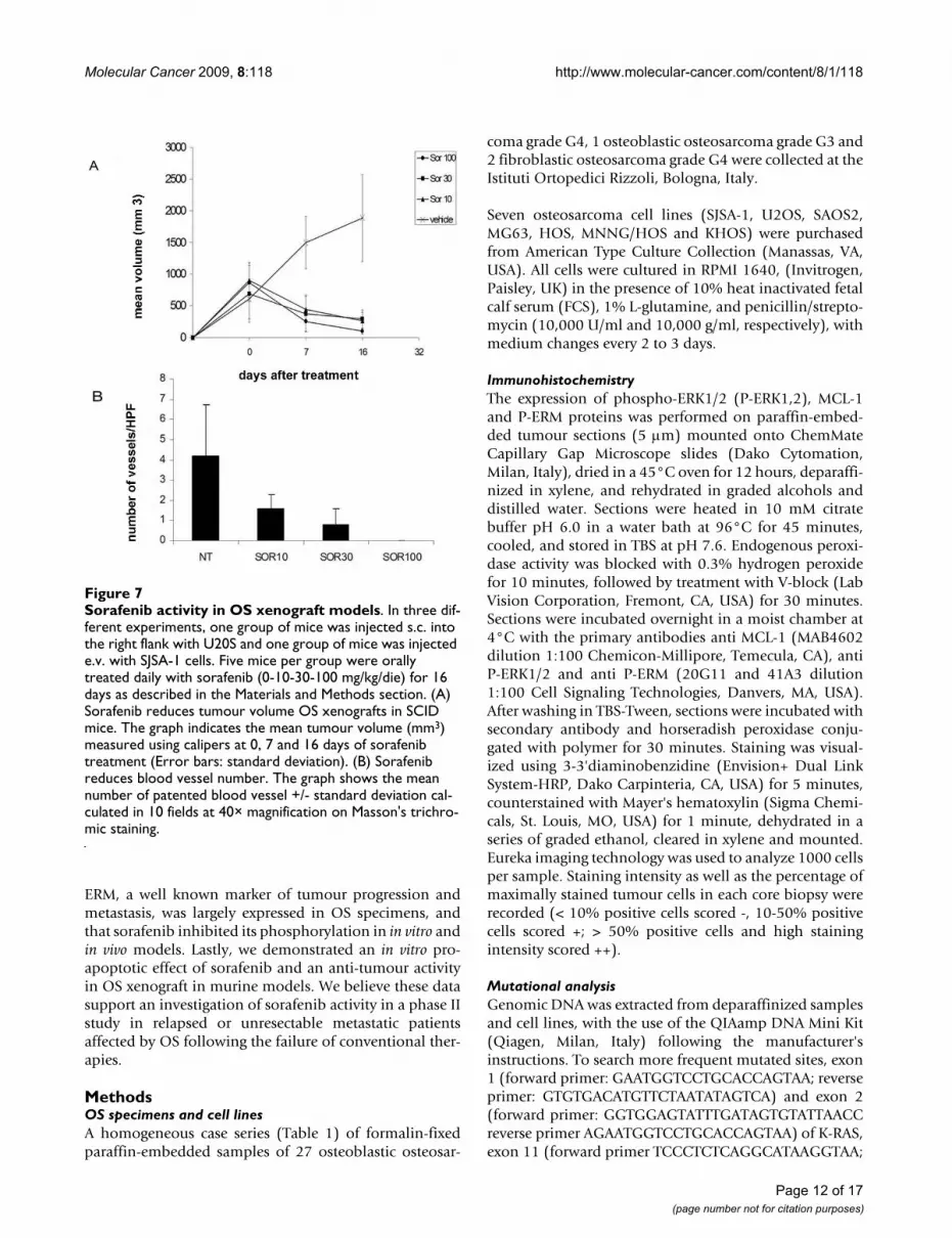
Sorafenib displays anti-tumoural activity in vivo against human OS xenograftsBased on their median level of MMP2 and VEGF-A pro-duction, and their previously demonstrated tumouri-genicity in mice [14], U2OS and SJSA-1 cell lines werechosen for in vivo studies. Sorafenib treatment dramati-cally reduced tumour volume of s.c. U2OS xenografts inSCID mice compared to untreated mice as shown in Fig-ure 7 (Panel A). Moreover, the number of patented bloodvessels was strikingly reduced in tumours of treated mice,as shown in Masson trichromic stained sections (Figure 7,panel B).
Histological analysis revealed that sorafenib-treatedxenografts had a lower tumour cell number, which mostlyshowed marked regressive nuclear changes as pyknosis(Figure 8, panels A-B). In treated mice, OS viable cellswere present on the edge of the lesion showing a general-ized shrinkage of the viable tissue thickness. A sharpincrease in the extracellular matrix is evident in histologi-cal sections from sorafenib-treated mice compared tonon-treated mice (Figure 8, panel C-D).
Sorafenib reduces metastasis formation in lungsOne million SJSA-1 cells were injected into the tail vein ofSCID mice giving rise to pulmonary colonies within 3weeks. Subsequent treatment with sorafenib for 16 daysinhibited tumour colony growth. In some of the treatedmice with the highest sorafenib dosage, a massive lungcollapse with pulmonary bleeding was observed atautopsy. The percentage of area occupied by lung foci ana-lyzed per optical field after hematoxylin and eosin stain-ing at lower magnification (40×) was 73% ± 14, 84% ±11,40% and 35% ± 7 in sorafenib 10, 30, 100 mg/kg/daytreated mice respectively. In Figure 8, panels E-F, repre-sentative sections of lung from untreated- and sorafenib-treated mice are shown.
Sorafenib down-regulates P-ERK 1/2, MCL-1 and P-ERM expression on OS xenograftsMolecular targets of sorafenib were also evaluated inxenograft sections. Immunohistochemical analysis clearlydemonstrated the reduced phosphorylation of ERK1/2,with the percentage of positive cells diminishing from84% (score ++) in sections of untreated xenografts to15%, 30% (score +) and 6% (score -) in 10, 30 e 100 mg/kg/die sorafenib treated mice respectively (p < 0.05).Expression of MCL-1 is also slightly down-regulated fol-lowing sorafenib treatment. MCL-1 was expressed in 90%cells (scored ++) of untreated xenografts and in 75% (++),60% (++) and 40% (+) in 10, 30 and 100 mg/kg/die sor-afenib-treated mice respectively (p < 0.05). P-ERM is
Table 2: Concentrations of sorafenib inhibiting 50% of OS cell line proliferation.
Cell line IC50 (μM) Confidence Intervals (μM)
MG63 2.793 2.32-3.35
U2OS 4.677 2.99-6.61
SAOS-2 3.943 1.87-5.31
KHOS 2.417 1.88-3.10
HOS 4.784 3.99-5.48
SJSA-1 4.511 3.01-6.71
MNNG-HOS 4.814 3.13-6.39
IC50 values (μM) and relative 95% confidence intervals obtained after 72 hours of sorafenib exposure.* IC50: Sorafenib concentration inhibiting 50% of the cell line proliferation.
Page 6 of 17(page number not for citation purposes)

Molecular Cancer 2009, 8:118 http://www.molecular-cancer.com/content/8/1/118
Page 7 of 17(page number not for citation purposes)
Sorafenib induces apoptosis and inhibits anchorage independent cell growth of a representative OS cell lineFigure 2Sorafenib induces apoptosis and inhibits anchorage independent cell growth of a representative OS cell line. DNA content (A) and apoptosis (B) analysis after 72 h treatments. Soft agar assays as described in "materials and methods" section (C).

Molecular Cancer 2009, 8:118 http://www.molecular-cancer.com/content/8/1/118
strongly expressed along the inner layer of the plasmamembrane in a continuous fashion in 50% of viable cellsin untreated mice (scored ++). Instead, sections fromtreated xenografts displayed a weak P-ERM staining in adiscontinuous fashion (scored +).
DiscussionIt is well known that ERK 1/2 play a key role in differentneoplasia. Moreover, even in cancer like GISTs driven byother kinases, activation of KIT or PDGFR-α lead to phos-phorylation of ERK 1/2 [25] and these kinases mediate theproliferative advantage of the neoplastic cells. Imatinibblocks KIT signalling, causing the proliferative arrest ofgastrointestinal stromal tumours (GISTs), and the inacti-vation of ERK 1/2. However, resistance to imatinib is
accompanied by reactivation (phosphorylation) of ERK1/2. Among the new drugs with specific molecular targetssorafenib was shown to be effective in renal cell carci-noma and hepatocarcinoma, I through the inhibition ofERK1/2 pathway [17,18]. These results, along with theunsatisfactory outcome of relapsed and metastatic OScases led us to investigate the presence and role of soraf-enib targets in paraffin-embedded tissue from OS patientsas well as in several OS cell lines and, thereafter, to exploresorafenib activity in xenograft models of human OS.
We demonstrated that 66.6% of OS samples from patientsdisplayed an activated ERK 1/2 pathway suggesting that itmay be relevant in increased OS proliferation. There wasonly one prior datum [26] addressing the overexpressionof ERK 1/2 in OS. We observed a highly reproducible andconsistent expression of P-ERK1/2 among OS specimens.Furthermore, activated ERK 1/2 were only present in theneoplastic tissue and not in the normal tissue surroundingthe tumour. Its selective expression is a clue to a prolifer-ative role compared to normal tissue.
Another metabolic pathway often involved in tumourgrowth advantage regards the mechanisms preventingapoptosis. Among sorafenib off-targets, we investigatedand found the antiapoptotic protein MCL-1 activated in84% of OS specimens, emphasizing its role as a possiblemechanism of survival after chemotherapy. This result isintriguing as it may represent both a reason to test soraf-enib activity independently from ERK 1/2 expression anda possible future target itself in OS.
Mutational status of the target is critical to the effectiveinhibition by small inhibitors in at least two cancers: nonsmall cell lung cancers [27] and GIST [25]. ConstitutiveERK activation is common in human cancers and is oftenthe result of activating mutations of B-RAF and K-RAS. AB-RAF mutation occur in approximately 8% of humantumours (melanoma, papillary thyroid cancer and coloncancer) and in over 80% of cases it is represented by a sin-gle base-pair substitution in exon 15 at codon 600(V600E) [28]. Therefore, we evaluated the presence of B-RAF mutations in our series. We demonstrated B-RAFmutations in 13.3% of cases: three mutations have alreadybeen described, whilst H608L is a novel point mutation ofunknown functional significance. On the contrary, wecould not detect any mutations in B-RAF exon 11 or in K-RAS exon 1 and 2. Since sorafenib is active in wild type B-RAF, and mutated forms represent only a minority, thisfinding does not stand against its clinical application inOS.
Ezrin was recently pointed out as one of the major deter-minants of metastatic behaviour in OS [29]. We investi-gated the expression of active ERM complexes (thephosphorylated form) in order to show if the ezrin path-
Sorafenib induced down-regulation of P-ERK 1/2 (A), MCL-1 (B) and P-ERM (C) on U2OSFigure 3Sorafenib induces down-regulation of P-ERK 1/2 (A), MCL-1 (B) and P-ERM (C) on U2OS. Effect on P-ERM is independent from PDGFR, C-KIT and ERK pathways (D). Cells were cultured for 24 hours with escalating doses (5, 10, 20 μM) of sorafenib, 10 μM STI571, 10 μM UO126 or left untreated. After protein extraction P-ERK 1/2 (A), MCL-1, (B), P-ERM (C and D) immunoblotting was carried out.
Page 8 of 17(page number not for citation purposes)

Molecular Cancer 2009, 8:118 http://www.molecular-cancer.com/content/8/1/118
way was active in the OS case series examined. Interest-ingly, for the first time, we showed ERM activation in 70%of cases and in all the OS cell lines tested.
Our data strengthen the role of ezrin in OS and the needto further explore the targeting of ezrin in this neoplasia.
In vitro preclinical models of human OS cell lines allowedus to test sorafenib activity. All the 7 cell lines we studiedclearly showed that sorafenib inhibits OS cell growth. Thisevent is not due to cell cycle arrest but to the induction ofapoptosis, probably via a mechanism involving the MCL-
1 downregulation, as already demonstrated in acute mye-loid leukemia [16]. Indeed, MCL-1 silencing with specificsiRNA induced an increase of apoptotic cells in OS in vitromodels.
Furthermore, sorafenib activity in OS could be mediatedby P-ERK 1/2 and P-ERM downregulation involved in pro-liferation and metastasization respectively [15,29].
Since the UO126-induced inhibition of the ERK pathwaydoes not affect ERM phosphorylation we can affirm thatsorafenib is able to down-regulate signalling through ERM
MCL-1 down-regulation induces apoptosis in OS cell linesFigure 4MCL-1 down-regulation induces apoptosis in OS cell lines. MCL-1 protein expression (A) and annexin V/PI staining (B) in MG63 and SAOS-2 after MCL-1 silencing with siRNA.
Page 9 of 17(page number not for citation purposes)

Molecular Cancer 2009, 8:118 http://www.molecular-cancer.com/content/8/1/118
in an ERK-independent manner. This effect is alsoPDGFR-independent. Indeed, treatment of OS cell lineswith STI571 does not change the phosphorylation statusof ERM. Our findings unveiled the ERM pathway to be anovel molecular target of sorafenib, and prompted us tofurther investigate this molecular mechanism of action.
Matrix metalloproteinases are one of the main causes ofthe invasive phenotype of tumour cells. It is noteworthythat MMP2 (gelatinase A) has been implicated in invasionand metastasis in several cancers [10,11]. We demon-strated that sorafenib is able to inhibit MMP 2 productionby OS cell lines, consistent with ERK1/2 involvement inthe induction of MMPs [30,31]. Furthermore, the reduc-tion of MMP2 production may determine a diminishedinvasiveness potential of OS. This finding is an intriguingaspect of sorafenib use in the clinical setting of OS.
VEGF, the principal stimulator of angiogenesis, is alsoinvolved in the metastatic behaviour of OS [9]. Weshowed sorafenib induces a consistent reduction of VEGFproduction in OS cell lines, most likely due to ERK1/2inhibition. Indeed, VEGF mRNA was blocked by theERK1/2 pathway inhibition [32].
Therefore, the anti-tumoural activity of sorafenib in OSmay also be caused by inhibition of the blood supply dueto the reduction of new blood vessel formation, asobserved in CAM assays, confirming its antiangiogenicactivity.
A xenograft OS model allowed us to verify whether soraf-enib would modify the growth of OS cell lines in vivo. Ourresults clearly show sorafenib had a major impact on thisendpoint. OS cell lines inoculated in SCID mice grow at avery high rate, causing death of the recipients in a shorttime. Sorafenib strongly reduced tumour dimensions after16 days of treatment even at a lower dosage (10 mg/Kg/die). Two aspects must be stressed: sorafenib treatmentbegan with established masses, just as in human OSrelapses where tumours are also often dimensionally con-spicuous. Secondly, we observed significant tumourshrinkage after a relatively short course of therapy. This isexpected to be the usual response to chemotherapy drugs,but not necessarily to small inhibitors as TK inhibitorsmay be efficient in prolonging survival without any signif-icant tumour shrinkage [33]. Dimensional tumourresponse might imply a major antitumour effect of thisdrug in OS. Finally, lungs are by far the most frequentmetastatic site in OS. In our xenograft model, Sorafenibwas shown to reduce mouse death rate and we demon-strated a reduction in the dimension and number of lungnodules. On OS xenografts, immunohistochemistry anal-ysis revealed that ERK1/2, MCL-1 and ERM were consist-ently inhibited, confirming the sorafenib-inducedmechanisms of action. As in renal cell carcinoma and inhepatocarcinoma, the antiangiogenic properties of soraf-enib may play a major role in its anti-tumoural effect inOS as well. However, elevated VEGF production is not arequirement for sorafenib activity in OS, since sorafenibwas also effective in the SJSA-1 xenografts which producelower levels of VEGF compared to other OS cell lines.
ConclusionDue to the discouraging results of present therapies inrelapsed OS, our work was primarily focused on searchingfor molecular cues useful for new therapeutic approachesas target therapies. We identified a consistent expressionof activated ERK1/2, MCL-1 in a homogeneous OS caseseries. These molecular players represent suitable targetsof sorafenib. In particular, sorafenib caused in vitro and invivo down-regulation of MCL-1 and inhibition of theERK1/2 pathway. For the first time, we demonstrated that
ELISA test for MMP2 and VEGF quantification on osteosar-coma cell line supernatantsFigure 5ELISA test for MMP2 and VEGF quantification on osteosarcoma cell line supernatants. The concentration of secreted MMP2 (a) and VEGF (B) on 48 h conditioned supernatants of untreated (Black bars) and 5 μM sorafenib treated (white bars) OS cells was shown per 106 cells as Mean +/- standard deviation (Y error bars) of 2 different experiments in triplicate. a, p < 0.05 versus untreated cells; b, p < 0.05 versus sorafenib.
Page 10 of 17(page number not for citation purposes)

Molecular Cancer 2009, 8:118 http://www.molecular-cancer.com/content/8/1/118
Page 11 of 17(page number not for citation purposes)
Sorafenib inhibits angiogenesis in CAM modeFigure 6Sorafenib inhibits angiogenesis in CAM mode. A. Culture medium (n = 4). A1: 10× magnification, A2: 20× magnification; B. Osteosarcoma cell line conditioned medium (n = 6). B1:10× magnification, B2: 20× magnification; C. Conditioned medium of sorafenib treated cells (n = 5). C1:10× magnification, C2: 20× magnification; D. Osteosarcoma conditioned medium plus soraf-enib 1 μM (n = 7). D1:10× magnification, D2: 20× magnification.
Molecular Cancer 2009, 8:118 http://www.molecular-cancer.com/content/8/1/118
ERM, a well known marker of tumour progression andmetastasis, was largely expressed in OS specimens, andthat sorafenib inhibited its phosphorylation in in vitro andin vivo models. Lastly, we demonstrated an in vitro pro-apoptotic effect of sorafenib and an anti-tumour activityin OS xenograft in murine models. We believe these datasupport an investigation of sorafenib activity in a phase IIstudy in relapsed or unresectable metastatic patientsaffected by OS following the failure of conventional ther-apies.
MethodsOS specimens and cell linesA homogeneous case series (Table 1) of formalin-fixedparaffin-embedded samples of 27 osteoblastic osteosar-
coma grade G4, 1 osteoblastic osteosarcoma grade G3 and2 fibroblastic osteosarcoma grade G4 were collected at theIstituti Ortopedici Rizzoli, Bologna, Italy.
Seven osteosarcoma cell lines (SJSA-1, U2OS, SAOS2,MG63, HOS, MNNG/HOS and KHOS) were purchasedfrom American Type Culture Collection (Manassas, VA,USA). All cells were cultured in RPMI 1640, (Invitrogen,Paisley, UK) in the presence of 10% heat inactivated fetalcalf serum (FCS), 1% L-glutamine, and penicillin/strepto-mycin (10,000 U/ml and 10,000 g/ml, respectively), withmedium changes every 2 to 3 days.
ImmunohistochemistryThe expression of phospho-ERK1/2 (P-ERK1,2), MCL-1and P-ERM proteins was performed on paraffin-embed-ded tumour sections (5 μm) mounted onto ChemMateCapillary Gap Microscope slides (Dako Cytomation,Milan, Italy), dried in a 45°C oven for 12 hours, deparaffi-nized in xylene, and rehydrated in graded alcohols anddistilled water. Sections were heated in 10 mM citratebuffer pH 6.0 in a water bath at 96°C for 45 minutes,cooled, and stored in TBS at pH 7.6. Endogenous peroxi-dase activity was blocked with 0.3% hydrogen peroxidefor 10 minutes, followed by treatment with V-block (LabVision Corporation, Fremont, CA, USA) for 30 minutes.Sections were incubated overnight in a moist chamber at4°C with the primary antibodies anti MCL-1 (MAB4602dilution 1:100 Chemicon-Millipore, Temecula, CA), antiP-ERK1/2 and anti P-ERM (20G11 and 41A3 dilution1:100 Cell Signaling Technologies, Danvers, MA, USA).After washing in TBS-Tween, sections were incubated withsecondary antibody and horseradish peroxidase conju-gated with polymer for 30 minutes. Staining was visual-ized using 3-3'diaminobenzidine (Envision+ Dual LinkSystem-HRP, Dako Carpinteria, CA, USA) for 5 minutes,counterstained with Mayer's hematoxylin (Sigma Chemi-cals, St. Louis, MO, USA) for 1 minute, dehydrated in aseries of graded ethanol, cleared in xylene and mounted.Eureka imaging technology was used to analyze 1000 cellsper sample. Staining intensity as well as the percentage ofmaximally stained tumour cells in each core biopsy wererecorded (< 10% positive cells scored -, 10-50% positivecells scored +; > 50% positive cells and high stainingintensity scored ++).
Mutational analysisGenomic DNA was extracted from deparaffinized samplesand cell lines, with the use of the QIAamp DNA Mini Kit(Qiagen, Milan, Italy) following the manufacturer'sinstructions. To search more frequent mutated sites, exon1 (forward primer: GAATGGTCCTGCACCAGTAA; reverseprimer: GTGTGACATGTTCTAATATAGTCA) and exon 2(forward primer: GGTGGAGTATTTGATAGTGTATTAACCreverse primer AGAATGGTCCTGCACCAGTAA) of K-RAS,exon 11 (forward primer TCCCTCTCAGGCATAAGGTAA;
Sorafenib activity in OS xenograft modelsFigure 7Sorafenib activity in OS xenograft models. In three dif-ferent experiments, one group of mice was injected s.c. into the right flank with U20S and one group of mice was injected e.v. with SJSA-1 cells. Five mice per group were orally treated daily with sorafenib (0-10-30-100 mg/kg/die) for 16 days as described in the Materials and Methods section. (A) Sorafenib reduces tumour volume OS xenografts in SCID mice. The graph indicates the mean tumour volume (mm3) measured using calipers at 0, 7 and 16 days of sorafenib treatment (Error bars: standard deviation). (B) Sorafenib reduces blood vessel number. The graph shows the mean number of patented blood vessel +/- standard deviation cal-culated in 10 fields at 40× magnification on Masson's trichro-mic staining.
Page 12 of 17(page number not for citation purposes)

Molecular Cancer 2009, 8:118 http://www.molecular-cancer.com/content/8/1/118
Page 13 of 17(page number not for citation purposes)
Sorafenib reduces tumour mass in OS xenografts in SCID miceFigure 8Sorafenib reduces tumour mass in OS xenografts in SCID mice. Histological sections derived from sorafenib untreated (a) and treated (b) xenografts stained with Hematoxylin and Eosin show massive necrosis and reduced cellularity. Sorafenib reduces blood vessel number. 40× magnification on Masson's trichromic staining of sorafenib untreated (c) and treated (d) xenografts. Sorafenib reduces lung OS foci. Pictures show representative examples (10× magnification) of OS foci in lung sections of untreated (e) and sorafenib-treated mice (f).

Molecular Cancer 2009, 8:118 http://www.molecular-cancer.com/content/8/1/118
reverse primer: ATCAAAGGAAATATTCACTGTTCG) andexon 15 (forward primer: TGCTTGCTCTGATAG-GAAAATG; reverse primer: AGCATCTCAG-GGCCAAAAAT) of B-RAF were amplified by PCR.
PCR products were then purified using QIAquick PCRpurification kit (Qiagen Milan, Italy) and sense and anti-sense sequences were obtained by using forward andreverse internal primers respectively. Each exon wassequenced using the BigDye Terminator Cycle sequencefollowing the PE Applied Biosystem strategy and AppliedBiosystems ABI PRISM3100 DNA Sequencer (AppliedBiosystem, Forster City, CA, USA). All mutations wereconfirmed performing two independent PCR amplifica-tions and their somatic origin was demonstrated, exclud-ing the presence of the same mutation in the surroundingnormal tissue.
Drugs and reagentsSorafenib (BAY-9006), provided by Bayer Pharmaceuti-cals Corporation, West Haven, CT, USA, was dissolved inPolyethylene Glycol 400 (PEG-400 Sigma-Aldrich, St.Louis, MO, USA) at a final concentration of 10 mM, andstored at -20°. The drug was diluted in RPMI 1640, (Inv-itrogen, Paisley, UK) to the desired concentration for invitro studies. Vehicle was added to cultures as a solventcontrol. For in vivo experiments sorafenib tosylate was pre-pared fresh every day dissolving it in Cremophor EL(Sigma-Aldrich, St. Louis, MO, USA)/95% ethanol(50:50) following 20 minutes sonication. MEK-specificinhibitor UO126 (Promega Corporation, Madison, WI,USA) was prepared at an initial concentration of 10 mMin DMSO, stored at -80°C and used at a final concentra-tion of 10 μM within 7 days. STI571 (purchased fromSequoia Research Products, Pangbourne UK) was storedin a 10 mM stock solution in dimethyl sulfoxide (DMSO)at -80°C.
Cell growth assayCell viability was determined with Cell Titer-Glo® lumi-nescent cell viability kit (Promega Corporation, Madison,WI, USA) on OS cell lines after treatment with escalatingdoses of sorafenib (from 2.5 to 10 μM) at different timepoints (from 24 to 72 hours). This method is based on themesurement of ATP production by cells, proportional tothe number of viable cells, detected by luciferin-luciferasereaction. The luminescent signal developed was measuredat 560 nm by DTX880 spectrofluorimeter multimodedetection microplate reader (Beckam Coulter, Fullerton,CA, USA).
The IC50 value (concentration inhibiting 50% of the cellgrowth compared with PEG-400 control) and the relativeconfidential range were calculated for each cell line after
72 hours of sorafenib treatment using GraphPad Prismsoftware version 5.0.
DNA content analysis and detection of apoptosisFollowing trypsinization, harvested cells were washedwith chilled PBS. Cells were then fixed with 4 ml of chilled70% ethanol and stored at -20°C. Following washingwith chilled PBS, cells were pelleted and resuspended in500 μl of PBS containing propidium iodide (PI, Sigma,0.06 μg/ml) and RNase T1 (Roche, 1000 U/ml). FlowCytometry was performed with FACS calibur employingthe Cell Quest software. Cells with DNA content less thanthat of G0/G1-phase cells were considered to be apoptotic(sub-G0). Apoptosis was measured using the ApoAlertAnnexin V-APC kit (Bender MedSystems Inc. Burlingame,CA). Cells were seeded in appropriate cell culture condi-tions in 60 mm plates. The following day, medium wasreplaced with fresh medium containing 10% FBS and theappropriate concentration of sorafenib. After 72 hours ofincubation at 37°C, both adherent and non-adherentcells were harvested, washed once with cold PBS (0.15mol/L, pH 7.2) and twice with binding buffer (150 mMNaCl2, 10 mM CaCl2, 10 mM Hepes). Cells were centri-fuged at 3000 rpm for 5 min and resuspended in 1× bind-ing buffer at a density of 1.0 × 10⁶ cells per mL; 100 μL ofthe resuspended cells were incubated with APC conju-gated annexin V and PI (0.5 μg/ml) for 15 min at RT in thedark. One hundred μL of 1 × binding buffer were added tothe samples and the analysis was performed by FACSusing Cell Quest Research Software (Becton Dickinson)and winMDI.2.8.
Soft agar assayTwo thousand cells in 0.5 ml of 0.5% SeaPlaque Agaroselow melting temperature (Lonza Rockland, ME, USA)with RPMI supplemented with 20% FBS and scalar con-centrations of sorafenib were plated onto the top of theexisting 1% bottom noble agar in each well of 24-well tis-sue culture plates. Plates were incubated at 37°C in ahumidified atmosphere with 10% CO2 for three weeks.Medium was replaced with fresh medium and drug every3 days. At the end of 3 weeks, colonies were stained with0.05% crystal violet solution.
CAM (chick chorioallantoic membrane) assayFertilized chicken embryos were incubated for 3 days at37°C at 70% humidity. A small hole was made over theair sac at the end of the egg and a second hole was madedirectly over the embryonic blood vessels. After 7 days,cortisone acetate-treated filter disks (5 mm) were satu-rated with a) culture medium with 0,5% FBS; b) superna-tant of 106 U2OS cells harvested after 72 h, c) same as b)plus 1 μM sorafenib and d) supernatant of U2OS cellstreated for 72 h with 1 μM sorafenib. After 3 days CAMswere fixed with 4% parafolmaldehyde for 10 min at room
Page 14 of 17(page number not for citation purposes)

Molecular Cancer 2009, 8:118 http://www.molecular-cancer.com/content/8/1/118
temperature, filter disks were excised and pictures weretaken with a QIcam FAST1394 digital color camera (QIm-aging) connected to the stereomicroscope (model SZX9;Olympus).
Western Blot analysisFive to ten million cells were washed with 1× PBS andlysed with lysis buffer (50 mM TrisHCl pH 7.5, 250 mMNaCl, 2 mM EDTA, 50 mM NaF, 0.1 mM Na3VO4, 0.5%Nonidet-P40, 1 mM DTT) and a cocktail of proteaseinhibitors (Sigma-Aldrich) for 15 minutes at 4°C and cen-trifuged at 14000 rpm for 15 minutes. The protein con-centration of cell lysates was measured using the Bio-RadDC Protein Assay kit (Bio-Rad Laboratories, Hercules, CA,USA) and 20 μg of proteins were resolved on 10% sodiumdodecyl sulfate-polyacrylamide gel electrophoresis (SDS-PAGE) and electrotransferred to 0.45 μm PVDF (Amer-sham Pharmacia Biotech, Piscataway, NJ, USA) at 180 mAfor 90 minutes at 4°C. Nonspecific sites were blocked byincubating for 1 hour with 5% non fat dry milk (Bio-RadLaboratories, Hercules, CA, USA) in 1× Tris-bufferedsaline-Tween (TBST) (20 mM Tris-HCl pH 7.5, 500 mMNaCl, 0.1% Tween-20). Membranes were first incubatedovernight with primary antibody and then with 1 μg/mlhorseradish peroxidase conjugated secondary antibody(Amersham Pharmacia Biotech, Piscataway, NJ, USA) in1× TBST with 1% non-fat dry milk. The anti-ERK1/2, P-ERK1/2 and MCL-1, P-ERM, ERM and β-actin were pur-chased from Cell signaling (Cell Signaling Technologies,Danvers, MA, USA). After each incubation step, mem-branes were washed 3 times for 15 minutes with 1× TBST,and revealed with a chemiluminescence reagent (Lumi-GLO, Cell Signaling Technologies, Danvers, MA, USA)and exposed to autoradiography film.
ELISA assaysSub-confluent OS cell lines were treated with sorafenib (5μM) or PEG-400. The conditioned medium was collected48 hours later, cleared by centrifugation at 14,000 rpm(4°C) for 5 minutes for ELISA analysis using Quantikinekits (R&D Systems, Minneapolis, MN) following manu-facturer's instructions to quantify the amount of VEGFand metalloproteinase production. Cells were detachedwith trypsin and counted. All experiments were per-formed three times in triplicate and the mean and stand-ard deviations were calculated. Results were done afternormalization based on cell number.
MCL-1 silencingValidated small interfering RNAs (siRNA) targeting MCL-1 (SI02781205) and appropriate non-silencing control(AllStars Negative Control siRNA, 1027280) were synthe-sized by Qiagen. RNAi liposomes were generated usingHyperFect Transfection Reagent (Qiagen) complexed withsiRNA (final concentration 5 nM) in RPMI following the
appropriate protocol. Samples were analyzed using West-ern Blot and FACS analysis at various time-points.
Mice Xenograft modelsCB.17 severe combined immunodeficient (SCID) femalemice (5-6 weeks old; purchased from Charles Rivers(Lecco, Italy) were used for in vivo experiments. Animalswere maintained at the animal facilities of CIOS (Torino,Italy) and handled according to institutional regulations,under sterile conditions in cage micro isolators. In threedifferent experiments, one group of mice was injectedsubcutaneously (s.c.) into the right flank with 2.5 × 10⁶
U20S in 50% growth factor-reduced BD Matrigel base-ment membrane matrix (BD Biosciences, San Jose, CA),and one group was injected intravenously (i.v.) with 105
SJSA-1. When the right flank xenografts were establishedat about 500 mm3 and after 3 weeks for i.v. injected mice,the animals (5 mice per group) were treated daily witheither sorafenib (10-30-100 mg/kg/die) or vehicle by oralgavage for 16 days and then sacrificed. S.c. xenograftdiameters were measured every 7 days using calipers.Tumour volumes (V) were calculated using the followingformula: V = A × B2/2 (A = largest diameter; B = smallestdiameter). Lungs were examined macroscopically andmicroscopically for the presence of OS foci. For histologi-cal and immunohistochemical evaluations, lung and sub-cutaneous xenografts were collected and fixed in 10%formalin and embedded in paraffin. Sections of 4 μm-ticks were stained with Hematoxylin and Eosin and withMasson's trichromic staining for conventional histology.Patented blood vessels (vessels containing blood cells)were counted on Masson's trichromic stained s.c.xenograft sections from 3 different mice per group. Themean of identified patented vessels+/-standard deviationof 10 optical fields per slide were calculated. Lung fociwere counted under optical microscope at 40× magnifica-tion on hematoxylin and eosin stained lung sections from3 different mice per group. The surface occupied by OScells was calculated as a percentage of the whole opticalfield.
StatisticsXenograft volume and immunohistochemical quantifica-tions were analyzed using Student's T test. Categorical var-iables were analyzed using Fisher's exact test. P values <0.05 were considered statistically significant.
Competing interestsThe authors declare that they have no competing interests.
Authors' contributionsYP drafted the manuscript, designed the study, carried outthe in vitro assays, the molecular studies and the in vivoexperiments. GG and MA1conceived the study, helped inits design and drafted the manuscript. GC1 carried out in
Page 15 of 17(page number not for citation purposes)

Molecular Cancer 2009, 8:118 http://www.molecular-cancer.com/content/8/1/118
vivo studies, drafted the manuscript and participated inthe coordination of the study. MM and BT carried outimmunohistochemical analysis. SB and GC3 conceived,designed and performed the histochemical analysis onmice. MT participated in the molecular and immunohis-tochemical studies. AB carried out CAM assay. LG per-formed FACS analysis. GM participated in in vivo studies.FB supervised CAM assays; PP, FF, SF and MA4 partici-pated in the study and collected samples from patients. Allauthors read and approved the final manuscript.
AcknowledgementsThe authors wish to thank Marco Macagno from the Center for Molecular Biotechnology (MBC) and Center of Experimental Research and Medical Sciences (CERMS) for his help in mice oral gavage, Radhika Srinivasan for editorial assistance, FAMARCO S.p.A. and SUSA TRASPORTI S.p.A. for white Leghorn chicken eggs, Bayer Healthcare for providing Sorafenib. Y.P was funded by Regione Piemonte "Assegni di ricerca-Contenimento del brain drain". This work was supported by Regione Piemonte "Ricerca final-izzata" and "Piattaforma tecnologica DRUIDI"; "Associazione Italiana Ricerca Cancro" (AIRC) and "Associazione Donatrici Italiane Sangue Cor-done Ombelicale" (ADISCO).
References1. Ferrari S, Smeland S, Mercuri M, Bertoni F, Longhi A, Ruggieri P, Alve-
gard TA, Picci P, Capanna R, Bernini G, Müller C, Tienghi A, WiebeT, Comandone A, Böhling T, Del Prever AB, Brosjö O, Bacci G, Sae-ter G, Italian and Scandinavian Sarcoma Groups: Neoadjuvantchemotherapy with high-dose Ifosfamide, high-dose meth-otrexate, cisplatin, and doxorubicin for patients with local-ized osteosarcoma of the extremity: a joint study by theItalian and Scandinavian Sarcoma Groups. J Clin Oncol 2005,23:8845-52.
2. Ferrari S, Briccoli A, Mercuri M, Bertoni F, Picci P, Tienghi A, DelPrever AB, Fagioli F, Comandone A, Bacci G: Postrelapse survivalin osteosarcoma of the extremities: prognostic factors forlong-term survival. J Clin Oncol 2003, 21:710-15.
3. Ozaki T, Flege S, Kevric M, Lindner N, Maas R, Delling G, Schwarz R,von Hochstetter AR, Salzer-Kuntschik M, Berdel WE, Jürgens H,Exner GU, Reichardt P, Mayer-Steinacker R, Ewerbeck V, Kotz R,Winkelmann W, Bielack SS: Osteosarcoma of the pelvis: experi-ence of the Cooperative Osteosarcoma Study Group. J ClinOncol 2003, 21:334-41.
4. Fagioli F, Aglietta M, Tienghi A, Ferrari S, Brach del Prever A, VassalloE, Palmero A, Biasin E, Bacci G, Picci P, Madon E: High-dose chem-otherapy in the treatment of relapsed osteosarcoma: an Ital-ian sarcoma group study. J Clin Oncol 2002, 20:2150-6.
5. Serra M, Pasello M, Manara MC, Scotlandi K, Ferrari S, Bertoni F, Mer-curi M, Alvegard TA, Picci P, Bacci G, Smeland S: May P-glycopro-tein status be used to stratify high-grade osteosarcomapatients? Results from the Italian/Scandinavian SarcomaGroup 1 treatment protocol. Int J Oncol 2006, 29:1459-68.
6. Hattinger CM, Reverter-Branchat G, Remondini D, Castellani GC,Benini S, Pasello M, Manara MC, Scotlandi K, Picci P, Serra M:Genomic imbalances associated with methotrexate resist-ance in human osteosarcoma cell lines detected by compar-ative genomic hybridization-based techniques. Eur J Cell Biol2003, 82:483-93.
7. Smithey BE, Pappo AS, Hill DA: C-kit expression in pediatricsolid tumours: a comparative immunohistochemical study.Am J Surg Pathol 2002, 26:486-92.
8. McGary EC, Weber K, Mills L, Doucet M, Lewis V, Lev DC, Fidler IJ,Bar-Eli M: Inhibition of platelet-derived growth factor-medi-ated proliferation of osteosarcoma cells by the novel tyro-sine kinase inhibitor STI571. Clin Cancer Res 2002, 8:3584-91.
9. Kaya M, Wada T, Akatsuka T, Kawaguchi S, Nagoya S, Shindoh M,Higashino F, Mezawa F, Okada F, Ishii S: Vascular endothelialgrowth factor expression in untreated osteosarcoma is pre-
dictive of pulmonary metastasis and poor prognosis. Clin Can-cer Res 2000, 6:572-7.
10. Yonemura S, Matsui T, Tsukita S, Tsukita S: Rho-dependent and -independent activation mechanisms of ezrin/radixin/moesinproteins: an essential role for polyphosphoinositides in vivo.J Cell Sci 2002, 15:2569-80.
11. Coussens LM, Fingleton B, Matrisian LM: Matrix metalloprotein-ase inhibitors and cancer: trials and tribulations. Science 2002,295:2387-92.
12. Egeblad M, Werb Z: New functions for the matrix metallopro-teinases in cancer progression. Nat Rev Cancer 2002, 2:161-74.
13. Fromigué O, Hamidouche Z, Marie PJ: lockade of the RhoA-JNK-c-Jun-MMP2 cascade by atorvastatin reduces osteosarcomacell invasion. J Biol Chem 2008, 283:30549-56.
14. Perissinotto E, Cavalloni G, Leone F, Fonsato V, Mitola S, Grignani G,Surrenti N, Sangiolo D, Bussolino F, Piacibello W, Aglietta M:Involvement of chemokine receptor 4/stromal cell-derivedfactor 1 system during osteosarcoma tumour progression.Clin Canc Res 2005, 11:490-497.
15. Wilhelm SM, Carter C, Tang L, Wilkie D, McNabola A, Rong H, ChenC, Zhang X, Vincent P, McHugh M, Cao Y, Shujath J, Gawlak S, Eve-leigh D, Rowley B, Liu L, Adnane L, Lynch M, Auclair D, Taylor I,Gedrich R, Voznesensky A, Riedl B, Post LE, Bollag G, Trail PA: BAY43-9006 exhibits broad spectrum oral antitumour activityand targets the RAF/MEK/ERK pathway and receptor tyro-sine kinases involved in tumour progression and angiogen-esis. Cancer Res 2004, 64:7099-109.
16. Rahmani M, Davis EM, Bauer C, Dent P, Grant S: Apoptosisinduced by the kinase inhibitor BAY 43-9006 in human leuke-mia cells involves down-regulation of MCL-1 through inhibi-tion of translation. J Biol Chem 2005, 280:35217-27.
17. Escudier B, Eisen T, Stadler WM, Szczylik C, Oudard S, Siebels M,Negrier S, Chevreau C, Solska E, Desai AA, Rolland F, Demkow T,Hutson TE, Gore M, Freeman S, Schwartz B, Shan M, Simantov R,Bukowski RM, TARGET Study Group: Sorafenib in advancedclear-cell renal-cell carcinoma. N Engl J Med 2007, 356:125-34.
18. Llovet JM, Ricci S, Mazzaferro V, Hilgard P, Gane E, Blanc JF, deOliveira AC, Santoro A, Raoul JL, Forner A, Schwartz M, Porta C,Zeuzem S, Bolondi L, Greten TF, Galle PR, Seitz JF, Borbath I, Häuss-inger D, Giannaris T, Shan M, Moscovici M, Voliotis D, Bruix J, SHARPInvestigators Study Group: Sorafenib in advanced hepatocellu-lar carcinoma. N Engl J Med 2008, 24(359):378-90.
19. Gupta-Abramson V, Troxel AB, Nellore A, Puttaswamy K, RedlingerM, Ransone K, Mandel SJ, Flaherty KT, Loevner LA, O'Dwyer PJ,Brose MS: Phase II trial of sorafenib in advanced thyroid can-cer. J Clin Oncol 2008, 26:4714-9.
20. Ikeda S, Sumii H, Akiyama K, Watanabe S, Ito S, Inoue H, Takechi H,Tanabe G, Oda T: Amplification of both c-myc and c-raf-1oncogenes in a human osteosarcoma. Jpn J Cancer Res 1989,80:6-9.
21. Sulzbacher I, Birner P, Trieb K, Träxler M, Lang S, Chott A: Expres-sion of platelet-derived growth factor-AA is associated withtumour progression in osteosarcoma. Mod Pathol 2003,16:66-71.
22. Yang SY, Yu H, Krygier JE, Wooley PH, Mott MP: High VEGF withrapid growth and early metastasis in a mouse osteosarcomamodel. Sarcoma 2007:95628.
23. Fromigué O, Haÿ E, Modrowski D, Bouvet S, Jacquel A, Auberger P,Marie PJ: RhoA GTPase inactivation by statins induces oste-osarcoma cell apoptosis by inhibiting p42/p44-ERK 1/2s-Bcl-2 signaling independently of BMP-2 and cell differentiation.Cell Death Differ 2006, 13:1845-56.
24. Thallinger C, Wolschek MF, Maierhofer H, Skvara H, Pehamberger H,Monia BP, Jansen B, Wacheck V, Selzer E: MCL-1 is a novel thera-peutic target for human sarcoma: synergistic inhibition ofhuman sarcoma xenotransplants by a combination of MCL-1 antisense oligonucleotides with low-dose cyclophospha-mide. Clin Cancer Res 2004, 10:4185-91.
25. Heinrich MC, Corless CL, Demetri GD, Blanke CD, von Mehren M,Joensuu H, McGreevey LS, Chen CJ, Abbeele AD Van den, Druker BJ,Kiese B, Eisenberg B, Roberts PJ, Singer S, Fletcher CD, Silberman S,Dimitrijevic S, Fletcher JA: Kinase Mutations and ImatinibResponse in Patients With Metastatic Gastrointestinal Stro-mal Tumour. J Clin Oncol 2003, 21:4342-4349.
Page 16 of 17(page number not for citation purposes)
http://www.ncbi.nlm.nih.gov/entrez/query.fcgi?cmd=Retrieve&db=PubMed&dopt=Abstract&list_uids=2496060

Molecular Cancer 2009, 8:118 http://www.molecular-cancer.com/content/8/1/118
Publish with BioMed Central and every scientist can read your work free of charge
"BioMed Central will be the most significant development for disseminating the results of biomedical research in our lifetime."
Sir Paul Nurse, Cancer Research UK
Your research papers will be:
available free of charge to the entire biomedical community
peer reviewed and published immediately upon acceptance
cited in PubMed and archived on PubMed Central
yours — you keep the copyright
Submit your manuscript here:http://www.biomedcentral.com/info/publishing_adv.asp
BioMedcentral
26. Do SI, Jung WW, Kim HS, Park YK: The expression of epidermalgrowth factor receptor and its downstream signaling mole-cules in osteosarcoma. Int J Oncol 2009, 34:797-803.
27. Lynch TJ, Bell DW, Sordella R, Gurubhagavatula S, Okimoto RA,Brannigan BW, Harris PL, Haserlat SM, Supko JG, Haluska FG, LouisDN, Christiani DC, Settleman J, Haber DA: Activating mutationsin the epidermal growth factor receptor underlying respon-siveness of non-small-cell lung cancer to gefitinib. N Engl J Med2004, 350:2129-39.
28. Davies H, Bignell GR, Cox C, Stephens P, Edkins S, Clegg S, Teague J,Woffendin H, Garnett MJ, Bottomley W, Davis N, Dicks E, Ewing R,Floyd Y, Gray K, Hall S, Hawes R, Hughes J, Kosmidou V, Menzies A,Mould C, Parker A, Stevens C, Watt S, Hooper S, Wilson R, JayatilakeH, Gusterson BA, Cooper C, Shipley J, Hargrave D, Pritchard-JonesK, Maitland N, Chenevix-Trench G, Riggins GJ, Bigner DD, PalmieriG, Cossu A, Flanagan A, Nicholson A, Ho JW, Leung SY, Yuen ST,Weber BL, Seigler HF, Darrow TL, Paterson H, Marais R, Marshall CJ,Wooster R, Stratton MR, Futreal PA: Mutations of the BRAFgene in human cancer. Nature 2002, 417:949-54.
29. Khanna C, Wan X, Bose S, Cassaday R, Olomu O, Mendoza A, YeungC, Gorlick R, Hewitt SM, Helman LJ: The membrane-cytoskele-ton linker ezrin is necessary for osteosarcoma metastasis.Nat Med 2004, 10:182-186.
30. Spallarossa P, Altieri P, Garibaldi S, Ghigliotti G, Barisione C, MancaV, Fabbi P, Ballestrero A, Brunelli C, Barsotti A: Matrix metallopro-teinase-2 and -9 are induced differently by doxorubicin inH9c2 cells: The role of MAP kinases and NAD(P)H oxidase.Cardiovasc Res 2006, 69:736-45.
31. Werle M, Schmal U, Hanna K, Kreuzer J: MCP-1 induces activa-tion of MAP-kinases ERK, JNK and p38 MAPK in humanendothelial cells. Cardiovasc Res 2002, 56:284-92.
32. Lee CC, Chen SC, Tsai SC, Wang BW, Liu YC, Lee HM, Shyu KG:Hyperbaric oxygen induced VEGF expression through ERK,JNK and c-Jun/AP-1 activation in human umbilical veinendothelial cells. Journal of Biomedical Science 2006, 13:143-156.
33. Leary A, Johnston SR: Small Molecule Signal TransductionInhibitors for the Treatment of Solid Tumours. Cancer Invest2007, 25:347-365.
Page 17 of 17(page number not for citation purposes)