Protection from intrauterine growth retardation in Tibetans at high altitude
Upload
independentCategory
view
1download
0
Intrauterine Pulmonary Hypertension Impairs Angiogenesis in vitro: Role of
VEGF-NO Signaling.
Jason Gien MD, 1 Gregory J Seedorf, BS, 2 Vivek Balasubramaniam MD, 2 Neil
Markham, BS, 2 Steven H. Abman, MD 2
From the Pediatric Heart Lung Center, Sections of Neonatology1 and Pulmonary Medicine2, Department of Pediatrics, University of Colorado School of Medicine, Denver, CO Running Title: Angiogenesis in PPHN. Correspondence: Dr Jason Gien P18-4402K Mail Stop 8317 12800 East 19TH Ave. PO Box 6511 Aurora, CO 80045 Phone: 303 724 4065 Fax: 303 724 4072 Email: [email protected] Grant Support: iNO therapeutics grant for advancing newborn medicine. NIH R01 HL068702-05A2 Role of VEGF in Perinatal Pulmonary Hypertension Descriptor Number: 103 Word Count: 3670 Prolonged intrauterine pulmonary hypertension decreases fetal lung vascular
growth in vivo. Mechanisms that regulate vascular growth during normal lung
development and impair angiogenesis in severe neonatal pulmonary hypertension
(PPHN) are poorly understood. Our findings suggest that pulmonary artery
endothelial cells from PPHN lambs maintain and abnormal in vitro phenotype and
that impaired VEGF-NO signaling within the endothelial cell, contributes to
abnormal vascular growth in PPHN. This provides a novel in vitro system to study
endothelial dysfunction in PPHN.
AJRCCM Articles in Press. Published on September 6, 2007 as doi:10.1164/rccm.200705-750OC
Copyright (C) 2007 by the American Thoracic Society.
1
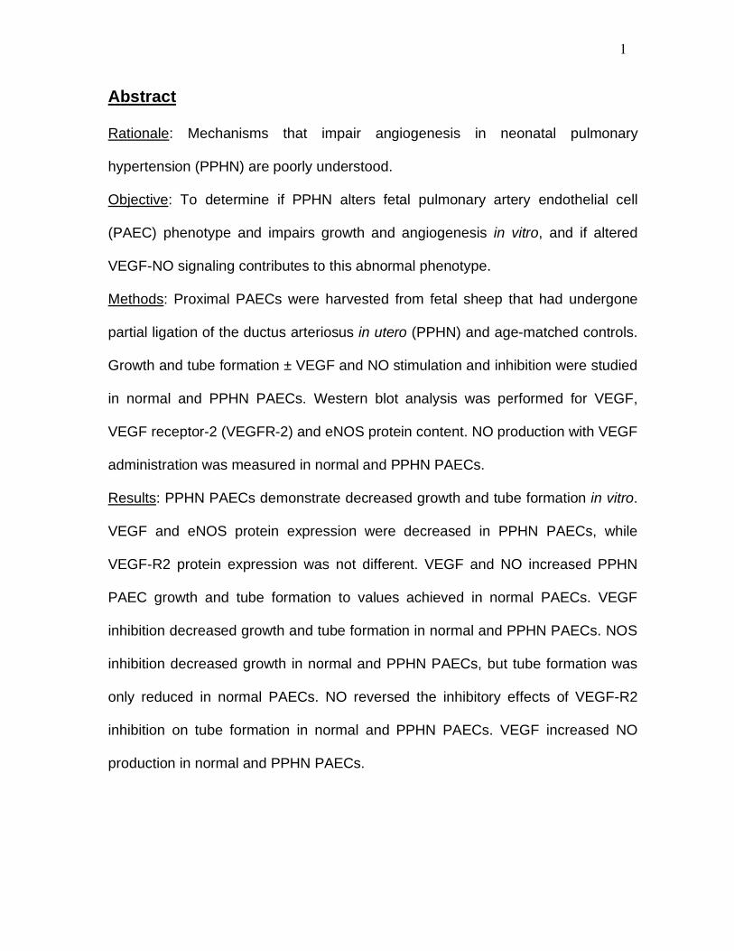
Abstract
Rationale: Mechanisms that impair angiogenesis in neonatal pulmonary
hypertension (PPHN) are poorly understood.
Objective: To determine if PPHN alters fetal pulmonary artery endothelial cell
(PAEC) phenotype and impairs growth and angiogenesis in vitro, and if altered
VEGF-NO signaling contributes to this abnormal phenotype.
Methods: Proximal PAECs were harvested from fetal sheep that had undergone
partial ligation of the ductus arteriosus in utero (PPHN) and age-matched controls.
Growth and tube formation ± VEGF and NO stimulation and inhibition were studied
in normal and PPHN PAECs. Western blot analysis was performed for VEGF,
VEGF receptor-2 (VEGFR-2) and eNOS protein content. NO production with VEGF
administration was measured in normal and PPHN PAECs.
Results: PPHN PAECs demonstrate decreased growth and tube formation in vitro.
VEGF and eNOS protein expression were decreased in PPHN PAECs, while
VEGF-R2 protein expression was not different. VEGF and NO increased PPHN
PAEC growth and tube formation to values achieved in normal PAECs. VEGF
inhibition decreased growth and tube formation in normal and PPHN PAECs. NOS
inhibition decreased growth in normal and PPHN PAECs, but tube formation was
only reduced in normal PAECs. NO reversed the inhibitory effects of VEGF-R2
inhibition on tube formation in normal and PPHN PAECs. VEGF increased NO
production in normal and PPHN PAECs.

2
Conclusions: PPHN in utero causes sustained impairment of PAEC phenotype in
vitro, with reduced PAEC growth and tube formation and down-regulation of VEGF
and eNOS protein. VEGF and NO enhanced growth and tube formation of PPHN
PAECs.
Abstract 249 words
Key terms: Persistent pulmonary hypertension of the newborn, angiogenesis,
vasculogenesis, vascular endothelial growth factor, nitric oxide, endothelial nitric
oxide synthase, endothelial cells, lung vascular development.
3
Introduction Persistent pulmonary hypertension of the newborn (PPHN), is a clinical
syndrome characterized by elevated pulmonary vascular resistance (PVR) after
birth, which causes extra-pulmonary right to left shunting across the foramen ovale
and ductus arteriosus (DA), leading to severe hypoxemia (1). Although PPHN is
associated with diverse cardiopulmonary disorders, mechanisms contributing to
high PVR include elevated pulmonary vascular tone, hypertensive remodeling of
the vessel wall, and in severe cases, decreased vascular growth due to impaired
angiogenesis (2).
Laboratory studies of experimental models of PPHN suggest that endothelial
cell dysfunction, including decreased production of vasodilators, such as nitric oxide
(NO) and increased release of vasoconstrictors, such as endothelin-1 (ET-1),
contributes significantly to the pathophysiology of high PVR. (3-9). Although
treatment with inhaled NO is effective in improving outcomes of many newborns
with PPHN, some infants fail to respond to inhaled NO, and require ECMO therapy
with increased morbidity and mortality (10-13). PPHN associated with poor
responsiveness to inhaled NO is often seen in the setting of impaired vascular
growth, as seen in patients with lung hypoplasia, such as congenital diaphragmatic
hernia and primary lung hypoplasia (12). In the setting of lung hypoplasia,
decreased arterial number plays a prominent role in maintaining high PVR, that is
refractory to therapy. Due to the lack of developmentally relevant in vivo and in vitro
4
models, mechanisms responsible for endothelial cell dysfunction and impaired
angiogenesis in severe PPHN remain poorly understood.
Past studies utilizing partial ligation of the ductus arteriosus (DA) in utero in
late gestation fetal sheep provide a useful animal model for studying the
pathogenesis and treatment of PPHN (3,14-16). In this model, partial ligation of the
ductus arteriosus increases pulmonary artery pressure without causing sustained
elevations of pulmonary blood flow or hypoxemia (3). After chronic intrauterine DA
constriction, PVR remains elevated at birth, pulmonary artery pressure elevated
and pulmonary blood flow reduced, despite ventilation with high levels of
supplemental oxygen (3). Endothelial cell dysfunction, as defined by altered
production of vasoactive mediators, contributes to elevated vascular tone in PPHN,
but may also decrease lung vascular growth. Recent studies in this model have
demonstrated that chronic intrauterine pulmonary hypertension impairs lung
angiogenesis and causes lung hypoplasia. (19). Mechanisms through which fetal
pulmonary hypertension inhibits lung vascular growth are uncertain, but may be
related to decreased production of pro-angiogenic growth factors, such as vascular
endothelial growth factor (VEGF), or altered responsiveness to mitogenic stimuli
(20,21).
We recently reported that impaired alveolarization and vessel growth in
chronic intrauterine pulmonary hypertension is associated with decreased VEGF
protein expression (19) and that VEGF treatment improved hemodynamics and
lung vascular growth (20). PPHN in vivo is further characterized by decreased

5
eNOS protein content and impaired NO production (5,6). Decreased NO production
has been implicated in contributing to elevated vascular tone as well as
hypertensive remodeling in PPHN (6,35). Alterations in pro-angiogenic growth
factors may be responsible for impaired angiogenesis in PPHN, however the
mechanisms by which hemodynamic stress (hypertension) impairs endothelial cell
function and angiogenesis in the developing lung remain unknown.
Therefore, we hypothesized that chronic intrauterine pulmonary hypertension
directly alters endothelial cell phenotype, impairing VEGF-NO signaling within the
endothelial cell and decreasing the ability of the developing endothelium to
proliferate and form new vessels.
6
Methods
Isolation and culture of fetal ovine pulmonary arterial endothelial cells. All
procedures and protocols were reviewed and approved by the Animal Care and
Use Committee at the University of Colorado Health Sciences Center, Denver, CO.
The left and right pulmonary arteries were isolated from late-gestation normal fetal
sheep (mixed-breed Columbia-Rambouillet pregnant ewes at 135 days gestation
(n=4), term = 147 days) and from fetal sheep that had undergone partial ligation of
the ductus arteriosus in utero 7-10 days prior to euthanasia (PPHN)(n=4)(as
previously described, 3). Proximal PAECs were isolated as previously described
(7) and endothelial cell phenotype confirmed by positive immunostaining for
standard endothelial cell markers. Cells from passage 4 and 5 were used for each
of the study experiments and cells from each animal were kept separate
throughout all passages and for all experiments.
Cell growth. Fetal PAECs from normal and PPHN lambs were plated at 2 x
105 cells/well and allowed to adhere overnight. Cells were grown in DMEM media
supplemented with 10% FBS under 3% oxygen conditions. Daily cell counts were
performed for 5 days using a Beckman Coulter cell counter, after removing cells
from wells by 0.25% trypsin/0.53mM EDTA digestion.
The effects of vascular endothelial growth factor (VEGF; 50ng/ml), S-Nitroso-N-
acetylpenicillamine (SNAP (1mM), NO donor, Calbiochem) nitric oxide gas (10 ppm),
7
SU5416 (a VEGF receptor inhibitor, 10 M), nitro-L-arginine (LNA; a nitric oxide
synthase inhibitor; 4mM) and combinations of these agents, on cell growth and tube
formation were compared between PAECs from normal and PPHN fetal sheep. Doses
for each agent were based on preliminary experiments (VEGF, NO gas, SNAP and
LNA) and previously published data (SU5416)(31). For each agent the lowest dose of
agent for which a given effect was seen was used. NO gas was used for the growth
studies as preliminary experiments revealed NO gas at 10ppm to be more effective
than SNAP at stimulating cell growth in PPHN PAECs. SNAP both at various doses
(1mM, 5mM, 10mM), with single treatment or with repeated administrations every 24
hours was less effective than NO gas at stimulating PAEC growth in PPHN cells (data
no shown). SNAP (1 m) was used for the tube formation studies as NO gas was less
effective at enhancing tube formation than SNAP when continuously administered at
10ppm (data not shown). Growth studies with treatment were performed in DMEM
supplemented with 5% serum, as this was the lowest serum concentration that
supported fetal PAEC proliferation.
Tube Formation Assay: The ability of fetal PAECs to form vascular
structures in vitro was assayed by plating PAEC’s on EHS Matrigel (BD
Pharmingen, San Jose, CA). PAECs from normal and PPHN animals were seeded
at a density of 5 x 10⁴ cells/well in serum free DMEM supplemented with and
without VEGF (50ng/ml), SNAP (1 m), LNA (4mM), SU5416 (10 m) and SU5416
(10 m) with SNAP (1 m) in combination. PAECs were incubated for 6 hours under
8
3% oxygen conditions. Branch point counting was performed in blinded fashion
under 10X magnification from each of 4 wells, as previously described (23).
Western Blot Analysis: PAECs from normal and PPHN animals were
grown on 150mm cloning dishes in DMEM supplemented with 10% serum. At
90% confluence, cell lysates were collected as previously described (22). Protein
content was determined by the BCA assay (Pierce Biotechnology Inc (catalog #
23225) Rockford, IL), using bovine serum albumin as the standard. 20 g of
protein sample per lane was resolved by SDS polyacrylamide gel
electrophoresis, and proteins from the gel were transferred to nitrocellulose
membrane. VEGF (Santa Cruz Biotech SC152), VEGF-R2 (Santa Cruz Biotech
SC504), eNOS (BD610297) (eNOS/NOS III) and -actin (Sigma,St. Louis,
A5316) were detected as previously described (46,47) using appropriate
controls and molecular weight as identified by the manufacturer for the protein of
interest. Densitometry was performed using NIH Image (v1.61). Changes in
protein expression were analyzed after normalizing for -actin expression.
Nitric Oxide Assay: Determined using the DAF-FM Nitric Oxide indicator
(Molecular Probes, Eugene, OR #D-23844). Nitric oxide was measured by
fluorescence after a 1 hour VEGF (50ng/ml) exposure as previously described
(36,37).

9
Proliferation/Apoptosis was assessed in normal and PPHN PAECs.
Normal and PPHN PAECs were plated on chamber slides and immunostaining
performed for Ki67 (1:250; Santa Cruz Biotech SC15402) and active caspase-3
(1:250 Chemicon, Temecula, CA AB2362). Comparison was made between
normal and PPHN PAECs.
Statistical analysis. Data are presented as means ± SEM, with data from all 4
animals in the normal and PPHN groups combined for analysis. Statistical analysis
was performed with the Prism 4 software package (GraphPad Software, San Diego,
CA). Statistical comparisons were made using analysis of variance for growth and
tube formation assays with Bonferroni post test analysis. Unpaired t test was used for
western blot analysis. P < 0.05 was considered significant.
10
Results
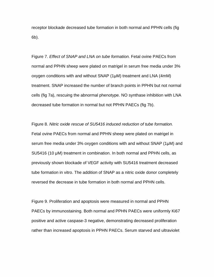
Decreased growth and tube formation in fetal PAECS from PPHN Lambs.
In comparison with controls, PAEC number from PPHN lambs was decreased by
28% (p<0.05) and 35% (p<0.001) on days 3 and 5 respectively (Fig 1). The ability
of PAECs to spontaneously form vascular networks was also decreased in PPHN
cells (Fig 2). In comparison with normal fetal PAECs the number of branch points
was decreased by 24% (p<0.001) in PAECs harvested from PPHN lambs (Fig 2)
Decreased eNOS and VEGF protein expression in fetal PAECs from PPHN
lambs. Western blot analysis of PAEC cell lysates from normal and PPHN lambs
demonstrated decreased endothelial nitric oxide synthase (eNOS) and vascular
endothelial growth factor (VEGF) protein. In comparison with controls, eNOS
protein expression was decreased by 38% in PPHN PAECs (p<0.01)(Fig 3a) and
VEGF protein expression by 48% in PPHN PAECs (p<0.05) (Fig 3b). VEGF-R2
expression was not different between normal and PPHN PAECs. (Fig 3c).
Effect of VEGF and SU5416 on endothelial cell growth. VEGF treatment
increased growth in PAECs harvested from both normal and PPHN lambs. VEGF
treatment of normal PAECs increased cell number by 21% (p<0.05). VEGF
treatment of PPHN PAECs increased cell number by 33% (p<0.01) achieving
values similar to that measured in the normal PAECs (Fig 4a). Treatment with the
11
VEGF receptor blocker, SU5416, decreased cell number by 32% in normal
(p<0.001) and 25% in the PPHN PAECs (p<0.05) (Fig 4b).
Effect of NO gas and LNA on endothelial cell growth. NO gas (10ppm)
treatment increased endothelial PAEC number by 17% (p<0.05) in normal and by
21% in PPHN PAECs (p<0.05) (Fig 5a). NOS inhibition with LNA treatment
decreased cell number by 25% and by 29% in normal and PPHN PAECs,
respectively (p<0.05; for each comparison) (Fig 5b). NO gas did not rescue growth
of control and PPHN PAECs from the inhibitory effects of SU5416 treatment.
Growth remained decreased by 32% in normal (p<0.001) and 31% in PPHN
PAECs (p<0.01) with combined treatment with SU5416 and NO gas (figure not
shown).
Effect of VEGF and SU5416 on tube formation. VEGF treatment had no
effect on the number of branch points in normal PAECs, but increased the number
of branch points by 31% in the PPHN cells (p<0.001; Fig 6a). VEGF treatment fully
reversed the decreased angiogenesis seen in vitro in PPHN PAECs to values
achieved by control PAECs. VEGF receptor blockade with SU5416 decreased the
number of branch points in both normal and PPHN PAECs. The number of branch
points was decreased by 29% in normal PAECs (p<0.001; Fig 6b). The reduction
in tube formation by normal PAECs after SU5416 treatment was similar to the
12
untreated values measured in PAECs from PPHN lambs. In PAEC from PPHN
lambs, SU5416 further decreased tube formation by 21% (p<0.01;fig 6b).
Effect of SNAP and LNA on tube formation The addition of SNAP as an NO
donor increased the number of branch points by 34% in PPHN PAECs (p<0.001;
Fig 7a). SNAP had no effect on the number of branch points in normal PAECs.
NOS inhibition with LNA decreased the number of branch points by 24% (p<0.001)
in normal PAECs, but did not decrease tube formation further in PPHN PAECs (Fig
7b).
NO rescue of SU5416 -induced reduction of tube formation. Blockade of
VEGF receptor activity with SU5416 treatment decreased PAEC tube formation in
vitro. The addition of SNAP as an NO donor completely reversed the effects of
SU5416 on tube formation, as demonstrated by the increase in tube formation to
control values in both normal and PPHN PAECs (Fig 8).
Effect of VEGF treatment on NO production. VEGF treatment (50ng/ml)
increased NO production in both normal and PPHN PAECs by 36% (p<0.01) and
33% (p<0.01) respectively.
Proliferation and Apoptosis in normal and PPHN PAECs. Normal and
PPHN PAECs were uniformly positive for Ki67 by immunostaining when studied at
13
both 50% and 90% confluence. In contrast, normal and PPHN PAECs were
negative for active caspase-3 protein. Serum starved and ultraviolet light treated
normal PAECs were used as positive controls and were strongly positive for active
caspase-3. In addition to negative active caspase-3 staining, no condensed nuclei
were seen after DAPI staining in normal and PPHN PAECs (Fig 9).
14
Discussion
In addition to high PVR due to increased vascular tone and hypertensive
remodeling, severe PPHN is often characterized by reduced vascular growth.
Previous studies have shown that pulmonary hypertension during late gestation
impairs fetal pulmonary vascular growth in vivo (19), but mechanisms by which
high pressure inhibits angiogenesis in the developing lung are unknown. Whether
PPHN causes functional abnormalities of endothelial cells such as changes in
proliferation and angiogenesis and whether these changes in endothelial cell
phenotype persist in vitro have not been previously studied. We found that fetal
PAECs harvested from lambs with experimental PPHN maintain an abnormal
phenotype in vitro, as characterized by decreased PAEC growth and tube
formation when compared with normal gestational age-matched controls. Since
VEGF and NO signaling can regulate endothelial cell growth and angiogenesis, we
measured VEGF, eNOS and VEGFR-2 protein expression in normal and PPHN
PAECs and demonstrate that VEGF and eNOS protein expression are markedly
reduced in PAECs from PPHN lambs. VEGF and NO treatment with either NO gas
or SNAP rescue the abnormal in vitro phenotype of the PPHN PAECs and restore
their growth rate and ability to form vascular networks on matrigel to normal. In
addition, inhibition of endogenous VEGF receptor and NOS activities decreased
growth and tube formation in normal PAECs to values seen in PPHN PAECs. NO
treatment with SNAP rescued the effects of VEGF receptor inhibition on tube
formation in both groups. These findings are the first data to demonstrate that
15
endothelial cells from an experimental model of PPHN maintain an abnormal
phenotype in vitro, including impaired proliferation and tube formation, as well as
dysfunctional VEGF-NO signaling, suggesting that this abnormal endothelial cell
phenotype directly contributes to decreased vascular growth in severe PPHN.
Previous in vivo studies in this model have demonstrated that increased
vascular tone and impaired vasoreactivity are partly due to endothelial dysfunction
(3,15). More recently, we reported that prolonged intrauterine pulmonary
hypertension also impairs angiogenesis, decreases alveolarization and reduces
lung to body weight ratios in fetal sheep, suggesting that pulmonary hypertension
itself impairs angiogenesis in the developing lung and causes lung hypoplasia (19).
Our studies parallel and extend these in vivo observations by demonstrating
persistent abnormalities of endothelial cell growth and tube formation in vitro, and
suggesting that impaired endothelial cell function likely mediates the reduction in
vascular growth observed in severe PPHN. This study also further supports the
hypothesis that decreased VEGF and NO signaling contribute to persistent
abnormalities in PAEC phenotype in the fetal sheep model of PPHN.
VEGF is a potent endothelial cell mitogen and regulator of angiogenesis. As
well as being characterized by impaired angiogenesis and lung hypoplasia, this
model of PPHN, also demonstrates decreased VEGF protein expression (20).
VEGF treatment improves hemodynamics and lung vascular growth in this

16
experimental model of PPHN (21). In vivo inhibition of VEGF receptors in normal
fetal sheep results in impaired vascular growth and pulmonary hypertension (20).
Rodent and rabbit models of hyperoxia induced lung injury are characterized by
decreased blood vessel growth and alveolar simplification (32-35). Associated with
the lung injury is decreased VEGF protein expression. In this model of hyperoxia
induced lung injury treatment with recombinant human VEGF protein (rhVEGF) or
more recently intratracheal adenoviral mediated VEGF gene therapy has been
shown to improve lung vascular growth and alveolarization (28,30). VEGF
treatment was also able to reverse the abnormal in vitro phenotype in PPHN
PAECs. These findings suggest that VEGF is an important regulator of PAEC
growth and angiogenesis in the developing fetal and postnatal lung and that
disruption of VEGF signaling pharmacologically, by hyperoxia or by hypertension
reduces distal lung vascular and alveolar growth.
In addition to regulating vascular tone, NO, which is produced by eNOS,
also plays an important role in angiogenesis and has been shown to mediate many
of the downstream functions of VEGF (24,25). This model of PPHN is
characterized by marked down-regulation of eNOS protein and impaired NO
production (5,6). Previous studies in this model have emphasized the role of
impaired NO production or activity, with a reduction in pulmonary vasodilation to
oxygen, pharmacologic agents and birth related stimuli in fetal sheep (3,39,40). NO
is also an important regulator of smooth muscle cell proliferation (41,42).

17
Decreased NO production has been associated with smooth muscle cell
hyperplasia, which contributes to hypertensive remodeling in PPHN (6,35). More
recently the importance of nitric oxide in regulating vascular and alveolar growth in
the developing lung has been demonstrated in eNOS deficient mice. Lungs from
eNOS -/- mice exhibit marked disruption of lung vascular and alveolar structures
(27). Other studies further demonstrate the susceptibility of eNOS deficient mice to
hypoxia, as mild hypoxia reduces vascular and alveolar growth in neonatal eNOS -
/- mice but not wild-type mice (43). Treatment with inhaled NO has been shown to
improve alveolarization and angiogenesis in experimental models of
bronchopulmonary dysplasia due to hyperoxia or SU 5416 administration in vivo
(26.29). Intratracheal adenoviral mediated eNOS gene therapy has recently been
shown to preserve alveolar development and promote lung capillary formation in
neonatal rat pups exposed to hyperoxia (30). Treatment of PAECs harvested from
PPHN lambs with NO (either SNAP or NO gas) reversed the abnormal growth and
tube formation in vitro providing direct cellular evidence to support this previous in
vivo work.
This is the first study to show persistent abnormalities in PAECs from PPHN
fetal sheep and suggests that decreased VEGF and eNOS protein expression
within the endothelial cell itself may contribute to this abnormal phenotype. Our
finding of decreased eNOS protein expression in PPHN PAECs differs from
Konduri et al (44) who reported decreased eNOS protein from isolated arteries (7),

18
but not from PAECs from fetal sheep (44). Differences between the results of these
studies are unclear but may be due to differences in oxygen tension (3% in our
studies versus room air) as well as other methodologies. We chose 3% oxygen for
our studies based on preliminary data as well as past studies from our lab that
have shown improved PAEC function in low oxygen conditions that mimic the
intrauterine environment (22). Tirosh and coworkers showed decreased NO
production in fetal PAECs from PPHN sheep in response to increased oxygen
tension but did not report on eNOS content (40). Our study is the only work to
measure VEGF protein in PPHN PAECs and demonstrate marked decreases in
VEGF content. Thus, these findings suggest that decreased VEGF and eNOS
protein expression may account for abnormal VEGF-NO signaling within the
endothelial cell itself which may be responsible for impaired vascular growth in
PPHN.
Differences seen in PAEC growth were due to decreased proliferation rather
than increased apoptosis as PPHN PAECs demonstrated positive Ki67 staining
and negative staining for markers of apoptosis (activated caspase-3 and
condensed nuclei with DAPI staining). Along with these findings cell viability was
always in excess of 95% when assessed by tryphan blue exclusion on a Beckman-
Coulter cell counter thus supporting the finding of decreased proliferation in PPHN
PAECs. While similar effects were seen with respect to PAEC growth in normal
and PPHN PAECs with VEGF and NO stimulation and SU5416 and LNA inhibition,
19
differences were seen in the responses of normal and PPHN PAECs to stimulation
and inhibition with respect to tube formation. VEGF and NO treatment with SNAP
increased tube formation in only PPHN PAECs and while VEGF receptor inhibition
decreased tube formation in both normal and PPHN PAECs. NOS inhibition
decreased tube formation in only normal PAECs. We speculate that the lack of
further impairment of tube formation in PPHN PAECs with NOS inhibition is due to
already down regulated eNOS expression. Treatment with SNAP as a NO donor
rescued the angiogenic capacity of SU5416 treated both normal and hypertensive
PAECs demonstrating an intact VEGF-eNOS signaling pathway in both normal and
PPHN PAECs. By demonstrating increased NO release in response to VEGF
stimulation in normal and PPHN PAECs, we further confirm intact VEGF-NO
signaling within both normal and PPHN PAECs. Angiogenesis is a complex
process, which involves the formation of new blood vessels from preexisting
vessels. For new blood vessels to form in the developing lung, endothelial cells
must proliferate and form vascular tubes. Our current study demonstrates
distinctive yet overlapping roles for VEGF-NO regulation of angiogenesis within the
developing lung as well as the critical role the VEGF-NO signaling pathway plays
in stimulating endothelial cells to proliferate and form new blood vessel within the
developing lung.
Potential limitations of this study include the concern that these studies used
fetal PAECs harvested from large vessels, and differences may exist in the
20
behavior of these cells as compared to microvascular PAECs. Since
microvascular PAECs may primarily be involved in lung angiogenesis during
development in vivo, future studies are needed to compare and contrast
microvascular PAEC function from normal and PPHN fetal sheep in parallel
fashion to these current studies. These studies looked exclusively at in vitro,
endothelial cell function. In vivo the mesenchyme and epithelium are important
regulators of endothelial cell growth and tube formation. The effect of the
mesenchyme and epithelium on the endothelium were not assessed in this in vitro
model.
In conclusion, we found that chronic intrauterine pulmonary hypertension
directly alters endothelial cell phenotype, which persists in vitro, as characterized
by decreased endothelial cell growth and tube formation. Based on these findings,
PAECs harvested from fetal lambs with chronic pulmonary hypertension provide a
unique in vitro model for studying basic mechanisms of endothelial dysfunction in
PPHN. In addition, this study demonstrates that impaired VEGF and NO signaling
contribute to the abnormal PAEC phenotype in vitro. Since exogenous VEGF and
NO treatment (either SNAP or NO gas) rescues the abnormal PPHN PAEC
phenotype in vitro, we speculate that treatment strategies that up-regulate VEGF
and NO signaling pathways may enhance angiogenesis in vivo in patients with
severe PPHN, and may be especially important in the setting of lung hypoplasia.

21
References
1) Levin DL, Heymann MA, Kitterman JA, Gregory GA, Phibbs RH, Rudolph AM.
Persistent pulmonary hypertension of the newborn infant. J Pediatr. 1976
Oct;89(4):626-30.
2) Geggel RL, Reid LM. The structural basis of PPHN. Clin Perinatol. 1984
Oct;11(3):525-49
3) Abman SH, Shanley PF, Accurso FJ. Failure of postnatal adaptation of the
pulmonary circulation after chronic intrauterine pulmonary hypertension in fetal
lambs. J Clin Invest. 1989 Jun;83(6):1849-58.
4) Villanueva ME, Zaher FM, Svinarich DM, Konduri G Decreased Gene
Expression of Endothelial Nitric Oxide Synthase in Newborns with Persistent
Pulmonary Hypertension. Pediatric Research: 44(3)Sept 338-343,1998
5) Villamor E, Le Cras TD, Horan MP, Halbower AC, Tuder RM, and Abman SH.
Chronic intrauterine pulmonary hypertension impairs endothelial nitric oxide
synthase in the ovine fetus. Am J Physiol Lung Cell Mol Physiol 16: L1013-L1020,
1997.
6) Shaul PW, Yuhanna IS, German Z,Chen Z, Steinhorn RH, Morin FC. Pulmonary
endothelial NO synthase gene expression is decreased in fetal lambs with
pulmonary hypertension. Am J Physiol Lung Cell Mol Physiol 16:L1005-
L1012,1997.

22
7) Konduri G, Yang Shi, JO, Pritchard, Jr, KA. Decreased association of HSP90
impairs endothelial nitric oxide synthase in fetal lambs with persistent pulmonary
hypertension Am J Physiol Heart Circ Physiol 285: H204–H211, 2003.
8) Murata T, Sato K, Hori M, Ozaki H, Karaki H. Decreased Endothelial Nitric-oxide
Synthase (eNOS) Activity Resulting from Abnormal Interaction between eNOS and
Its Regulatory Proteins in Hypoxia-induced Pulmonary Hypertension. J Biolog
Chem 277.46, 44085–44092, 2002
9) Wedgwood S,Black SM. Endothelin-1 decreases endothelial NOS expression
and activity through ETA receptor-mediated generation of hydrogen peroxide. Am
J Physiol Lung Cell Mol Physiol 288: L480–L487, 2005.
10) Kinsella JP, Neish SR, Shaffer E, Abman SH. Low-dose inhalation nitric oxide
in persistent pulmonary hypertension of the newborn. Lancet. 1992 Oct
3;340(8823):819-20.
11) Davidson D, Barefield ES, Kattwinkel J, Dudell G, Damask M, Straube R,
Rhines J, Chang CT. Inhaled nitric oxide for the early treatment of persistent
pulmonary hypertension of the term newborn: a randomized, double-masked,
placebo-controlled, dose-response, multicenter study. The I-NO/PPHN Study
Group. Pediatrics. 1998 Mar;101(3 Pt 1):325-34.
12) Goldman AP, Tasker RC, Haworth SG, Sigston PE, Macrae DJ. Four patterns
of response to inhaled nitric oxide for persistent pulmonary hypertension of the
newborn. Pediatrics. 1996 Oct;98(4 Pt 1):706-13

23
13) Farrow KN, Fliman P, Steinhorn RH. The diseases treated with ECMO: focus
on PPHN. Semin Perinatol. 2005 Feb;29(1):8-14
14) Wild LM, Nickerson PA, Morin FC III. Ligating the ductus arteriosus before birth
remodels the pulmonary vasculature of the lamb. Pediatr Res. 1989
Mar;25(3):251-7.
15) Morin FC III. Ligating the ductus arteriosus before birth causes persistent
pulmonary hypertension in the newborn lamb. Pediatr Res. 1989 Mar;25(3):245-50.
16) Belik J, Keeley FW, Baldwin F, Rabinovitch M. Pulmonary hypertension and
vascular remodeling in fetal sheep. Am J Physiol. 1994 Jun;266(6):H2303-9.
17) Ivy D; Parker TA.; Ziegler JW.; Galan HL.; Kinsella JP.; Tuder RM.; Abman SH.
Prolonged Endothelin A Receptor Blockade Attenuates Chronic Pulmonary
Hypertension in the Ovine Fetus. J Clin Invest. 99(6):1179-1186, March 15, 1997.
18) Ivy D; Ziegler JW.; Dubus MF.; Fox J.; Kinsella JP.; Abman SH. Chronic
Intrauterine Pulmonary Hypertension Alters Endothelin Receptor Activity in the
Ovine Fetal Lung. Pediatr Res. 39(3):435-442, March 1996.
19) Grover TR, Parker TA, Balasubramaniam V, Markham NE, Abman SH. Pulmonary
hypertension impairs alveolarization and reduces lung growth in the ovine fetus Am J
Physiol Lung Cell Mol Physiol 288: L648-L654, 2005
20) Grover TR, Parker TA, Zenge JP, Markham NE, Kinsella JP, Abman SH.
Intrauterine hypertension decreases lung VEGF expression and VEGF inhibition
causes pulmonary hypertension in the ovine fetus. Am J Physiol Lung Cell Mol
Physiol. 2003 Mar;284(3):L508-17.
24
21) Grover TR, Parker TA, Abman SH. Vascular endothelial growth factor
improves pulmonary vascular reactivity and structure in an experimental model of
chronic pulmonary hypertension in fetal sheep. Chest. 2005 Dec;128 (6
Suppl):614S.
22) Balasubramaniam V, Maxey AM, Fouty BW, Abman SH. Nitric Oxide
Augments Fetal Pulmonary Artery Endothelial Cell Angiogenesis in vitro. Am J
Physiol Lung Cell Mol Physiol 2006 Jun;290(6):L1111-6.
23) Donovan D, Brown NJ, Bishop ET, Lewis CE. Comparison of three in vitro
human ‘angiogenesis’ assays with capillaries formed in vitro Angiogenesis 4:113-
121, 2001
24) Papapetropoulos A, García-Cardeña G, Madri JA, Sessa WC. Nitric Oxide
Production Contributes to the Angiogenic Properties of Vascular Endothelial
Growth Factor in Human Endothelial Cells, J Clin Invest 1997; 100: 3131 –3139.
25) Morbidelli L, Chang CH, Douglas JG et al. Nitric oxide mediates mitogenic
effect of VEGF on coronary venular endothelium. Am J Physiol 1996;270:H411–
H415.
26) Lin YJ; Markham NE.; Balasubramaniam V; Tang JR; Maxey A; Kinsella JP.;
Abman SH. Inhaled Nitric Oxide Enhances Distal Lung Growth after Exposure to
Hyperoxia in Neonatal Rats. Pediatric Research. 58(1):22-29, July 2005.
27) Han RN, Stewart DJ. Defective lung vascular development in endothelial nitric
oxide synthase-deficient mice. Trends Cardiovasc Med. 2006 Jan;16(1):29-34.

25
28) Kunig AM, Balasubramaniam V, Markham NE, Seedorf G, Gien J, Abman
SH. Recombinant human VEGF treatment transiently increases lung edema but
enhances lung structure after neonatal hyperoxia. Am J Physiol Lung Cell Mol
Physiol. 2006 Nov;291 (5):L1068-7
29) Tang JR, Markham NE, Lin YJ, McMurtry IF, Maxey A, Kinsella JP, Abman
SH. Inhaled nitric oxide attenuates pulmonary hypertension and improves lung
growth in infant rats after neonatal treatment with a VEGF receptor inhibitor. Am
J Physiol Lung Cell Mol Physiol. 2004 Aug;287(2):L344-51
30) Thebaud B, Ladha F, Michelakis ED, Sawicka M, Thurston G, Eaton F,
Hashimoto K, Harry G, Haromy A, Korbutt G, Archer SL. Vascular endothelial
growth factor gene therapy increases survival, promotes lung angiogenesis, and
prevents alveolar damage in hyperoxia-induced lung injury: evidence that
angiogenesis participates in alveolarization. Circulation. 2005 Oct 18;112(16):
2477-86.
31) Ma L, Francia G, Viloria-Petit A, Hicklin DJ, du Manoir J, Rak J Kerbel RS. In
vitro Procoagulant Activity Induced in Endothelial Cells by Chemotherapy and
Antiangiogenic Drug Combinations: Modulation by Lower-Dose Chemotherapy
Cancer Research 2005 Jun;65, 5365-5373.
32) Klekamp JG, Jarzecka K, Perkett EA. Exposure to hyperoxia decreases the
expression of vascular endothelial growth factor and its receptors in adult rat
lungs.Am J Pathol. 1999 Mar;154(3):823-31
26
33) Hosford GE, Olson DM. Effects of hyperoxia on VEGF, its receptors, and
HIF-2alpha in the newborn rat lung. Am J Physiol Lung Cell Mol Physiol. 2003
Jul;285(1):L161-8.
34) Maniscalco WM, Watkins RH, D'Angio CT, Ryan RM. Hyperoxic injury
decreases alveolar epithelial cell expression of vascular endothelial growth factor
(VEGF) in neonatal rabbit lung. Am J Respir Cell Mol Biol. 1997 May;16(5):557-
67.
35) Ambalavanan N, Mariani G, Bulger A, Philips III JB. Role of nitric oxide in
regulating neonatal porcine pulmonary artery smooth muscle cell proliferation.
Biol Neonate. 1999 Nov;76(5):291-300.
36) Nakatsubo N, Kojima H, Kikuchi K, Nagoshi H, Hirata Y, Maeda D, Imai Y,
Irimura T, Nagano T. Direct evidence of nitric oxide production from bovine aortic
endothelial cells using new Fluorescence indicators: diaminofluoresceins. FEBS
Letters 427 (1998) 263-266.
37) R. Berkels, C. Dachs, R. Roesen, W. Klaus. Simultaneous measurement of
intracellular Ca2+ and nitric oxide: a new method. Cell Calcium (2000) 27 (5),
281–286
38) Voelkel NF, Vandivier RW, Tuder RM. Vascular endothelial growth factor in
the lung. Am J Physiol Lung Cell Mol Physiol. 2006 Feb;290(2):L209-21. Review.
39) McQueston JA, Kinsella JP, Ivy DD, McMurtry IF, Abman SH.
Chronic pulmonary hypertension in utero impairs endothelium-dependent
vasodilation. Am J Physiol. 1995 Jan;268(1 Pt 2):H288-94
27
40) Tirosh R, Resnik ER, Herron J, Sukovich DJ, Hong Z, Weir EK, Cornfield DN.
Acute normoxia increases fetal pulmonary artery endothelial cell cytosolic Ca2+
via Ca2+-induced Ca2+ release. Pediatr Res. 2006 Sep;60(3):258-63
41) Garg UC, Hassid A. Nitric oxide-generating vasodilators and 8-bromo-cyclic
guanosine monophosphate inhibit mitogenesis and proliferation of cultured rat
vascular smooth muscle cells. J Clin Invest 1989;83:1774–1777.
42)Thomae KR, Nakayama DK, Billiar TR, Simmons RL, Pitt BR, Davies P. The
effect of nitric oxide on fetal pulmonary artery smooth muscle growth. J Surg Res
1995;59:337–343
43) Balasubramaniam V, Tang JR, Maxey A, Plopper CG, Abman SH. Mild
hypoxia impairs alveolarization in the endothelial nitric oxide synthase-deficient
mouse. Am J Physiol Lung Cell Mol Physiol. 2003 Jun;284(6):L964-71
44) Konduri GG, Bakhutashvili I, Eis A, Pritchard KA.Oxidant Stress from
Uncoupled Nitric Oxide Synthase Impairs Vasodilation in Fetal Lambs with
Persistent Pulmonary Hypertension. Am J Physiol Heart Circ Physiol, Apr 2007;
292: H1812 - H1820
45) Zhao YD, Courtman DW, Ng DS, Robb MJ, Deng YP, Trogadis J, Han RN,
Stewart DJ. Microvascular regeneration in established pulmonary hypertension
by angiogenic gene transfer. Am J Respir Cell Mol Biol. 2006 Aug;35(2):182-9
46) Balasubramaniam V, Maxey AM, Morgan DB, Markham NE, Abman SH.
Inhaled NO restores lung structure in eNOS-deficient mice recovering from

28
neonatal hypoxia Am J Physiol Lung Cell Mol Physiol, Jul 2006; 291: L119 -
L127.
47) Le Cras, TD, Xue C, Rengasamy A, and Johns RA. Chronic hypoxia
upregulates endothelial and inducible NO synthase gene and protein expression
in rat lung. Am J Physiol Lung Cell Mol Physiol 270: L164-L170, 199

Figure Legends
Figure 1. Decreased growth in fetal PAECs from PPHN Sheep. Fetal ovine
PAECs from normal and PPHN lambs were plated in 10% FBS under 3% oxygen
conditions and counted daily for 5 days. In comparison with normal PAECs
proliferation of endothelial cells from PPHN lambs were decreased over time in
culture from day 3 to day 5. Error bars represent SD from mean.
Figure 2. Decreased tube formation in fetal PAECs from PPHN Sheep. Fetal
ovine PAECs from normal and PPHN lambs were plated on matrigel in serum
free media under 3% oxygen conditions. The ability of PPHN cells to form
vascular networks is markedly impaired when compared with normal controls.
Error bars represent SD from mean.
Figure 3. Decreased eNOS and VEGF protein expression in fetal PAECs from
PPHN Sheep. Western blot analysis on cell lysates from PAECs from PPHN
sheep demonstrate decreased eNOS (fig3a) and VEGF (fig3b) protein when

compared with normal controls. There was no difference in KDR (fig3c) protein
expression. Error bars represent SD from mean.
Figure 4. Effect of VEGF and SU5416 on endothelial cell growth. Ovine PAEC
from normal and PPHN lambs were grown in 5% FBS under 3% oxygen
conditions with and without VEGF (50ng/ml) and SU5416 (10 M). VEGF
treatment stimulated growth of PAECs in both normal and PPHN cells (fig 4a)
and VEGF receptor blockade with SU5416 decreased growth in both normal and
PPHN PAECs (fig4b). Error bars represent SD from mean.
Figure 5. Effect of NO gas and LNA on endothelial cell growth. Fetal ovine
PAECs from normal and PPHN lambs were grown in 5% FBS under 3% oxygen
conditions with and without NO gas (10ppm) and LNA (4mM). As shown, NO
treatment stimulated growth (fig 5a) and NO synthase inhibition with LNA
decreased growth in normal and PPHN PAECs (fig 5b). Error bars represent SD
from mean.
Figure 6. Effect of VEGF and SU5416 on tube formation. Fetal ovine PAECs
from normal and PPHN sheep were plated on matrigel in serum free media under
3% oxygen conditions with and without VEGF treatment (50ng/ml) and SU5416
(10 M) treatment. VEGF treatment increased the number of branch points in
PPHN but not normal cells (fig 6a), rescuing the abnormal phenotype. VEGF
receptor blockade decreased tube formation in both normal and PPHN cells (fig
6b).
Figure 7. Effect of SNAP and LNA on tube formation. Fetal ovine PAECs from
normal and PPHN sheep were plated on matrigel in serum free media under 3%
oxygen conditions with and without SNAP (1 M) treatment and LNA (4mM)
treatment. SNAP increased the number of branch points in PPHN but not normal
cells (fig 7a), rescuing the abnormal phenotype. NO synthase inhibition with LNA
decreased tube formation in normal but not PPHN PAECs (fig 7b).
Figure 8. Nitric oxide rescue of SU5416 induced reduction of tube formation.
Fetal ovine PAECs from normal and PPHN sheep were plated on matrigel in
serum free media under 3% oxygen conditions with and without SNAP (1 M) and
SU5416 (10 M) treatment in combination. In both normal and PPHN cells, as
previously shown blockade of VEGF activity with SU5416 treatment decreased
tube formation in vitro. The addition of SNAP as a nitric oxide donor completely
reversed the decrease in tube formation in both normal and PPHN cells.
Figure 9. Proliferation and apoptosis were measured in normal and PPHN
PAECs by immunostaining. Both normal and PPHN PAECs were uniformly Ki67
positive and active caspase-3 negative, demonstrating decreased proliferation
rather than increased apoptosis in PPHN PAECs. Serum starved and ultraviolet

light treated normal PAECs were used as controls for apoptosis and were
strongly positive for activated caspase-3.
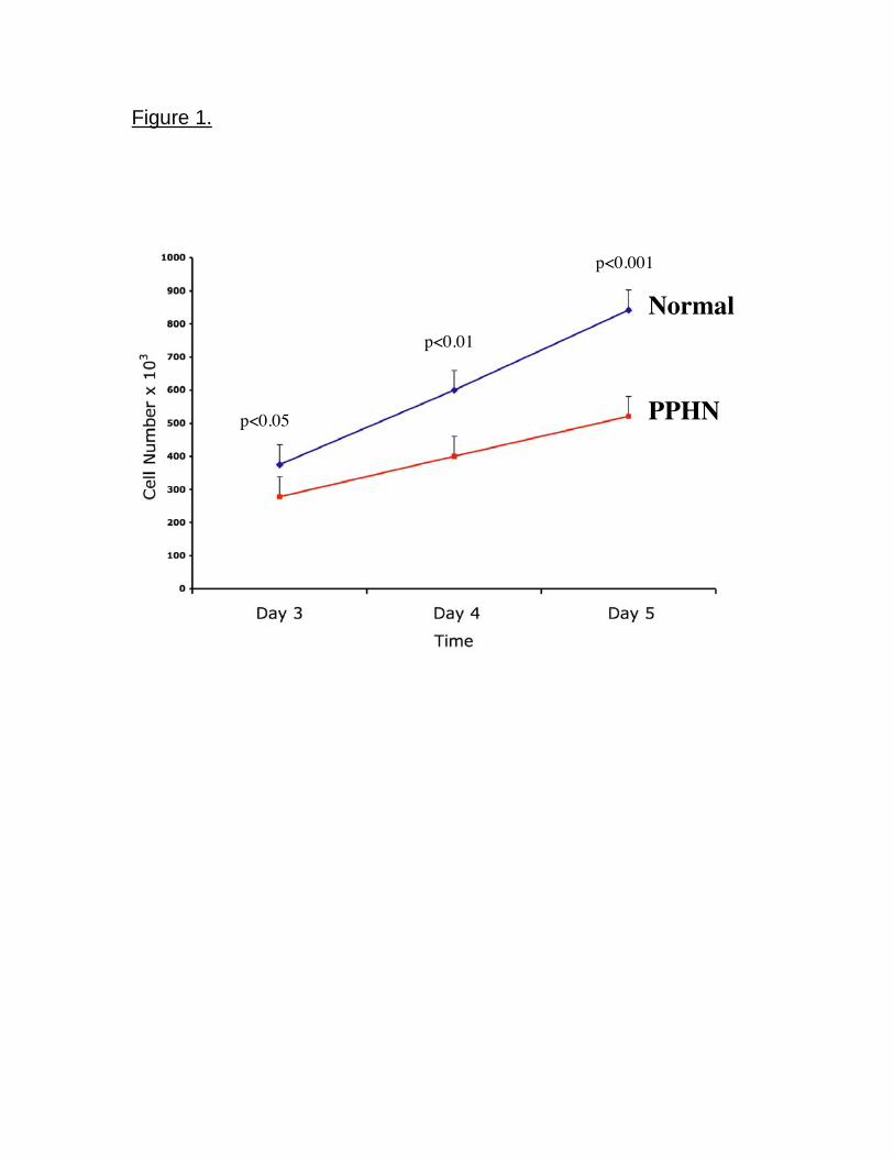
Figure 1.
Normal
PPHN p<0.05
p<0.01
p<0.001
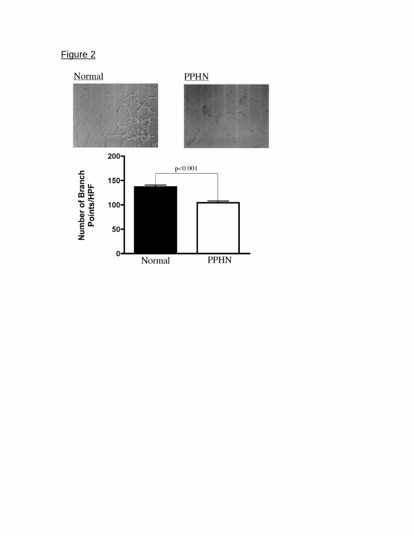
Figure 2
Figure 3a
Figure 3b
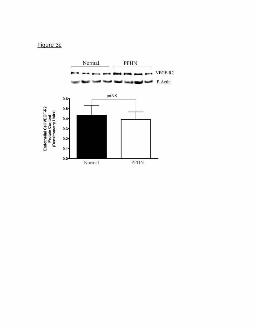
Figure 3c

Figure 4a.
Figure 4b.

Figure 5a.
Figure 5b.

Figure 6a.
Figure 6b.
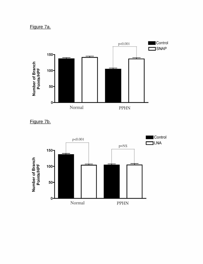
Figure 7a.
Figure 7b.

Figure 8
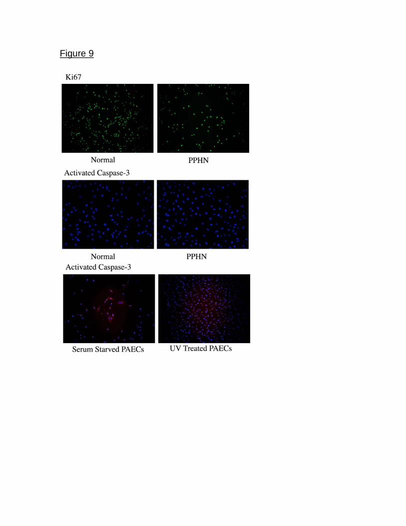
Figure 9

Intrauterine Pulmonary Hypertension Impairs Angiogenesis in vitro: Role of
VEGF-NO Signaling.
Jason Gien MD, Gregory J Seedorf, BS, Vivek Balasubramaniam MD, Neil
Markham, BS, Steven H. Abman, MD
Online Data Supplement
Methods
Isolation and culture of fetal ovine pulmonary arterial endothelial cells. All
procedures and protocols were reviewed and approved by the Animal Care and Use
Committee at the University of Colorado Health Sciences Center, Denver, CO.
Pulmonary arteries were isolated from late-gestation fetal sheep (mixed-breed
Columbia-Rambouillet pregnant ewes at 135 days gestation, term = 147 days) and
from fetal sheep that had undergone partial ligation of the ductus arteriosus in utero 7-
10 days prior to euthanasia (PPHN). In this model, ductal ligation causes sustained
elevation in pulmonary artery pressure without a change in blood flow or oxygenation
(3). The ewe and fetus were killed with high dose intravenous pentobarbital sodium.
The left and right main pulmonary arteries were isolated by direct visualization,
branching vessels were tied with silk suture, and the endothelial surface exposed to
0.1% collagenase for 5 minutes. The vascular surface was washed with D-valine media
(Cellgro; Mediatech Inc, Herndon, VA) containing 10% fetal bovine serum (FBS), 1%
antibiotic-antimycotic agent (Cellgro; Mediatech Inc, Herndon, VA). Cells were
separated from the remaining collagenase by centrifugation for 8 minutes at 1200
RPM. The supernatant was removed and the cell pellet resuspended in D-valine
media. The pellet was dispersed, and cells were plated in 6 well dishes coated with
0.1% gelatin. All tissue culture plastic used in the below mentioned experiments were
coated with 0.1% gelatin. After 24 hours, D-valine media was removed and replaced
with Dulbecco’s Modification of Eagles Medium (DMEM; Sigma, St. Louis, MO) with
10% FBS, 1% antibiotic-antimycotic agent and 1% L glutamine. Cells were allowed to
grow to confluence in humidified incubators (Forma Scientific, Marietta, OH) at 37¡C in
room air, 5% CO2. Endothelial cell phenotype was confirmed at each passage by
positive immunostaining for von Willebrands Factor (vWF), eNOS, vascular endothelial
(VE)-cadherin and VEGFR-2 (KDR); the positive uptake of diacetylated-low density
lipoprotein and the absence of staining for desmin. Cells from passage 4 and 5 were
used for each of the study experiments.
The effects of vascular endothelial growth factor (VEGF; 50ng/ml), S-Nitroso-
N-acetylpenicillamine (SNAP (1mM), NO donor, Calbiochem) nitric oxide gas (10
ppm), SU5416 (a VEGF receptor inhibitor, 10 M), nitro-L-arginine (LNA; a nitric
oxide synthase inhibitor; 4mM) and combinations of these agents, on cell growth
were compared between PAEC from normal and PPHN sheep. Doses for each
agent were based on preliminary experiments (VEGF, NO gas, SNAP and LNA)
and previously published data (SU5416)(31). For each agent the lowest dose of
agent for which a given effect was seen was used. These studies were performed in
DMEM supplemented with 5% serum, as this was the lowest serum concentration
that supported fetal PAEC proliferation.
Effect of SNAP and NO gas on PAEC growth and tube formation. Preliminary
experiments were performed to determine the effects of NO on endothelial cell growth
and tube formation. We compared the effects of NO gas at 10ppm and SNAP, a NO
donor at various doses (1mM, 5mM, 10mM), with single treatment or repeated
administrations every 24 hours, and found NO gas be more effective than SNAP in
stimulating endothelial cell growth. SNAP spontaneously degrades and releases NO
when kept in solution and is depleted after 24 hours. As our experiments on PAEC
growth were performed over several days, NO gas was used to provide both a
continuous and constant dose exposure. NO gas was less effective at enhancing tube
formation when continuously administered at 10 ppm (data not shown). For this reason,
NO gas was used to determine the effect of nitric oxide as well as SU5416 (10 M) +
NO on PAEC growth, while SNAP was used to determine the effect of nitric oxide as
well as SU5416 (10 M) + NO on tube formation.
Cell growth. Fetal PAEC from normal and PPHN lambs were plated at 2 x
105
cells/well and allowed to adhere overnight. Cells were plated and grown in
DMEM media supplemented with 10% FBS. Cells were grown in 3% oxygen, as
preliminary experiments, as well as previous studies in our lab have shown
improved fetal PAEC function in low oxygen tension conditions that mimic the
intrauterine environment (22). Daily cell counts were performed for 5 days using a
Beckman Coulter cell counter, after removing cells from wells by 0.25%
trypsin/0.53mM EDTA digestion.
Stimulatory effects of VEGF and NO on cell growth. PAECs were seeded at
a density of 2 x 10⁵
cells/well in 6 well dishes. Cells were allowed to adhere
overnight and baseline counts performed on day 1. Cells were allowed to proliferate
to day 3 and at this time point, cells were treated with VEGF or NO gas. This time
point was chosen for stimulation, as at day 3 differences in growth were seen
between normal and PPHN cells. Cell counts were then performed on day 5 (after
2 days of treatment), and comparisons were made between treated and untreated
cells.
Inhibitory effects of SU5416 and L-NA on cell growth. Cells were seeded at a
density of 2 x 10⁵
cells/well and allowed to adhere overnight. Baseline counts were
performed on day 1 prior to treatment with SU5416 and L-NA. Counts were performed
on day 3 and comparisons were made between treated and untreated cells after 2 days
of treatment.
To assess the ability of NO gas to rescue SU5416 inhibition of cell proliferation,
cells were seeded at a density of 2 x 10⁵
cells/well. Cells were allowed to adhere
overnight and baseline counts performed on day 1. Cells were then exposed to the
combination of SU5416 and NO gas. Cell counts were then performed on day 3 and a
comparison made between normal and PPHN PAECs with respect to cell number.
Tube Formation Assay: The ability of fetal PAECs to form vascular structures in
vitro was assayed by plating PAEC’s on EHS Matrigel. (BD Pharmingen, San Jose,
CA) EHS matrigel was pipetted into 24 well tissue culture dishes (250 l/well) and
allowed to polymerize at 37¡C for one hour. PAEC from normal and PPHN animals
were seeded at a density of 5 x 10⁴
cells/well in serum free DMEM supplemented with
and without VEGF (50ng/ml), SNAP (1 M), LNA (4mM), SU5416 (10 M) and SU5416
(10 M) with SNAP (1 M) in combination. PAECs were incubated for 6 hours under
3% oxygen conditions, since maximal PAEC tube formation occurred at this time.

Branch point counting was performed in blinded fashion under 10X magnification from
each of 4 wells, as previously described (23).
Western Blot Analysis: PAECs from normal and PPHN animals were grown
on 150mm cloning dishes in DMEM supplemented with 10% serum. At 90%
confluence, cells were washed with ice cold PBS x 2 and lysed in
radioimmunoprecipitation (RIPA) buffer (PBS, 1% Nonidet P-40, 0.5%, sodium
deoxycholate, 0.1% SDS, PMSF [10mg/ml], aprotinin [16 l/ml], and sodium
orthovanadate [1mM]). Cell lysate was then scraped off the dishes. The cell lysate
was sonicated and frozen down. On thawing, the cell lysate was sonicated once
more and centrifuged at 10,000 x g for 30 min at 4¡C. The supernatant was removed
and protein content in the supernatant was determined by the BCA assay (Pierce
Biotechnology Inc (catalog # 23225) Rockford, IL), using bovine serum albumin as
the standard. Briefly, 20 _g of protein sample per lane was resolved by SDS
polyacrylamide gel electrophoresis, and proteins from the gel were transferred to
nitrocellulose membrane.
VEGF: Blots were blocked for 30 minutes in 5% nonfat dry milk dissolved in
buffer 1 (10mM tris-hcl,150mM NaCl, 0.05% tween-20, PH 8.0) The VEGF blot was
incubated for 1 hour in room air with (Santa Cruz Biotech SC152) (1:500) diluted in
5% nonfat dry milk in buffer 1. After washing, the VEGF blot was then incubated for 1
hour at room temperature with goat anti-rabbit IgG-HRP antibody (Santa Cruz
Biotech, SC2054). VEGF bands were then visualized by enhanced
chemiluminenscence (ECL+ kit; Amersham Pharmacia Biotech, Buckinghamshire,
UK). VEGF was run as a control for easy detection of the VEGF band.
eNOS/VEGF-R2: Blots were blocked for 30 minutes in 2% ECL advance
(Amersham Pharmacia Biotech, Buckinghamshire, UK) dissolved in PBS with 0.5%
Tween 20. The KDR blot was incubated overnight at 4¡C with SC504 (1:250; Santa
Cruz Biotech) diluted in 2% ECL advance. The eNOS blot was incubated for 1 hour in
room air with BD610297 (eNOS/NOS III)(1:1000) diluted in 2% ECL advance. After
washing, the KDR blot was then incubated for 1 hour at room temperature with goat
anti-rabbit IgG-HRP antibody (Santa Cruz Biotech, SC2054) and the eNOS blot
incubated for 1 hour at room temperature with goat anti-mouse HRP conjugated
secondary (Chemicon)(1:20000). eNOS and KDR were visualized using the ECL
advance kit, identified by molecular weight as identified by the manufacturer for the
protein of interest.
All blots were then stripped and reprobed with an antibody to B-actin (Sigma,St.
Louis, A5316). Densitometry was performed using NIH Image (v1.61). Changes in
protein expression were analyzed after normalizing for B-actin expression.
Nitric Oxide Assay: Determined using the DAF-FM Nitric Oxide indicator (Molecular
Probes, Eugene, OR #D-23844). 5 x 10³ normal and PPHN PAECs were plated in a 96
well plate in DMEM with 10% FBS under 3% oxygen conditions. Cells were allowed to
adhere overnight after which media was switched to DMEM with 0.5% FBS for 24
hours followed by serum free DMEM for 24 hours. Normal and PPHN PAECs were
then incubated with DAF-FM with and without VEGF (50ng/ml) in PBS for 1 hour. PBS
was then removed from each well and NO production measured in response to VEGF
stimulation using a microplate reader with fluorescence excitation and emission
maxima of 495 and 515 nm, respectively. Comparisons were made between normal
and PPHN cells with respect to NO production.
Proliferation/Apoptosis was assessed in normal and PPHN PAECs. Normal and PPHN
PAECs were plated on chamber slides and fixed with 4% paraformaldehyde at 50%
and 90% confluence for 15 minutes. Control slides for apoptosis were obtained by
serum starving normal PAECs for 72 hrs or exposing normal PAECs to ultraviolet (UV)
irradiation for 15 minutes to induce apoptosis. After serum starvation and UV irradiation
slides were fixed with 4% paraformaldehyde for 15 minutes. Slides were then
permeabilized with ice cold methanol in -20C for 15min. After 3 washes with PBS,
slides were again permeabilized with 0.1% Triton-X. 3 further washes with PBS were
performed and slides blocked with 2% horse serum for 1 hour. 3 more washes were
performed with 0.1%BSA in PBS (PBSA) and the slides were then incubated with
primary antibody Ki67 (1:250; Santa Cruz Biotech SC15402) and active Caspase-3
(1:250 Chemicon, Temecula, CA AB2362) for 1 hour. After 3 more washes with PBSA
slides were incubated with secondary antibody Alexa-Flour goat anti-rabbit 488 (1:500)
(Invitrogen Carlsbad CA #A11034) for 1 hour. Slides were washed 3 more times with
PBSA and mounted with Vectashield Hardmount with DAPI (Carpinteria CA #H1500).
Comparison was made between normal and PPHN PAECs.
Statistical analysis. Data are presented as means ± SEM, with data from all 4 animals
in the normal and PPHN groups combined for analysis. Statistical analysis was performed
with the Prism 4 software package (GraphPad Software, San Diego, CA). Statistical
comparisons were made using analysis of variance for growth and tube formation assays
with Bonferroni post test analysis. Unpaired t test was used for western blot analysis. P <
0.05 was considered significant.
Copyright © 2022 FDOKUMEN