Defective Neurogenesis in Citron Kinase Knockout Mice by Altered Cytokinesis and Massive Apoptosis
Upload
independentCategory
view
0download
0
Molecular Biology of the CellVol. 17, 3423–3434, August 2006
CP110 Cooperates with Two Calcium-binding Proteinsto Regulate Cytokinesis and Genome StabilityWilliam Y. Tsang,* Alexander Spektor,* Daniel J. Luciano,* Vahan B. Indjeian,†Zhihong Chen,‡ Jeffery L. Salisbury,§ Irma Sanchez,* and Brian David Dynlacht*
*Department of Pathology, New York University School of Medicine, New York, NY 10016; †Department ofMolecular and Cellular Biology, Harvard University, Cambridge, MA 02138; ‡Esai Research Institute,Andover, MA 01810; and §Tumor Biology Program, Mayo Clinic, Rochester, MN 55905
Submitted May 1, 2006; Revised May 18, 2006; Accepted May 25, 2006Monitoring Editor: Trisha Davis
The centrosome is an integral component of the eukaryotic cell cycle machinery, yet very few centrosomal proteins havebeen fully characterized to date. We have undertaken a series of biochemical and RNA interference (RNAi) studies toelucidate a role for CP110 in the centrosome cycle. Using a combination of yeast two-hybrid screens and biochemicalanalyses, we report that CP110 interacts with two different Ca2�-binding proteins, calmodulin (CaM) and centrin, in vivo.In vitro binding experiments reveal a direct, robust interaction between CP110 and CaM and the existence of multiplehigh-affinity CaM-binding domains in CP110. Native CP110 exists in large (�300 kDa to 3 MDa) complexes that containboth centrin and CaM. We investigated a role for CP110 in CaM-mediated events using RNAi and show that its depletionleads to a failure at a late stage of cytokinesis and the formation of binucleate cells, mirroring the defects resulting fromablation of either CaM or centrin function. Importantly, expression of a CP110 mutant unable to bind CaM also promotescytokinesis failure and binucleate cell formation. Taken together, our data demonstrate a functional role for CaM bindingto CP110 and suggest that CP110 cooperates with CaM and centrin to regulate progression through cytokinesis.
INTRODUCTION
The centrosome is the microtubule-nucleating center in mosteukaryotic cells (Doxsey, 2001). It is composed of a pair oforthogonally arranged centrioles and surrounding pericen-triolar material from which microtubules emanate and elon-gate. During cell cycle progression, centrosome duplicationcommences as cells enter S phase, coincident with the initi-ation of DNA replication. As a cell progresses through G2and enters mitosis, centrosomes separate and migrate toopposite poles to establish the mitotic spindle. The processesof centrosome duplication and separation, known collec-tively as the centrosome cycle, are precisely coordinatedwith the cell cycle to ensure proper chromosome segregationand cell division. Defects in the centrosome cycle often giverise to chromosome mis-segregation, genetic instability, an-euploidy, cancer, cell cycle arrest, or death (Lingle et al.,1998; Pihan et al., 1998; Sluder and Nordberg, 2004; Badanoet al., 2005).
In addition to its essential function in microtubule orga-nization, the centrosome is thought to be crucial for cytoki-nesis. Acentriolar Drosophila cell lines exhibit incompletecytokinesis and rapidly become binucleate and polyploid(Debec, 1978; Debec and Abbadie, 1989). In mammals, sur-gical removal of centrosomes results in cytokinesis failure
without affecting spindle formation and chromosome segre-gation (Hinchcliffe et al., 2001; Khodjakov and Rieder, 2001).Live cell imaging experiments have revealed a transientrepositioning of the mother centriole to the intercellularbridge, called the midbody, and this translocation is pre-sumably necessary for signaling the completion of cytokine-sis (Piel et al., 2001). Recent mass spectrometric analyses ofpurified centrosomes have identified many putative mam-malian centrosomal proteins (Andersen et al., 2003). Al-though a function has not yet been assigned to many of theseproteins, a handful of centrosomal proteins have beenshown to play a role in cytokinesis. Suppression of �-tubu-lin, centrin, or centriolin using RNA interference (RNAi)results in persistent intercellular bridges between dividingcells or coalescence of emerging daughter cells, ultimatelyleading to the generation of syncytia, binucleate, or multinu-cleate cells (Shu et al., 1995; Salisbury et al., 2002; Gromley etal., 2003; Fabbro et al., 2005; Zou et al., 2005). Likewise,displacement of a centrosomal protein, AKAP450, by over-expression of a dominant-negative form of the protein resultsin abnormal cytokinesis and induces polyploidy (Keryer et al.,2003). Although these findings have unequivocally establisheda requirement of the centrosome in cytokinesis, the precisemechanisms by which centrosomal proteins regulate comple-tion of this event remain largely elusive.
We previously identified CP110 as a centrosomal proteinof 110 kDa in a screen for cyclin-dependent kinase (CDK)substrates (Chen et al., 2002). Depletion of CP110 by RNAiabolishes centrosome reduplication in S-phase–arrested cellsand induces premature centrosome separation, suggestingthat this protein may positively regulate centrosome dupli-cation and negatively control centrosome separation. Reduc-tion of CP110 levels or expression of a CP110 mutant lackingmost putative CDK phosphorylation sites promoted un-
This article was published online ahead of print in MBC in Press(http://www.molbiolcell.org/cgi/doi/10.1091/mbc.E06–04–0371)on June 7, 2006.
Address correspondence to: Brian David Dynlacht ([email protected]).
Abbreviations used: CaM, calmodulin; CDK, cyclin-dependent ki-nase; RNAi, RNA interference; siRNA, small interfering RNA.
© 2006 by The American Society for Cell Biology 3423

scheduled centrosome separation, and cell lines stably ex-pressing this phospho-acceptor mutant exhibit polyploid 4Nor 8N DNA content. However, these studies did not addresshow polyploidy arises from a defect in CP110 and whetherthe mutant phenotypes result from abnormal cytokinesis.
To further explore a potential role for CP110 in cytokinesisand to elucidate components of pathways involving CP110that are critical for its function, we initiated studies to iden-tify CP110-interacting proteins. Using a combination ofyeast two-hybrid screens and biochemical analysis, we dem-onstrate that CP110 interacts with two Ca2�-binding pro-teins, calmodulin (CaM) and centrin, in vivo. In vitro exper-iments establish that the association between CP110 andCaM is direct and robust. In contrast, CP110 appears tointeract indirectly with centrin, and these three proteins(CP110, CaM, and centrin) form high-molecular-weight na-tive complexes in vivo. Ablation of CP110 by RNAi results inovert tetraploidy and binucleate cells, hallmarks of cytoki-nesis failure. These phenotypes are reminiscent of defectsinduced by abrogation of centrin or CaM function, consis-tent with the notion that both CaM and centrin are essentialfor the completion of cytokinesis. To more precisely delin-eate the defects caused by CP110 loss, we performed live cellimaging and showed that CP110 plays a role in cytokinesis.Furthermore, we generated a mutant form of CP110 thatinteracts with centrin but binds poorly to CaM. Expressionof such a mutant induces the formation of binucleate cells.Our results reveal the functional significance of CaM bind-ing to CP110 and suggest that CP110 regulates progressionthrough cytokinesis by cooperating with CaM and centrin.
MATERIALS AND METHODS
Cell Culture and PlasmidsHeLa, 293T, T98G, U2OS, IMR90, and Saos2 cells were grown in DMEMsupplemented with 10% fetal bovine serum at 37°C in a humidified 5% CO2atmosphere. Bacterial expression plasmids for GST and GST-centrin produc-tion were pGEXGP-1 (Amersham Pharmacia, Piscataway, NJ) and pRLS201(Salisbury et al., 2002), respectively. pCDNA3 and pCDNA3-GFP-CaM (gen-erous gift from D. C. Chang) were used to express GFP and GFP-CaM,respectively, in mammalian cells. To generate Flag-tagged CP110 fusion pro-teins for in vitro translation studies, CP110 fragments encoding residues1-991, 1-991(�67-82) (also named 1-991�1), 1-991(�67-82, �781-820, �914-933)(also named 1-991�123), 1-630, 1-565, 1-565(�67-82) (also named 1-565�1),1-530, 1-223, 200-565, 350-991, 620-991, 620-991(�781-820, �914-933) (alsonamed 620-991�23) were amplified by PCR using Pfu Turbo polymerase(Stratagene, La Jolla, CA) and subcloned into the BglII and NotI sites ofpRSET-B vector. All constructs were verified by DNA sequencing. The frag-ments encoding residues 1-991 and 1-991�123 were also subcloned into amammalian expression vector pCBF (generous gift from M. Cole).
AntibodiesAntibodies used in this study included polyclonal rabbit anti-CP110 (Chen etal., 2002), anti-centrin (mouse monoclonal 20H5 for immunoprecipitation andimmunoblotting; hCetn2.4 for immunofluorescence; Salisbury et al., 2002),anti-CaM (Upstate Biotechnology, Lake Placid, NY), anti-GFP (Roche, India-napolis, IN), anti-�-tubulin, anti-�-tubulin, and anti-�-tubulin (all from Sig-ma-Aldrich, St. Louis, MO), anti-calnexin (BD Transduction Laboratories,Lexington, KY), anti-giantin (Covance, Madison, WI), and anti-kendrin andanti-CG-NAP (generous gifts from M. Takahaski and Y. Ono).
Yeast Two-Hybrid ScreenThe yeast two-hybrid screen was performed exactly as described previously(Walhout and Vidal, 2001). The full-length cDNA of CP110 was cloned intothe BglII and NotI of pPC97 bait vector. The resultant plasmid was trans-formed into the yeast reporter strain, MaV103, together with a human fetalbrain cDNA library cloned into pPC86 prey vector (Invitrogen, Carlsbad, CA).A total of 380,000 independent colonies were screened, and a total of 149positive colonies were picked and tested for the presence of inserts. Ninety-eight of 149 gave single PCR products and were subsequently sequenced. Theidentity of each clone was determined by performing BLAST searches againstthe human genome.
Cell Cycle Synchronization and FACS AnalysisT98G cells were synchronized by serum deprivation and restimulation asdescribed (Chen et al., 2002). FACS analysis was performed as reportedpreviously (Woo et al., 1997).
Immunoprecipitation, Immunoblotting, andImmunofluorescence MicroscopyCells were lysed with buffer containing 50 mM HEPES, pH 7, 250 mM NaCl,5 mM EDTA, pH 8, 0.1% NP-40, 1 mM DTT, 0.5 mM phenylmethylsulfonylfluoride (PMSF), 2 �g/ml leupeptin, 2 �g aprotinin, 10 mM NaF, 50 mM�-glycerophosphate, and 10% glycerol at 4°C for 30 min. In experimentsinvolving calcium, 2.5 mM CaCl2 (�Ca2�) or 5 mM EGTA (�Ca2�) was alsoadded to the lysis buffer as indicated. After centrifugation, 2 mg of theresulting supernatant was incubated with an appropriate antibody at 4°C for1 h and collected using protein A- or G-Sepharose. The resin was washed withlysis buffer, and the bound polypeptides were analyzed by SDS-PAGE andimmunoblotting. Typically, 50–100 �g of lysate was loaded into the input (IN)lane. Indirect immunofluorescence detection was performed as described(Chen et al., 2002), using a Nikon Eclipse E800 microscope (100� objectivelens; Melville, NY) equipped with a Photometrics Coolsnap HQ CCD camera(Tucson, AZ).
Differential Interference Contrast and Real-TimeVideomicroscopyHeLa cells were plated on glass-bottom dishes (MatTek, Ashland, MA) andtransfected with either rhodamine-labeled and CP110 small interfering RNA(siRNA) oligonucleotides, or rhodamine-labeled and nonspecific controlsiRNA oligonucleotides 12 h later at 30–50% confluency. Seventy-two hoursafter transfection, cells were transferred to a live cell-imaging chamber on aZeiss Axiovert 200 epifluorescence microscope (Thornwood, NY) equippedwith Nomarski optics. Cells were imaged every 5 min for 12 h using a 40�phase-contrast lens on an inverted microscope. Images were captured usingattached Retiga EX CCD (QImaging, Burnaby, BC, Canada) and OpenLab4.0.1 software (Improvision, Lexington, MA).
Centrin and CaM Interaction AssaysBacterially expressed GST or GST-tagged centrin was incubated with gluta-thione-agarose beads (Sigma) in lysis buffer at 4°C for 1 h. The beads werewashed with lysis buffer and were incubated with cell extract either at roomtemperature for 1 h or at 4°C for 2 h. After washing beads with lysis buffer,bound proteins were analyzed by SDS-PAGE and immunoblotting. For CaMinteraction assays, CaM agarose (Sigma) was incubated at 4°C with cellextracts for 2 h, washed, and subjected to SDS-PAGE and immunoblotting.Alternatively, CaM agarose was incubated at room temperature for 1 h withCP110 that was in vitro translated and labeled with 35S-methonine. CP110 wasin vitro translated according to the manufacturer’s protocol using the TNT QuickCoupled Transcription/Translation System (Promega, Madison, WI). Proteinswere resolved by SDS-PAGE and visualized by autoradiography.
RNAiSynthetic siRNA oligonucleotides were obtained from Dharmacon (Boulder,CO). Transfection of siRNAs using Oligofectamine (Invitrogen) was performedaccording to the manufacturer’s instructions. The 21-nucleotide siRNA sequencefor the nonspecific control was 5�-AATTCTCCGAACGTGTCACGT-3�. The 21-nucleotide siRNA sequence for CP110 was 5�-AAGCAGCATGAGTATGC-CAGT-3�. The siRNA for centrin silencing was described previously (Salisbury etal., 2002). For long-term depletion of CP110, we used a plasmid (pBS/U6)expressing shRNA as described (Sui et al., 2002). Briefly, the sense sequence (withDNA oligos 5�-GGGATCATCAACTAGTGGCTA-3� and 5�-AGCTTAGCCAC-TAGTTGATGATCCC-3�) was inserted into ApaI and HindIII sites of pBS/U6.The inverted sequence (with DNA oligos 5�-AGCTTAGCCACTAGTTGAT-GATCCCTTTTTTG-3� and 5�-AATTCAAAAAGGGGATCATCAACTAGTG-GCTA-3�) was then inserted into HindIII and EcoRI sites of the intermediateplasmid to generate the final product, pBS/U6-CP110. U2OS cells were trans-fected with either pBS/U6 or pBS/U6-CP110 and a plasmid carrying a G418-resistant marker using calcium phosphate. After 48 h, the cells were split andtreated with G418 for 1 mo. Stable transfectants were cloned, and single-cellcolonies were selected and confirmed by Western blot assay.
Superose 6 Gel Filtration AnalysisTwo milligrams of extract were chromatographed over a Superose 6 gelfiltration column (Amersham Pharmacia) in lysis buffer (50 mM HEPES, pH7, 250 mM NaCl, 5 mM EDTA, pH 8, 0.1% NP-40, 0.5 mM PMSF, and 10%glycerol). Equal volumes of each fraction were precipitated with TCA andanalyzed by SDS-PAGE and immunoblotting. In immunoprecipitation exper-iments involving Superose 6 fractions, an equivalent of 10 mg of cell extractwas loaded onto the column to collect the fractions.
W. Y. Tsang et al.
Molecular Biology of the Cell3424

RESULTS
Biochemical Interaction between CP110 and CaMTo understand how CP110 functions at the centrosome andto identify potential upstream and downstream regulators,we performed a stringent yeast two-hybrid screen usingfull-length CP110 as bait. From �380,000 transformants, 98positive clones were identified, and their inserts were se-quenced. Remarkably, 82 clones carried cDNAs that en-coded CaM (Figure 1A). Several other well- or partiallycharacterized proteins were also identified, but each wasisolated once or in some cases a few times (Figure 1A). Wefocused on CaM because this protein has been previouslyimplicated in centrosome function (Matsumoto and Maller,2002).
CaM belongs to the ubiquitous, EF-hand family of smallCa2�-binding proteins that modulate a wide spectrum ofcellular processes such as cell growth, division, prolifera-tion, motility, and differentiation, in addition to centrosomefunction (Takuwa et al., 1995; Li et al., 1999; Chin and Means,2000). To begin exploring whether the observed interactionbetween CP110 and CaM is physiologically relevant inmammalian cells, we examined the interaction of endoge-nous proteins by immunoprecipitation of crude cell extractwith an antibody specific for CP110 (Chen et al., 2002).Immunoprecipitation of CP110 from 293T cell extract andsubsequent immunoblot analysis suggested that CP110 andCaM association is robust and specific (Figure 1B). Compa-rable results were also obtained using other immortal (HeLa,U2OS, T98G) and normal diploid (IMR90) human cell lines.Furthermore, we generated a U2OS cell line stably express-ing a GFP-CaM fusion protein to examine whether immu-noprecipitates of the epitope-tagged protein contain endoge-nous CP110. We observed that endogenous CP110 specificallycoimmunoprecipitates with recombinant GFP-CaM (Figure1C). In addition, endogenous CP110 from 293T cell extractsbinds specifically to a CaM agarose conjugate under lowcalcium (Figure 1D). Furthermore, we showed that CP110and CaM interact in the presence or absence of calcium fromextracts supplemented with either EGTA (�Ca2�) or cal-cium (�Ca2�), suggesting that CP110 may contain bothCa2�-dependent and Ca2�-independent CaM-binding do-mains (Figure 1E).
CP110 and CaM Associate throughout the Cell CycleWe investigated the CP110-CaM association using immuno-fluorescence to determine if this interaction is restricted to aspecific portion of the cell cycle. During interphase, CaM iswidely distributed throughout the cell, but during mitosis,
the protein dramatically localizes to the spindle poles andthe spindle microtubules, consistent with previous reports(Li et al., 1999; Figure 2A). Because CP110 is a centrosomalprotein, we examined whether these proteins colocalize dur-ing mitosis. We found that CP110 and a portion of CaMcolocalized, substantially overlapping during metaphase,anaphase, and telophase (Figure 2A). To determine whetherCP110 and CaM interact before mitosis, we synchronizedcells by serum deprivation and immunoprecipitated CP110from extracts of cells synchronously traversing the cell cycle.CP110 and CaM were coimmunoprecipitated from extractsof cells in G0, G1, S, and G2 (Figure 2B). Interestingly, al-though CP110 levels vary during the cell cycle, the amountof immunoprecipitated CaM remains relatively unchanged(Figure 2B), suggesting that the levels of centrosomal CaMassociated with CP110 may be constant throughout the cellcycle. Thus, we conclude that CP110 and CaM associate atthe centrosome throughout the cell cycle, although the in-teraction is most easily visualized at the cellular level duringmitosis.
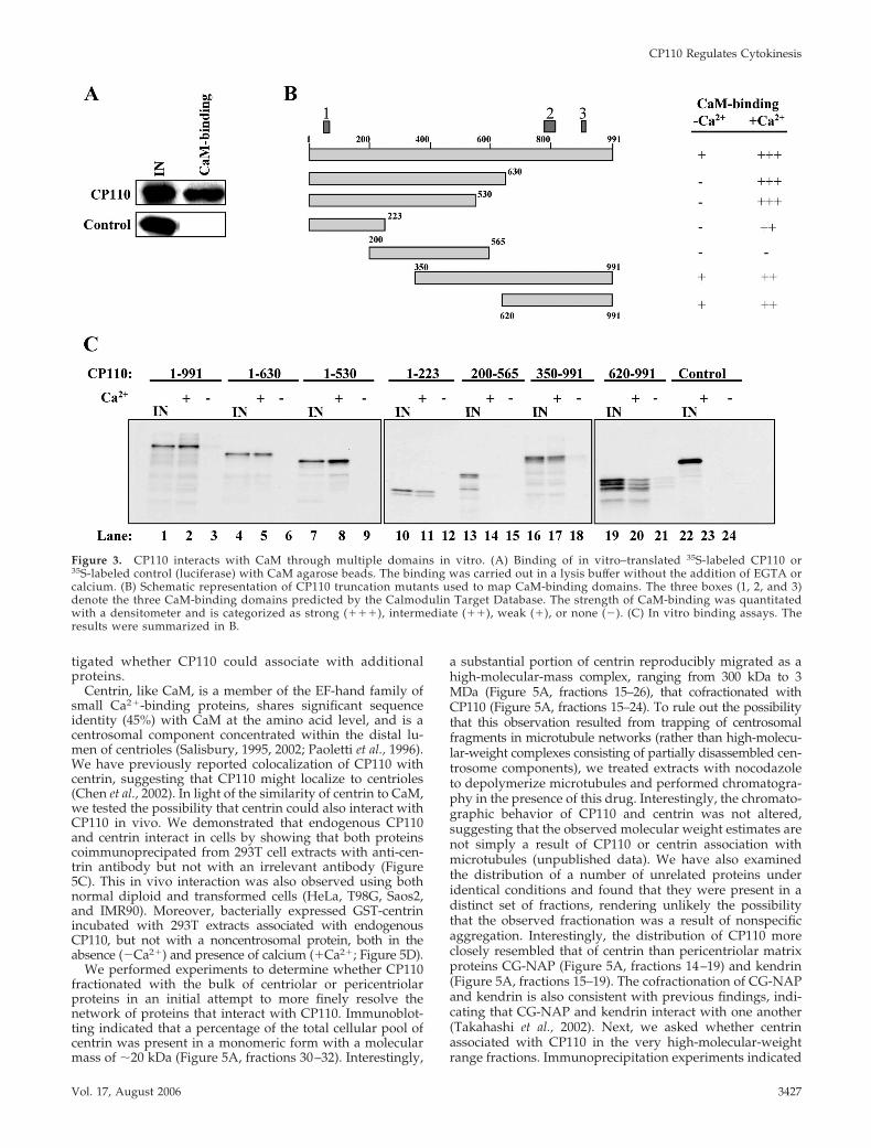
CP110 Has Multiple CaM-binding DomainsTo determine whether the interaction between CP110 andCaM is direct or is mediated by other proteins, we incubatedin vitro-translated, radiolabeled CP110 with CaM agarose,and we observed robust binding (Figure 3A). No interactionwas detected between CaM agarose and control proteins,strongly suggesting that the binding between CaM andCP110 is direct and specific (Figure 3A).
The Calmodulin Target Database (Yap et al., 2000) pre-dicts the existence of three putative CaM-binding do-mains within CP110 (residues 64 – 82, red box 1; residues781– 821, red box 2; and residues 909 –924, red box 3;Figures 3B and 4A). To identify the region(s) that confer(s)binding to CaM, we generated a series of epitope-taggedtruncation mutants and examined their ability to bindCaM in vitro. To examine calcium-dependent binding,each reaction was supplemented with either EGTA(�Ca2�) or calcium (�Ca2�). These experiments demon-strated that CP110 contains independent amino-terminal(1–223) and carboxy-terminal CaM-binding domains(620 –991) that are Ca2�-dependent (Figure 3, B and C,lanes 11, 12 and 20, 21), consistent with database predic-tions. In contrast, the region spanning residues 200 –565does not associate with CaM irrespective of calcium ad-dition (Figure 3, B and C, lanes 14 and 15). We attemptedto refine the identification of CaM-binding domains bydeleting smaller regions of CP110. Interestingly, when thepredicted CaM-binding domains were deleted singly or in
Figure 1. CaM interacts with CP110 in vivo.(A) The number of instances that each anno-tated protein was identified in the yeast two-hybrid screen is shown. (B) Western blottingof endogenous CP110 and CaM after immu-noprecipitation with anti-CP110 antibody orcontrol (anti-calnexin) antibody from 293Tcell extracts. IN, input. (C) Western blotting ofendogenous CP110 and recombinant GFP-CaM after immunoprecipitation with anti-GFP antibody using extracts from GFP-CaM-(GFP-CaM) and GFP-expressing cells (GFP).(D) Western blot detection of endogenousCP110 and control (giantin) after binding ofCaM agarose with 293 extracts (extract) or
with lysis buffer (�). (E) Western blot detection of endogenous CP110 after binding of CaM agarose with 293 extracts supplemented witheither EGTA (�Ca2�) or calcium (�Ca2�).
CP110 Regulates Cytokinesis
Vol. 17, August 2006 3425

combination, calcium-dependent binding to CaM was es-sentially abolished (Figure 4, A and B, compare lane 2 tolanes 5 and 8; compare lane 11 to lane 14). We detected aweak, residual interaction between CaM and full-lengthCP110 or carboxy-terminal fragments when calcium isabsent (Figure 3, B and C, lanes 3, 18, and 21, and Figure4, A and 4, lanes 3 and 18), and this interaction wasobserved even when the putative CaM-binding domainswere deleted (Figure 4, A and B, lanes 9 and 21), suggest-ing that additional Ca2�-independent CaM binding sites,not revealed by the database, may be present at the car-boxy terminus. Thus, our data show that CP110 can binddirectly to CaM via Ca2�-dependent and -independentdomains, in accordance with experiments in which CaMwas shown to interact with endogenous CP110 in cellextracts in the absence or presence of calcium (Figure 1E).To determine the overall impact of ablating all threeCP110 CaM-binding sites in vivo, we expressed thisCP110 mutant in cells and determined whether it couldassociate with endogenous CaM. Remarkably, removal of
all three sites nearly abolished its interaction with CaM invivo (see Figure 9D), indicating that this assay may besomewhat more specific than the in vitro binding assayand prompting us to test the functional impact of abridg-ing the CP110-CaM interaction in vivo (see below).
Native CP110 Associates with CaM and Centrinin High-Molecular-Weight ComplexesOur experiments identified CaM as a physiologically rel-evant interacting protein. We asked whether CaM was themajor protein associated with CP110 or if additional pro-teins could interact with this centriolar protein. We deter-mined the native molecular weight of CP110 – containingcomplexes by fractionating whole cell extracts using size-exclusion chromatography. Interestingly, a substantialportion of CP110 reproducibly migrated as a high-molec-ular-mass complex, ranging from 300 kDa to 3 MDa (Fig-ure 5A, fractions 15–24). Certain high molecular weightfractions (fractions 18 –19) contained CP110-CaM com-plexes (Figure 5B), but given the mass of CaM, we inves-
Figure 2. Colocalization and coimmunoprecipitation of CP110 and CaM across the cell cycle. (A) U2OS cells were processed forimmunofluorescence with anti-CaM antibody (red) and anti-CP110 antibody (green). DNA was stained with DAPI (blue). A representativeinterphase, metaphase, anaphase, and telophase cell are illustrated. Yellow color in merged images indicates substantial colocalization ofCP110 and CaM. Insets show magnified view of centrosomes. Bar, 10 �m; insets, 3 �m. (B) T98G cells were synchronized by serum starvationand restimulated with serum to initiate cell cycle reentry. Cell lysates from different cell cycle stages were collected. Western blots ofendogenous CP110 and CaM after immunoprecipitation with anti-CP110 antibody or control (anti-calnexin) antibody.
W. Y. Tsang et al.
Molecular Biology of the Cell3426
tigated whether CP110 could associate with additionalproteins.
Centrin, like CaM, is a member of the EF-hand family ofsmall Ca2�-binding proteins, shares significant sequenceidentity (45%) with CaM at the amino acid level, and is acentrosomal component concentrated within the distal lu-men of centrioles (Salisbury, 1995, 2002; Paoletti et al., 1996).We have previously reported colocalization of CP110 withcentrin, suggesting that CP110 might localize to centrioles(Chen et al., 2002). In light of the similarity of centrin to CaM,we tested the possibility that centrin could also interact withCP110 in vivo. We demonstrated that endogenous CP110and centrin interact in cells by showing that both proteinscoimmunoprecipated from 293T cell extracts with anti-cen-trin antibody but not with an irrelevant antibody (Figure5C). This in vivo interaction was also observed using bothnormal diploid and transformed cells (HeLa, T98G, Saos2,and IMR90). Moreover, bacterially expressed GST-centrinincubated with 293T extracts associated with endogenousCP110, but not with a noncentrosomal protein, both in theabsence (�Ca2�) and presence of calcium (�Ca2�; Figure 5D).
We performed experiments to determine whether CP110fractionated with the bulk of centriolar or pericentriolarproteins in an initial attempt to more finely resolve thenetwork of proteins that interact with CP110. Immunoblot-ting indicated that a percentage of the total cellular pool ofcentrin was present in a monomeric form with a molecularmass of �20 kDa (Figure 5A, fractions 30–32). Interestingly,
a substantial portion of centrin reproducibly migrated as ahigh-molecular-mass complex, ranging from 300 kDa to 3MDa (Figure 5A, fractions 15–26), that cofractionated withCP110 (Figure 5A, fractions 15–24). To rule out the possibilitythat this observation resulted from trapping of centrosomalfragments in microtubule networks (rather than high-molecu-lar-weight complexes consisting of partially disassembled cen-trosome components), we treated extracts with nocodazoleto depolymerize microtubules and performed chromatogra-phy in the presence of this drug. Interestingly, the chromato-graphic behavior of CP110 and centrin was not altered,suggesting that the observed molecular weight estimates arenot simply a result of CP110 or centrin association withmicrotubules (unpublished data). We have also examinedthe distribution of a number of unrelated proteins underidentical conditions and found that they were present in adistinct set of fractions, rendering unlikely the possibilitythat the observed fractionation was a result of nonspecificaggregation. Interestingly, the distribution of CP110 moreclosely resembled that of centrin than pericentriolar matrixproteins CG-NAP (Figure 5A, fractions 14–19) and kendrin(Figure 5A, fractions 15–19). The cofractionation of CG-NAPand kendrin is also consistent with previous findings, indi-cating that CG-NAP and kendrin interact with one another(Takahashi et al., 2002). Next, we asked whether centrinassociated with CP110 in the very high-molecular-weightrange fractions. Immunoprecipitation experiments indicated
Figure 3. CP110 interacts with CaM through multiple domains in vitro. (A) Binding of in vitro–translated 35S-labeled CP110 or35S-labeled control (luciferase) with CaM agarose beads. The binding was carried out in a lysis buffer without the addition of EGTA orcalcium. (B) Schematic representation of CP110 truncation mutants used to map CaM-binding domains. The three boxes (1, 2, and 3)denote the three CaM-binding domains predicted by the Calmodulin Target Database. The strength of CaM-binding was quantitatedwith a densitometer and is categorized as strong (���), intermediate (��), weak (�), or none (�). (C) In vitro binding assays. Theresults were summarized in B.
CP110 Regulates Cytokinesis
Vol. 17, August 2006 3427

that CP110 indeed associated with centrin in a subset of thehigh-molecular-weight fractions in which the proteins coe-luted (Figure 5E, fractions 16–18). Thus, we believe thatCP110 forms very large complexes with centrin, CaM, and acohort of uncharacterized proteins.
Because CP110 and centrin associate under physiologi-cal conditions, we tested whether this interaction wasdirect or mediated through additional proteins. We per-
formed binding assays by incubating in vitro–translatedCP110 with either GST-tagged or in vitro–translated cen-trin. These studies failed to reveal significant bindingbetween CP110 and centrin, indicating that the associa-tion is most likely mediated through additional, unknownprotein(s). Thus, CP110 interacts with two calcium-bind-ing proteins, CaM and centrin, in a fundamentally differ-ent manner.
Figure 4. CP110 CaM-binding mutants 1-565�1 and 1-991�123 interact poorly with CaM in vitro. (A) Schematic representation of CP110truncation mutants and CaM-binding domains. The three boxes (1, 2, and 3) denote the three CaM-binding domains predicted by theCalmodulin Target Database. The strength of CaM-binding was quantitated with a densitometer and is categorized as strong (���),intermediate (��), weak (�), or none (�). (B) In vitro binding assays. The results were summarized in A.
Figure 5. Centrin interacts with CP110 in vivoand cofractionates with CP110 and CaM in high-molecular-weight complexes. (A) Cell extract waschromatographed on a Superose 6 gel filtrationcolumn, and the resulting fractions were Westernblotted with antibodies against CP110, centrin,kendrin, CG-NAP, or CaM. Estimated molecularweights are indicated at the top of the panel. (B)Western blotting of endogenous CP110 and CaMafter immunoprecipitation with anti-CP110 anti-body using fractions 18 –19 (Fr 18 –19) from theSuperose 6 column. (C) Western blot of endoge-nous CP110 and centrin after immunoprecipitationwith anti-centrin antibody or control (anti-caln-exin) antibody using extracts from 293T cells. (D)Western blotting of endogenous CP110 and con-trol (giantin) after binding of either GST or GST-centrin prebound to glutathione agarose beadswith 293T extracts. The buffer used for prebindingand the 293T extracts were supplemented witheither EGTA (�Ca2�) or calcium (�Ca2�). (E)Western blotting of endogenous CP110 and cen-trin after immunoprecipitation with anti-centrinantibody using 293T cell extract (Extract), fractions 16 –18 (Fr 16 –18), or fractions 30 –32 (Fr 30 –32) from the Superose 6 column.
W. Y. Tsang et al.
Molecular Biology of the Cell3428

Phenotypic Effect of CP110 Depletion and Induction ofBinucleate Cell FormationWe have previously reported that cells stably expressing amutant version of CP110 that lacks the majority of CDKphosphorylation sites exhibit marked tetraploidy (Chen etal., 2002). To determine the effects of long-term depletion ofCP110, we derived several independent, stable clones ex-pressing short hairpin RNAs (shRNA) targeting the CP110coding sequence. As a control, we generated several linesharboring only the integrated expression vector. Suppres-sion of CP110 with the shRNA resulted in a �50% reductionin CP110 protein levels when compared with the control(Figure 6A). Remarkably, clones expressing the CP110shRNA displayed polyploidy and exhibited tetraploid 4N or8N DNA content, in contrast to controls that were diploid(Figure 6B). We confirmed that these cells were in tetraploidG1 and G2 state using a nocodazole block (Chen et al., 2002and unpublished data). Thus, normal cell cycle progressionappears to be exquisitely sensitive to the dosage of CP110protein, because a twofold reduction in protein levels hasdramatic effects on genomic stability.
Recent studies demonstrated a requirement for centrinin centriole duplication and cytokinesis in human cells(Salisbury et al., 2002). The occurrence of tetraploidy incells stably depleted of CP110 suggested that loss of thisprotein could provoke cytokinesis defects. To examinethis possibility and to comprehensively describe the phe-notypic consequences of CP110 depletion, we used RNAito acutely deplete CP110 and centrin individually. Weshowed that treatment of cells with a centrin-specificsiRNA resulted in a substantial reduction of centrin ex-pression with no discernible effect on CP110 or CaMprotein levels, and conversely, depletion of CP110 did notalter centrin or CaM expression (Figure 6C).
Microscopic examination of siRNA-treated cells led toseveral important conclusions. First, diminution of CP110led to unscheduled centrosome separation, as expected(Chen et al., 2002). Second, immunofluorescent detection ofboth centrin and CP110 indicated that depletion of CP110had no apparent impact on the localization of at least threecentrosomal markers, namely, centrin, �-tubulin, and C-NAP (Figure 7 and unpublished data). Furthermore, sup-
Figure 6. RNAi-mediated suppression of CP110 induces polyploidy and results in the formation of binucleate cells. (A) Western blotdetection of CP110 proteins in U2OS cells transfected with a control or CP110 shRNA expression vector. �-tubulin was used as a loadingcontrol. (B) FACS analysis of control or CP110 shRNA-expressing cells. (C) Western blotting of CP110, centrin, and CaM in HeLa cells treatedwith control, centrin, CP110, or centrin and CP110 siRNAs. �-tubulin was used as a loading control. (D) The percentages of cells withmultipolar spindles were determined. About 50 mitotic cells were scored for each condition, and the experiments were repeated at least twice.(E) The percentages of binucleate cells were determined. About 200 cells were scored for each condition, and the experiments were repeatedat least three times.
CP110 Regulates Cytokinesis
Vol. 17, August 2006 3429

pression of centrin expression did not affect CP110 localiza-tion, suggesting that these proteins are independentlyrecruited and anchored to the centrosome. Third, we fre-quently observed multipolar spindles in cells with dimin-ished CP110 (Figure 6D). This result is strikingly reminiscentof centrin-depleted cells in which multipolar mitotic figurespredominated (Salisbury et al., 2002; Figure 6D). Remark-ably, like centrin depletion, CP110 depletion also resulted ina significant increase in binucleate cells (Figure 6E). Impor-tantly, this phenotype was also observed both with a second,distinct CP110 siRNA and in normal diploid IMR90 cells.Interestingly, we note that CP110-depleted binucleate cellsoften display centrosomal clustering, and these centrosomesappear to be functional microtubule-organizing centers (Fig-ure 7). Indeed, loss of CP110 had no apparent effect on denovo microtubule nucleation (unpublished data). We alsotested in parallel the impact of centrin depletion. In agree-ment with previous findings, depletion of centrin resultedin a fivefold increase in the incidence of binucleate cells(Figure 6E).
Loss of CP110 Results in Cytokinesis Failure
Next, we investigated in greater detail how binucleatecells could arise as a result of CP110 depletion. Depletionof at least four other centrosomal proteins (centriolin,�-tubulin, CEP55, and centrobin) besides centrin results incytokinesis defects (Shu et al., 1995; Gromley et al., 2003;Fabbro et al., 2005; Zou et al., 2005). Furthermore, giventhe fact that CaM plays a well-established role in cytoki-nesis (Moser et al., 1997; Lippincott and Li, 1998; Osmanand Cerione, 1998), we asked whether binucleate cellswith a polyploid DNA content arose after CP110 deple-tion as a consequence of cytokinesis failure using real-time videomicroscopy. We monitored siRNA-transfectedcells with rhodamine-labeled oligonucleotides and useddifferential interference contrast (DIC) to image live HeLacells progressing through mitosis (Figure 8). Cells treated
with a nonspecific control duplex progressed throughmitosis and cleavage, after which daughter cells separatedfrom one another, as expected. In striking contrast, cellstreated with CP110 siRNAs progressed through mitosisbut failed at a late stage in cytokinesis, leading to rapidfusion of emerging daughter cells and binucleate cellformation. We observed a similar block in cells treatedwith a centrin siRNA (unpublished data). We note thatunlike the centriolin knockdown, which also gives rise tocytokinesis defects, we did not detect a frequent occur-rence of long intercellular bridges.
Immunofluorescence experiments indicated that microtu-bules were not globally disrupted by CP110 knockdown, nordid we observe inappropriate localization of centrosomalmarkers (centrin, �-tubulin, and C-NAP), suggesting thatcatastrophic disruption of centrosome assembly or microtu-bule arrays is not likely to underlie the cytokinesis defect(unpublished data). We conclude that CP110 plays a role incytokinesis and that loss of this protein results in cytokinesisfailure.
Functional Interactions between CP110 and Centrin
Given that CP110 and centrin depletion have profound ef-fects on centrosome function and cytokinesis, we initiated aseries of experiments to examine functional interactions be-tween CP110 and centrin in vivo. We reasoned that if thetwo proteins function in concert in a linear manner, it mightbe expected that depletion of both proteins through RNAiwould not have additive effects on binucleate cell formation.We compared the extent of induction of the binucleate phe-notype in cells singly depleted of CP110 or centrin and cellsdepleted of both proteins. Western blotting confirmed thatboth CP110 and centrin protein levels were significantlyreduced (Figure 6C). Simultaneous depletion resulted in asignificant enrichment of binucleate cells (Figure 7), andinterestingly, the percentage of binucleate cells closely ap-proximated what we observed when either CP110 or centrin
Figure 7. Ablation of CP110 and centrin results in the formation of binucleate cells. HeLa cells transiently transfected with control, CP110,centrin, or CP110 and centrin siRNAs stained with antibodies to �-tubulin (green), �-tubulin (red), and with DAPI (blue). Only DAPI andmerged images are shown. Bar, 10 �m; insets, 2 �m.
W. Y. Tsang et al.
Molecular Biology of the Cell3430

was knocked down individually (Figure 6E). A similar ob-servation was also made in IMR90 cells. As a control, weshowed that the frequency of binucleate cell formation in cellscotransfected with both control and centrin siRNAs was com-parable to cells treated with centrin siRNA alone. These obser-vations indicate that CP110 and centrin interact functionally,consistent with the possibility that interference with CP110 orcentrin deregulates a common step in cytokinesis.
Ectopic Expression of the CP110 CaM-binding MutantInduces Binucleate Cell FormationOur in vitro binding data suggested that removal of CaM-binding motifs dramatically reduced the ability of CP110to interact with CaM (Figure 4). To address the functionalsignificance of CaM-binding to CP110 in vivo, we ectopi-cally expressed Flag-tagged CP110 CaM-binding mutants(Flag-1-565�1 and Flag-1-991�123) in human cells andinvestigated the impact on cell cycle progression andcompletion of cytokinesis. These two mutants were cho-sen because they exhibited the most dramatic decreases inaffinity for CaM in vitro. Immunofluorescence microscopyrevealed that only a single mutant, Flag-1-991�123 (inaddition to Flag-1-991), properly localized to the centro-some (Figure 9A and unpublished data), and therefore,
we studied this mutant in detail. Western blot analysisindicated a three- to fourfold increase in the levels ofepitope-tagged CP110 relative to the endogenous protein(Figure 9B). Flag-1-991�123 exhibited significantly re-duced affinity for CaM compared with the wild-type con-trol in coimmunoprecipitation experiments (Figure 9D).Interestingly, both the wild type and CaM-binding mu-tant proteins interacted equally well with centrin (Figure9C), rendering less likely the possibility that the mutantprotein fails to interact with CaM owing to global mis-folding. Remarkably, expression of the CaM-binding mu-tant did not grossly affect progression through G1 or Sphase (unpublished data), although it resulted in a signif-icant (p � 0.01) and reproducible elevation in the numberof binucleate cells when compared with vector and wild-type controls (Figure 9E). In conclusion, our studiesstrongly support the notion that disrupting the interactionbetween CP110 and CaM induces cytokinesis failure andbinucleate cell formation and that CP110 interacts withCaM and centrin in fundamentally different ways. Wehave therefore for the first time established a clear andimportant functional link between CaM-binding to CP110and cytokinesis.
Figure 8. Cells depleted of CP110 exhibit a late cytokinesis defect. HeLa cells transfected with rhodamine-labeled and nonspecific controlsiRNA oligonucleotides or rhodamine-labeled and CP110 siRNA oligonucleotides were observed by DIC time-lapse videomicroscopy. Atleast 80 mitotic events were scored for each condition. A representative cell treated with control and two representative cells treated withCP110 siRNAs are illustrated. Times from the onset of metaphase are indicated. The fluorescent images were taken before mitosis. Arrowsindicate transfected cells and their daughter cells. Bar, 10 �m.
CP110 Regulates Cytokinesis
Vol. 17, August 2006 3431

DISCUSSION
A detailed molecular description of the events involved incontrolling the mammalian centrosome cycle has not yetbeen achieved. Beyond the events surrounding the dupli-cation and function of this organelle, recent experimentsinvolving surgical removal of this organelle and laserablation also suggest that the centrosome plays a role inthe initiation and completion of cytokinesis (Hinchcliffe etal., 2001; Khodjakov and Rieder, 2001; Piel et al., 2001). Inter-estingly, several centrosomal proteins participate in cytokine-sis, including centrin, centriolin, �-tubulin, CEP55, and cen-trobin (Shu et al., 1995; Salisbury et al., 2002; Gromley et al.,2003; Keryer et al., 2003; Fabbro et al., 2005; Zou et al., 2005).Clearly, elucidating the molecular architecture of the centro-some and deciphering the role of critical centrosomal pro-teins are essential for understanding not only centrosomefunction but also cytokinesis.
In an effort to begin identifying the proteins that play animportant role in both processes, we have focused on CP110,which we have shown is important for centrosome redupli-cation and separation. Here, we have begun identifyingCP110-interacting proteins and have found that two calcium-binding proteins, CaM and centrin, associate with high-molecular-weight, native CP110 complexes. CaM, a multi-functional Ca2�-binding protein and an important cell cycleand cytokinesis regulator (Moser et al., 1997; Lippincott andLi, 1998; Osman and Cerione, 1998), has also been implicatedin centrosome function and has been shown to bind othercentrosomal proteins (Flory et al., 2000; Takahashi et al.,2002). Centrin, like CaM, is a member of the EF-hand familyof small Ca2�-binding proteins. It shares significant se-quence identity (45%) with CaM at the amino acid level andis a centrosomal component concentrated within the distallumen of centrioles (Salisbury, 1995; Paoletti et al., 1996;
Salisbury et al., 2002). Our studies show that although CP110binds to CaM directly, the association between CP110 andcentrin does not appear to be direct and therefore may bemediated through other proteins (Kilmartin, 2003). Further,we demonstrate that a CP110 mutant unable to bind CaMcan still interact with centrin. Thus, two highly related cal-cium-binding proteins interact with CP110 in fundamentallydifferent ways. Mass spectrometry should allow us to gainfurther insight into the molecular composition of the large,native CP110-CaM-centrin complexes and to identify addi-tional proteins that connect CP110 to centrin.
Our observation that the association between CP110 andCaM is cell cycle–independent is especially interesting, be-cause CaM does not appear to be concentrated at the cen-trosome during interphase. One likely explanation is that asmall portion of total cellular CaM is always present at thecentrosome. Indeed, CaM is an integral component of thecentral plaque of the yeast microtubule-nucleating center,the spindle pole body (Geiser et al., 1993; Spang et al., 1996;Sundberg et al., 1996). Recent proteomic characterization hasbegun to shed light on the structure and composition of thehuman centrosome and has also detected the presence ofCaM in this organelle during interphase (Andersen et al.,2003). In addition, CaM has been shown to interact withtwo pericentriolar matrix proteins, kendrin and CG-NAP(Witczak et al., 1999; Flory et al., 2000). Further electronmicroscopic studies will be needed to define precisely thelocalization CaM at an ultrastructural level.
Several lines of evidence point toward a function forCP110 in cytokinesis, and the interaction between CP110,centrin, and CaM appears to play a pivotal role here. First,CP110 binds to CaM and centrin, both of which have beenimplicated in regulating this event (Li et al., 1999; Salisburyet al., 2002). Second, expression of a nonphosphorylated
Figure 9. Ectopic expression of a CP110CaM-binding mutant in vivo results in binu-cleate cell formation. (A) U2OS cells tran-siently transfected with a Flag-1-991 or Flag-1-991�123 expression vector stained withantibodies to �-tubulin (green), Flag (red),and with DAPI (blue). Bar, 10 �m; insets: 2�m. (B) Western blot detection of CP110 pro-teins in HeLa cells transfected with a control,Flag-1-991 or Flag-1-991�123 expression vec-tor. The blots labeled CP110 and Flag wereprobed with anti-CP110 and anti-Flag anti-bodies, respectively. �-tubulin was used as aloading control. (C) Western blotting of Flag-CP110 and endogenous centrin after immu-noprecipitation with anti-centrin antibodyusing 293T cell extract expressing Flag (con-trol), Flag-1-991 or Flag-1-991�123 proteins.IN, input. (D) Western blotting of Flag-CP110 and endogenous CaM after immuno-precipitation with anti-Flag antibody using293T cell extract expressing Flag (control),Flag-1-991 or Flag-1-991�123 proteins. IN,input. (E) HeLa cells were transfected with aG418-resistant marker along with a control,Flag-1-991 or Flag-1-991�123 expression vec-tor. The percentages of binucleate cells weredetermined after 5–6 d of selection in thepresence of G418. About 500 cells werescored for each condition, and the experi-ments were repeated at least twice. Asteriskdenotes that the percentage of binucleate cells resulting from expression of the mutant (Flag-1-991�123) is significantly higher than that ofcontrol (p � 0.01) or wild-type (p � 0.01) based on a two-tailed Student’s t test.
W. Y. Tsang et al.
Molecular Biology of the Cell3432

CP110 mutant induces overt polyploidy, a common hall-mark of cytokinesis failure (Chen et al., 2002). In addition,long-term disruption of CP110 with shRNA leads topolyploidy and phenocopies the CP110 phospho-mutant,whereas short-term disruption results in a binucleate phe-notype, another outcome of cytokinesis malfunction. Thestriking similarity between the phenotypes resulting fromCP110, CaM, or centrin ablation indicates that these proteinsare likely to be connected by a common biological function.Interestingly, when CP110 and centrin are disabled together,we do not observe additive increases in the percentage ofbinucleate cells. These experiments suggest functional inter-actions between these two proteins and raise the possibilitythat CP110, centrin, and CaM could function in a geneticpathway that regulates the centrosome cycle and progres-sion through cytokinesis.
We have further demonstrated a functional link betweenCP110 and CaM and its relevance to cytokinesis by studyingthe effects of inactivating three high-affinity CaM-bindingdomains within CP110. This mutant, 1-991�123, localizes tothe centrosome, binds poorly to CaM in vitro and in vivo,and induces binucleate cell formation in vivo. The pheno-type resulting from expression of this mutant is less pro-nounced than that observed after depletion of CP110 withsiRNAs. One possible explanation is that the effect of ex-pressing this mutant is dampened by the presence of endog-enous CP110. Another possibility is that suppression ofCP110 expression disrupts its interaction with both CaM andcentrin, whereas overexpression of the CaM-binding mutantstrictly abolishes the CP110-CaM interaction. Indeed, theCP110 CaM-binding mutant is fully capable of interactingwith centrin (Figure 9C). It will be interesting to map thecentrin-binding domains within CP110 to create a CP110mutant refractory to binding both CaM and centrin. Such amutant could produce a more potent binucleate phenotype.
Our observation of binucleate cells subsequent to CP110depletion is reminiscent of findings in which acentrosomalcells were choreographed through mitosis (Piel et al., 2001).Acentriolar Drosophila cells exhibit hallmarks of incompletecytokinesis, including intercellular bridges, binucleate for-mation, and polyploidy, arguing for a role for centrosomesin the process of cell abscission at the conclusion of cytoki-nesis (Debec, 1978; Debec and Abbadie, 1989). Recent re-ports have revealed several proteins whose depletion couldresult in cytokinesis defects, and it is intriguing that fouradditional centriolar proteins other than CP110 (centrin, cen-triolin, CEP55, and centrobin) appear to play a role in cyto-kinesis (Salisbury et al., 2002; Gromley et al., 2003; Fabbro etal., 2005; Zou et al., 2005). However, unlike centriolin orCEP55 silencing, which appears to result in long intercellu-lar bridges, suppression of CP110 or centrobin expressionleads to failed abscission without such bridges. This couldsuggest that centrosomes play multiple, distinct roles duringcytokinesis. Although the basis for cytokinesis defectsbrought on by the loss of CP110 remains unknown, ourfindings suggest that abnormal cytokinesis does not resultfrom an inability to nucleate or organize microtubules. Inter-estingly, recent evidence suggests that centriolin may play arole in anchoring vesicle-targeting and vesicle-fusion proteincomplexes at the midbody during abscission (Gromley et al.,2005). On the basis of the conserved phenotypes resulting fromdepletion of CP110 and these centriolar proteins, it should bepossible to begin dissecting the mechanisms that connect cen-trosome function and cytokinesis.
Although we have not yet fully defined the pathway(s)that link the function of centrosomal proteins such as CP110or centrin with faithful completion of cytokinesis, the direct
and robust interaction between CP110 and CaM offers sev-eral potential avenues for further exploration. Another in-teresting possibility involves interactions with a cytoskeletalnetwork, and the identification of �-spectrin and �-actinin asCP110-interacting proteins in our two-hybrid screen may beparticularly revealing in this regard. Furthermore, we havebegun to biochemically fractionate centrosomal compo-nents, suggesting possible proteomic approaches for theidentification of centrosome subassemblies. Future studiesthat combine genetic and proteomic approaches will allowus to unravel the complex functional relationships betweendifferent centrosomal components and provide importantclues for elucidating their role in cytokinesis.
ACKNOWLEDGMENTS
We are grateful to M. Vidal for help with the yeast two-hybrid screen. Wethank M. Takahaski and Y. Ono for the gift of anti-kendrin and anti-CG-NAPantibodies, M. Cole for the gift of pCBF plasmid DNA, and D. C. Chang forthe gift of pCDNA3-GFP-CaM plasmid DNA. We are especially grateful to P.Asp in our laboratory for invaluable assistance with fast-performance liquidchromatography purification and to members of M. Philips’ laboratory forassistance with real-time videomicroscopy. We thank all members of theDynlacht laboratory for constructive advice and encouragement. This workwas supported in part by an Irma T. Hirschl Career Scientist Award to B.D.D.,for which he is grateful. W.Y.T. was supported by a Natural Sciences andEngineering Research Council of Canada postdoctoral fellowship and anAlberta Heritage Foundation for Medical Research full-time postdoctoralfellowship.
REFERENCES
Andersen, J. S., Wilkinson, C. J., Mayor, T., Mortensen, P., Nigg, E. A., andMann, M. (2003). Proteomic characterization of the human centrosome byprotein correlation profiling. Nature 426, 570–574.
Badano, J. L., Teslovich, T. M., and Katsanis, N. (2005). The centrosome inhuman genetic disease. Nat. Rev. Genet. 6, 194–205.
Chen, Z., Indjeian, V. B., McManus, M., Wang, L., and Dynlacht, B. D. (2002).CP110, a cell cycle-dependent CDK substrate, regulates centrosome duplica-tion in human cells. Dev. Cell 3, 339–350.
Chin, D., and Means, A. R. (2000). Calmodulin: a prototypical calcium sensor.Trends Cell Biol. 10, 322–328.
Debec, A. (1978). Haploid cell cultures of Drosophila melanogaster. Nature 274,255–256.
Debec, A., and Abbadie, C. (1989). The acentriolar state of the Drosophila celllines 1182. Biol. Cell 67, 307–311.
Doxsey, S. (2001). Re-evaluating centrosome function. Nat. Rev. Mol. CellBiol. 2, 688–698.
Fabbro, M. et al. (2005). Cdk1/Erk2- and Plk1-dependent phosphorylation ofa centrosome protein, Cep55, is required for its recruitment to midbody andcytokinesis. Dev. Cell 9, 477–488.
Flory, M. R., Moser, M. J., Monnat, R. J., Jr., and Davis, T. N. (2000). Identi-fication of a human centrosomal calmodulin-binding protein that shareshomology with pericentrin. Proc. Natl. Acad. Sci. USA 97, 5919–5923.
Geiser, J. R., Sundberg, H. A., Chang, B. H., Muller, E. G., and Davis, T. N.(1993). The essential mitotic target of calmodulin is the 110-kilodalton com-ponent of the spindle pole body in Saccharomyces cerevisiae. Mol. Cell. Biol. 13,7913–7924.
Gromley, A., Jurczyk, A., Sillibourne, J., Halilovic, E., Mogensen, M., Groisman,I., Blomberg, M., and Doxsey, S. (2003). A novel human protein of thematernal centriole is required for the final stages of cytokinesis and entry intoS phase. J. Cell Biol. 161, 535–545.
Gromley, A., Yeaman, C., Rosa, J., Redick, S., Chen, C. T., Mirabelle, S., Guha,M., Sillibourne, J., and Doxsey, S. J. (2005). Centriolin anchoring of exocystand SNARE complexes at the midbody is required for secretory-vesicle-mediated abscission. Cell 123, 75–87.
Hinchcliffe, E. H., Miller, F. J., Cham, M., Khodjakov, A., and Sluder, G. (2001).Requirement of a centrosomal activity for cell cycle progression through G1into S phase. Science 291, 1547–1550.
Keryer, G., Witczak, O., Delouvee, A., Kemmner, W. A., Rouillard, D., Tasken,K., and Bornens, M. (2003). Dissociating the centrosomal matrix protein
CP110 Regulates Cytokinesis
Vol. 17, August 2006 3433

AKAP450 from centrioles impairs centriole duplication and cell cycle pro-gression. Mol. Biol. Cell 14, 2436–2446.
Khodjakov, A., and Rieder, C. L. (2001). Centrosomes enhance the fidelity ofcytokinesis in vertebrates and are required for cell cycle progression. J. CellBiol. 153, 237–242.
Kilmartin, J. V. (2003). Sfi1p has conserved centrin-binding sites and anessential function in budding yeast spindle pole body duplication. J. Cell Biol.162, 1211–1221.
Li, C. J., Heim, R., Lu, P., Pu, Y., Tsien, R. Y., and Chang, D. C. (1999).Dynamic redistribution of calmodulin in HeLa cells during cell division asrevealed by a GFP-calmodulin fusion protein technique. J. Cell Sci. 112(Pt 10),1567–1577.
Lingle, W. L., Lutz, W. H., Ingle, J. N., Maihle, N. J., and Salisbury, J. L. (1998).Centrosome hypertrophy in human breast tumors: implications for genomicstability and cell polarity. Proc. Natl. Acad. Sci. USA 95, 2950–2955.
Lippincott, J., and Li, R. (1998). Sequential assembly of myosin II, an IQGAP-like protein, and filamentous actin to a ring structure involved in buddingyeast cytokinesis. J. Cell Biol. 140, 355–366.
Matsumoto, Y., and Maller, J. L. (2002). Calcium, calmodulin, and CaMKIIrequirement for initiation of centrosome duplication in Xenopus egg extracts.Science 295, 499–502.
Moser, M. J., Flory, M. R., and Davis, T. N. (1997). Calmodulin localizes to thespindle pole body of Schizosaccharomyces pombe and performs an essentialfunction in chromosome segregation. J. Cell Sci. 110(Pt 15), 1805–1812.
Osman, M. A., and Cerione, R. A. (1998). Iqg1p, a yeast homologue of themammalian IQGAPs, mediates cdc42p effects on the actin cytoskeleton. J. CellBiol. 142, 443–455.
Paoletti, A., Moudjou, M., Paintrand, M., Salisbury, J. L., and Bornens, M.(1996). Most of centrin in animal cells is not centrosome-associated andcentrosomal centrin is confined to the distal lumen of centrioles. J. Cell Sci.109(Pt 13), 3089–3102.
Piel, M., Nordberg, J., Euteneuer, U., and Bornens, M. (2001). Centrosome-dependent exit of cytokinesis in animal cells. Science 291, 1550–1553.
Pihan, G. A., Purohit, A., Wallace, J., Knecht, H., Woda, B., Quesenberry, P.,and Doxsey, S. J. (1998). Centrosome defects and genetic instability in malig-nant tumors. Cancer Res. 58, 3974–3985.
Salisbury, J. L. (1995). Centrin, centrosomes, and mitotic spindle poles. Curr.Opin. Cell Biol. 7, 39–45.
Salisbury, J. L., Suino, K. M., Busby, R., and Springett, M. (2002). Centrin-2 isrequired for centriole duplication in mammalian cells. Curr. Biol. 12, 1287–1292.
Shu, H. B., Li, Z., Palacios, M. J., Li, Q., and Joshi, H. C. (1995). A transientassociation of gamma-tubulin at the midbody is required for the completionof cytokinesis during the mammalian cell division. J. Cell Sci. 108(Pt 9),2955–2962.
Sluder, G., and Nordberg, J. J. (2004). The good, the bad and the ugly: thepractical consequences of centrosome amplification. Curr. Opin. Cell Biol. 16,49–54.
Spang, A., Grein, K., and Schiebel, E. (1996). The spacer protein Spc110ptargets calmodulin to the central plaque of the yeast spindle pole body. J. CellSci. 109(Pt 9), 2229–2237.
Sui, G., Soohoo, C., Affar el, B., Gay, F., Shi, Y., Forrester, W. C., and Shi, Y.(2002). A DNA vector-based RNAi technology to suppress gene expression inmammalian cells. Proc. Natl. Acad. Sci. USA 99, 5515–5520.
Sundberg, H. A., Goetsch, L., Byers, B., and Davis, T. N. (1996). Role ofcalmodulin and Spc110p interaction in the proper assembly of spindle polebody components. J. Cell Biol. 133, 111–124.
Takahashi, M., Yamagiwa, A., Nishimura, T., Mukai, H., and Ono, Y. (2002).Centrosomal proteins CG-NAP and kendrin provide microtubule nucleationsites by anchoring gamma-tubulin ring complex. Mol. Biol. Cell 13, 3235–3245.
Takuwa, N., Zhou, W., and Takuwa, Y. (1995). Calcium, calmodulin and cellcycle progression. Cell Signal. 7, 93–104.
Walhout, A. J., and Vidal, M. (2001). High-throughput yeast two-hybridassays for large-scale protein interaction mapping. Methods 24, 297–306.
Witczak, O., Skalhegg, B. S., Keryer, G., Bornens, M., Tasken, K., Jahnsen, T.,and Orstavik, S. (1999). Cloning and characterization of a cDNA encoding anA-kinase anchoring protein located in the centrosome, AKAP450. EMBO J. 18,1858–1868.
Woo, M. S., Sanchez, I., and Dynlacht, B. D. (1997). p130 and p107 use aconserved domain to inhibit cellular cyclin-dependent kinase activity. Mol.Cell. Biol. 17, 3566–3579.
Yap, K. L., Kim, J., Truong, K., Sherman, M., Yuan, T., and Ikura, M. (2000).Calmodulin target database. J. Struct. Funct. Genom. 1, 8–14.
Zou, C., Li, J., Bai, Y., Gunning, W. T., Wazer, D. E., Band, V., and Gao, Q.(2005). Centrobin: a novel daughter centriole-associated protein that is re-quired for centriole duplication. J. Cell Biol. 171, 437–445.
W. Y. Tsang et al.
Molecular Biology of the Cell3434
Copyright © 2022 FDOKUMEN